Trichiasis surgery for trachoma - Light for the World · Trichiasis surgery for trachoma. Update...
78
ISBN 978 92 4 154867 0 Trichiasis surgery for trachoma Trichiasis surgery for trachoma
Transcript of Trichiasis surgery for trachoma - Light for the World · Trichiasis surgery for trachoma. Update...

ISBN 978 92 4 154867 0
Trichiasis surgery for trachoma
Trichiasis surgery for trachoma
Final-PBD5
vendredi 6 décembre 2013 08:31:47

WHO Library Cataloguing-in-Publication Data
Trichiasis surgery for trachoma.
Update from « Final assessment of Trichiasis surgeons » and « Trichiasis surgery for trachoma,
the bilamellar tarsal rotation procedure » (WHO/PBL/93.29)
1.Trachoma – prevention and control. 2.Blindness – prevention and control. 3.Trachoma -
surgery. 4.Trichiasis - surgery. 5.Eyelid diseases. 6.Eyelids – surgery. 7.Teaching materials.
I.World Health Organization.
ISBN 978 92 4 154867 0 (NLM classification: WW 215)
© World Health Organization 2013
All rights reserved. Publications of the World Health Organization are available on the WHO web site
(www.who.int) or can be purchased from WHO Press, World Health Organization, 20 Avenue Appia, 1211
Geneva 27, Switzerland (tel.: +41 22 791 3264; fax: +41 22 791 4857; e -mail: [email protected]).
Requests for permission to reproduce or translate WHO publications –whether for sale or for non-
commercial distribution– should be addressed to WHO Press through the WHO web site
(www.who.int/about/licensing/copyright_form/en/index.html).
The designations employed and the presentation of the material in this publication do not imply the
expression of any opinion whatsoever on the part of the World Health Organization concerning the legal
status of any country, territory, city or area or of its authorities, or concerning the delimitation of its frontiers
or boundaries. Dotted lines on maps represent approximate border lines for which there may not yet be full
agreement.
The mention of specific companies or of certain manufacturers’ products does not imply that they are
endorsed or recommended by the World Health Organization in preference to others of a similar nature that
are not mentioned. Errors and omissions excepted, the names of proprietary products are distinguished by
initial capital letters.
All reasonable precautions have been taken by the World Health Organization to verify the information
contained in this publication. However, the published material is being distributed without warranty of any
kind, either expressed or implied. The responsibility for the interpretation and use of the material lies with the
reader. In no event shall the World Health Organization be liable for damages arising from its use.
Printed in France
Final-PBD5
vendredi 6 décembre 2013 08:31:48

Trichiasis Surgery for Trachoma
Update of
« Trichiasis surgery for trachoma, the bilamellar tarsal rotation procedure »
and
« Final assessment of Trichiasis surgeons »

ACKNOWLEDGEMENTS
The World Health Organization wishes to acknowledge:
the support provided by the Fred Hollows Foundation and
Sight Savers International;
the collaboration of
S. Merbs, S. Resnikoff, A. Kello, S.P. Mariotti, G. Greene, S. West
in preparing and developing this manual;
T .Phelps for the illustrations;
S. Bakayoko and E. Gower for some images;
M. Burton, B. Gaynor and S. Llewelyn for their reviews.
Contents
Overview . . . . . . . . . . . . . . . . . . . . . . . . . . . . . . . . . . . . . . . . . . . . . . . . . . . . . . . . . . . . . . . . . . . 1
Section One
1 . Introduction . . . . . . . . . . . . . . . . . . . . . . . . . . . . . . . . . . . . . . . . . . . . . . . . . . . . . . . . . . . . . . 1
2 . The Anatomy of the Eye and the Eyelid . . . . . . . . . . . . . . . . . . . . . . . . . . . . . . . . . . . . . . . . 2
3 . Trachoma and its Effect on the Eye . . . . . . . . . . . . . . . . . . . . . . . . . . . . . . . . . . . . . . . . . . . 4
4 . History and Examination for Upper Eyelid Trichiasis . . . . . . . . . . . . . . . . . . . . . . . . . . . . . 5
5 . Indications for Eyelid Surgery . . . . . . . . . . . . . . . . . . . . . . . . . . . . . . . . . . . . . . . . . . . . . . . . 7
6 . Fitness of Patient for Surgery . . . . . . . . . . . . . . . . . . . . . . . . . . . . . . . . . . . . . . . . . . . . . . . . 8
7 . Facilities and Surgical Materials . . . . . . . . . . . . . . . . . . . . . . . . . . . . . . . . . . . . . . . . . . . . . . 9
8 . Sterilization . . . . . . . . . . . . . . . . . . . . . . . . . . . . . . . . . . . . . . . . . . . . . . . . . . . . . . . . . . . . . 11
9 . Preparation . . . . . . . . . . . . . . . . . . . . . . . . . . . . . . . . . . . . . . . . . . . . . . . . . . . . . . . . . . . . . 13
10 . Injecting Local Anaesthetic . . . . . . . . . . . . . . . . . . . . . . . . . . . . . . . . . . . . . . . . . . . . . . . . . 16
11 . Surgical Procedure . . . . . . . . . . . . . . . . . . . . . . . . . . . . . . . . . . . . . . . . . . . . . . . . . . . . . . . . 18
11 .1 Bilamellar Tarsal Rotation . . . . . . . . . . . . . . . . . . . . . . . . . . . . . . . . . . . . . . . . . . . . . 18
11 .2 Trabut . . . . . . . . . . . . . . . . . . . . . . . . . . . . . . . . . . . . . . . . . . . . . . . . . . . . . . . . . . . . . 34
12 . Postoperative Care . . . . . . . . . . . . . . . . . . . . . . . . . . . . . . . . . . . . . . . . . . . . . . . . . . . . . . . . 47
13 . Results . . . . . . . . . . . . . . . . . . . . . . . . . . . . . . . . . . . . . . . . . . . . . . . . . . . . . . . . . . . . . . . . . 48
Section TwoFor Trainers
14 . Introduction . . . . . . . . . . . . . . . . . . . . . . . . . . . . . . . . . . . . . . . . . . . . . . . . . . . . . . . . . . . . . 49
15 . Final Assessment of TT Surgeons . . . . . . . . . . . . . . . . . . . . . . . . . . . . . . . . . . . . . . . . . . . . 51
16 . Checklist . . . . . . . . . . . . . . . . . . . . . . . . . . . . . . . . . . . . . . . . . . . . . . . . . . . . . . . . . . . . . . . 58
APPENDIX I . Final Assessment: Cuenod Nataf . . . . . . . . . . . . . . . . . . . . . . . . . . . . . . . . . . 66
APPENDIX II . References . . . . . . . . . . . . . . . . . . . . . . . . . . . . . . . . . . . . . . . . . . . . . . . . . . . 72


page 1
Trichiasis Surgery for Trachoma
Overview
The second edition of this manual combines and updates material contained in three previous manuals on bilamellar tarsal rotation procedure, trabut procedure, and the final assessment of candidate trichiasis surgeons .
This manual is designed to provide specific information for trachomatous trichiasis (TT) trainers who are training others to undertake surgery for entropion trachomatous trichiasis (TT) . Other approaches are not addressed . The manual is divided into two parts . The first part covers specifics designed for training TT surgeon candidates, and serves as a resource document . The trainer can elect to have trainees read the material directly, use this manual as a guide for creating a training presentation, or use it in other ways to assist in the training . The manual contains both knowl-edge that should be imparted during training and a description of the skills that need to be developed and assessed during practice and surgery sessions . The second part is designed only for the trainers of the surgeon trainees and covers selection and final assessment of the trainees .
Section One
1. INTRODUCTION
OBJECTIVES FOR SECTION ONE: In this section, the manual will provide specific details on training potential TT surgeons to undertake bilamellar tarsal rotation and/or Trabut surgery for trichiasis .
1 .1 Objectives (a) To learn to identify patients who require surgery for trichiasis (b) To be able to perform successful bilamellar tarsal rotation and/or Trabut operations
to correct trichiasis (c) To be able to assess results and manage complications of the bilamellar tarsal and/or
Trabut procedures

page 2
2. THE ANATOMY OF THE EYE AND THE EYELID
OBJECTIVE: TO BE ABLE TO CORRECTLY NAME THE PARTS OF THE EYE AND THE EYELID
2 .1 The eye (Fig. 1a) (a) The CORNEA is the clear window in the front of the eye . (b) The CONJUNCTIVA is a thin transparent layer covering the eye and the inner
parts of the eyelid . (c) The PUNCTUM is a hole at the nasal end on the inside of each eyelid (upper and
lower), through which tears drain to the nose .
2 .2 The eyelid (Fig. 1b) The EYELASHES come from roots 2 mm deep . They emerge just above the EYELID MARGIN, and normally point away from the cornea . In normal upper eyelids, the eyelid margin is visible beneath the lashes at the edge of the eyelid . In TT eyes, the eyelid margin is often not visible as it, and the base of the eyelashes, are tucked behind the eyelid (Fig . 1c & 1d) . (a) The SKIN covers the outer surface of the eyelid . (b) The orbicularis MUSCLE lies under the skin . (c) The TARSAL PLATE is a thick, fibrous layer, which lies under the muscle and
keeps the eyelid stiff . It is 1 cm high in the upper eyelid . (d) The CONJUNCTIVA is a shiny transparent layer, which covers the inner surface of
the eyelid and goes onto the globe . It is easily seen on the everted upper eyelid . Normally vessels are seen in the conjunctiva . This may be partially or totally replaced by scarring, white stellate scars, or fibrous bands in cases of severe scarring .
PRACTICE: TRAINEES WILL OBSERVE THE PARTS OF THE EYE IN EACH OTHER AND PRACTICE EVERTING THE EYELID TO OBSERVE THE TARSAL CONJUNCTIVA .

page 3
Fig. 1a.Image of Normal eye
Fig. 1b.Sagittal drawing of normal eye
Fig. 1c.Image of abnormal TT eye
Fig. 1d.Sagittal drawing of abnormal eye showing
entropion and trichiasis
Fig. 1. ANATOMY OF THE EYE

page 4
3. TRACHOMA AND ITS EFFECT ON THE EYE
OBJECTIVE: TO DESCRIBE THE VARIOUS STAGES OF TRACHOMA AND HOW TRICHIASIS DEVELOPS
3 .1 Trachoma TRACHOMA is an infectious disease caused by a bacterium, Chlamydia trachomatis . It usually starts in childhood, even as early as the first year of life . The disease is characterized by repeated acute episodes of infection throughout childhood and early adulthood .
3 .2 Inflammation Trachoma is an inflammation of the tarsal conjunctiva and tarsal plate seen on eversion of the upper eyelid . Inflammation is characterized by the formation of follicles, white dots or bumps that contain cells . The inflammation may be intense enough to thicken the conjunctiva, obscuring the normal pattern of conjunctival blood vessels and even obscuring follicles .
3 .3 Trichiasis The chronic inflammation when repeated throughout life leads to scarring of the tarsal plate and conjunctiva of the inside of the eyelid . This turns the eyelid margin inward (ENTROPION) and maybe severe enough to turn the eyelashes inwards . When the eyelashes rub on the eye, the condition is called TRACHOMATOUS TRICHIASIS, “TT” (Figs 1c and 1d) . This is mainly a problem of the upper eyelid . The purpose of the surgery is to correct the entropion and trichiasis by rotating the eyelid margin outward, directing the eyelashes away from the globe . There may be other causes of trichiasis and of entropion besides trachoma . Correction of other causes of trichiasis or entropion, such as metaplastic lashes, is not covered in this manual .
3 .4 Corneal scarring When the eyelid is abnormal, with distorted glands and abnormal secretions as well as an abnormal eyelid margin and trichiasis, rubbing of the eyelashes on the cornea disturbs the normal corneal surface and causes scarring (CORNEAL OPACITY) . This leads to gradual loss of vision and eventually to blindness . Surgery can restore a modest amount of vision but cannot help those with severe vision loss . In these cases, surgery prevents pain and further vision loss .

page 5
4. HISTORY AND EXAMINATION FOR UPPER EYELID TRICHIASIS
OBJECTIVE: TO BE ABLE TO CARRY OUT SCREENING FOR TRICHIASIS WITH QUESTIONS ABOUT EYE PROBLEMS AND TO DEMONSTRATE THE ELEMENTS OF AN EXAMINATION
4 .1 History: These questions indicate someone who may have a TT problem (a) Ask the patient if they have a problem with their eyes . (b) Ask the patient if they (or another person helps) pull out their eyelashes or
EPILATE . (c) Ask the patient if they have PAIN in their eyes . (d) Ask the patient if they have TEARING or watery eyes . (e) Ask the patient if they have a problem seeing in bright sunlight .
4 .2 Examination of the eyelid (a) Examine the patient indoors or in the shade because bright sunlight produces
shadows that make the edge of the eyelid difficult to see . Also, patients may be very sensitive to sunlight .
(b) Ask the patient to look straight ahead with his eyes open in the normal way . (c) Use a torch, and shine it up on the edge of the eyelid FROM BELOW . (d) Look up at the eyelid FROM BELOW, and examine the edge of the eyelid, where
the lashes emerge . A 2 .5X magnifying loupe is helpful to clearly see the trichiasis . (e) Have the patient look up . Sometimes, it is easier to see dark lashes against a white
conjunctiva . While still looking from below and from the side, look for lashes that are pointed downward . There may be need to also examine the eye from the side to see if the lash actually touches the eye .
4 .3 Examination of the cornea Look directly at the cornea and see if a white or hazy area is present, especially one that covers part of the pupil .

page 6
4 .4 Examination for defective eyelid closure If the eyelid does not close properly, either because of trachoma or because of previous surgery, a more complicated operation will be needed . Defective eyelid closure is present if the eyelids do not meet completely when the eyes are gently closed, as if going to sleep . The white of the eye will still be seen in between the eyelids . The way to examine for defective closure is to ask the patient to close both eyes gently, and then shine the torch from below to look for an exposed eye (not covered by the eyelids) . THESE PATIENTS NEED REFERRAL TO AN EXPERIENCED TT SURGEON OR AN OPHTHALMOLOGIST, whether or not they have trichiasis .
PRACTICE: TRAINEES WILL PRACTICE ON EACH OTHER, TAKING HISTORY AND DOING THE EXAMINATION FOLLOWING THE DETAILED PROTOCOL ABOVE .

page 7
5. INDICATIONS FOR EYELID SURGERY
OBJECTIVE: TO BE ABLE TO DESCRIBE WHICH TT CASES ARE ELIGIBLE FOR SURGERY
All patients should be offered surgery for entropion trichiasis . If the patient is not complaining and has only one or two eyelashes (either nasally or temporally) rubbing on the conjunctiva (not the cornea), then patient consultation and the possibility of other approaches can be offered . If the patient does not desire surgery, they must be instructed to return if the pain gets worse, or if vision begins to worsen .
5 .1 Definite indications for eyelid surgery in the community are: (a) one or more eyelashes which turn in and touch the cornea when the patient looks
straight ahead (b) evidence of corneal damage from trichiasis (c) severe discomfort from trichiasis (d) TT patient requesting surgery
5 .2 Contraindications to performing surgery in the community (a) Defective eyelid closure or repeat trichiasis after surgery (b) Childhood . Children need surgery in hospital, possibly with a general anaesthetic . (c) Poor general health (see section 6) (d) TT of the lower eyelid . This is rare but it does occur and will require more
assessment by an ophthalmologist . (e) The cases above require referral to an ophthalmologist for management .
PRACTICE: TRAINER WILL PROVIDE ORALLY A SERIES OF CASES, TO WHICH THE TRAINEES MUST RESPOND CORRECTLY AS TO WHETHER THEY SHOULD BE OFFERED SURGERY OR WHETHER OTHER OPTIONS SHOULD BE DISCUSSED .

page 8
6. FITNESS OF PATIENTS FOR SURGERY
OBJECTIVE: TO BE ABLE TO ASSESS THE FITNESS OF TT PATIENTS FOR SURGERY
The procedure must cause only minimal risk to the general health of the patient .
6 .1 The patient must be questioned for general fitness. (a) Ask the patient if he or she has any SHORTNESS OF BREATH that results
in difficulty lying flat for 30 minutes . These symptoms may indicate evidence of HEART FAILURE .
(b) Ask the patient if he or she knows if they have DIABETES (“sugar”), or HIGH BLOOD PRESSURE, and if they are taking medication for these conditions .
(c) Very rarely, a person may be ALLERGIC to local anaesthetic, or have a BLEEDING DISORDER . Ask the patient if he or she has received surgery before or experienced any problems with injections of local anaesthetic or with excessive bleeding if cut (this does not relate to menstrual bleeding) .
(d) Does the patient have difficulty in cooperating and following instructions? Be certain this is not simply an issue of differences in dialect or language between the patient and surgeon .
IF HEART FAILURE, known but untreated DIABETES or untreated HYPERTENSION, ALLERGY TO LOCAL ANAESTHETIC, OR A BLEEDING ABNORMALITY EXISTS, THE OPERATION SHOULD NOT BE DONE IN A COMMUNITY CLINIC . Refer the patient to a doctor for management of the condition first, and to consider whether the operation can be performed under medical supervision in hospital . If the patient seems to be unable to follow instructions, the patient may not be able to give a true informed consent and may not be able to cooperate during surgery . Engage the patient in sufficient discussion to decide if the procedure can go forward .
PRACTICE: TRAINEES WILL PRACTICE WITH EACH OTHER, ASKING QUESTIONS TO IDENTIFY CONDITIONS SUGGESTING THAT THE PATIENT SHOULD NOT HAVE SURGERY AND SHOULD BE REFERRED, AND DESCRIBING THE APPROPRIATE ACTION INDICATED BASED ON THE ANSWER GIVEN .
page 9
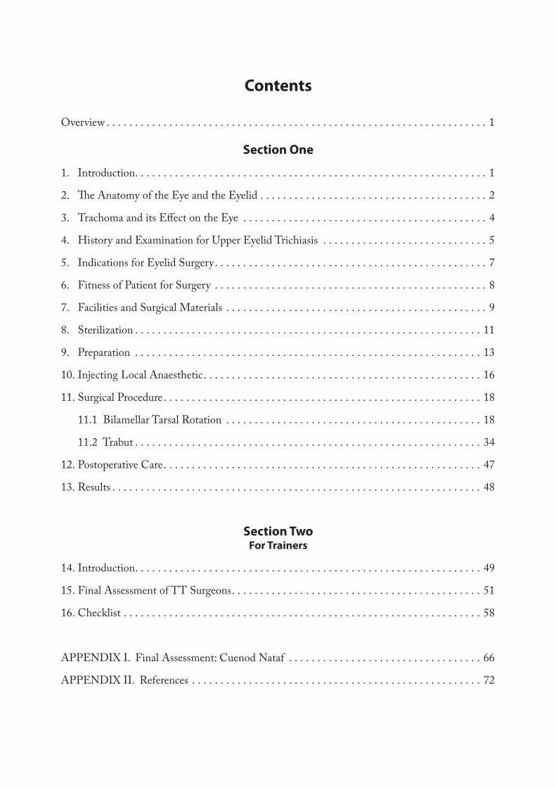
7. FACILITIES AND SURGICAL MATERIALS
OBJECTIVE: TO BE ABLE TO LIST KEY INSTRUMENTS, SUTURES, AND CONSUMABLES REQUIRED FOR SURGERY
7 .1 Facilities required The operating room should be: (a) CLEAN (free from dust) with covered windows to avoid flies (b) WELL-LIT, using a focused light powered either by electricity or by a battery (c) LARGE ENOUGH to allow the patient to lie down and the surgeon to work (d) CLOSE TO WHERE PATIENTS LIVE to avoid the expense and inconvenience
of travel, and to retain a familiar environment Surgery may be performed by daylight, if necessary, but this is less satisfactory .7 .2 Surgical materials (a) Instruments required:
No. Item
11111111211
Autoclave or pressure cookerLarge metal bowl or plastic bucketKidney dishGalley potScalpel handle for a No . 15 bladeNeedle holder (with or without catch)Toothed forcepsScissors (straight with tapered ends)Small haemostat forceps (“mosquitos”)*Lid plate*Package of spring eye cutting needles
*These are not needed if using a TT or Waddell clamp, or Trabut plateOperating loupes, 2.5X magnification with good field depth, are important for good quality sur-gery if available.

page 10
(b) Consumables and supplies required:
1% Tetracycline eye ointment or topical azithromycinOral azithromycin, 1 gm doseAmethocaine eye drops (or similar topical anaesthetic)2% Lidoocaine local anaesthetic (preferably WITH 1:100,000 epinephrine)Sterile distilled water or normal saline10% Povidone iodine skin preparation, aquous solution without alcohol or detergents70% Alcohol21G disposable needles5 ml disposable syringesNo. 15 bladesSurgical gloves (appropriate size)Gauze/patchesZinc strapping 1/2 inchA sterile drape, approximately 1 metre by 1 metre in size, with a central hole approximately 10 cm by 10 cm, made of linen or sterilized paper . If this is not available, the inner paper containing the sterile gloves may be used with a hole cut into it for the eye .Mask and cap for surgeon4.0 silk on a reel or pre-packaged single arm needles with suture material . Absorbable su-tures can also be used (4/0 cat gut or 5/0 polygalactin 910)
PRACTICE: TRAINER SHOULD ASK TRAINEES, WITH BOOKLET CLOSED, TO CREATE THE LISTS OF REQUIRED INSTRUMENTS AND SUPPLIES AND DESCRIBE WHERE THEY WILL OBTAIN THE ITEMS .

page 11
8. STERILIZATION
OBJECTIVE: TO UNDERSTAND PRINCIPLES OF STERILITY, BE ABLE TO STERILIZE EQUIPMENT, AND PREPARE FOR STERILE SURGERY
TT surgery involves creating a wound and thus exposes the patient to the risk of infection, and the possibility of transmission of infection between surgeon and patient and subsequent patients if sterile practices are not followed . Trainees must understand the principles of sterility and sterile technique, that is, the way sterile materials are handled in order to keep them free of contamination by live organisms .
8 .1 Principles of sterility (a) ALL MATERIALS used as a part of the sterile field for an operation MUST BE
STERILE . For example, the drapes or towels that surround a patient’s face must be sterilized, not just washed .
(b) Surgical instruments may be sterilized the night before or immediately preceding the operation and taken directly from the sterilizer to the sterile operative field .
(c) Once an item is removed from a sterile wrapper or sterilizer, it must be used, discarded, or re-sterilized . Items should be considered unsterile if there is doubt about their sterility .
(d) If there is doubt about the timing of a sterilization process, the supplies are considered unsterile and must be re-sterilized .
(e) If an unsterile person or item touches a sterile object, the object is considered CONTAMINATED AND NOT STERILE . For example, if a TT surgeon’s shirt tail brushes next to a hemostat, that hemostat is now contaminated . Also, if a “sterile” surgeon brushes close to an unsterile object, the surgeon is now considered contaminated . For example, if the room is warm and the surgeon wipes his brow with a sterile glove, that glove must be removed and replaced with a new sterile glove .
(f ) All surgical team members should wash their hands using techniques described below before starting surgery, and change gloves after the care of each patient .
Because of the risk of disease transmission, particularly HIV, it is essential that instruments be sterilized before each operation . SURGERY MUST NOT BE PERFORMED IF THE INSTRUMENTS CANNOT BE PREPARED IN ONE OF THE WAYS DESCRIBED BELOW .

page 12
8 .2 Sterilization is defined as the destruction of all viruses, bacteria and spores. (a) Sterilization by steam Steam sterilization is performed under pressure for at least 15 minutes after the load reaches a temperature of 121 degrees Centigrade (250 degrees Fahrenheit), at a pressure of 1 atmosphere above atmospheric pressure (101 kPa, 15 lb/sq .in .) and after water vapour saturation . (b) Sterilization by dry heat Sterilization in an electric or gas oven is achieved after two hours at 170 degrees Centigrade (340 degrees Fahrenheit), allowing additional time prior to this for the load to equilibrate at that temperature .
PRACTICE: THE TRAINER SHOULD PROVIDE A SERIES OF ORAL EXAMPLES WHERE STERILITY MIGHT BE LOST AND THE TRAINEE MUST RECOGNIZE THE CONTAMINATION IN SUCH CASES .
EACH TRAINEE MUST USE A PRESSURE COOKER OR AUTOCLAVE WITH A SET OF INSTRUMENTS, AND DEMONSTRATE HOW TO LOAD, SET, AND PROPERLY UNLOAD THE INSTRUMENTS TO MAINTAIN STERILITY .

page 13
9. PREPARATION
OBJECTIVES: FIRST, TO BE ABLE TO EXPLAIN IN SIMPLE TERMS TO THE PATIENT WHAT TRICHIASIS IS, HOW THE OPERATION IS PERFORMED, AND WHAT THE PATIENT SHOULD EXPECT AFTER SURGERY . SECOND, TO BE ABLE TO SCRUB HANDS, PUT ON GLOVES WHILE MAINTAINING STERILITY, AND CREATE A STERILE FIELD FOR THE INSTRUMENTS
9 .1 Preoperative patient preparation (a) EXPLAIN to the patient what their condition is and how it will lead to vision loss . (b) EXPLAIN what the operation is for and what the patient can expect during the
operation and afterwards . (c) ASK him or her to sign, or to mark appropriately, a consent form . (d) Ensure the patient’s face is CLEAN and free of eye make-up . (e) Ask the patient to LIE DOWN on the operating table . (f ) EXPLAIN further that: (i) He or she should lie quietly and still during the procedure . (ii) He or she will receive numbing drops that might sting at first . (iii) He or she might feel the sting of the injection but this will not be for long . (iv) He or she should not feel pain during the operation and, if there is pain, he or
she should tell the surgeon . (v) Clean towels will cover the face and chest so the operation is clean . (vi) He or she must not move the towels, or try to touch the eye, or touch the
surgeon so the operation remains clean .
PRACTICE: EACH TRAINEE SHOULD PRACTICE THE EXPLANATIONS WITH OTHER TRAINEES WITH THE TRAINER OBSERVING . TRAINEES SHOULD PRETEND TO BE THE PATIENT ASKING QUESTIONS THEN PRETEND TO BE THE SURGEON EXPLAINING ANSWERS .

page 14
9 .2 Applying the local anaesthetic drops Ask the patient to look up . Pull down the lower eyelid and put in two drops of topical anesthetic (Fig . 2) . The dropper should not touch the eye or eyelid or your finger .
Fig. 2. APPLYING A LOCAL ANAESTHETIC DROP

page 15
9 .3 Sterile preparation of the surgeon’s (and assistant’s) hands and patient’s skin (a) Put on the surgical mask, cap, and possibly loupes before scrubbing hands . (b) SCRUB THE HANDS (both the surgeon’s and the assistant’s if present) with soap
and water for 5 minutes, then WASH with 10% povidone iodine (or an alternative skin antiseptic solution) and RINSE with sterile water . Hands can be dried using a sterile towel . Once hands are scrubbed they must not touch anything until covered with sterile gloves .
(c) PUT ON STERILE GLOVES (both surgeon and assistant) . Because of the risk of infection, GLOVES MUST BE WORN . The trainer should demonstrate how to put on gloves without contaminating them .
(d) Use a STERILE DRAPE to make a sterile field on a table . (e) Remove the instruments from the autoclave or pressure cooker using sterile gloves
or sterile forceps and place the sterile instruments in the sterile kidney dish on the sterile drape . These instruments are ready for use .
(f ) CLEAN THE PATIENT’S FACE . A gauze soaked in 10% povidone iodine solution is used to clean thoroughly the patient’s closed eyelids and surrounding area . Only touch the patients face with the gauze, not directly with the glove, or have the assistant perform this task .
PRACTICE: TRAINEES MUST DEMONSTRATE WASHING HANDS APPROPRIATELY, PUTTING ON GLOVES IN A STERILE FASHION, AND CREATING A STERILE FIELD FOR THE INSTRUMENTS .

page 16
10. INJECTING LOCAL ANAESTHETIC
OBJECTIVE: TO ANESTHETIZE THE UPPER EYELID WITH MINIMAL DISCOMFORT FOR THE PATIENT
The anaesthetic usually used is 2% LIDOCAINE WITH 1:100,000 EPINEPHRINE . Check the label to confirm the anesthetic and expiration date just before use .10.1 Keeping the lidocaine in the bottle sterile (a) CLEAN the rubber stopper of the bottle with a sterile swab soaked in antiseptic,
e .g . 10% povidone iodine, before perforating with the needle . (b) USE A NEW STERILE NEEDLE AND SYRINGE to draw up lidocaine . If
you need to draw up more, even for the same patient, use another new needle and syringe .
(c) If separate ampoules are used, open a fresh ampoule for each patient .
10 .2 The injection (a) Draw up 2-3 ml per eye . NEVER USE MORE THAN 5 ml for each eyelid
operation . If doing both eyes, then draw up 5 ml . (b) Inject the lidocaine into the upper eyelid (i) Stand beside the patient . If only one eyelid is to have surgery, CONFIRM
which eyelid requires surgery and on which side the patient has consented to have surgery .
(ii) Ask the patient to close their eyes . (iii) Draw the upper eyelid laterally with your fingers . (iv) Insert the needle into the muscle beneath the skin in front of the tarsal plate,
about 3 mm above the eyelid edge, parallel to the eyelid margin (Fig 3) . (v) Begin to SLOWLY inject the lidocaine . Slowly slide the needle through the
tissues as you continue to inject the lidocaine AHEAD OF YOUR NEEDLE . Proceed across the eyelid following the curve of the eyelid, 3 mm above the eyelid margin, injecting a total of 2 ml of local anaesthetic . The needle should be IN FRONT OF THE TARSAL PLATE, and should slide easily as you inject and advance the needle .
(vi) Massage the lidocaine into the eyelid for 1 minute with a swab and gentle finger pressure .
(vii) Slow injection is less painful for the patient .

page 17
(vii) Wait a total of 3 minutes until the lidocaine has taken effect . Test by gently pinching the skin of the eyelid with forceps . The patient should feel no pain, though he or she may feel movement .
(viii) If pain is felt, inject the remaining 1 ml of lidocaine . (ix) Usually 3 ml is sufficient . Never inject more than 5 ml in any one operation .DO NOT INJECT MORE THAN 5 ml FOR EACH EYELID .DO NOT INJECT INTO THE EYE .
Fig. 3. INJECTION OF LOCAL ANAESTHETIC*
*This and all other drawings are of the right eye,from the perspective of the surgeon
at the head of the bed .
page 18
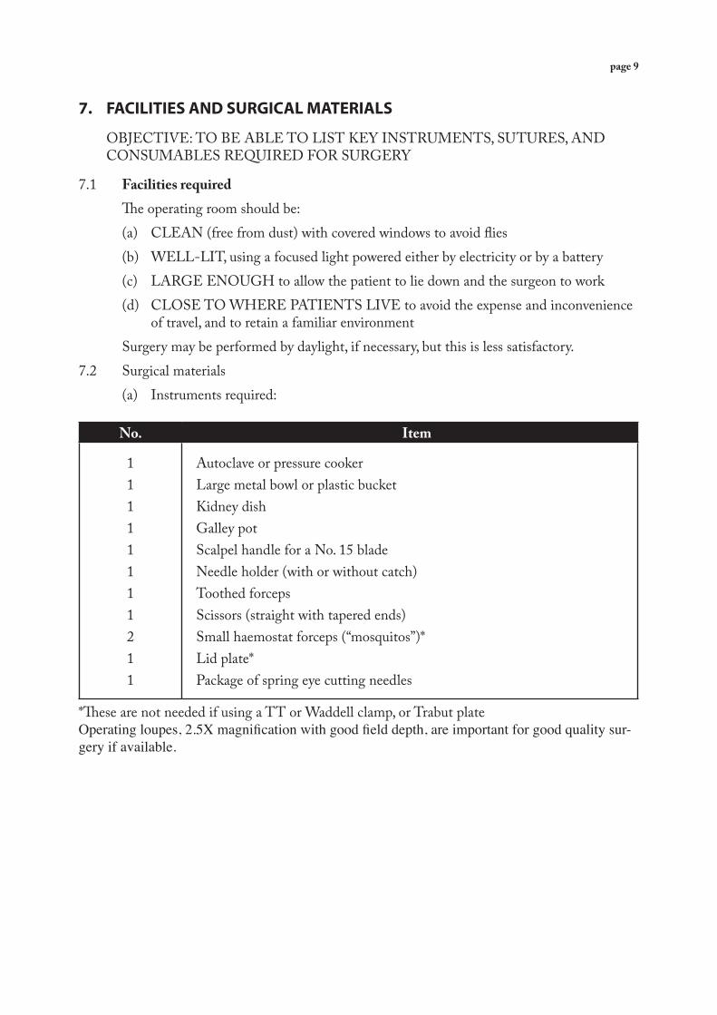
11. SURGICAL PROCEDURE
11 .1 Bilamellar tarsal rotation operation In the bilamellar tarsal rotation operation, a full thickness incision is made through the upper eyelid parallel to the eyelid margin . The portion of the eyelid containing the eyelashes is rotated outwards so that the eyelashes are no longer in contact with the cornea, and this position is secured with sutures . The operation is performed seated at the head of the patient (Fig . 4) . A sterile drape is placed over the face, with the eye visible through the central opening . The surgeon’s wrists should be steadied on the forehead during surgery . To help with the operation, an assistant (to hand the instruments) and a set of 2 .5X magnifying loupes (for better visibility) are useful but are not absolutely necessary .
Fig. 4. POSITION OF SURGEON AND PATIENT

page 19
11 .1 .1 Stabilizing the eyelid (Fig. 5a) (a) Place a haemostat at the nasal end of the upper eyelid, just lateral to the upper
lachrymal punctum, and close with just enough pressure to engage the first locking position . The tip of the haemostat should extend only 5 mm in from the eyelid margin .
(b) Place another haemostat at the temporal end of the upper eyelid, also extending no more than 5 mm in from the eyelid margin . If the haemostats extend much beyond 5 mm from the eyelid margin it will be difficult to evert the eyelid .
(c) The tips of both haemostats should be slightly angled in towards each other . (d) Confirm that the eyelid can be everted without difficulty . Do not force eversion or
the eyelid may tear . Reposition haemostats if eversion is not easy . (e) The haemostats should not be left closed on the eyelid for more than 15 minutes,
as they interrupt the blood flow to the eyelid and can cause eyelid necrosis and scarring .
(f ) If using the TT or Waddell clamp, haemostats are not used (Fig . 5b) . The TT clamp is placed so that the eyelid margin lines up with the groove on the plate and then secured . The Waddell clamp is placed so that the eyelid margin is up against the vertical piece on the clamp and then secured . The plate between the eyelid and the eye allows a full thickness incision to be made with either clamp .
Fig. 5a and b. EYELID FIXATION

page 20
11 .1 .2 Creating the incision (a) Incise the skin and muscle (Fig . 6a): (i) Hold the haemostats downwards so that the eyelid does not move . (ii) If using a separate eyelid plate, insert between the eyelid and the eye . Incise the
skin and muscle parallel to the eyelid margin and 3 mm ABOVE IT for the entire distance between the haemostats . The blade is held at right angles to the skin, and enters to a depth just superficial to the tarsal plate . REMEMBER THAT THE EYE IS BENEATH THE EYELID AND MUST NOT BE DAMAGED .
(b) If using the TT or Waddell clamp, the incision is made “full thickness” or through all layers down to the metal plate from one side of the clamp to the other (Fig . 6b) .
Fig. 6a and b. INCISION
page 21
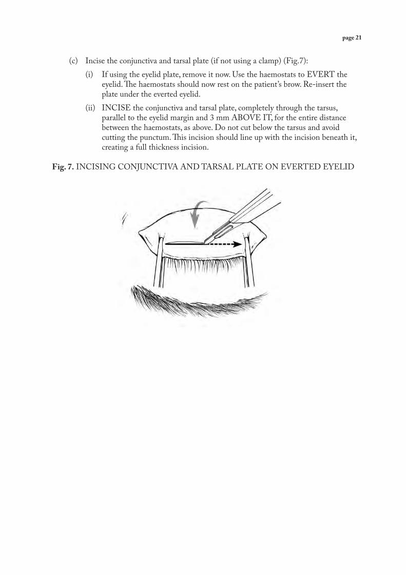
(c) Incise the conjunctiva and tarsal plate (if not using a clamp) (Fig .7): (i) If using the eyelid plate, remove it now . Use the haemostats to EVERT the
eyelid . The haemostats should now rest on the patient’s brow . Re-insert the plate under the everted eyelid .
(ii) INCISE the conjunctiva and tarsal plate, completely through the tarsus, parallel to the eyelid margin and 3 mm ABOVE IT, for the entire distance between the haemostats, as above . Do not cut below the tarsus and avoid cutting the punctum . This incision should line up with the incision beneath it, creating a full thickness incision .
Fig. 7. INCISING CONJUNCTIVA AND TARSAL PLATE ON EVERTED EYELID

page 22
(d) Unite the incisions (if not using a clamp) (Fig . 8): (i) Remove lid plate . Elevate the eyelid with the haemostats . (ii) With the eyelid still everted, insert the tips of the closed scissors into the
incision through the conjunctiva, tarsal plate, through remaining intact muscle, and out through the skin-muscle incision . DO NOT INSERT THE SCISSORS FROM THE SKIN SIDE of the incision . This can cause tearing of the tarsal plate if the incision was not full thickness .
(iii) Open the scissors across the eyelid: the blunt sides of the blades will separate apart intact muscle . Repeat along the incision if necessary until it is a full thickness incision . THIS STEP IS DONE ONLY TO ENSURE A FULL THICKNESS HOLE, NOT TO REPLACE A PROPER INCISION LENGTH .
Fig. 8. UNITING THE INCISIONS WITH SCISSORS
(e) Complete the incision medially and laterally: (i) Remove the haemostats or clamp (if desired, the clamp can be left on for
haemostasis during suture placement) . THE EYELID MAY BLEED PROFUSELY . PRESSURE WITH A SWAB FOR A MINUTE OR TWO WILL USUALLY CONTROL THE BLEEDING .
(i) Open the incision by grasping and elevating the skin of the eyelid just above the eyelashes, near where you intend to cut, with toothed forceps (Fig . 9) .
(ii) Using the scissors, completely divide the nasal and temporal edges of the tarsal plate (the portion formerly held in the haemostats), still cutting PARALLEL to the eyelid margin . Do not cut the punctum or beyond the edge of the tarsal plate nasally as the marginal artery may be cut and bleed . This step is NOT done to lenghten an improper first incision length .

page 23
Fig. 9. COMPLETING THE INCISION WITH SCISSORS
THE EYELID SHOULD NOW BE DIVIDED THROUGH ITS ENTIRE THICKNESS, 3 mm FROM AND PARALLEL TO THE EYELID MARGIN, REMAINING CONNECTED AT BOTH ENDS . ON AVERAGE, THE INCISION SHOULD BE 22mm IN LENGTH WHERE POSSIBLE .We shall refer to the 3 mm eyelid margin portion that contains the eyelashes as the EYELID MARGIN FRAGMENT, the remaining portion as the LARGER FRAGMENT (Fig . 10) .
Fig. 10. INCISED EYELID AND PARTS

page 24
11 .1 .3 Suturing the eyelid The purpose of the sutures is to re-attach the eyelid margin fragment in an outwardly rotated position, so that the eyelashes no longer rub on the cornea . This is achieved by anchoring the skin and muscle of the eyelid margin fragment near the lashes to the tarsus of the larger fragment, thus drawing the lash margin outwards and upwards . 4/0 silk is suitable for suturing, and absorbable sutures can also be used . The following description of suturing presumes use of a single armed needle . (a) Placing centre suture in the eyelid margin fragment (i) Look down at the SKIN SURFACE of the eyelid margin fragment . Mentally
divide the eyelid margin into five sections: three of those sections will be sutures and two will be the spaces between the sutures . The centre suture will be placed first . One suture will be placed on either side of the centre suture and will be spaced equidistant from the centre suture .
(ii) Prepare the needle holder: Mount the needle to point TOWARDS you . (iii) Grasp the skin of the eyelid margin fragment of the eyelid with a forceps . (iv) Starting just nasal from the centre of the fragment , pass the needle through
the skin about 1 mm ABOVE THE EYELASHES to emerge through the cut edge of the muscle layer IN THE FRONT OF (NOT THROUGH) THE TARSAL PLATE . Leave enough of the suture at the end to tie the knot (Fig . 11a) .
Fig. 11a. PLACEMENT OF THE CENTRE SUTURE
page 25
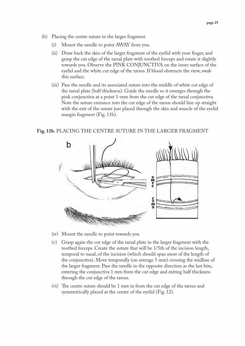
(b) Placing the centre suture in the larger fragment (i) Mount the needle to point AWAY from you . (ii) Draw back the skin of the larger fragment of the eyelid with your finger, and
grasp the cut edge of the tarsal plate with toothed forceps and rotate it slightly towards you . Observe the PINK CONJUNCTIVA on the inner surface of the eyelid and the white cut edge of the tarsus . If blood obstructs the view, swab this surface .
(iii) Pass the needle and its associated suture into the middle of white cut edge of the tarsal plate (half thickness) . Guide the needle so it emerges through the pink conjunctiva at a point 1-mm from the cut edge of the tarsal conjunctiva . Note the suture entrance into the cut edge of the tarsus should line up straight with the exit of the suture just placed through the skin and muscle of the eyelid margin fragment (Fig . 11b) .
Fig. 11b. PLACING THE CENTRE SUTURE IN THE LARGER FRAGMENT
(iv) Mount the needle to point towards you (v) Grasp again the cut edge of the tarsal plate in the larger fragment with the
toothed forceps . Create the suture that will be 1/5th of the incision length, temporal to nasal, of the incision (which should span most of the length of the conjunctiva) . Move temporally (on average 5 mm) crossing the midline of the larger fragment . Pass the needle in the opposite direction as the last bite, entering the conjunctiva 1 mm from the cut edge and exiting half thickness through the cut edge of the tarsus .
(vi) The centre suture should be 1 mm in from the cut edge of the tarsus and symmetrically placed at the centre of the eyelid (Fig . 12) .

page 26
Fig. 12. CONTINUATION OF CENTRE SUTURE
(c) Returning to the eyelid margin fragment to complete the centre suture (i) Mount the needle to point away from you . (ii) Grasp the skin of the eyelid margin fragment . (iii) Pass the needle through the muscle layer in front of the tarsal plate, to emerge
through the skin about 1 mm above the eyelashes . The entry point should correspond with the exit site of the suture in the larger fragment . THE TWO ARMS OF THE CENTRE SUTURE MUST BE PARALLELL TO EACH OTHER AND PERPENDICULAR TO THE EYELID MARGIN TO AVOID EYELID CONTOUR ABNORMALITIES (Fig . 13) .
(iv) Leave enough suture to tie a knot and cut the suture . These two ends will be tied later . Now proceed to do one of the side sutures exactly the same way .
Fig. 13. COMPLETION OF CENTRE SUTURE AND CUTTING SUTURE LINE
page 27
(d) Placing second (temporal) suture in eyelid fragment (i) Mount the needle to point towards you . Continue to hold the skin of the
eyelid margin fragment with forceps . (ii) Leave another 1/5th of the eyelid ( about 5 mm) between the temporal bite
of the centre suture and the first bite of the temporal suture . . Pass the needle through the skin about 1 mm ABOVE THE EYELASHES to emerge through the muscle layer IN THE FRONT OF (NOT THROUGH) THE TARSAL PLATE . Leave enough of the suture at the end to tie the knot . Return to the larger fragment .
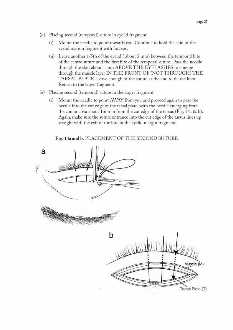
(e) Placing second (temporal) suture in the larger fragment (i) Mount the needle to point AWAY from you and proceed again to pass the
needle into the cut edge of the tarsal plate, with the needle emerging from the conjunctiva about 1mm in from the cut edge of the tarsus (Fig . 14a & b) . Again, make sure the suture entrance into the cut edge of the tarsus lines up straight with the exit of the bite in the eyelid margin fragment .
Fig. 14a and b. PLACEMENT OF THE SECOND SUTURE

page 28
(ii) Mount the needle to point towards you . Move temporally approximately 5 mm and you should be at the temporal end of the incision . Pass the needle through the conjunctiva 1 mm from the cut edge of the tarsus and exit half thickness through the cut edge of the tarsus . This second suture should be symmetric with the first suture and also 1 mm in from the cut edge of the tarsus . (f ) Returning to the eyelid margin fragment to complete the second suture (i) Finish the second suture by returning to the eyelid margin fragment . Mount
the needle to point away from you . Pass the needle through the muscle layer in front of the tarsal plate to emerge through the skin about 1 mm above the eyelashes and at the end of the incision (Fig . 15a & b) . Leave enough suture to tie a knot later and cut the suture . AGAIN, THE TWO ARMS OF THE TEMPORAL SUTURE MUST BE PARALLEL TO EACH OTHER AND TO THE CENTRE SUTURE, AND PERPENDICULAR TO THE EYELID MARGIN . THIS LINE UP OF SUTURES MUST BE EXACT TO AVOID EYELID CONTOUR ABNORMALITIES .
Fig. 15a and b. BOTH SUTURES IN PLACE

page 29
(g) Create the third (nasal) suture (i) Follow the directions for the second suture, only place the third suture on the
nasal side of the centre suture (Fig . 16a & b) . (ii) Leave another 1/5th of the eyelid (approximately 5 mm) between the nasal
bite of the centre suture and the first bite of the nasal suture . Pass the needle through the skin about 1 mm ABOVE THE EYELASHES to emerge through the muscle layer IN FRONT OF (NOT THROUGH) THE TARSAL PLATE . Leave enough of the suture at the end to tie the knot . Return to the larger fragment .
(iii) Mount the needle to point AWAY from you and proceed again to pass the needle into the cut edge of the tarsal plate, with the needle emerging from the conjunctiva about 1mm in from the cut edge . Again, make sure the suture entrance into the cut edge of the tarsus lines up straight with the exit of the first bite in the eyelid margin fragment .
Fig. 16a and b. PLACEMENT OF THIRD SUTURE
(iv) Mount the needle to point towards you . Move nasally approximately 5 mm (you should now be at the nasal end of the incision) . Create the third suture, which should be symmetric with the other sutures and also 1 mm in from the cut edge of the tarsus .
(v) Finish the Final suture by returning to the eyelid margin fragment . Mount the needle to point away from you . Pass the needle through the muscle layer in front of the tarsal plate to emerge through the skin about 1 mm above the eyelashes and at the end of the incision . AGAIN, THE TWO

page 30
ARMS OF THE NASAL SUTURE MUST BE PARALLEL TO EACH OTHER AND TO THE CENTRE AND TEMPORAL SUTURES AND PERPENDICULAR TO THE EYE LID MARGIN .
(vi) Leave enough suture to tie a knot at the end of suturing . Cut this final suture . The eyelid and suturing should now look like Figure 17 .
(vii) The sutures are now on the inside of the eyelid .
Fig. 17. THIRD SUTURE IN PLACE
(h) Tying the sutures (Fig . 18) (i) TIE THE CENTRAL SUTURE FIRST with a single throw . Then tie
the other two sutures in the same way . They should be tightened FIRMLY ENOUGH TO PRODUCE A SLIGHT OVERCORRECTION . Look at the eyelid margin from below to observe how the eyelid looks before further knotting (Fig . 19) .
Fig. 18. TYING SUTURES TO PRODUCE A SIGHT OVERCORRECTION

page 31
Fig. 19. EXAMPLES OF CORRECT EYELID SURGERY OUTCOMES
Immediate Post-op 6-Week Outcome

page 32
(ii) If the eyelid looks like those in Figure 20 (left side), either under corrected or over corrected, follow the instructions in the figure legend to adjust the tension and if necessary, remove and replace one or more sutures . If knots are too tight, there is a risk of eyelid necrosis .
Fig. 20. EXAMPLES OF EYELIDS WITH SURGICAL PROBLEMS
Immediate Post-op 6-Week Outcome
Problem: Over-rotation – cut edge of lower half of tarsus showingPossible Causes:Sutures are too tightIncision is too highSkin/muscle bites too close to the lashesTarsal bites too high
Problem: Under-rotation – lashes close to the eye nasallyPossible Causes:Sutures too looseIncomplete incision nasal sideSkin/muscle bites too close to the incisionTarsal bites too low
Result: Severe eyelid contour abnormalitySolution: Loosen sutures, if still present then replace the sutures with the skin/muscle bites and the tarsal bites closer to the incision
Result: Nasal recurrenceSolution: Tighten sutures, if still present then nasal incision on nasal side and replace sutures with skin/muscle bites and the tarsal bites farther from the incision

page 33
(iii) If the eyelid looks like the ones in Figure 19, with a uniform contour and a slight overcorrection along the entire eyelid, complete the knots with a single throw (a square knot) and cut the sutures 3 mm above the knot (Fig . 21) . This is long enough to permit ready removal, without being so long as to irritate the eye .
Fig. 21. SUTURES KNOTTED AND CUT
(i) Skin sutures: If the skin is not well approximated, two or three skin sutures can be placed by passing into the skin 1 mm from the cut edge, across the wound, and emerging from the skin again 1 mm from the other cut edge . They are tied without tension and cut .

page 34
11 .2 Trabut (images are shown for the right eye) In the Trabut operation the eyelid is fixed on the Trabut plate, incised through the conjunctiva and tarsal plate, parallel to the eyelid margin, and stopping at the orbicularis muscle . The muscle is dissected from the tarsal plate in both fragments, and the fragments are re-sutured so that the eyelid margin is rotated outwards and the eyelashes no longer touch the globe . An assistant (to hand the instruments) and a set of 2 .5X magnifying loupes (for better visibility) simplify the operation but are not absolutely necessary . The operation is performed seated at the head of the patient, as for BLTR (see Fig . 4) . A sterile drape is placed over the face, revealing the eye through the central opening .
11 .2 .1 Traction suture (a) A 4/0 silk suture with needle is used in conjunction with a Trabut plate to fix the
eyelid and keep it in everted position . Insert the needle three millimetres above the lashes through the skin and orbicularis of the upper eyelid and take approximately a 5mm bite horizontally starting from the temporal side .
(b) Leave a big loop and take a second five millimetre bite 2/3 of the way to the nasal side, coming out at the nasal side .
(c) The traction suture has two ends, temporally and nasally, with a loop of suture in the middle that covers the distance about 1/3 of the eyelid (Fig . 22) .
Fig. 22. PLACEMENT OF TRACTION SUTURES

page 35
11 .2 .2 Stabilize the upper eyelid on to the Trabut Plate (a) Hold the Trabut plate with the central tab facing towards you . (b) Pull the loop in the middle of the traction suture and hitch it onto the tab of the
Trabut plate (Fig . 23) .
Fig. 23. PLACING THE SUTURE OVER THE TRABUT PLATE
(c) Hold the Trabut plate on the eyelid with the tab facing away from you and continue to pull at the two suture ends until the Trabut plate is firmly in contact with the eyelid .

page 36
(d) Flip the plate towards you thereby everting the eyelid . The eyelid should should evert easily, if not rearrange the position of the Trabut plate and try again . Secure the suture around the tab (Fig . 24) .
Fig. 24. SECURING TRACTION SUTURES
(e) Fix the sutures with an artery forceps to the drape to secure the Trabut plate and the everted eyelid in position .

page 37
11 .2 .3 Incision (a) Holding the blade perpendicular to the conjunctiva, make a cut 3 mm from the
eyelid margin on the tarsal conjunctiva . Cut through the conjunctiva and the tarsal plate, but not through the muscle (Fig . 25) .
Fig. 25. INCISION THROUGH CONJUNCTIVA AND TARSAL PLATE
(b) Complete the cut with scissors, angling closer to the eyelid margin at the most temporal and nasal edge . DO NOT CUT THE PUNCTUM OR CUT THROUGH THE EYELID MARGIN (Fig . 26) .
Fig. 26. CUTTING WITH SCISSORS
(c) We shall call the fragment with the upper eyelashes the EYELID MARGIN FRAGMENT and the other fragment the LARGER FRAGMENT .

page 38
11 .2 .4 Dissection (a) Hold the cut edge of the eyelid margin fragment up, and using the blunt side of the
blade or scissors, gently dissect the orbicularis muscle away from the tarsal plate . Create a pocket between the orbicularis and the Tarsal plate about 2-3mm deep (Fig . 27) .
Fig. 27. DISSECTION OF EYELID FRAGMENT
Fig. 28a and b. DISSECTION OF THE LARGER FRAGMENT
(b) Once you have created the pocket, use the forceps to stabilize the cut edge of the larger fragment and dissect the orbicularis muscle away from the tarsal plate 5 mm . (Fig . 28a and b)

page 39
11 .2 .5 Sutures (a) Centre Suture (i) To start suturing, imagine the incision length of the larger fragment eyelid in
five sections, with three of them being the stitches and two of them being the space between the stitches .
(ii) Using the needle holder, mount the needle away from you . Use the forceps to pick up the eyelid margin fragment . Take the first suture bite, starting in the eyelid margin fragment about 1mm below the lashes on the skin side, through the skin and muscle to emerge in the pocket behind the tarsus, not through the tarsus (Fig . 29a, b & c) .
Fig. 29a, b and c. CENTRE SUTURE IN EYELID FRAGMENT

page 40
(iii) Grab the needle as it exits the pocket with the needle holder and continue straight down to the larger fragment . Grasp the cut edge of the larger fragment tarsal plate with the toothed forceps and rotate it slightly towards you (Fig . 30a) . Pass the needle into the white cut edge of the tarsal plate about in the middle (half thickness) . Gently guide the needle so it emerges from the tarsal plate through the conjunctiva at a point about 1 .5 mm from the cut edge (Fig 30b) .
(iv) Note the suture entrance into the cut edge of the tarsus should line up straight with the exit of the suture just placed through the eyelid margin fragment . Complete the first larger fragment suture by holding the tarsus with toothed forceps . Hold the needle so it is facing you and take a bite through the conjunctiva and in line with, but about 1/5th distance away from the suture exit, and pull the needle through the inside of the tarsus at half thickness . Gently guide the needle so it emerges through the cut edge .
Fig. 30a and b. CENTRE SUTURE IN LARGER FRAGMENT
(v) Keeping the needle straight, proceed to the eyelid margin fragment, and pass through at the bottom of the pocket behind the tarsus (not through the tarsus), about 1 .5mm from the cut edge, and exit at the eyelid margin below the lashes (Fig . 31a & b) . Pull the needle through and leaving enough suture to tie, cut the suture . The centre suture is complete .

page 41
Fig. 31a and b. COMPLETING THE CENTRE SUTURE
(b) Second Suture (i) Take another bite of the eyelid margin, below the lashes on the skin side, as in
the first suture, but at least 5 mm from the first suture . Proceed as described above for the first suture (Fig . 32a & b) .
Fig. 32a and b. STARTING AND COMPLETING THE SECOND SUTURE

page 42
(c) Third Suture (i) The third suture is done exactly as above only on the other side of the eyelid . (ii) At this point, there are six sutures exiting the eyelid fragment margin, equally
spaced (Fig . 33) .
Fig. 33. THIRD SUTURE

page 43
(d) Pulling the Sutures (i) Start to pull the sutures up towards the plate, which should draw the larger
fragment tarsus INTO the pocket of the eyelid margin fragment . Use the forceps or bottom of the blade handle to gently guide the larger fragment tarsus into the pocket .
Fig. 34a and b. PULLING SUTURES AND GUIDING FRAGMENT
(ii) Done correctly, the stitches should not be visible, and the line straight .

page 44
(e) Tie the sutures (i) TIE THE CENTRAL SUTURE with a square knot or 2 single throws .
Then tie the other two sutures in the same way . They should be tied FIRMLY ENOUGH TO PRODUCE A SLIGHT OVERCORRECTION . (Fig . 35) . The knots must not be so loose that the fragment will slip from the pocket . Cut the sutures 3mm above the knot .
Fig: 35. TYING KNOTS
(f ) Once the sutures are tied, remove the traction sutures and gently remove the Trabut plate . Place the eyelid in normal position (Fig . 36) .
(g) If the eyelid looks like Figure 19 (see in BLTR section), the surgery will likely be successful .
(h) If the eyelid looks like Figure 20 (see in BLTR section ) then adjust as suggested in the figures .
Fig. 36. SUTURES KNOTTED AND CUT

page 45
11 .3 Possible surgical difficulties
OBJECTIVE: TO BE ABLE TO DESCRIBE POSSIBLE SURGICAL DIFFICULTIES DURING SURGERY AND IMMEDIATELY AFTERWARDS, AND DESCRIBE WHAT TO DO
(a) Bleeding: If bleeding cannot be controlled by pressure with a gauze swab, the MARGINAL ARTERY, which runs along the eyelid margin, may have been severed . This usually occurs nasally, and blood will be seen springing from a single source . Locate this source, clip a haemostat onto it, and tie an absorbable suture just below the haemostat to close the artery . Otherwise, undersew the area with a suture . (b) Division of the eyelid margin: This is most unlikely with careful surgery but, should it occur, the cut portions of the eyelid margin fragment must be sutured together . Place one absorbable suture in the eyelid margin, so that its edges match exactly . Tie the suture without tension, with three single knots . Place one or two separate sutures on the outer surface of the tarsal plate . If the skin has also been divided, it may be sutured with one or two separate sutures . If the repair is satisfactory, proceed with the operation . If not, refer the patient to an ophthalmologist at once . (c) Overcorrection: If procedures have been followed carefully and the surgeon has looked at the eyelid before tying the knots, this should not be an issue . However, if the eyelid margin is grossly everted, remove the sutures and repeat the suturing . This time, tie the sutures with less tension to give the proper results, a mild degree of overcorrection . (d) Undercorrection: If the procedures have been followed carefully and the surgeon has looked at the eyelid before tying the knots this should not be an issue . If the eyelashes still touch the globe, remove the sutures and repeat the suturing . Tie the sutures with more tension to produce a mild degree of overcorrection,
PRACTICE: TRAINEES SHOULD CLOSE BOOKS AND LIST SURGICAL COMPLICATIONS AND SOLUTIONS

page 46
11 .4 Applying the antibiotic and dressing (a) Apply tetracycline ointment into conjunctival sac and onto the wound . (b) Pad the eye . A bandage may also be used . (c) Give a single dose of azithromycin, 1 gm dose, if available . Provide two 500-mg
tablets of acetaminophen (paracetamol) for pain . Inform the patient that pain may return after the injection wears off . The patient may take eight further tablets home, and take two every six hours if required .
(d) The patient is advised to rest quietly at home and to return after one day . If it is unlikely that the patient will return the following day, advise the patient to remove the pad after a day and to clean the wound with clean water and sterile gauze (supply the patient with sterile gauze) . The patient should return within 8-14 days for suture removal and/or a check of the wound .
11 .5 Safe disposal of sharps (a) In order to avoid accidents with used needles or blades, these must be properly
disposed of in designated sharps disposal containers .11 .6 Cleaning and resterilizing the instruments (a) After the operation has been performed, the instruments are cleaned with water and
detergent to remove any blood . (b) The clean instruments are then re-sterilized using steam or autoclave as described
previously .

page 47
12. POSTOPERATIVE CARE
Day 1: Check the wound (a) Remove the pad and clean the eye with gauze and saline . The eyelid may be swollen . (b) If oral azithromycin has not been given, apply tetracycline ointment between the
lower eyelid and the eyeball . Show the patient how this is done, so that he or she can apply ointment three times daily for seven days at home .
(c) If needed, provide a tablet of paracetamol .
Day 8-14: Remove the sutures (if absorbable sutures were used, the wound should still be checked)
(a) Clean the eye with gauze and saline . (b) Gently pull on knot with forceps . (c) Insert scissors or a blade under the knot so only ONE SIDE of the suture is cut .
DO NOT CUT BOTH SIDES OF THE LOOP because when you pull the knot away, half the suture will remain in the eyelid . Retained sutures are a major cause of infection and granulomas .
(d) Remove the skin sutures and the surgery sutures by gently pulling on the knotted end of the suture, as appropriate .
(e) Check for local infection: If pus is seen on the wound, remove any involved sutures and clean with gauze and
boiled water three times daily . (f ) Check for cellulitis: If there is pain, spreading redness, fever and raised pulse: GIVE ANTIBIOTICS,
for example AMPICILLIN, BY MOUTH AND REFER TO A DOCTOR URGENTLY . HOSPITAL ADMISSION MAY BE NEEDED .
(g) Check for eyelid closure defect: If the eyelids do not close properly when the patient tries to close them gently,
as if in sleep, or the cosmetic appearance is very distressing, REMOVE THE SUTURES AND MASSAGE THE UPPER EYELID DOWNWARDS . If this does not correct the problem, refer the patient to an ophthalmologist for a second operation to correct the excessive rotation . DEFECTIVE EYELID CLOSURE IS A SERIOUS CONDITION . Note this should have been corrected at close of surgery .

page 48
Six weeks to six months: (a) Granuloma formation: This looks like a red lump on the conjunctiva over the wound . It can be excised with
a scalpel or scissors after applying anaesthetic drops and everting the eyelid . Remove any remaining suture at the site .
(b) Necrosis of the eyelid margin: This is a defect in the eyelid margin, which is the result of poor blood supply caused
by too narrow a distal fragment . It will gradually heal without any treatment . The patient should be closely monitored for the possible development of an eyelid closure defect .
13. RESULTS
OBJECTIVE: TO LEARN HOW TO RECOGNIZE SUCCESSFUL AND NON-SUCCESSFUL OUTCOMES, AND HOW TO MANAGE ADVERSE OUTCOMES
Complete success is defined as NO EYELASHES RUBBING ON THE EYEBALL (in the absence of epilation or further surgery), WITH NO COMPLICATIONS by six months, and no severe eyelid contour abnormalities (see images Figure 20 for severe eyelid abnormalities) . Patient satisfaction with outcomes should also be assessed . The development of granuloma requires surgical excision for comfort as it can distort the eyelid and can cause chronic discharge . If a few inturning lashes at the medial or lateral edge of the eyelid persist despite surgery, they may not need to be managed by repeat surgery . Epilation is a possibility . If any lashes continue to rub on the cornea, if there is still sufficient trichiasis to cause severe discomfort, or if there is renewed corneal damage from persistent misdirected eyelashes that have been epilated, further surgery is required . REFER THE PATIENT TO AN OPHTHALMOLOGIST for further surgery .

page 49
Section TwoFor Trainers
14. INTRODUCTIONThis section is designed for the trainers of candidate TT surgeons, and covers selection and final assessment of the candidates . This section does not cover the logistics of setting up a training program . This is intended to be used by an experienced trichiasis surgeon trainer, preferably an ophthalmologist with theoretical background, to certify non-ophthalmic surgeons as competent to perform the BLTR or Trabut procedure on their own .
14 .1 Objectives. The objectives of the section are as follows: (a) identify good candidates for training (b) list and describe the knowledge that must be demonstrated and the procedures
that must be successfully completed before, during, and after, surgery in order for certification to be granted
(c) provide a checklist of the knowledge and procedures to assess during observation of the surgical process
(d) provide guidelines for scoring the checklist for purposes of certification
14 .2 Who Should Be Trained? Trainees are expected to be eye surgeons, physicians with surgical experience, eye care or surgical nurses, or eye care assistants . General medical assistants with some surgical background may be considered but may need more background in the anatomy and examination of the eye . Trainees should have: (a) previous experience with eye examination (b) experience in giving injections (c) knowledge of sterile surgical techniques (d) demonstrated ability in manual dexterity (stable hands and even stitches can be
demonstrated using a piece of thick material or an orange peel) (e) near vision of 20/20 corrected
14 .3 Expected training A minimum requirement is 22 eyes with TT for training, which will be needed for surgeries by the trainer and the transition to surgery by the trainee . There is also the assumption that the trainer or another surgeon has carried out TT surgery two weeks prior to the training program so that the trainees can practice removing sutures acceptably . During each section in this manual, there are objectives and practice sessions that can be held in a classroom setting the first day, with clinical days the next days . Below is a possible program of five days per trainee with a maximum of 6 trainees in any one session .

page 50
Day 1: Read the training booklet and carry out the exercises and practices sessions together .Day 2: The trainer and trainees examine patients, observe the trainer perform at least two eyes, and if deemed ready, the trainees assist in the operation of five more eyes with increasing responsibility . No more than 2 students per trainer should be observing at a time .Day 3 and 4: Trainees observe outcomes of the previous day and set up the entire operation for the day, from sterilization to closure, but under supervision . At least 10 eyes should be operated on Day 3 and Day 4, with a mix of right and left eyes . At least two eyes at the end should be done per trainee without the need for intervention . Trainees should also remove sutures from trainer’s cases done prior to the start of trainingIf at end of 10 eyes a trainee cannot perform surgery independently, then the trainer must inform the trainee that they cannot be certified and therefore cannot perform TT surgery . This is the most difficult step for many training program managers, but is ESSENTIAL from an ethical perspective . Trainees who cannot be independent or who fail final assessment must not be allowed to do surgery .Day 5: If the trainer feels the trainee is ready, Day 5 will be devoted to final assessment, where the trainer observes the trainees plan, discuss with the patient, and carry out surgery on 5 eyes without intervention and complete the certification checklist .In total, each trainee should have operated on at least 15 eyes, at least five independently as part of certification .

page 51
15. FINAL ASSESSMENT OF TT SURGEONS
15 .1 . Using this SectionThis section presumes that the original trainer is conducting the final certification . If, for some reason, the examiner is NOT the trainer, the examiner should begin by talking to the person who trained the trainees . In order to understand what the trainees have been taught, the examiner should discuss and review with the trainer the standard manual used for training, and observe the trainer performing two operations . It may be, for example, that the trainees were not taught the rationale behind placement of the haemostats, and it would be unfair to test them on information that they were not given . Understanding the material used by the trainer is key to the certification process . In addition, local practice must be taken into consideration . For example, while the use of loupes is highly recommended in the certification process, especially for older surgeons, it is not a requirement of this manual . The examiner can use knowledge of local practices to provide additional information or knowledge of procedures to the trainees during their first surgery .For certification, the examiner should observe each trainee carrying out five BLTR or Trabut procedures, i .e . surgery on five eyelids, with a mixture of right and left eyes . An initial surgery can be scheduled as a practice operation, during which the examiner can talk with the trainee, put him or her at ease, and provide additional information or demonstrate skills that the trainer may have omitted . The trainee must undertake the subsequent five procedures alone, without comment or intervention by the examiner (unless such intervention becomes necessary for the welfare of the patient) .
15 .2 . Qualifications for certificationIn order to become certified in the BLTR or Trabut surgical procedure, the trainee must have accomplished the following:• The trainees must have completed training in trichiasis surgery in a course of accepted
minimum depth and practical content (depending on national policy) and have done at least ten eyes independently .
• The trainee must have received a recommendation for certification from the instructor;• The trainee must have successfully performed five sequential trichiasis operations under
observation by the certification examiner, defined as fewer than 10 unsatisfactory marks on the checklist and none in the critical areas (those marked in yellow with a star,*) .
15 .3 . Knowledge and procedures to be assessedThis section focuses on pre surgery, then the BLTR surgery: Trabut assessment is described on page 62 and Cuenod nataf in appendix 1 . It includes a detailed description of each item on the checklist, and comprehensive guidelines for assessing the trainee . The knowledge base can be assessed at the time of the first operation, and need not be repeated for successive operations unless the examiner deems it necessary . All other assessments must be made at every operation . The items marked with a star (*) are critical and must be correctly performed in every case if the trainee is to achieve certification .

page 52
15 .4 . Before surgery1 . Assembly of materials before surgery. The trainee should demonstrate assembly of the
necessary materials and consumables, preparing and setting them up on a table before surgery . Such materials include the following, and others as locally appropriate:
Scalpel holder for blades Correct blade Needle holder Correct forceps 2 haemostats (if used), TT or Wadell clamp, or Trabut plate Scissors Correct needles Correct suture material (suture and needles may be combined) Syringe Needles Topical anaesthetic Anaesthetic for injection Skin preparation solution (e.g. povidine iodine) 1% TTC eye ointment or other postoperative antibiotic Single Dose of azithromycin Surgical gloves Sterile gauze Sterile drape/inner paper containing the sterile gloves Kidney dish or similar tray Galley pot Lid guard (if used)
2 . Knowledge of surgical material. The trainee should be able to identify each instrument or material and know what it is used for and why it is needed .

page 53
15 .5 . Sterilization of equipment before use1 . *Knowledge of sterile techniques. The examiner must ask questions on the definition
of sterile, why sterility is necessary, details of techniques for achieving sterility, and alternatives to use in the local setting if the usual technique is not available . For example, if the health centre is using an autoclave, the trainee must be able describe the use of the autoclave, the washing and cleaning of instruments before insertion, loading of the autoclave, the duration of autoclaving after a temperature of 121 °C is reached, and what to do if the autoclave is not working (e .g . pressure cooker sterilization of the instruments) .
2 . *Appropriate sterilization of all non-disposable instruments. The examiner should observe performance of the sterilization procedure and note whether sterility is achieved . This step can be combined with preparation above .
3 . *Maintenance of sterility of disposable items. The examiner should observe the use of sterile forceps to handle materials in order to maintain sterility .
15 .6 . Examination of the patient1 . Interacting with the patient. The examiner should observe the trainee interacting
appropriately with the patient and obtaining informed consent for the surgery (if this has not yet been done) . Locally appropriate customs for greeting should be observed before any examination is started or the patient is touched .
2 . Using a bright torch to examine the patient. Use of a bright light ensures that trichiasis is not missed . It is not easy to see a black lash touching the globe against a black pupil, and a bright light is essential .
3 . Looking at the eyelid from below to see whether there is trichiasis. The correct position must be used when assessing trichiasis . The patient should be in primary position (head level, with eyes looking straight ahead) and the examiner should be below the gaze to determine whether trichiasis is present .
4 . *Correctly identifying trichiasis. The examiner must certify that the trainee has correctly identified trichiasis, even if the condition is not so severe as to warrant surgery in the local setting .
5 . *Determining whether there is defective eyelid closure. The examiner must observe the trainee using proper examination technique and what would be done with the patient if a eyelid closure defect were found . In many settings, such patients must be referred to an ophthalmologist for appropriate surgery .
6 . Obtaining a relevant medical history from the patient, according to local practice. The trainee must confirm that the patient will be able to tolerate surgery; this should include ascertaining that the patient can lie flat on his or her back for 30 minutes, and whether the patient has other relevant problems, such as a blood disorder that may result in excessive bleeding, any condition that necessitates daily medication (ascertain what condition and what medication is being taken), shortness of breath, or heart problems .

page 54
7 . *Correct classification of the patient as a surgical patient for the trainee. The patient should have no other ocular condition that would complicate the surgery, such as eyelid closure defect or grossly infected eyelid, and must be fit to undergo surgery at the community level, under local anaesthesia .
15 .7 . Preoperative preparation1 . Explaining to the patient. The trainee explains to the patient what is going to happen . The
examiner should hear the trainee clearly state both the problem (e .g . eyelashes turning in) and the solution (corrective surgery) . The initial steps, such as injection of the local anaesthetic, should also be explained (e .g . the injection will cause some slight stinging, but the patient should feel no pain) . If the examiner does not speak the local language, this must be checked by another party .
2 . Use of loupes. The trainee should put on operating loupes . This may not be usual practice in some settings but is highly recommended .
3 . Administration of anaesthetic. The trainee administers the topical anaesthetic . The examiner should observe proper placement of the anaesthetic in the lower fornix, while the patient is looking up .
4 . Washing hands appropriately. The trainee should demonstrate proper surgical scrub technique, and the examiner will observe the duration and thoroughness of the scrub . The trainee should brush with soap and running water, and the sequence of brushing and use of disinfectant should be observed to ensure maximum aseptic conditions .
5 . *Use of sterile gloves to maintain sterility. The examiner should observe the trainee putting on surgical gloves and note whether his or her fingers, hands, or arms touch any part of the gloves that they should not touch .
6 . *Preparing patient’s face and eyelids. The examiner must observe the use of appropriate disinfectant, with care being taken to avoid too much entering the patient’s eyes . The technique of cleaning, a centrifugal pattern from the proximal side of the eyelid to the face, must be observed . If the trainee must return to clean the eyelid again, fresh gauze must be used to prevent any contaminant from the face being transferred to the eyelid area .
15 .8 . Injecting anaesthetic1 . Checking the label. The label on the bottle should be checked for the expiration date and
the name of the drug .2 . Maintaining sterility of the anaesthetic. The examiner observes that sterile techniques
are used to draw up the anaesthetic and, if the bottle is multi-dose, that sterility is maintained after the required amount has been drawn .
3 . Drawing up correct amount. No more than 5 ml of lidocaine per eyelid is needed, and the trainee should understand why this limit is important .

page 55
4 . *Re-checking that the correct eyelid is receiving the anaesthetic. This step is absolutely essential in a patient with a unilateral condition: with the surgeon at the head of the table, the affected eyelid will be on the opposite side relative to the original examination . If the trainee is in error, the examiner should halt the procedure and note performance as unsatisfactory .
5 . *Proper introduction of needle. The examiner must observe the proper procedure, with the needle being introduced temporal from the lateral canthus and 3 mm above the eyelid margin . Insertion should be in the plane of the upper eyelid, with care being taken to avoid poking the needle out through the eyelid or piercing the eyeball . Either adverse event must be immediately noted as unsatisfactory performance .
6 . *Proper injection of the anaesthetic. The needle should lie over the tarsal plate and in the plane of the eyelid . The trainee should inject the anaesthetic ahead of the sliding needle injecting the anaesthetic continuously .
7 . Ascertaining anaesthesia. The local anaesthetic, 2-3 ml with the first injection, should be massaged into the eyelid for about one minute using a swab and gentle finger pressure . After 3 minutes, the trainee should gently pinch the eyelid with forceps to ascertain whether the patient feels pain . If pain is felt, additional anaesthetic can be administered but no more than 5 ml should be given in total .
15 .9 . BLTR Operation1 . *Proper placement of haemostats/clamp. The examiner should observe proper placement
of the haemostats, and the trainee should be able to explain why correct placement is essential . Medial placement is critical to avoid damage to the punctum and the canaliculus . Placement should not be beyond 5 mm of the eyelid margin, in order to avoid tearing the eyelid during eversion .
2 . Proper placement of lid guard (eyelid plate), if used .3 . *Correct position, depth and extent of incision. The incision on the eyelid must be in the
correct position and of the correct depth, and must extend the correct distance across the eyelid:
The examiner should observe that the incision is parallel to the eyelid margin and about 3 mm above it . The incision should include only skin and muscle on the skin surface of the eyelid, just superficial to the tarsal plate . If the incision is too deep the eyeball is jeopardized .
4 . Proper eversion of the eyelid. There should be no tearing of the eyelid during eversion .5 . *Correct position, depth and extent of incision on the conjunctival surface. Again, the incision
should be 3 mm from the eyelid margin: it should meet the incision on the skin surface of the eyelid .
6 . Appropriate use of scissors to unite the incision. Scissors should be used to gently open the tissue . The examiner must ascertain that the scissors are used only to unite the incision and not to cut a substantial amount of tissue .
page 56
7 . Haemostats removed from the eyelid within 15 minutes. The trainee should understand the rationale for the time limit .
8 . *Satisfactory completion of incision. After using scissors, if necessary, to complete the incision, the examiner should use all the observations from the foregoing steps to assess the quality of the incision .
9 . *Knowledge of possible complications and their management. The trainee must show knowledge of at least the following three complications:
• Damage to the globe, either from improper injection or bad incision . Prevention using the lid guard or clamp is the best strategy . Damage could have catastrophic consequences; if it occurs, the eye should be patched and the patient referred immediately to an ophthalmologist .
• Excessive bleeding . If the wound is oozing, a compress may stop the bleeding . If bleeding persists and is spurting arterial blood, the marginal artery may have been cut; it should be clamped and a suture placed to stop the bleeding .
• Division of the eyelid margin . If this occurs, the cut portions must be sutured together appropriately before the operation proceeds further .
10 . Suturing • Correct mounting of needles for suture placement. The examiner must look for correct
placement of the needle on the needle holder . • Correct placement of sutures. The examiner looks for sutures having the correct depth
and bite in the tissues . • *Sutures correctly aligned on eyelid margin and larger fragments. Sutures should be
aligned to look straight, and avoid “gathering” of tissue; no suture should be more than 1 mm out of alignment .
11 . Pulling sutures • *Firm pulling of sutures. Sutures must be pulled and tied with sufficient firmness to
produce slight overcorrection, with eyelashes pointing away from the eyeball . The maximum correction can be about 3 mm if the incisions were correct .
• *Trainee must look at eyelid for under or overcorrection prior to tying the knots. Trainee should be familiar with steps to correct either adverse outcome prior to tying the knots .
• *Management of gross over- or under-correction. The trainee must know how to correct gross over- or under-correction . Over-correction must be rectified postoperatively by repeating the suturing, tying the sutures with less tension to reduce the over-correction as appropriate . Under-correction is corrected postoperatively by removing the original sutures and repeating the suturing, tying the sutures with greater tension to achieve slight over-correction .
• Appropriate skin sutures. The examiner looks for 1-mm bites, tied together gently .

page 57
15 .10 . Postoperative care1 . Provision of appropriate postoperative care. The examiner should observe the trainee
cleaning the area, applying ointment to the wound, patching the eye with the eyelids closed, placing adhesive tape diagonally across the patch (avoiding the mouth), and providing a single dose of oral azithromycin if available .
2 . Giving advice to the patient. The trainee should advise the patient on postoperative care, telling the patient to remove the patch the next day, wash the face and eye with soap and water to keep the wound clean, and apply the prescribed antibiotic ointment if no oral agithromycin was available . The patient should be advised to return for review and if necessary, suture removal after an appropriate interval . Finally, the trainee should describe some of the complications, including excessive bleeding and pain, and persistent post-operative swelling that indicates infection, and instruct the patient to return if these develop .
3 . *Knowledge of postoperative complications and their management. The trainee should discuss excessive bleeding and use of pressure to control it, as well as the possible need to reopen the wound . He or she should also discuss infection, the use of systemic antibiotics, signs of serious infection (cellulitis) and the need to refer the patient to a hospital if the problem does not resolve in 48-72 hours . The latter should be referred to an ophthalmologist . If granulomas occur, they can be removed .
15 .11 . Use of the checklistThe examiner should use the checklist for each trainee . All items should be scored as satisfactory or unsatisfactory for the first operation, but some of the questions on knowledge can be excluded for the subsequent operations . At the end of the five operations, the examiner calculates the total number of unsatisfactory marks for both starred and unstarred items . Any unsatisfactory mark for any starred item in any one operation is sufficient to deny certification and send the trainee for further training . A total of 10 unsatisfactory marks in the other, unstarred, items across the five operations is also sufficient to deny certification . A total of six to nine unsatisfactory scores should act as a warning: the examiner should discuss the problems with the trainee concerned and five additional operations should be performed with improving scores .

page 58
16. Checklist of procedures for certification of surgeon in Bilamellar Tarsal RotationExaminer: Please observe the trainee in all of the following procedures, and indicate whether each procedure is performed satisfactorily (tick “S”) or unsatisfactorily (tick “U”) . If the procedure is not performed, you must indicate “unsatisfactorily”, since none of these procedures can be omitted . Mark your observations at the end of each operation . At the end of all five operations, total the scores . Trainees MUST perform the procedures marked with a star (*) satisfactorily in order to be certified . Any unsatisfactory mark for any starred item in any one operation is sufficient to deny certification and send the trainee for further training . No unsatisfactory marks in the items with stars (*), and fewer than 10 unsatisfactory marks for all other unstarred items over all five operations, must be achieved for certification to be granted .A total of six to nine unsatisfactory scores in the unstarred items should act as a warning . The examiner should discuss the problems with the trainee and five additional operations should be performed with improving scores .

page 59
SURGEON’S NAME: ______________________________ DATE: _________________
EXAMINER’S NAME ______________________________
Bilamellar Tarsel Rotation
PROCEDURE
Lid 1OD/OS
Lid 2OD/OS
Lid 3OD/OS
Lid 4OD/OS
Lid 5OD/OS
S U S U S U S U S UAssembly of necessary materials before surgeryKnowledge of surgical materialsSterilization of equipment before use* Knowledge of sterile techniques* Appropriate sterilization by autoclave or pressure
cooker of all non-disposable instruments* Handling of sterilized instruments and items
(e .g . using sterile gloves, forceps, towels)Examination of the patientGreeted the patient appropriatelyUsed a bright torch to examine the lidLooked up at the lid from below to see trichiasis* Correctly identified trichiasis* Determined whether there was defective lid closureObtained a relevant medical history from the patient* Correctly classified patient as a surgical patientPreoperative preparationExplained to the patient what was wrong and what was going to happen during the procedureAdministered topical anaestheticWashed hands appropriately* Put on sterile gloves so as to maintain sterility* Prepared patient’s face and eyelids using skin
preparation solution (e .g . povidone iodine)Injecting anaestheticCheck the bottle labelAnaesthetic kept sterileCorrect amount of anaesthetic drawn up (e .g . not more than 5 ml of lidocaine)* Re-checked that correct eye was receiving anaesthetic* Needle inserted properly – never pointed
perpendicular to the eyelid skin

page 60
Bilamellar Tarsel Rotation
PROCEDURE
Lid 1 OD/OS
Lid 2 OD/OS
Lid 3 OD/OS
Lid 4 OD/OS
Lid 5 OD/OS
S U S U S U S U S U* Anaesthetic injected properly into eyelid (ahead of needle)Anaesthesia ascertained by checking patient’sresponse to painOperation* Proper placement of haemostats/clamp* Incision on eyelid correctly positioned and of correct depth
and extent; eyeball not piercedLid everted without tearing* Incision on conjunctiva and tarsal plate correctly
positioned and of correct depth and extentAppropriate use of scissors to unite incisionHaemostats not left on the lid for more than 15 minutes* Incision completed satisfactorily* Knowledge of possible operative complications and
their managementInformation on progress of surgery given to patient; ensured that patient was comfortable and well; reacted promptly to patient’s needsSuturingNeedles correctly mounted for suture placementSutures correctly placed (e .g . correct depth and bite in tissues)Sutures evenly spaced across the incision* Correct alignment of sutures on eyelid margin and larger
fragmentsPulling and knotting sutures
* Sutures pulled with sufficient firmness to produce slight over-correction, eyelashes pointed away from eye
* Review of suturing before tying knots* Knowledge of management of gross over- or
under- correctionSkin sutures appropriatePostoperative careAppropriate postoperative care given (e .g . dressings, ointment, azithromycin)Adequate postoperative advice given to the patient* Knowledge of postoperative complications and
their management
SCORE: No . of unsatisfactory* items ________ No . of other unsatisfactory items ________

page 61
Bilamellar Tarsel Rotation
COMMENTS BY EXAMINER:

page 62
Checklist of procedures for certification of surgeon in Trabut methodExaminer: Please review section 4 for clarification on all procedures that need to be observed . The difference between this section and section 4 is the use of the Trabut plate, incision depth, dissection, and suture placement .Observe the trainee in all the following procedures, and indicate whether the procedure is performed satisfactorily (tick “S”) or unsatisfactorily (tick “U”) . If the procedure is not performed, you must indicate “unsatisfactorily”, since none of these procedures can be omitted . Mark your observations at the end of each operation . At the end of all five operations, total the scores . Trainees MUST perform the procedures marked with a star (*) satisfactorily in order to be certified . Any unsatisfactory mark for any starred item in any one operation is sufficient to deny certification and send the trainee for further training . No unsatisfactory marks in the items with stars (*), and fewer than 10 unsatisfactory marks for all other items over all five operations must be achieved for certification to be granted .A total of six to nine unsatisfactory scores in the unstarred items should act as a warning . The examiner should discuss the problems with the trainee and five additional operations should be performed with improving scores .

page 63
SURGEON’S NAME: ______________________________ DATE: _________________
EXAMINER’S NAME: ______________________________
Trabut
PROCEDURE
Lid 1OD/OS
Lid 2OD/OS
Lid 3OD/OS
Lid 4OD/OS
Lid 5OD/OS
S U S U S U S U S UAssembly of necessary materials before surgeryKnowledge of surgical materialsSterilization of equipment before use* Knowledge of sterile techniques* Appropriate sterilization by autoclave or pressure
cooker of all non-disposable instruments* Handling of sterilized instruments and items
(e .g . using sterile gloves, forceps, towels)Examination of the patientGreeted the patient appropriatelyUsed a bright torch to examine the lidLooked up at the lid from below to see trichiasis* Correctly identified trichiasis* Determined whether there was defective lid closureObtained a relevant medical history from the patient* Correctly classified patient as a surgical patientPreoperative preparationExplained to the patient what was wrong and what was going to happen during the procedureAdministered topical anaestheticWashed hands appropriately* Put on sterile gloves so as to maintain sterility* Prepared patient’s face and eyelids using skin
preparation solution (e .g . povidone iodine)Injecting anaestheticCheck the bottle labelAnaesthetic kept sterileCorrect amount of anaesthetic was drawn up (e .g . not more than 5 ml of lidocaine)* Re-checked that correct eye was receiving anaesthetic* Needle inserted properly never pointed perpendicular
to eyelid skin

page 64
Trabut
PROCEDURE
Lid 1 OD/OS
Lid 2 OD/OS
Lid 3 OD/OS
Lid 4 OD/OS
Lid 5 OD/OS
S U S U S U S U S U* Anaesthetic injected properly (ahead of needle)Anaesthesia ascertained by checking patient’s response to painOperation* Proper placement of traction sutureProper eversion of the eyelid onto plate and stabilizationof lid* Incision on conjunctiva and tarsal plate correctly positioned
and not through muscleSatisfactory use of scissors for finishing cut* Dissection of muscle to achieve pockets* Knowledge of possible operative complications and their
managementInformation on progress of surgery given to patient and ensured that patient was comfortable and well; reacted promptly to patient’s needsSuturingNeedles correctly mounted for suture placementSutures correctly placed (e .g . correct depth and bite in tissues)* Sutures evenly spaced across the incision* Correct alignment of sutures on eyelid margin and
large fragments* Pulling in sutures, using forceps to guide larger fragment
tarsus into pocket . Tying sutures* Sutures pulled snugly so that the lid margin was everted
and eyelashes pointed away from eyeProper removal of Trabut plate* Knowledge of management of gross over- or
under- correctionPostoperative careAppropriate postoperative care given (e .g . dressings, ointment, azithromycin)Adequate postoperative advice given to the patient* Knowledge of postoperative complications and
their management
SCORE: No . of unsatisfactory* items ________ No . of other unsatisfactory items ________

page 65
Trabut
COMMENTS BY EXAMINER:

page 66
Appendix ICuenod Nataf methodWhile this document is intended for use in the certification of surgeons performing bilamellar tarsal rotation and trabut, it is recognized that other procedures are performed as well . This appendix briefly describes the Cuenod Nataf method, and provides a checklist for surgeons who are to be certified in the use of this method .Summary of method1 . Refer to this manual for basic issues prior to surgery and administration of local
anaesthetic .2 . Insert a lid plate firmly beneath the eyelid .3 . Grey line incision . Starting 1 mm laterally from the punctum, cut along the grey line as
far as the lateral margin of the lid . The split should be deep enough for the lash root to be visible .
4 . First lid incision . Make a horizontal incision through the skin and muscle at the upper tarsal border, from one end of the tarsus to the other, along the lid fold (2-5 mm above the lid margin) . The incision should go from directly above the punctum to above the lateral margin at the same height as the grey line split .
5 . Second lid incision . Determine the appropriate amount of skin to be removed from the lid . The goal is to remove enough skin for the loose folds to be flattened without stretching the skin . Make an arched incision through the skin and orbicularis muscle, joining the two ends of the incision with the ends of the previous incision . In young patients and recurrent cases, the second skin incision and skin removal may be unnecessary .
6 . Skin removal . Using skin forceps and scissors, remove the excess skin flap .7 . Expose the tarsus . Pick up the distal edge of the skin incision and dissect bluntly towards
the lid margin until the lash roots are seen, then upwards to the end of the tarsus to expose the whole tarsus .
8 . Tarsal incision, grooving and wedge removal . Using a scalpel, make an elliptical, angled (45°) incision on the tarsal plate 4 mm above the lid margin . Then make a second angled (45°) incision back to and 2-3 mm above the first incision, then remove the tarsal wedge . The length of each incision should be approximately the length of the tarsus . The depth of the wedge is 1-1 .5 mm, depending on the thickness of the tarsus . (In a conventional procedure, a triangular piece of the eyelid skin is removed at both the lateral cut ends of the skin incision to avoid puckering of the skin when sutured .)
9 . Suturing . Three to four equidistant mattress sutures are placed along the width of the lid . Beginning at the lid margin, through the skin orbicularis, go through the cut distal
tarsus, through the proximal tarsal fragment, and come out through the proximal tarsal fragment . Then go back through the tarsus at a distance of 3-4 mm from the first bite, driving the needle through the cut tarsal fragment and coming out through the skin orbicularis at the lid margin . Then, apply continuous skin sutures to close the skin wound

page 67
and cut the edge of the suture . Pull up the ends of the tarsal sutures . Each suture should be tightened by placing the blunt edge of a pair of forceps at the junction of the sutures and the lid margin and pulling to achieve adequate firmness .
10 . Haemostasis is maintained throughout the procedure . If sutures are not tied, do not trim suture ends but tape them to the patient’s forehead, ensuring that adequate pressure is applied to maintain positioning of the lid .
11 . Apply antibiotic ointment to the upper lid and lightly patch the wound and eye . Provide a single dose of azithromycin . After 24 hours, the correction is assessed . If correction is excessive, the knots are loosened; if it is less than desired, the knots are pulled tighter . The sutures are removed on the seventh day .
Equipment Operating loupe Scalpel holder for blades Correct blade Needle holder Correct forceps Scissors Correct needles Correct suture material (suture and needles may be combined) Syringe Needles Topical anaesthetic Skin preparation solution (e.g. povidone iodine) 1% TTC eye ointment or other postoperative antibiotic Azithromycin 1 gm oral dose Surgical gloves Sterile gauze Sterile drape/inner paper containing the sterile gloves Kidney dish (or similar tray) Galley pot Lid guard (if used)

page 68
Checklist of procedures for certification of surgeon in Cuenod Nataf methodExaminer: Please observe the trainee in all the following procedures, and indicate whether each procedure is performed satisfactorily (tick “S”) or unsatisfactorily (tick “U”) . If the procedure is not performed, you must indicate “unsatisfactorily”, since none of these procedures can be omitted . Mark your observations at the end of each operation . At the end of all five operations, total the scores . Trainees MUST perform the procedures marked with a star (*) satisfactorily in order to be certified . Any unsatisfactory mark for any starred item in any one operation is sufficient to deny certification and send the trainee for further training . No unsatisfactory marks in the items with stars (*) and fewer than 10 unsatisfactory marks for all five operations must be achieved for certification to be granted .A total of six to nine unsatisfactory scores in the unstarred items should act as a warning . The examiner should discuss the problems with the trainee and five additional operations should be performed with improving scores .

page 69
SURGEON’S NAME: ______________________________ DATE: _________________
EXAMINER’S NAME: ______________________________
Cuenod Nataf
PROCEDURE
Lid 1OD/OS
Lid 2OD/OS
Lid 3OD/OS
Lid 4OD/OS
Lid 5OD/OS
S U S U S U S U S UAssembly of necessary materials before surgeryKnowledge of surgical materialsSterilization of equipment before use* Knowledge of sterile techniques* Appropriate sterilization by autoclave or pressure
cooker of all non-disposable instruments* Handling of sterilized instruments and items
(e .g . using sterile gloves, forceps, towels)Examination of the patientGreeted the patient appropriatelyUsed a bright torch to examine the lidLooked up at the lid from below to see whether there was trichiasis* Correctly identified trichiasis* Determined whether there was defective lid closureObtained a relevant medical history from the patient* Correctly classified patient as a surgical patientPreoperative preparationExplained to the patient what was wrong and what was going to happen during the procedureAdministered topical anaestheticWashed hands appropriately* Put on sterile gloves so as to maintain sterility .* Prepared patient’s face and eyelids using skin prepara-
tion solution (e .g . povidone iodine)Injecting anaestheticCheck the bottle labelAnaesthetic kept sterileCorrect amount of anaesthetic drawn up (e .g . not more than 5 ml of lidocaine)* Re-checked that correct eye was receiving anaesthetic* Needle inserted properly

page 70
Cuenod Nataf
PROCEDURE
Lid 1 OD/OS
Lid 2 OD/OS
Lid 3 OD/OS
Lid 4 OD/OS
Lid 5 OD/OS
S U S U S U S U S U*Anaesthetic injected properly (ahead of needle)Anaesthesia ascertained by checking patient’s responseto painOperation* Lid plate properly placed* Proper incision along the grey line (appropriate placement,
depth and length)* Proper skin incisions, with appropriate judgement of
amount of excess skin to removeAppropriate dissection of skin orbicularis to expose tarsus* Adequate removal of tarsal wedge* Knowledge of possible operative complications and their
management (question asked on first surgery only)Reacted promptly to patient’s needs if necessarySuturingNeedles correctly mounted for suture placementSutures correctly placed(e .g . correct depth and bite in tissues)* Sutures evenly spaced across the incision* Correct alignment of sutures on proximal and
distal fragmentsTying sutures* Sutures pulled tightly, without tissue damage, so that the
lid margin is everted and eyelashes point away from the eye .
* Evaluation of correction before knotting sutures
* If sutures not tied, sutures taped to forehead with sufficient tension . (This can be done only in areas where patient can return to the clinic daily .)
* Knowledge of management of gross over- or under- correction
Postoperative careAppropriate postoperative care given (e .g . dressings, ointment, azithromycin )Adequate postoperative advice given to the patient* Knowledge of postoperative complications and
their management
SCORE: No . of unsatisfactory* items ________ No . of other unsatisfactory items ________

page 71
Cuenod Nataf
COMMENTS BY EXAMINER:

page 72
Appendix IIReferences1 . A controlled trial of surgery for trachomatous trichiasis of the upper lid .Reacher MH,
Muñoz B, Alghassany A, Daar AS, Elbualy M, Taylor HR .Arch Ophthalmol . 1992 May;110(5):667-74 .PMID: 1580842 .
2 . Results of community-based eyelid surgery for trichiasis due to trachoma .Bog H, Yorston D, Foster A .Br J Ophthalmol . 1993 Feb;77(2):81-3 .PMID: 8435423 .
3 . Surgery for trichiasis by ophthalmologists versus integrated eye care workers: a randomized trial .Alemayehu W, Melese M, Bejiga A, Worku A, Kebede W, Fantaye D .Ophthalmology . 2004 Mar;111(3):578-84 . PMID: 15019339 .
4 . Trachomatous trichiasis clamp vs standard bilamellar tarsal rotation instrumentation for trichiasis surgery: results of a randomized clinical trial . Gower EW, West SK, Harding JC, Cassard SD, Munoz BE, Othman MS, Kello AB, Merbs SL . JAMA Ophthalmol . 2013 Mar;131(3):294-301 .PMID: 23494035 .
5 . The trachomatous trichiasis clamp: a surgical instrument designed to improve bilamellar tarsal rotation procedure outcomes .Merbs SL, Kello AB, Gelema H, West SK, Gower EW .Arch Ophthalmol . 2012 Feb;130(2):220-3 .PMID: 22332216 .
6 . Single-dose azithromycin prevents trichiasis recurrence following surgery: randomized trial in Ethiopia . West SK, West ES, Alemayehu W, Melese M, Munoz B, Imeru A, Worku A, Gaydos C, Meinert CL, Quinn T .Arch Ophthalmol . 2006 Mar;124(3):309-14 .PMID: 16534049 .
7 . Final Assessment of Trichiasis Surgeons .Geneva: World Health Organisation, 2005 .8 . Trichiasis surgery for trachoma: the bilamellar tarsal rotation procedure . Reacher M,
Foster A, Huber J . Geneva: World Health Organization, 1993 .9 . Absorbable versus silk sutures for surgical treatment of trachomatous trichiasis in
Ethiopia: A randomised controlled trial Rajak SN, Habtamu E, Weiss HA, Kello AB, Gebre T, Genet A, Bailey RL, Mabey DC, Kaw TT, Gilbert CE, Emerson PM, Burton MJ . PLoS Med 2011; 8(12): e1001137 .PMID: 22180732


![Population-based prevalence survey of follicular trachoma ......inwards and scratch the cornea (trachomatous trichiasis, TT), leading to corneal opacity and blindness [2]. The World](https://static.fdocuments.us/doc/165x107/606ee2729bde5e77a30c4adf/population-based-prevalence-survey-of-follicular-trachoma-inwards-and-scratch.jpg)