Treatment effects of the edgewise Herbst appliance: A...
12
ORIGINAL ARTICLE Treatment effects of the edgewise Herbst appliance: A cephalometric and tomographic investigation Ryan VanLaecken, a Chris A. Martin, b Terry Dischinger, c Thomas Razmus, d and Peter Ngan e Watertown, SD, Morgantown, WV, and Lake Oswego, Ore Introduction: The crown Herbst appliance was introduced in the late 1980s because of shortcomings of the banded Herbst. In edgewise Herbst treatment, a fixed appliance is used with the crown Herbst to maximize the skeletal effects of treatment. Treatment response to the edgewise Herbst appliance has not been reported in the literature. Our objective was to investigate skeletal and dental changes in patients with Class II malocclusions treated with the edgewise Herbst appliance. Methods: Fifty-two consecutive patients were treated with the edgewise Herbst appliance; 32 (18 girls, 14 boys) met the criterion of 16 months out of Herbst treatment and were included in the study. Mean treatment time with this appliance was 8.0 1.8 months. Patients in the mixed dentition received additional treatment with 2 4 appliances until proper overbite, overjet, and torque on the incisors and permanent first molars were achieved. Patients in the permanent dentition were treated with full appliances to finalize the occlusion. Cephalometric measurements were taken at pretreatment, posttreatment, and 16 months after removal of the Herbst appliance, and the results were compared with 32 untreated Class II subjects from the Bolton Brush Study, matched for sex, age, and cephalometric dentofacial morphology. Data were analyzed with ANOVA, Tukey-Kramer multiple comparison tests, and 2-tailed t tests. Results: After 8 months of Herbst treatment, incisal relationship was overcorrected to an end-to-end incisal relationship and improved 8.4 mm, compared with the control group. The maxilla moved backward 1.4 mm at Point A, and the mandible moved forward 1.7 mm. The maxillary incisors moved lingually 1.7 mm, and the mandibular incisors were proclined 3.6 mm. The molars were corrected to a Class III relationship with a change of 7.2 mm compared with the control group. The mandible moved downward and forward. However, the condyle showed only 0.2 mm forward movement in the fossa. Sixteen months after appliance removal, the molars had relapsed into a Class I relationship, for a net change of 2.4 mm compared with the control group. Net overjet gain was 2.7 mm. Net restraint of maxillary growth was 1.3 mm, and net forward movement of the mandible was 1.0 mm. The maxillary incisors had no net movement, and the mandibular incisors had a net forward movement of 0.3 mm. Overall, skeletal change contributed 85% of the net overjet correction. Conclusions: Class II treatment with the edgewise Herbst appliance is accompanied by both skeletal and dental changes. The changes are stable, with significant skeletal differences remaining 16 months after appliance removal. The forward and downward movement of the mandible with minimal changes in the position of the condyles in the fossae suggests a combination of condylar growth and remodeling of the glenoid fossa with treatment. (Am J Orthod Dentofacial Orthop 2006;130:582-93) T he Herbst appliance was introduced in the early 1900s by Emil Herbst as a fixed bite-jumping device for Class II treatment. 1 Pancherz 2,3 rein- troduced the Herbst in the 1970s as a banded appliance and called attention to the possibilities of stimulating mandibular condylar growth. The crown Herbst was introduced in the late 1980s in response to breakage problems and shortcomings of the banded Herbst ap- pliance. 4-7 The crown Herbst is worn full time, unlike a removable functional appliance. The active treatment time is relatively short and requires little or no patient cooperation, and the appliance is streamlined, making hygiene simple. Research showed that the Herbst ap- pliance can correct Class II skeletal problems by a Private practice, Watertown, SD. b Assistant professor, Department of Orthodontics, School of Dentistry, West Virginia University, Morgantown, WV. c Private practice, Lake Oswego, Ore. d Professor and chair, Department of Diagnostic Sciences, School of Dentistry, West Virginia University, Morgantown, WV. e Professor and chair, Department of Orthodontics, School of Dentistry, West Virginia University, Morgantown, WV. Reprint requests to: Dr Peter Ngan, Department of Orthodontics, West Virginia University, School of Dentistry, 1076 Health Science Center, PO Box 9480, Morgantown, WV 26506; e-mail, [email protected]. Submitted, October 2004; revised and accepted, January 2005. 0889-5406/$32.00 Copyright © 2006 by the American Association of Orthodontists. doi:10.1016/j.ajodo.2005.01.030 582
Transcript of Treatment effects of the edgewise Herbst appliance: A...

ORIGINAL ARTICLE
Treatment effects of the edgewise Herbstappliance: A cephalometric andtomographic investigationRyan VanLaecken,a Chris A. Martin,b Terry Dischinger,c Thomas Razmus,d and Peter Ngane
Watertown, SD, Morgantown, WV, and Lake Oswego, Ore
Introduction: The crown Herbst appliance was introduced in the late 1980s because of shortcomings of thebanded Herbst. In edgewise Herbst treatment, a fixed appliance is used with the crown Herbst to maximizethe skeletal effects of treatment. Treatment response to the edgewise Herbst appliance has not beenreported in the literature. Our objective was to investigate skeletal and dental changes in patients with Class IImalocclusions treated with the edgewise Herbst appliance. Methods: Fifty-two consecutive patients weretreated with the edgewise Herbst appliance; 32 (18 girls, 14 boys) met the criterion of 16 months out of Herbsttreatment and were included in the study. Mean treatment time with this appliance was 8.0 � 1.8 months.Patients in the mixed dentition received additional treatment with 2 � 4 appliances until proper overbite,overjet, and torque on the incisors and permanent first molars were achieved. Patients in the permanentdentition were treated with full appliances to finalize the occlusion. Cephalometric measurements were takenat pretreatment, posttreatment, and 16 months after removal of the Herbst appliance, and the results werecompared with 32 untreated Class II subjects from the Bolton Brush Study, matched for sex, age, andcephalometric dentofacial morphology. Data were analyzed with ANOVA, Tukey-Kramer multiple comparisontests, and 2-tailed t tests. Results: After 8 months of Herbst treatment, incisal relationship wasovercorrected to an end-to-end incisal relationship and improved 8.4 mm, compared with the controlgroup. The maxilla moved backward 1.4 mm at Point A, and the mandible moved forward 1.7 mm. Themaxillary incisors moved lingually 1.7 mm, and the mandibular incisors were proclined 3.6 mm. Themolars were corrected to a Class III relationship with a change of 7.2 mm compared with the control group.The mandible moved downward and forward. However, the condyle showed only 0.2 mm forward movementin the fossa. Sixteen months after appliance removal, the molars had relapsed into a Class I relationship, fora net change of 2.4 mm compared with the control group. Net overjet gain was 2.7 mm. Net restraint ofmaxillary growth was 1.3 mm, and net forward movement of the mandible was 1.0 mm. The maxillary incisorshad no net movement, and the mandibular incisors had a net forward movement of 0.3 mm. Overall, skeletalchange contributed 85% of the net overjet correction. Conclusions: Class II treatment with the edgewiseHerbst appliance is accompanied by both skeletal and dental changes. The changes are stable, withsignificant skeletal differences remaining 16 months after appliance removal. The forward and downwardmovement of the mandible with minimal changes in the position of the condyles in the fossae suggests acombination of condylar growth and remodeling of the glenoid fossa with treatment. (Am J Orthod
Dentofacial Orthop 2006;130:582-93)aPrivate practice, Watertown, SD.bAssistant professor, Department of Orthodontics, School of Dentistry, WestVirginia University, Morgantown, WV.cPrivate practice, Lake Oswego, Ore.dProfessor and chair, Department of Diagnostic Sciences, School of Dentistry,West Virginia University, Morgantown, WV.eProfessor and chair, Department of Orthodontics, School of Dentistry, WestVirginia University, Morgantown, WV.Reprint requests to: Dr Peter Ngan, Department of Orthodontics, West VirginiaUniversity, School of Dentistry, 1076 Health Science Center, PO Box 9480,Morgantown, WV 26506; e-mail, [email protected], October 2004; revised and accepted, January 2005.0889-5406/$32.00Copyright © 2006 by the American Association of Orthodontists.
doi:10.1016/j.ajodo.2005.01.030582
The Herbst appliance was introduced in the early1900s by Emil Herbst as a fixed bite-jumpingdevice for Class II treatment.1 Pancherz2,3 rein-
troduced the Herbst in the 1970s as a banded applianceand called attention to the possibilities of stimulatingmandibular condylar growth. The crown Herbst wasintroduced in the late 1980s in response to breakageproblems and shortcomings of the banded Herbst ap-pliance.4-7 The crown Herbst is worn full time, unlike aremovable functional appliance. The active treatmenttime is relatively short and requires little or no patientcooperation, and the appliance is streamlined, makinghygiene simple. Research showed that the Herbst ap-
pliance can correct Class II skeletal problems by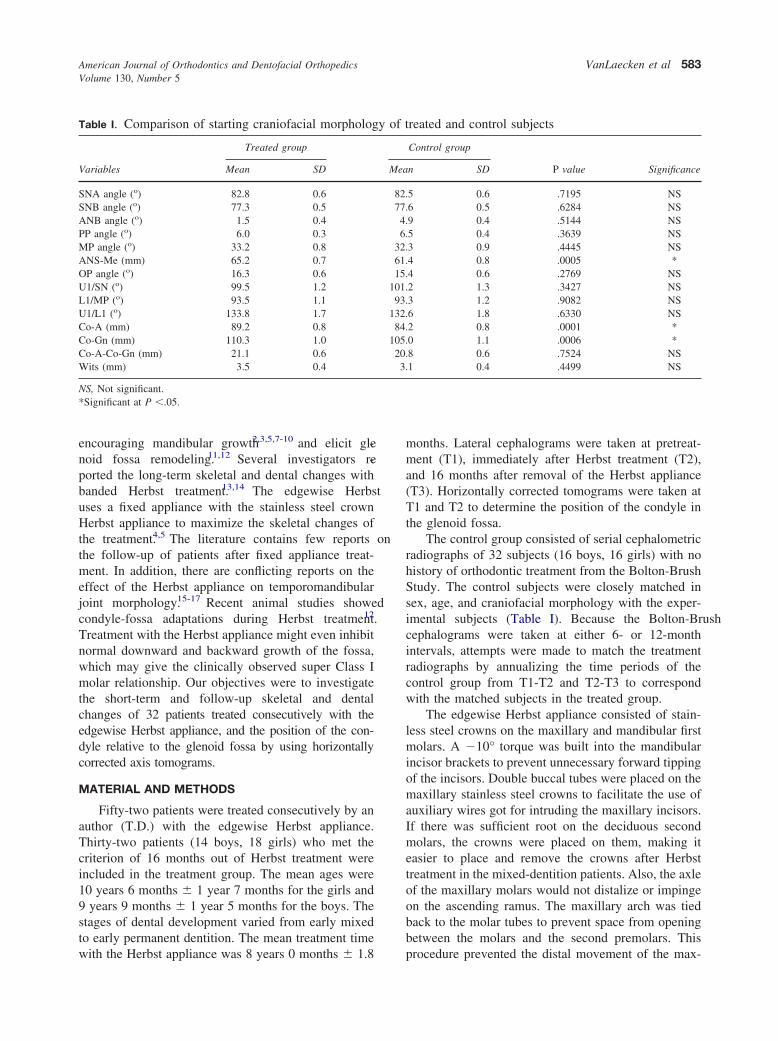
American Journal of Orthodontics and Dentofacial OrthopedicsVolume 130, Number 5
VanLaecken et al 583
encouraging mandibular growth2,3,5,7-10 and elicit gle-noid fossa remodeling.11,12 Several investigators re-ported the long-term skeletal and dental changes withbanded Herbst treatment.13,14 The edgewise Herbstuses a fixed appliance with the stainless steel crownHerbst appliance to maximize the skeletal changes ofthe treatment.4,5 The literature contains few reports onthe follow-up of patients after fixed appliance treat-ment. In addition, there are conflicting reports on theeffect of the Herbst appliance on temporomandibularjoint morphology.15-17 Recent animal studies showedcondyle-fossa adaptations during Herbst treatment.12
Treatment with the Herbst appliance might even inhibitnormal downward and backward growth of the fossa,which may give the clinically observed super Class Imolar relationship. Our objectives were to investigatethe short-term and follow-up skeletal and dentalchanges of 32 patients treated consecutively with theedgewise Herbst appliance, and the position of the con-dyle relative to the glenoid fossa by using horizontallycorrected axis tomograms.
MATERIAL AND METHODS
Fifty-two patients were treated consecutively by anauthor (T.D.) with the edgewise Herbst appliance.Thirty-two patients (14 boys, 18 girls) who met thecriterion of 16 months out of Herbst treatment wereincluded in the treatment group. The mean ages were10 years 6 months � 1 year 7 months for the girls and9 years 9 months � 1 year 5 months for the boys. Thestages of dental development varied from early mixedto early permanent dentition. The mean treatment time
Table I. Comparison of starting craniofacial morpholog
Variables
Treated group
Mean SD
SNA angle (o) 82.8 0.6SNB angle (o) 77.3 0.5ANB angle (o) 1.5 0.4PP angle (o) 6.0 0.3MP angle (o) 33.2 0.8ANS-Me (mm) 65.2 0.7OP angle (o) 16.3 0.6U1/SN (o) 99.5 1.2L1/MP (o) 93.5 1.1U1/L1 (o) 133.8 1.7Co-A (mm) 89.2 0.8Co-Gn (mm) 110.3 1.0Co-A-Co-Gn (mm) 21.1 0.6Wits (mm) 3.5 0.4
NS, Not significant.*Significant at P �.05.
with the Herbst appliance was 8 years 0 months � 1.8
months. Lateral cephalograms were taken at pretreat-ment (T1), immediately after Herbst treatment (T2),and 16 months after removal of the Herbst appliance(T3). Horizontally corrected tomograms were taken atT1 and T2 to determine the position of the condyle inthe glenoid fossa.
The control group consisted of serial cephalometricradiographs of 32 subjects (16 boys, 16 girls) with nohistory of orthodontic treatment from the Bolton-BrushStudy. The control subjects were closely matched insex, age, and craniofacial morphology with the exper-imental subjects (Table I). Because the Bolton-Brushcephalograms were taken at either 6- or 12-monthintervals, attempts were made to match the treatmentradiographs by annualizing the time periods of thecontrol group from T1-T2 and T2-T3 to correspondwith the matched subjects in the treated group.
The edgewise Herbst appliance consisted of stain-less steel crowns on the maxillary and mandibular firstmolars. A �10° torque was built into the mandibularincisor brackets to prevent unnecessary forward tippingof the incisors. Double buccal tubes were placed on themaxillary stainless steel crowns to facilitate the use ofauxiliary wires got for intruding the maxillary incisors.If there was sufficient root on the deciduous secondmolars, the crowns were placed on them, making iteasier to place and remove the crowns after Herbsttreatment in the mixed-dentition patients. Also, the axleof the maxillary molars would not distalize or impingeon the ascending ramus. The maxillary arch was tiedback to the molar tubes to prevent space from openingbetween the molars and the second premolars. This
reated and control subjects
Control group
P value Significancen SD
5 0.6 .7195 NS6 0.5 .6284 NS9 0.4 .5144 NS5 0.4 .3639 NS3 0.9 .4445 NS4 0.8 .0005 *4 0.6 .2769 NS2 1.3 .3427 NS3 1.2 .9082 NS6 1.8 .6330 NS2 0.8 .0001 *0 1.1 .0006 *8 0.6 .7524 NS1 0.4 .4499 NS
y of t
Mea
82.77.
4.6.
32.61.15.
101.93.
132.84.
105.20.3.
procedure prevented the distal movement of the max-

American Journal of Orthodontics and Dentofacial OrthopedicsNovember 2006
584 VanLaecken et al
illary molars and the subsequent distalization of themandible and seating of the condyle, thus decreasingthe orthopedic effect because the condyle was notunloaded in the joint to allow the maximum orthopediceffect. A 2-mm half-round Remenium cantilever wasplaced between the first molar and the interproximalarea of the canine and the first premolar in the mandib-ular arch. An axle was placed at the mesial end of thecantilever, and a .022 � 028-in archwire tube wasplaced below the axle. Most Class II subjects had deepbites, and the archwire tubes were positioned below theaxle. Positioning the archwire tube below the axle wasimportant in controlling tooth movement and levelingthe mandibular arch because of the reciprocal force ofintrusion of the mandibular incisors that resulted in atip-back force to the mandibular first molar due to thecantilever construction. This tip-back controlled themovement of the mandibular molar. If the patient hadan open-bite tendency, the archwire tube was placedabove the axle to aid in the eruption of the mandibularincisors and the closing of the open bite. A transpalatalarch was not incorporated in the appliance to allow themaxillary first molars to rotate as the Class II relation-ship was corrected. A mandibular lingual arch was notpart of the appliance to allow easier placement of theappliance and ease of recementation of the crown if itloosens. However, a stop off the crown on the mandib-ular molar in the permanent dentition to the secondpremolar and in the mixed dentition to the deciduousfirst molar was placed and bonded to the teeth toprevent tipping of the cantilever arm or rotation of themolar lingually. If the second premolar was not presentor the deciduous first molar would be lost duringmixed-dentition treatment, a lingual arch was placeduntil the brackets could be placed with an edgewisearchwire to support the cantilever arm. The mandible
Fig 1. Lateral cephalograms of
was advanced initially to an end-to-end position and
reactivated 3 mm every 10 weeks until an overcorrectedClass III canine relationship was achieved with themaxillary canines in an end-to-end relationship with themandibular first premolar. In more severe cases, themaxillary canine was in a full-tooth Class III inrelationship to the mandibular first premolar and theincisors in an anterior crossbite.
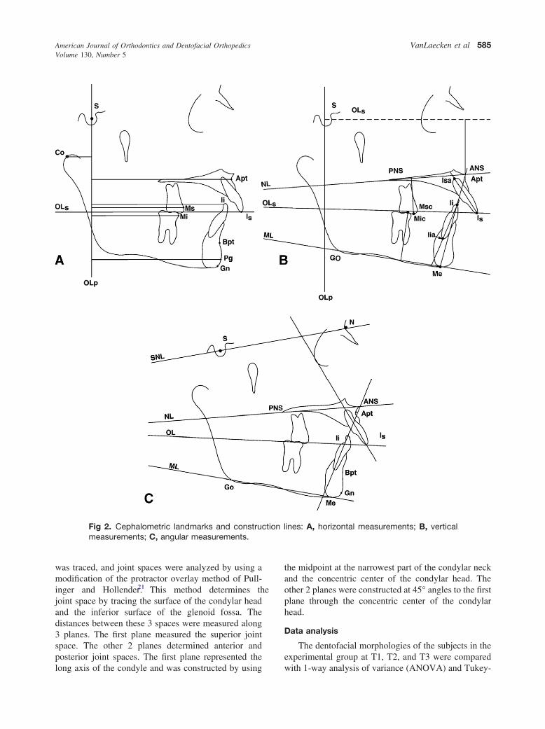
Cephalometric analysis
Figure 1 shows the lateral cephalometric radio-graphs of a typical subject taken at T1, T2, and T3. Thecephalometric systems described by Pancherz18,19 wereused to analyze the treatment and posttreatmentchanges. The landmarks used are shown in Figure 2.The magnification factor of the lateral cephalogramswas found to be similar for the treated and controlgroups (6% for both). Therefore, no standardizationwas needed. Registration of the lateral cephalogramswas performed on a 0.003-in matte cephalometricacetate tracing film. For all cephalometric landmarkswith right and left images, the midpoint bisecting the 2images was used. The measurement for each variablewas made with a cephalometric protractor or an elec-tronic caliper. Angular measurements were evaluated tothe nearest 0.5° by using a cephalometric protractor.Sagittal and vertical measurements were evaluated tothe nearest 0.1 mm. Analysis of the sagittal, skeletal,and dental changes were recorded along the occlusalplane (OLs) and to the occlusal plane perpendicular(OLp) from the first cephalogram; this formed thereference grid. The grid was then transferred to subse-quent cephalograms by superimposing the tracings onthe midsagittal cranial structure.
Figure 3 shows the pretreatment and posttreatmenttomograms of a typical subject. The tomographic mea-surements were determined as described by Croft
al subject: A, T1; B, T2; C, T3.
typicet al20 (Table II, Fig 4). Each tomogram (right and left)
American Journal of Orthodontics and Dentofacial OrthopedicsVolume 130, Number 5
VanLaecken et al 585
was traced, and joint spaces were analyzed by using amodification of the protractor overlay method of Pull-inger and Hollender.21 This method determines thejoint space by tracing the surface of the condylar headand the inferior surface of the glenoid fossa. Thedistances between these 3 spaces were measured along3 planes. The first plane measured the superior jointspace. The other 2 planes determined anterior andposterior joint spaces. The first plane represented the
Fig 2. Cephalometric landmarks and construmeasurements; C, angular measurements.
long axis of the condyle and was constructed by using
the midpoint at the narrowest part of the condylar neckand the concentric center of the condylar head. Theother 2 planes were constructed at 45° angles to the firstplane through the concentric center of the condylarhead.
Data analysis
The dentofacial morphologies of the subjects in theexperimental group at T1, T2, and T3 were compared
ines: A, horizontal measurements; B, vertical
ction lwith 1-way analysis of variance (ANOVA) and Tukey-

Po
American Journal of Orthodontics and Dentofacial OrthopedicsNovember 2006
586 VanLaecken et al
Kramer multiple comparison tests. The starting formsof the treated and control samples were compared witha 2-tailed t test. The skeletal and dental changesbetween the treated and control samples at the varioustimes were compared with a 2-tailed t test. The tomo-grams were compared with the Student t test to deter-mine whether there was a change in condylar positionfrom T1 to T2.
Error measurements
The errors in locating, superimposing, and measur-ing the changes of the landmarks by 1 examiner weremeasured on the cephalograms of 10 subjects. Allcephalograms were measured twice with 3 weeks
Fig 3. Tomograms: A
Table II. Tomographic measurements of variables
Name Symbol
Right posterior joint space RPRight superior joint space RSRight anterior joint space RALeft posterior joint space LPLeft superior joint space LSLeft anterior joint space LA
Fig 4. Landmarks and construction lines for tomogrammeasurements.
between sessions. For all the cephalometric variables,
differences between the independent repeated measure-ments of each subject at T1, T2, and T3 and superim-position errors were calculated. The error of measure-ment and location of anatomical landmarks werecalculated by the Dahlberg formula22
ME �� d2
2h
where d is the difference between 2 registrations of apair and h is the number of double registrations. Thegreatest mean error for all linear measurements did notexceed 0.7 mm. The greatest mean error for angularmeasurements did not exceed 1.5°. In the combinederrors in tracing and superimposition of landmarks atT2-T1, T3-T2, and T3-T1, the greatest mean error didnot exceed 0.6 mm. The error in tomographic measure-ments at T1 and T2 did not exceed 0.2 mm.
RESULTSSagittal changes
Changes in subjects in the treatment and controlgroups from T2-T1, T3-T2, and T3-T1 are compared inTables III through V. Compared with the control group,treatment induced backward movement of the maxil-lary base (OLp-A pt) from T1 to T2 (1.4 mm) andforward movement (0.1 mm) during the follow-up pe-riod (T2-T3), with a net restraint of forward growth of1.3 mm (T1-T3). The position of the condyle (OLp-Co)was found to move forward 0.1 mm with treatment, but
ght; B, T1 left; C, T2.
Definition
sition of right condyle in relation to superior wall of the eminencesition of right condyle in relation to anterior wall of eminencesition of right condyle in relation to the posterior wall of eminencesition of left condyle in relation to posterior wall of eminencesition of left condyle in relation to superior wall of eminencesition of left condyle in relation to anterior wall of eminence
, T1 ri
PoPoPoPoPo
there was no change during the follow-up period, with
American Journal of Orthodontics and Dentofacial OrthopedicsVolume 130, Number 5
VanLaecken et al 587
a net movement of 0.1 mm. The effective maxillarylength (Co-A pt) was restrained 1.9 mm compared withthe control group during treatment; there was an in-crease of 0.3 mm during the follow-up period, for a netrestraint of 1.6 mm. Effective mandibular length (Co-Gn) increased 1.9 mm during treatment and decreased0.9 mm during follow-up, for a net increase of 1.0 mmcompared with the control group. The position of themaxilla relative to the mandible (Wits) decreased 6.9mm during treatment and increased 2.8 mm duringobservation, for a net decrease of 4.1 mm. For theangular measurements, the position of the maxilla rela-tive to the cranial base (SNA) had a decrease duringtreatment (1.3°) compared with the control group and anincrease of 0.2° during the follow-up period, for a netdecrease of 1.1°. The treatment induced forward move-ment of the mandible (1.5°) relative to the cranial base
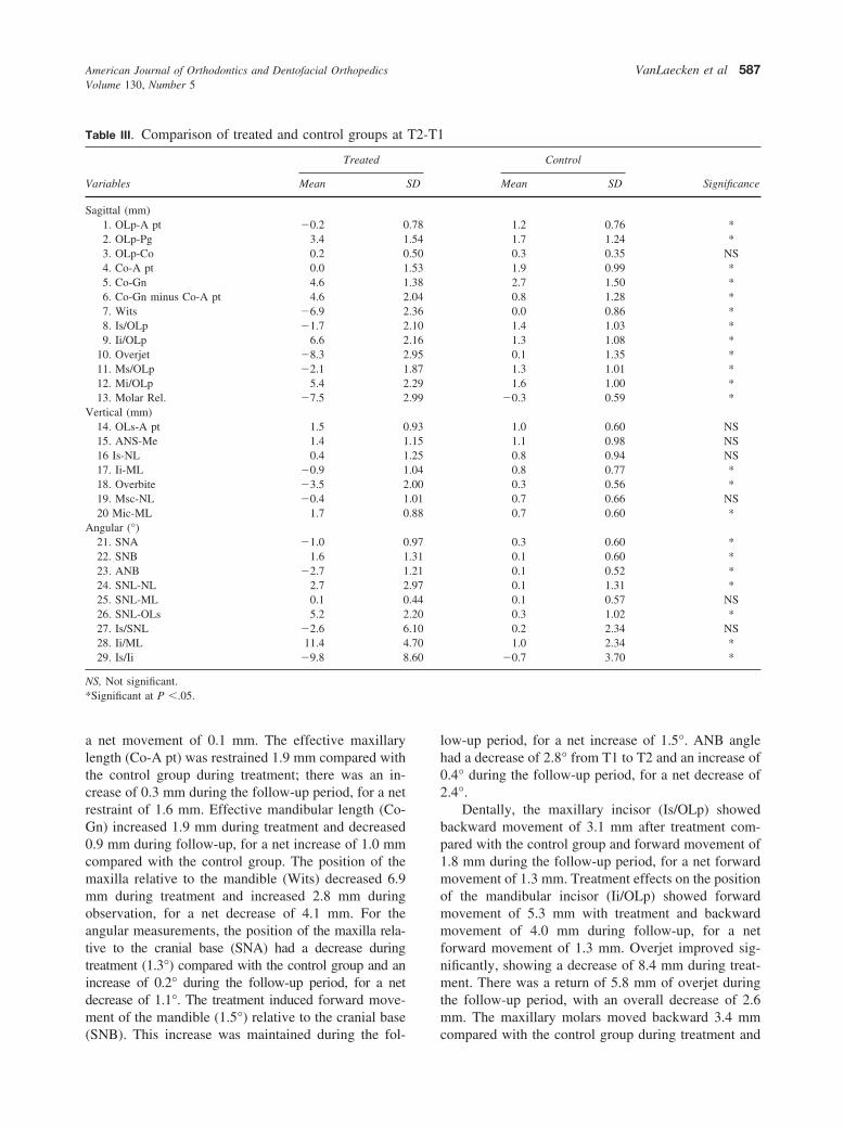
Table III. Comparison of treated and control groups at
Variables
Treated
Mean
Sagittal (mm)1. OLp-A pt �0.2 02. OLp-Pg 3.4 13. OLp-Co 0.2 04. Co-A pt 0.0 15. Co-Gn 4.6 16. Co-Gn minus Co-A pt 4.6 27. Wits �6.9 28. Is/OLp �1.7 29. Ii/OLp 6.6 2
10. Overjet �8.3 211. Ms/OLp �2.1 112. Mi/OLp 5.4 213. Molar Rel. �7.5 2
Vertical (mm)14. OLs-A pt 1.5 015. ANS-Me 1.4 116 Is-NL 0.4 117. Ii-ML �0.9 118. Overbite �3.5 219. Msc-NL �0.4 120 Mic-ML 1.7 0
Angular (°)21. SNA �1.0 022. SNB 1.6 123. ANB �2.7 124. SNL-NL 2.7 225. SNL-ML 0.1 026. SNL-OLs 5.2 227. Is/SNL �2.6 628. Ii/ML 11.4 429. Is/Ii �9.8 8
NS, Not significant.*Significant at P �.05.
(SNB). This increase was maintained during the fol-
low-up period, for a net increase of 1.5°. ANB anglehad a decrease of 2.8° from T1 to T2 and an increase of0.4° during the follow-up period, for a net decrease of2.4°.
Dentally, the maxillary incisor (Is/OLp) showedbackward movement of 3.1 mm after treatment com-pared with the control group and forward movement of1.8 mm during the follow-up period, for a net forwardmovement of 1.3 mm. Treatment effects on the positionof the mandibular incisor (Ii/OLp) showed forwardmovement of 5.3 mm with treatment and backwardmovement of 4.0 mm during follow-up, for a netforward movement of 1.3 mm. Overjet improved sig-nificantly, showing a decrease of 8.4 mm during treat-ment. There was a return of 5.8 mm of overjet duringthe follow-up period, with an overall decrease of 2.6mm. The maxillary molars moved backward 3.4 mm
Control
SignificanceMean SD
1.2 0.76 *1.7 1.24 *0.3 0.35 NS1.9 0.99 *2.7 1.50 *0.8 1.28 *0.0 0.86 *1.4 1.03 *1.3 1.08 *0.1 1.35 *1.3 1.01 *1.6 1.00 *
�0.3 0.59 *
1.0 0.60 NS1.1 0.98 NS0.8 0.94 NS0.8 0.77 *0.3 0.56 *0.7 0.66 NS0.7 0.60 *
0.3 0.60 *0.1 0.60 *0.1 0.52 *0.1 1.31 *0.1 0.57 NS0.3 1.02 *0.2 2.34 NS1.0 2.34 *
�0.7 3.70 *
T2-T1
SD
.78
.54
.50
.53
.38
.04
.36
.10
.16
.95
.87
.29
.99
.93
.15
.25
.04
.00
.01
.88
.97
.31
.21
.97
.44
.20
.10
.70
.60
compared with the control group during treatment and

American Journal of Orthodontics and Dentofacial OrthopedicsNovember 2006
588 VanLaecken et al
moved forward 2.2 mm during follow-up, for a netbackward movement of 1.2 mm. The mandibular mo-lars moved forward 3.8 mm and backward 3.0 mmduring the follow-up, for a net forward movement of0.8 mm. The molar relationship was altered signifi-cantly, with a change of 7.2 mm during treatmentresulting from forward movement of the mandibularmolars and backward movement of the maxillary mo-lars. During the follow-up period, the molar relation-ship relapsed 4.7 mm, giving a net change of 2.0 mm.The maxillary incisor angle (Is/SNL) moved lingually2.8° during treatment and moved labially 8.2° duringfollow-up, for a net labial movement of 5.4°. Themandibular incisor angle (Ii/ML) proclined 10.4° dur-ing treatment and moved back 8.0° during follow-up,for a net proclination of 2.4°. The interincisal angledecreased 9.1° during treatment and increased 1.2°
Table IV. Treated subjects vs control subjects at T3-T2
Variables
Treated
Mean
Sagittal (mm)1. OLp-A pt 1.4 02. OLp-Pg 0.9 13. OLp-Co 0.3 04. Co-A pt 1.8 15. Co-Gn 1.5 26. Co-Gn minus Co-A pt �0.3 27. Wits 2.7 18. Is/OLp 3.5 29. Ii/OLp �2.3 1
10. Overjet 5.8 211. Ms/OLp 3.7 212. Mi/OLp �0.9 213. Molar Rel. 4.6 2
Vertical (mm)14. OLs-A pt 1.2 015. ANS-Me 1.1 116 Is-NL 1.1 117. Ii-ML 0.6 118. Overbite 1.6 119. Msc-NL 1.1 120 Mic-ML 0.5 1
Angular (°)21. SNA 0.3 022. SNB 0.0 123. ANB 0.4 124. SNL-NL �0.6 125. SNL-ML 0.0 026. SNL-OLs �3.4 227. Is/SNL 7.9 528. Ii/ML �7.8 329. Is/Ii 1.0 6
NS, Not significant.*Significant at P �.05.
during follow-up, for a net decrease of 7.9°.
Vertical changes
The vertical position of the maxilla (OLs-A pt.)showed downward movements of 2.5 mm comparedwith the controls during treatment and 0.2 mm duringfollow-up, for a net downward movement of 2.7 mm.The lower facial height (ANS-Me) increased 0.3 mmduring treatment and decreased 0.5 mm during follow-up, for a net decrease of 0.2 mm. The palatal plane(SNL/NL) exhibited downward movement of 2.6° withtreatment and upward movement of 0.7° during follow-up, for a net downward movement of 1.9°. The occlusalplane (SNL/OLs) had clockwise tipping of 4.9° duringtreatment and counterclockwise tipping of 3.2° duringfollow-up, for a net clockwise tipping of 1.7°.
Dentally, the maxillary incisor (Is/NL) was intruded0.4 mm during treatment and extruded 0.7 mm during
Control
SignificanceMean SD
1.3 0.91 NS1.6 1.42 NS0.3 0.44 NS1.5 1.59 NS2.4 2.34 *0.9 1.19 NS
�0.1 0.84 *1.7 1.06 *1.7 1.10 *0.0 0.59 *1.5 1.23 *2.1 1.60 *
�0.6 1.73 *
1.0 0.84 NS1.6 1.59 *0.4 0.67 NS0.5 1.80 NS0.2 0.46 *0.9 0.79 NS0.8 0.90 NS
0.1 0.55 NS0.0 0.48 NS0.0 0.52 NS0.1 1.12 NS0.1 0.68 NS
�0.2 1.04 *�0.3 1.78 *
0.2 2.39 *�0.2 3.02 NS
SD
.89
.84
.42
.46
.17
.32
.31
.22
.91
.53
.16
.21
.42
.88
.31
.43
.14
.41
.07
.20
.65
.05
.04
.50
.51
.39
.97
.20
.41
follow-up, for a net extrusive movement of 0.3 mm.

American Journal of Orthodontics and Dentofacial OrthopedicsVolume 130, Number 5
VanLaecken et al 589
The mandibular incisor (Ii/ML) exhibited intrusivemovement of 1.7 mm during treatment and extrusivemovement of 0.1 mm during follow-up, for a netintrusive movement of 1.6 mm. Overbite decreased 3.8mm compared with the controls during treatment andincreased 1.4 mm during follow-up, for a net decreaseof 2.4 mm. The maxillary molar (Msc/NL) was in-truded 1.1 mm during treatment and extruded 0.2 mmduring follow-up, for a net intrusion of 0.9 mm. Themandibular molar (Mic/NL) was extruded 1.0 mmduring treatment and intruded 0.3 mm during follow-up, for a net extrusive movement of 0.7 mm.
Sex differences
During treatment, only 3 of the 29 variables showedsignificant differences between the sexes (data notshown). Compared with the controls, the maxilla
Table V. Treated subjects vs control subjects at T3-T1
Variables
Treated
Mean
Sagittal (mm)1. OLp-A pt 1.2 12. OLp-Pg 4.3 23. OLp-Co 0.5 04. Co-A pt 1.8 15. Co-Gn 6.1 26. Co-Gn minus Co-A pt 4.3 27. Wits �4.1 28. Is/OLp 1.8 29. Ii/OLp 4.2 2
10. Overjet �2.4 111. Ms/OLp 1.6 112. Mi/OLp 4.4 213. Molar Rel. �2.8 1
Vertical (mm)14. OLs-A pt 2.7 115. ANS-Me 2.6 116 Is-NL 1.5 117. Ii-ML �0.3 118. Overbite �1.9 119. Msc-NL 0.7 120. Mic-ML 2.2 1
Angular (°)21. SNA �0.7 122. SNB 1.6 123. ANB �2.3 124. SNL-NL 2.1 225. SNL-ML 0.1 026. SNL-OLs 1.8 227. Is/SNL 5.3 628. Ii/ML 3.6 229. Is/Ii �4.5 9
NS, Not significant.*Significant at P �.05.
moved down 0.2 mm in the girls and 0.9 mm in the
boys. Lower facial height increased 0.7 mm in the girlsand decreased 0.2 mm in the boys. The mandibularmolars erupted 1.2 mm in female group and 0.7 mm inthe male group For the follow-up period, 6 variableshad significant differences between the sexes. Com-pared with the controls, the effective length of themaxilla increased 0.7 mm in girls and decreased 0.1mm in boys. SNA angle increased 0.3° in girls and 0.2°in boys. For the vertical variables, lower facial heightdecreased 0.9 mm in girls, with no change in boys. Themaxillary incisors erupted 1.0 mm in girls and 0.4 mmin boys. For the net changes, 5 variables showedsignificant differences between the sexes. Comparedwith the control group, the net forward movements ofthe mandibular incisors were 4.6 mm in the femalegroup and 1.8 mm in the male group. The net down-ward movements of the maxilla were 1.1 mm in the
Control
SignificanceMean SD
2.5 1.15 *3.3 1.89 *0.6 0.50 NS3.4 1.88 *5.1 2.49 *1.7 1.71 *0.0 1.17 *3.3 1.53 *3.0 1.65 NS0.3 1.06 *3.3 1.69 *3.7 1.92 NS
�0.4 0.93 *
2.0 1.00 NS2.3 1.55 *1.1 1.06 NS1.3 1.83 *0.6 0.79 *1.6 0.90 NS1.5 1.05 NS
0.4 0.57 NS0.1 0.83 *0.2 0.73 *0.2 1.10 *0.0 0.65 NS0.1 0.75 *
�0.1 2.27 *1.2 2.43 *
�0.9 3.40 *
SD
.07
.05
.64
.62
.10
.07
.10
.26
.29
.78
.90
.01
.18
.27
.89
.89
.20
.76
.28
.17
.2
.36
.40
.73
.50
.61
.61
.84
.66
male group and 0.7 mm in the female group. Lower

-T2 (t
American Journal of Orthodontics and Dentofacial OrthopedicsNovember 2006
590 VanLaecken et al
facial height increased 0.3 mm in the female group anddecreased 0.1 mm in the male group. The verticalintrusion of maxillary molars was 1.6 mm in the malegroup compared with 0.6 mm in the female group.Vertical eruptions of the mandibular molars were 0.9
Fig 5. Skeletal and dental contributions to ovcontrol); B, T3-T2 (treatment vs control); C, T3
mm in the male group and 0.5 mm in the female group.
Skeletal and dental contributions of overjetand molar correction
Figure 5 shows the skeletal and dental contributionsto overjet and molar correction. During Herbst treat-
nd molar corrections: A, T2-T1 (treatment vsreatment vs control).
erjet a
ment, overjet correction was 8.4 mm compared with the

American Journal of Orthodontics and Dentofacial OrthopedicsVolume 130, Number 5
VanLaecken et al 591
control group; 36.9% of this was due to skeletalchanges and 63.1% to dental changes. During thefollow-up, overjet increased by 5.8 mm; 13.8% was dueto skeletal changes and 86.2% to dental changes. Thenet change in overjet was 2.7 mm; 85% was due toskeletal changes and 15% to dental changes. DuringHerbst treatment, the molars were corrected 7.2 mm toa super Class I or Class III relationship compared withthe control group; 43.1% was due to skeletal changesand 56.9% to dental changes. During follow-up, themolar relationship relapsed to a Class I relationshipwith a change of 5.2 mm; 15.4% was due to skeletalchanges and 84.6% to dental changes. The net changein molar relationship was 2.4 mm, with 95.8% due toskeletal changes and 4.2% to dental changes.
Tomographic measurement changes
Tomographic measurement changes for T1 to T2are shown in Table VI. Five variables had significantdifferences. The right and left superior joint spacesshowed increases of 0.24 and 0.23 mm, respectively,indicating downward displacement of the condyle inthe fossa during treatment. The right and left posteriorjoint spaces had increases of 0.33 and 0.29 mm,respectively, and the right and left anterior joint spaceshad decreases of 0.27 and 0.23 mm, respectively,indicating anterior positioning of the condyle in thefossa during treatment. No sex differences were foundfor the tomographic measurements.
DISCUSSION
This retrospective study was performed with 32 youngpatients with skeletal Class II malocclusions treated con-secutively with edgewise crown Herbst appliances. Theaverage treatment time was about 8 months. The patientswere followed for another 16 months after removal ofthe Herbst appliances. During the observation period,the mixed-dentition patients were treated with 2 � 4
Table VI. Tomographic measurements and differences
Variables (mm)
T1
Mean SD Mean
Tomograms1. RS 2.33 0.38 2.572. RP 2.07 0.25 2.403. RA 1.90 0.10 1.634. LS 2.39 0.22 2.615. LP 2.23 0.41 2.526. LA 2.02 0.07 1.79
NS, Not significant.*Significant at P �.05.
appliances until proper torque on the incisors and proper
overbite and overjet were achieved. In the permanent-dentition patients, full appliances were placed to completethe treatment. All patients were in retention at T3. Theeffect of the edgewise Herbst treatment could be esti-mated by deducting the growth changes obtained froma matched control sample in Tables III through V.
With 8 months of Herbst treatment, all treatmentsubjects developed super Class I or Class III malocclu-sions, the result of many factors including posteriormovement of the maxilla, increased horizontal compo-nent of condylar growth, anterior displacement of themandible, and most likely remodeling of the glenoidfossa. The position of the maxilla did not change signifi-cantly. This means that its normal forward growth wassignificantly restrained. Similar headgear effects werereported by Pancherz,3 Burkhardt et al,7 and Hägg etal.10 This effect was enhanced by edgewise treatment inwhich the incisors were tied back to the molars.Mandibular growth was stimulated beyond what nor-mally occurs in Class II Division 1 growing children.Paulsen23 reported newly formed bone on the posteriorpart of the condyle as a response to hypertophicchondrocytes, and on the posterior part of the ramus asa response to mechanically induced changes in thecondyle. In our study, the appliance was advanced to anend-to-end incisal relationship and eventually overcor-rected to a negative overjet. Omblus et al24 reportedimprovement in jaw-base relationships with the step-by-step advancement of the mandible using removablefunctional appliance. Rabie et al25 reported similarresults using the headgear-Herbst appliance. The for-ward growth of the mandible with step-by-step ad-vancement of the mandible was 3.1 mm in 6 monthscompared with 3.4 mm per year in our study.
In this study, we also showed downward andforward displacement of the mandible along with ac-celerated horizontal growth. However, the supracondy-lar position of the condyle in the glenoid fossa was
2-T1
T2-T1
SignificanceSD Difference SD
0.40 0.24 0.07 *0.20 0.33 0.03 *0.08 �0.27 0.09 *0.24 0.23 0.07 NS0.39 0.29 0.03 *0.04 �0.23 0.04 *
from T
T2
similar to normal growth at the end of Herbst treatment.

American Journal of Orthodontics and Dentofacial OrthopedicsNovember 2006
592 VanLaecken et al
That means that the anteroinferior translation of themandible was most likely accompanied by appositionalgrowth in the glenoid fossa. The glenoid fossa musthave relocated to a more downward and forwardposition after Herbst treatment. This is supported byrecent animal studies that show that, in condyle-fossamodifications during Herbst treatment, growth modifi-cation in the glenoid fossa was in an inferior andanterior direction.11,12,26 Those authors suggested thatHerbst treatment might even inhibit normal downwardand backward growth of the fossa; this can result in theclinically observed super Class I molar relationship. Inour study, we also found that the position of thecondyle in the fossa after Herbst treatment did notchange more than 0.2 mm in any direction of the jointspace. This agrees with other tomogram and magneticresonance imaging studies that reported only minorcondyle position changes.12,17
The combination of restraining maxillary growthand enhancing mandibular growth with Herbst treat-ment resulted in significant improvements in overjet,molar, and jaw-base relationships. Of the 8.4 mm ofoverjet change, 37% was contributed by skeletalchanges and 63% by dental changes. Of 7.2 mm ofmolar correction, 43% was contributed by skeletalchanges and 56% by dental changes. This agrees withmost other studies that reported about 50% of thechanges from skeletal changes.7,10,16
Vertically, average overbite increased by 2 mm.This could be because the maxilla (A-pt) moved infe-riorly more than normal growth, and the maxillarymolars were temporary intruded. However, the man-dibular molars erupted more than normal growth. Themandibular plane was maintained during treatment.
During the 16 months of follow-up, the maxillaresumed forward and downward growth similar to thecontrol group. The mandibular horizontal growth wasless than normal growth. Wieslander9 stated that, with-out retention devices, initial skeletal effects were diffi-cult to maintain. Hägg et al10 reported the use ofheadgear and removable activators after Herbst treat-ment to maintain positive growth pattern. They sug-gested that increasing the length of treatment enhancessuccessful outcomes. No orthopedic retention devicewas used in this study. In the mixed-dentition patients,maxillary and mandibular lingual arches with clasps onthe maxillary lateral teeth were used as retainers.Lingual arches are important for stability of the ortho-pedic correction. Overbite correction was maintainedby the lingual wires to prevent relapse of the Class IItreatment. The torque on the maxillary incisors wasalso maintained to prevent Class II relapse. The arch
form was maintained, and the “e” space was preserved.In the permanent dentition, maxillary and mandibularvacuum-formed retainers were used.
Significant dental changes were found with theedgewise Herbst treatment including retraction of themaxillary incisors and proclination of the mandibularincisors. These changes were found to return to T1levels during the observation period. We believe thatthe use of super-torque brackets on the maxillaryincisors and negative-torque brackets on the mandibu-lar incisors aids in this recovery of incisors to pretreat-ment levels. Paulsen23 reported that 80% of the anchor-age loss was recovered after removal of the appliance.The maxillary molars were significantly distalized andintruded. Some changes relapsed during the observa-tion period. The net amount of molar distalization was1.7 mm more than normal growth. The net amount ofmolar intrusion was 0.9 mm more than normal growth.
The net effect of the edgewise Herbst treatment isprimarily skeletal (85% of the overjet correction and96% of the molar correction). This contrasts with 70%reported by Hagg et al.10 The increase in skeletal effectmight be related to the osseous adaptive changes in theglenoid fossa that are presumed to be more stable thanmandibular positional changes as in the case of remov-able functional appliances.
CONCLUSIONS
1. Correction of overjet and molar relationship byedgewise Herbst treatment was a combination ofposterior movement of the maxilla and the maxil-lary teeth, increased horizontal component of con-dylar growth, anterior displacement of the mandi-ble, and possibly remodeling of the glenoid fossa.
2. During the 16 months of post-Herbst treatment,part of the initial skeletal correction was lostwithout retention. However, the net effects of thetreatment were found to be mostly skeletal, sug-gesting the advantage of edgewise treatment com-bined with Herbst treatment to maximize theskeletal outcome.
REFERENCES
1. Herbst E. Dresissigjahrige erahrungen mit dem retentionsscharnier.Zahnarztl Rundschau 1934;43:1515-24, 1563-8, 1611-6.
2. Pancherz H. Treatment of Class II malocclusions by jumping thebite with the Herbst appliance: a cephalometric investigation.Am J Orthod 1979;76:432-42.
3. Pancherz H. The Herbst appliance—its biological effects andclinical use. Am J Orthod 1985;87:1-20.
4. Dischinger TG. Edgewise bioprogressive Herbst appliance.J Clin Orthod 1989;23:608-7.
5. Dischinger TG. Edgewise Herbst appliance. J Clin Orthod
1995;29:738-42.
American Journal of Orthodontics and Dentofacial OrthopedicsVolume 130, Number 5
VanLaecken et al 593
6. Langford NM Jr. The Herbst appliance. J Clin Orthod 1980;15:58-61.
7. Burkhardt DR, McNamara JA Jr, Bacetti T. Maxillary molardistalization or mandibular advancement: a cephalometric com-parison of comprehensive orthodontic treatment including thependulum and the Herbst appliances. Am J Orthod DentofacialOrthop 2003;123:108-16.
8. Wieslander L. Intensive treatment of severe Class II malocclu-sion with a headgear-Herbst appliance in the early mixeddentition. Am J Orthod 1984;86:1-13.
9. Wieslander L. Long-term effect of treatment with the headgear-Herbst appliance in the mixed dentition. Am J Orthod Dentofa-cial Orthop 1993;104:319-29.
10. Hägg U, Du X, Rabie ABM. Initial and late treatment effects ofheadgear-Herbst appliance with mandibular step-by-step ad-vancement. Am J Orthod Dentofacial Orthop 2002;122:477-85.
11. Woodside DG, Metaxas A, Altuna G. The influence of functionalappliance on glenoid fossa remodeling. Am J Orthod DentofacialOrthop 1987;92:181-98.
12. Voudouris JC, Woodside DG, Altuna G, Gerassimos A, BourquePJ, Lacouture CY. Condyle-fossa modifications and muscleinteractions during Herbst treatment. Part 2. Results and conclu-sions. Am J Orthod Dentofacial Orthop 2003;124:13-29.
13. Pancherz H, Fischer S. Amount and direction of temporoman-dibular joint growth changes in Herbst treatment: a cephalomet-ric long-term investigation. Angle Orthod 2003;73:493-501.
14. Schaefer AT, McNamara JA Jr, Franchi L, Baccetti T. Acephalometric comparison of treatment with Twin-block andstainless steel crown Herbst appliances followed by fixed appli-ance therapy. Am J Orthod Dentofacial Orthop 2004;126:7-15.
15. Baltromejus S, Ruf S, Pancherz H. Effective temporomandibularjoint growth and chin position changes: activator versus Herbsttreatment. A cephalometric roentgenographic study. Eur J Orthod
2002;24:627-37.16. Popowich K, Major PW. Effect of Herbst treatment on temporo-mandibular joint morphology: a systematic literature review.Am J Orthod Dentofacial Orthop 2003;123:388-94.
17. Paulsen HU, Karle A, Bakke M, Heskind A. CT scanning andradiographic analysis of temporomandibular joints and cephalo-metric analysis in a case of Herbst treatment in late puberty. EurJ Orthod 1995;17:165-75.
18. Pancherz H. The mechanism of Class II correction in Herbstappliance treatment: a cephalometric investigation. Am J Orthod1982;82:104-13.
19. Pancherz H. Vertical dentofacial changes during Herbst appli-ance treatment: a cephalometric investigation. Swed Dent J1982(Suppl);15:189-96.
20. Croft RS, Buschang PH, English J, Meyer R. A cephalometricand tomographic evaluation of Herbst treatment in the mixeddentition. Am J Orthod Dentofacial Orthop 1999;116:435-43.
21. Pullinger A, Hollender L. Assessment of condyle position: acomparison of transcranial radiographs and linear tomograms.Oral Surg Oral Med Oral Pathol 1985;60:329-34.
22. Dahlberg G. Statistical methods for medical and biologicalstudents. London: George Allen and Unwin Ltd; 1940. p. 122-32.
23. Paulsen HU. Morphological change of the TMJ condyle of 100patients treated with the Herbst appliance in the period of pubertyto adulthood: a long-term radiographic study. Eur J Orthod 1997;19:647-68.
24. Omblus J, Malmgren O, Pancherz H, Hensen K. Long-termeffects of Class II correction in Herbst and Bass therapy. EurJ Orthod 1997;19:185-93.
25. Rabie ABM, Chayanupatkul A, Hägg U. Stepwise advancementusing functional appliance. Experimental perspectives. SeminOrthod 2003;9:41-6.
26. Rabie ABM, Zhao Z, Shen G, Hägg U, Robinson W. Osteogen-esis in the glenoid fossa in response to mandibular advancement.
Am J Orthod Dentofacial Orthop 2001;119:390-400.