Transcranial Magnetic Stimulation and Aphasia Rehabilitation
9
SPECIAL COMMUNICATION Transcranial Magnetic Stimulation and Aphasia Rehabilitation Margaret A. Naeser, PhD, Paula I. Martin, BS, Michael Ho, PhD, Ethan Treglia, MS, CCC-SLP, Elina Kaplan, BS, Shahid Bashir, PhD, Alvaro Pascual-Leone, MD, PhD ABSTRACT. Naeser MA, Martin PI, Ho M, Treglia E, Kaplan E, Bashir S, Pascual-Leone A. Transcranial magnetic stimulation and aphasia rehabilitation. Arch Phys Med Rehabil 2012;93(1 Suppl 1):S26-34. Repetitive transcranial magnetic stimulation (rTMS) has been reported to improve naming in chronic stroke patients with nonfluent aphasia since 2005. In part 1, we review the rationale for applying slow, 1-Hz, rTMS to the undamaged right hemisphere in chronic nonfluent aphasia patients after a left hemisphere stroke; and we present a transcranial magnetic stimulation (TMS) protocol used with these patients that is associated with long-term, improved naming post-TMS. In part 2, we present results from a case study with chronic nonfluent aphasia where TMS treatments were followed immediately by speech therapy (constraint-induced language therapy). In part 3, some possible mechanisms associated with improvement after a series of TMS treatments in stroke patients with aphasia are discussed. Key Words: Aphasia; Rehabilitation; Stroke; Transcranial magnetic stimulation. © 2012 by the American Congress of Rehabilitation Medicine R EPETITIVE TRANSCRANIAL magnetic brain stimula- tion has been studied worldwide since 1985 1 as a potential treatment for some disorders associated with stroke, including paralysis or hemispatial visual neglect, as well as to treat other disorders such as depression and epilepsy. 2 This article pres- ents an overview of repetitive transcranial magnetic stimulation (rTMS) where this new technology is explained in relationship to treatment of aphasia. In part 1, we present the rationale for using rTMS on the right hemisphere (RH) in chronic nonfluent aphasia patients with left hemisphere (LH) stroke. We also present a transcranial magnetic stimulation (TMS) protocol associated with long-term, improved naming post-TMS treat- ment, and review a related functional magnetic resonance (fMRI) study. In part 2, we briefly review a case study where TMS was combined with constraint-induced language therapy (CILT). In part 3, we conclude with a review of possible mechanisms underlying language improvement post-TMS. TMS is a noninvasive procedure that uses magnetic fields to create electric currents in discrete brain areas. 3,4 TMS involves discharging a current through a coil of copper wire that is held over the subject’s scalp. The current pulse flowing through the coil generates a rapidly fluctuating magnetic field that pene- trates the scalp and skull unimpeded, and induces a changing electrical field in the cerebral cortex below the coil. The phys- iologic response appears to be caused by current flow in the cortical tissue, which leads to neuronal depolarization, exciting or inhibiting the cortex. 5 The participant feels a light tap on the scalp, may feel a twitch of the face muscles, and hears a brief, loud click as the current passing through the coil tightens the copper wire. Participants report that this is not unpleasant. The stimulation of the brain itself is painless. When rTMS is applied as multiple stimuli (trains) of appro- priate frequency, intensity, and duration, rTMS can lead to increases or decreases in excitability of the affected cortex that last beyond the duration of the train itself. 6 Slow rTMS, where 1 magnetic pulse is applied every second (1Hz), delivered to the motor cortex can give rise to a lasting decrease in cortico- spinal excitability. 7,8 Conversely, fast rTMS (5, 10, or 20Hz) can induce a transient increase in cortical excitability. 9 The maximum output of a TMS device can reach up to 2.5T. To achieve focal brain stimulation, rTMS is often applied with a figure 8-shaped stimulation coil (7cm in diameter), where the area of the brain cortex affected is approximately 1cm 3 , located in the center where the 2 wings of the figure 8-shaped coil meet. From the Veterans Affairs Boston Healthcare System and the Harold Goodglass Boston University Aphasia Research Center, Department of Neurology, Boston University School of Medicine, Boston, MA (Naeser, Martin, Ho, Treglia, Kaplan); Berenson-Allen Center for Noninvasive Brain Stimulation, Department of Neurology, Harvard Medical School and Beth Israel Deaconess Medical Center, Boston, MA (Bashir, Pascual-Leone); Institut Guttman de Neurorehabilitació, Institut Universitari de la Universitat Autonoma de Barcelona, Badalona, Spain (Pascual-Leone). Supported by the National Institutes of Health (grant no. RO1 DC05672) from the National Institute on Deafness and Other Communication Disorders (NIDCD), Bethesda, MD, and a grant from the Medical Research Service, Department of Veterans Affairs, Washington, DC; a K24 National Institutes of Health award (award no. RRO18875); the Harvard-Thorndike General Clinical Research Center (grant no. NCRR MO1 RR01032); the Harvard Clinical and Translational Science Center (grant no. UL1 RR025758); and a NIDCD grant to the Harold Goodglass BU Aphasia Research Center (grant no. P30 DC05207). No commercial party having a direct financial interest in the results of the research supporting this article has or will confer a benefit on the authors or on any organi- zation with which the authors are associated. Clinical Trials Registration No: NCT00608582. Correspondence to Margaret Naeser, PhD, Aphasia Research Center (12-A), VA Boston Healthcare System, 150 S Huntington Ave, Boston, MA 02130, e-mail: [email protected]. Reprints are not available from the author. 0003-9993/12/9301S-00185$36.00/0 doi:10.1016/j.apmr.2011.04.026 List of Abbreviations AF arcuate fasciculus BA Brodmann’s area BDAE Boston Diagnostic Aphasia Exam BNT Boston Naming Test CILT constraint-induced language therapy FDI first dorsal interosseus muscle fMRI functional magnetic resonance imaging IFG inferior frontal gyrus LH left hemisphere MT motor threshold POp pars opercularis PTr pars triangularis RH right hemisphere ROI region of interest RT response time rTMS repetitive transcranial magnetic stimulation SLP speech-language pathologist SMA supplementary motor area SMG supramarginal gyrus TMS transcranial magnetic stimulation vPMC ventral premotor cortex S26 Arch Phys Med Rehabil Vol 93, Suppl 1, January 2012
Transcript of Transcranial Magnetic Stimulation and Aphasia Rehabilitation

S26
SPECIAL COMMUNICATION
Transcranial Magnetic Stimulation and Aphasia RehabilitationMargaret A. Naeser, PhD, Paula I. Martin, BS, Michael Ho, PhD, Ethan Treglia, MS, CCC-SLP,
Elina Kaplan, BS, Shahid Bashir, PhD, Alvaro Pascual-Leone, MD, PhDdocteico
1tsc
im
ABSTRACT. Naeser MA, Martin PI, Ho M, Treglia E,Kaplan E, Bashir S, Pascual-Leone A. Transcranial magneticstimulation and aphasia rehabilitation. Arch Phys Med Rehabil2012;93(1 Suppl 1):S26-34.
Repetitive transcranial magnetic stimulation (rTMS) hasbeen reported to improve naming in chronic stroke patientswith nonfluent aphasia since 2005. In part 1, we review therationale for applying slow, 1-Hz, rTMS to the undamagedright hemisphere in chronic nonfluent aphasia patients after aleft hemisphere stroke; and we present a transcranial magneticstimulation (TMS) protocol used with these patients that isassociated with long-term, improved naming post-TMS. In part2, we present results from a case study with chronic nonfluentaphasia where TMS treatments were followed immediately byspeech therapy (constraint-induced language therapy). In part3, some possible mechanisms associated with improvementafter a series of TMS treatments in stroke patients with aphasiaare discussed.
Key Words: Aphasia; Rehabilitation; Stroke; Transcranialmagnetic stimulation.
© 2012 by the American Congress of RehabilitationMedicine
REPETITIVE TRANSCRANIAL magnetic brain stimula-tion has been studied worldwide since 19851 as a potential
treatment for some disorders associated with stroke, includingparalysis or hemispatial visual neglect, as well as to treat otherdisorders such as depression and epilepsy.2 This article pres-ents an overview of repetitive transcranial magnetic stimulation(rTMS) where this new technology is explained in relationshipto treatment of aphasia. In part 1, we present the rationale forusing rTMS on the right hemisphere (RH) in chronic nonfluentaphasia patients with left hemisphere (LH) stroke. We alsopresent a transcranial magnetic stimulation (TMS) protocolassociated with long-term, improved naming post-TMS treat-
From the Veterans Affairs Boston Healthcare System and the Harold GoodglassBoston University Aphasia Research Center, Department of Neurology, BostonUniversity School of Medicine, Boston, MA (Naeser, Martin, Ho, Treglia, Kaplan);Berenson-Allen Center for Noninvasive Brain Stimulation, Department of Neurology,Harvard Medical School and Beth Israel Deaconess Medical Center, Boston, MA(Bashir, Pascual-Leone); Institut Guttman de Neurorehabilitació, Institut Universitaride la Universitat Autonoma de Barcelona, Badalona, Spain (Pascual-Leone).
Supported by the National Institutes of Health (grant no. RO1 DC05672) from theNational Institute on Deafness and Other Communication Disorders (NIDCD),Bethesda, MD, and a grant from the Medical Research Service, Department ofVeterans Affairs, Washington, DC; a K24 National Institutes of Health award (awardno. RRO18875); the Harvard-Thorndike General Clinical Research Center (grant no.NCRR MO1 RR01032); the Harvard Clinical and Translational Science Center (grantno. UL1 RR025758); and a NIDCD grant to the Harold Goodglass BU AphasiaResearch Center (grant no. P30 DC05207).
No commercial party having a direct financial interest in the results of the researchsupporting this article has or will confer a benefit on the authors or on any organi-zation with which the authors are associated.
Clinical Trials Registration No: NCT00608582.Correspondence to Margaret Naeser, PhD, Aphasia Research Center (12-A), VA
Boston Healthcare System, 150 S Huntington Ave, Boston, MA 02130, e-mail:[email protected]. Reprints are not available from the author.
0003-9993/12/9301S-00185$36.00/0doi:10.1016/j.apmr.2011.04.026
Arch Phys Med Rehabil Vol 93, Suppl 1, January 2012
ment, and review a related functional magnetic resonance(fMRI) study. In part 2, we briefly review a case study whereTMS was combined with constraint-induced language therapy(CILT). In part 3, we conclude with a review of possiblemechanisms underlying language improvement post-TMS.
TMS is a noninvasive procedure that uses magnetic fields tocreate electric currents in discrete brain areas.3,4 TMS involvesischarging a current through a coil of copper wire that is heldver the subject’s scalp. The current pulse flowing through theoil generates a rapidly fluctuating magnetic field that pene-rates the scalp and skull unimpeded, and induces a changinglectrical field in the cerebral cortex below the coil. The phys-ologic response appears to be caused by current flow in theortical tissue, which leads to neuronal depolarization, excitingr inhibiting the cortex.5 The participant feels a light tap on the
scalp, may feel a twitch of the face muscles, and hears a brief,loud click as the current passing through the coil tightens thecopper wire. Participants report that this is not unpleasant. Thestimulation of the brain itself is painless.
When rTMS is applied as multiple stimuli (trains) of appro-priate frequency, intensity, and duration, rTMS can lead toincreases or decreases in excitability of the affected cortex thatlast beyond the duration of the train itself.6 Slow rTMS, where
magnetic pulse is applied every second (1Hz), delivered tohe motor cortex can give rise to a lasting decrease in cortico-pinal excitability.7,8 Conversely, fast rTMS (5, 10, or 20Hz)an induce a transient increase in cortical excitability.9 The
maximum output of a TMS device can reach up to 2.5T. Toachieve focal brain stimulation, rTMS is often applied with afigure 8-shaped stimulation coil (7cm in diameter), where thearea of the brain cortex affected is approximately 1cm3, locatedn the center where the 2 wings of the figure 8-shaped coileet.
List of Abbreviations
AF arcuate fasciculusBA Brodmann’s areaBDAE Boston Diagnostic Aphasia ExamBNT Boston Naming TestCILT constraint-induced language therapyFDI first dorsal interosseus musclefMRI functional magnetic resonance imagingIFG inferior frontal gyrusLH left hemisphereMT motor thresholdPOp pars opercularisPTr pars triangularisRH right hemisphereROI region of interestRT response timerTMS repetitive transcranial magnetic stimulationSLP speech-language pathologistSMA supplementary motor areaSMG supramarginal gyrusTMS transcranial magnetic stimulation
vPMC ventral premotor cortex
R
n(
siBori
pfcw
lifieimpha
mtsspbb
S27TRANSCRANIAL MAGNETIC STIMULATION FOR APHASIA, Naeser
PART 1: RATIONALE FOR TMS, AND A TMSTREATMENT PROTOCOL FOR APHASIA
ationaleFunctional imaging studies of language in patients with
onfluent aphasia frequently reveal an increased activationpossible overactivation) in RH language homologues.10-14 It is
possible that unusually high activation in the RH is related totranscallosal disinhibition leading only to partial, or incom-plete, recovery. Such increased RH activation would representmaladaptive plasticity, and lead to a dead-end, inefficient strat-egy for recovery.2,10,12,14,15
Several studies have suggested that for long-term recovery,RH recruitment may be less efficient than restoring the LHnetwork. Patients with better recovery have been observed tohave higher activation in left superior temporal gyrus and leftsupplementary motor area (SMA).16-18 A study using perfu-ion-weighted imaging in acute stroke patients has shownmproved naming to be associated with reperfusion of leftrodmann’s area (BA) 37.19 As early as 2 weeks poststrokenset, better performance on a verbal fluency test (and betterecovery) was found to be associated with activation of the leftnferior frontal gyrus (IFG).20
After speech-language therapy in some chronic stroke pa-tients, new LH activation has been associated with improvedlanguage.21-24 However, in some studies, new RH activationhas also been observed after speech-language therapy.25-28 RHarticipation in the acute recovery stage of LH stroke may beollowed later, by LH activation corresponding to further re-overy; the RH may play a larger role in supporting recoveryhen there is greater damage to LH language areas.29 It thus
remains unclear whether recovery in aphasia is mediated pri-marily from LH undamaged language or perilesional regions,or from RH language homologues, or from both. It is possiblethat different mechanisms may be engaged by different indi-viduals. Regardless, there seems to be potential for brain reor-ganization and improved language, even in chronic, poststrokeaphasia.15,30-32
Naeser et al33 have hypothesized that suppression of a cor-tical region of interest (ROI) in the RH, with 1-Hz rTMS couldresult in a decrease of overactivation in that ROI, and in somepatients, lead to an overall modulation of the bilateral neuralnetwork for naming. This may include reactivation of someareas within the damaged LH, and ultimately a functionallanguage improvement. This notion is consistent with the phe-nomenon of Paradoxical Functional Facilitation34 that suggestsdirect or indirect neural damage or disruption of a specific areain the central nervous system may result in facilitation ofbehavior across functional neural networks.35
TMS Treatment Protocol With Nonfluent AphasiaPatients
The studies by Naeser et al,33,36,37 have included chronicaphasia patients who are at least 6 months postsingle, unilateralLH stroke. They were right handed, native English speakers,and ranging in age from 40 to 73 years (allowing �age 80). Ifthere was a history of seizures, they were well controlled withmedication, and the patient had not had a seizure for at least 1year prior to entry. Slow, 1Hz rTMS, however, has been usedto help treat seizures.38 Patients had nonfluent speech, with a 1to 4 word phrase length as measured with elicited propositionalspeech on the Cookie Theft Picture, Boston Diagnostic Apha-sia Exam (BDAE).39,40 Patients named at least 3 pictures out of60 on the Boston Naming Test (BNT),41 but not more than 47
(to allow for potential improvement). The primary languageoutcome measures were the BNT and naming subtests on theBDAE.
In addition, prior to any rTMS sessions, a baseline namingability for Snodgrass & Vanderwart42 pictures was established.During the baseline Snodgrass & Vanderwart naming testing,ten, 20-item Snodgrass & Vanderwart picture lists were admin-istered. Across the 10 Snodgrass & Vanderwart lists, the base-line mean number of Snodgrass & Vanderwart pictures namedcorrectly was calculated, as well as the baseline mean responsetime (RT).
Phase 1 TMS: locate the best-response RH ROI to suppresswith TMS. During Phase 1 of TMS in the studies by Naeseret al,43 the best-response RH cortical ROI was located for eachpatient. This ROI was defined as that ROI, which when tar-geted with 1Hz rTMS for 10 minutes, resulted in an immediatesignificant improvement in naming, as compared with baselinenaming. (This improvement in naming is only temporary, dur-ing the Phase 1 TMS protocol.) This ROI later becomes thetargeted location for rTMS during Phase 2 TMS (20min ofrTMS for 10d), for that patient.
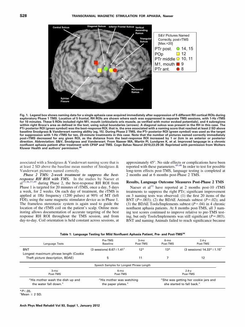
In the protocol of Naeser,43 during Phase 1 TMS, the effectof slow, 1-Hz rTMS for 10 minutes was used to suppressactivity in each of at least 4 different RH frontal ROIs inseparate TMS sessions. A total of 600 magnetic pulses at 90%of motor threshold (MT) for the left first dorsal interosseusmuscle (FDI) was delivered to each RH ROI using the Super-Rapid High Frequency Magnetic Stimulator.a Published guide-ines for safety parameters of rTMS are based on stimulationntensities expressed as a percent of the individual’s MT.44-46 Agure-8 shaped rTMS coil with a 7-cm outside diameter onach wing was used. The RH cortical ROIs that were examinedncluded the right primary motor cortex representation for theouth (orbicularis oris muscle, as verified with motor evoked
otentials), and at least 3 subregions within right Broca’s areaomologue, as described below. These subregions were labeledccording to sulcal and gyral boundaries36,47,48 (fig 1).
The vertical ascending ramus is traditionally the most com-on sulcus used to separate the pars opercularis (POp) from
he pars triangularis (PTr) within Broca’s area. However, inome cases, a diagonal sulcus is present. Cytoarchitectonictudies have observed, for example, that a diagonal sulcus wasresent in every second hemisphere, and it can either mark theorder between BA 44 (likely POp) and BA 45 (likely PTr), ore located inside BA 44.47 Thus, when a diagonal sulcus is
present, without cytoarchitectonics, it is not possible to knowwhether the diagonal sulcus is actually a border between POpand PTr, or if it is within POp. Taking this into consideration,when a diagonal sulcus is present in an aphasia patient who isparticipating in TMS treatment, it is important to carefullyexamine at least 4 subregions within right Broca’s homologue,as shown in figure 1.36
The exact location of the best response RH ROI to suppresswith 1Hz rTMS can vary somewhat, from patient to patient,and the ROI needs to be firmly established for each case.Suppression of POp often impairs naming and/or increasesRT.43 The best response area is often reported to be locatedimmediately anterior to the POp, for example, the right poste-rior PTr.33,36,37,49,50
A frameless stereotaxic systemb is used in combination withthe patient’s 3-dimensional magnetization-prepared rapid gra-dient-echo (MPRAGE) magnetic resonance imaging (MRI)scan, in order to guide the rTMS coil placement onto thetargeted ROI. After each 10-minute rTMS application to aspecific RH ROI during Phase 1, a 20-item Snodgrass &Vanderwart set of pictures is presented for the patient to name.
A best response RH ROI is defined as that ROI which isArch Phys Med Rehabil Vol 93, Suppl 1, January 2012
PaaFTlird
arl2
R
toB(niiB
n Be
S28 TRANSCRANIAL MAGNETIC STIMULATION FOR APHASIA, Naeser
associated with a Snodgrass & Vanderwart naming score that isat least 2 SD above the baseline mean number of Snodgrass &Vanderwart pictures named correctly.
Phase 2 TMS: 2-week treatment to suppress the best-response RH ROI with TMS. In the studies by Naeser etal33,36,37,49 during Phase 2, the best-response RH ROI from
hase 1 is targeted for 20 minutes of rTMS, once a day, 5 daysweek, for 2 weeks. On each day of treatment, the rTMS is
pplied at 1Hz frequency (1200 pulses) at 90% of MT (leftDI), using the same magnetic stimulator device as in Phase 1.he frameless stereotaxic system is again used to guide the
ocation of the rTMS coil on the patient’s scalp. Online mon-toring allows documentation of accurate targeting of the bestesponse RH ROI throughout the TMS session, and fromay-to-day. Coil orientation is held constant across sessions, at
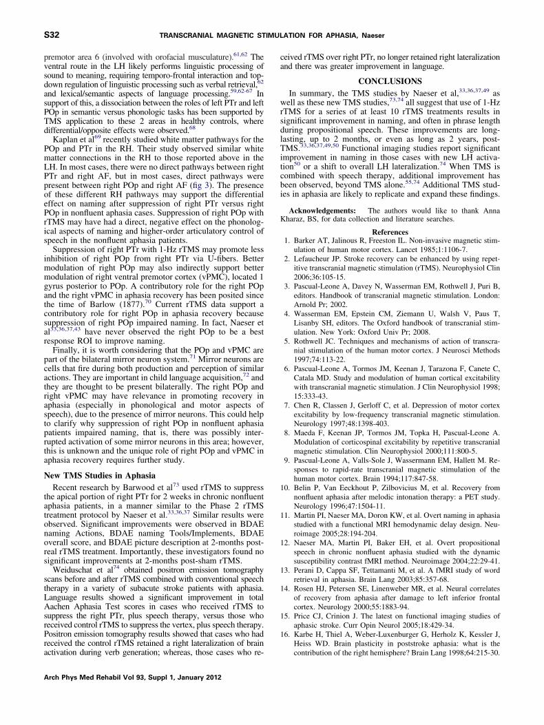
Fig. 1. Legend box shows naming data for a single aphasia case acquexploratory Phase 1 TMS. Location of 5 frontal, RH ROIs are shownfor 10 minutes. These 5 ROIs included right M1, mouth (orbicularis owithin right Broca’s area as defined in the text, using sulcal boundaPTr posterior ROI (green symbol) was the best-response ROI, that is,baseline Snodgrass & Vanderwart naming ability (eg, 15). During Phfor suppression with 1-Hz rTMS for ten, 20-minute treatments in thpost-rTMS decreased for any given ROI, as the distance from thedirection. Abbreviation: S&V, Snodgrass and Vanderwart. From Nanonfluent aphasia patient after treatment with CPAP and TMS. CogKluwer Health and authors’ permission.36
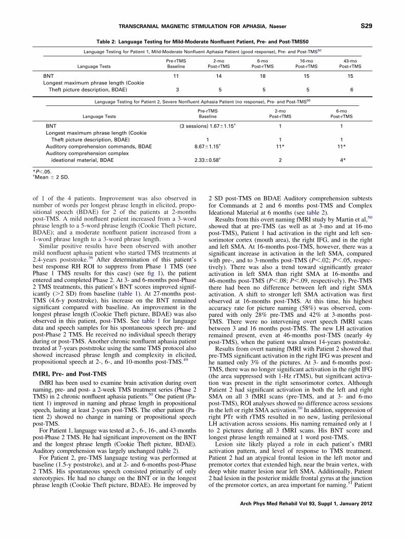
Table 1: Language Testing for Mild Non
Language TestsPre-TMSBaseline
BNT (3 sessions) 8.67�1Longest maximum phrase length (Cookie
Theft picture description, BDAE) 5
Speech Samples fo
3-moPost-TMS
6Pos
“His mother wash the dish up andthe water fall down.”
“His mother wthe paper p
*P�.05.†Mean � 2 SD.
Arch Phys Med Rehabil Vol 93, Suppl 1, January 2012
pproximately 45°. No side effects or complications have beeneported with these parameters.45,46 In order to test for possibleong-term effects post-TMS, language testing is completed at
months and at 6 months post-Phase 2 TMS.
esults, Language Outcome Measures Post-Phase 2 TMSNaeser et al33 have reported at 2 months post-10 rTMS
reatments to suppress the right PTr; significant improvementn 3 naming tests was observed: (1) the first 20 items of theNT (P�.003); (2) the BDAE Animals subtest (P�.02); and
3) the BDAE Tools/Implements subtest (P�.04) in 4 chroniconfluent aphasia patients. At 8 months post-TMS, all 3 nam-ng test scores continued to improve relative to pre-TMS test-ng, but only Tools/Implements was still significant (P�.003).NT and naming Animals failed to reach significance because
immediately after suppression of 5 different RH cortical ROIs duringre each was suppressed in separate TMS sessions, with 1-Hz rTMSuscle, as verified with motor evoked potentials), and 4 subregions
(arrows). A diagonal sulcus was present in the RH in this case. Therea associated with a naming score that reached at least 2 SD above
TMS, the PTr posterior ROI (green symbol) was used as the targetse. Note that the number of pictures named correctly immediatelyt-response ROI increased by 1 or 2cm in an anterior or posteriorMA, Martin PI, Lundgren K, et al. Improved language in a chronic
hav Neurol 2010;23:29-38. Reprinted with permission from Wolters
t Aphasia Patient, Pre- and Post-TMS36
3-moPost-TMS
6-moPost-TMS
2.4-yPost-TMS
12* 13* (3 sessions) 14.33*�1.15†
11 7 12
est Phrase Length
2.4-yPost-TMS
atching”
“She was getting her cookie jars andshe started to fall back.”
iredwheris mriesthe aase 2is cabes
eser
fluen
.41†
r Long
-mot-TMS
as wlates.
rLtl
aPpd2
*
S29TRANSCRANIAL MAGNETIC STIMULATION FOR APHASIA, Naeser
of 1 of the 4 patients. Improvement was also observed innumber of words per longest phrase length in elicited, propo-sitional speech (BDAE) for 2 of the patients at 2-monthspost-TMS. A mild nonfluent patient increased from a 3-wordphrase length to a 5-word phrase length (Cookie Theft picture,BDAE); and a moderate nonfluent patient increased from a1-word phrase length to a 3-word phrase length.
Similar positive results have been observed with anothermild nonfluent aphasia patient who started TMS treatments at2.4-years poststroke.36 After determination of this patient’sbest response RH ROI to suppress from Phase 1 TMS (seePhase 1 TMS results for this case) (see fig 1), the patiententered and completed Phase 2. At 3- and 6-months post-Phase2 TMS treatments, this patient’s BNT scores improved signif-icantly (�2 SD) from baseline (table 1). At 27-months post-TMS (4.6-y poststroke), his increase on the BNT remainedsignificant compared with baseline. An improvement in thelongest phrase length (Cookie Theft picture, BDAE) was alsoobserved in this patient, post-TMS. See table 1 for languagedata and speech samples for his spontaneous speech pre- andpost-Phase 2 TMS. He received no individual speech therapyduring or post-TMS. Another chronic nonfluent aphasia patienttreated at 7-years poststroke using the same TMS protocol alsoshowed increased phrase length and complexity in elicited,propositional speech at 2-, 6-, and 10-months post-TMS.49
fMRI, Pre- and Post-TMSfMRI has been used to examine brain activation during overt
naming, pre- and post- a 2-week TMS treatment series (Phase 2TMS) in 2 chronic nonfluent aphasia patients.50 One patient (Pa-tient 1) improved in naming and phrase length in propositionalspeech, lasting at least 2-years post-TMS. The other patient (Pa-tient 2) showed no change in naming or propositional speechpost-TMS.
For Patient 1, language was tested at 2-, 6-, 16-, and 43-monthspost-Phase 2 TMS. He had significant improvement on the BNTand the longest phrase length (Cookie Theft picture, BDAE).Auditory comprehension was largely unchanged (table 2).
For Patient 2, pre-TMS language testing was performed atbaseline (1.5-y poststroke), and at 2- and 6-months post-Phase2 TMS. His spontaneous speech consisted primarily of onlystereotypies. He had no change on the BNT or in the longest
Table 2: Language Testing for Mild-Mode
Language Testing for Patient 1, Mild-Moderate Nonflu
Language TestsPre-rTMSBaseline
BNT 11Longest maximum phrase length (Cookie
Theft picture description, BDAE) 3
Language Testing for Patient 2, Severe Nonfluen
Language Tests
BNT (3 sessLongest maximum phrase length (Cookie
Theft picture description, BDAE)Auditory comprehension commands, BDAE 8Auditory comprehension complex
ideational material, BDAE 2
P�.05.†Mean � 2 SD.
phrase length (Cookie Theft picture, BDAE). He improved by o
2 SD post-TMS on BDAE Auditory comprehension subtestsfor Commands at 2 and 6 months post-TMS and ComplexIdeational Material at 6 months (see table 2).
Results from this overt naming fMRI study by Martin et al,50
showed that at pre-TMS (as well as at 3-mo and at 16-mopost-TMS), Patient 1 had activation in the right and left sen-sorimotor cortex (mouth area), the right IFG, and in the rightand left SMA. At 16-months post-TMS, however, there was asignificant increase in activation in the left SMA, comparedwith pre-, and to 3-months post-TMS (P�.02; P�.05, respec-tively). There was also a trend toward significantly greateractivation in left SMA than right SMA at 16-months and46-months post-TMS (P�.08; P�.09, respectively). Pre-TMSthere had been no difference between left and right SMAactivation. A shift to stronger left SMA activation was firstobserved at 16-months post-TMS. At this time, his highestaccuracy rate for picture naming (58%) was observed, com-pared with only 28% pre-TMS and 42% at 3-months post-TMS. There were no intervening overt speech fMRI scansbetween 3 and 16 months post-TMS. The new LH activationremained present, even at 46-months post-TMS (nearly 4ypost-TMS), when the patient was almost 14-years poststroke.
Results from overt naming fMRI with Patient 2 showed thatpre-TMS significant activation in the right IFG was present andhe named only 3% of the pictures. At 3- and 6-months post-TMS, there was no longer significant activation in the right IFG(the area suppressed with 1-Hz rTMS), but significant activa-tion was present in the right sensorimotor cortex. AlthoughPatient 2 had significant activation in both the left and rightSMA on all 3 fMRI scans (pre-TMS, and at 3- and 6-mopost-TMS), ROI analyses showed no difference across sessionsin the left or right SMA activation.50 In addition, suppression ofight PTr with rTMS resulted in no new, lasting perilesionalH activation across sessions. His naming remained only at 1
o 2 pictures during all 3 fMRI scans. His BNT score andongest phrase length remained at 1 word post-TMS.
Lesion site likely played a role in each patient’s fMRIctivation pattern, and level of response to TMS treatment.atient 2 had an atypical frontal lesion in the left motor andremotor cortex that extended high, near the brain vertex, witheep white matter lesion near left SMA. Additionally, Patienthad lesion in the posterior middle frontal gyrus at the junction
Nonfluent Patient, Pre- and Post-TMS50
phasia Patient (good response), Pre- and Post-TMS50
2-moost-rTMS
6-moPost-rTMS
16-moPost-rTMS
43-moPost-rTMS
14 18 15 15
5 5 5 6
asia Patient (no response), Pre- and Post-TMS50
MSline
2-moPost-rTMS
6-moPost-rTMS
1.67�1.15† 1 1
1 11.15† 11* 11*
0.58† 2 4*
rate
ent A
P
t Aph
Pre-rTBase
ions)
1.67�
.33�
f the premotor cortex, an area important for naming.51 Patient
Arch Phys Med Rehabil Vol 93, Suppl 1, January 2012
2dim
B
sm
aa
TN
csms
E, etOS P
S30 TRANSCRANIAL MAGNETIC STIMULATION FOR APHASIA, Naeser
2 also had a lesion located inferior and posterior to Wernicke’sarea, in BA 21 and 37. Patient 1 had no lesions in these 3 areas.
The significant increase in activation of the left SMA post-TMS in Patient 1, who improved in naming, is compatible withprevious fMRI studies that observed new left SMA activationto be present in aphasia patients with a better outcome.16,17
This improved LH activation is also compatible with previousstudies that have observed better outcome after language ther-apy to be associated with new perilesional LH activation.21-24
Based on their results, Martin et al50 have suggested minimumcriteria for entry into TMS treatment as follows: (1) the patientshould have a mean score of at least 3 pictures named correctly onthe BNT (without phonemic cueing), tested across 3 test sessions,and (2) the patient should not produce stereotypies in spontaneousspeech. Patient 2 would not have met these minimum criteria forentry, as his mean BNT score across 3 test sessions pre-TMS �SD was only 1.67�1.15 (range, 1–3); and he produced stereotyp-ies during spontaneous speech. One severe nonfluent global apha-sia patient with a 1-word phrase length, who did not producestereotypies, had a good response to the TMS protocol of Naeseret al.37 She named 4 pictures on the BNT pre-TMS; 7 pictures, at-months post-TMS; and 12 pictures, at 8-months post-TMS. Theegree of improvement resulted in the patient’s ability to havendividual speech therapy sessions, leading to continued improve-ent in naming and communication.
PART 2: TMS PLUS CILT
ackground and RationaleCILT is an intensive speech therapy program shown to
ignificantly improve naming after a series of 10 CILT treat-ents.52-54 During CILT, patients are only allowed to respond
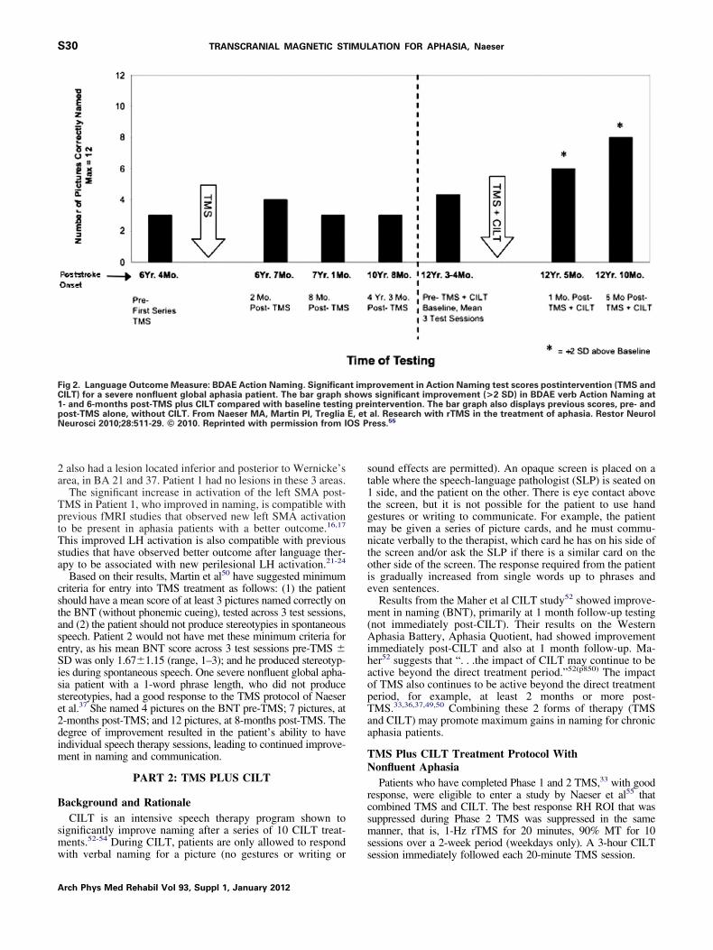
Fig 2. Language Outcome Measure: BDAE Action Naming. SignificanCILT) for a severe nonfluent global aphasia patient. The bar graph s1- and 6-months post-TMS plus CILT compared with baseline testinpost-TMS alone, without CILT. From Naeser MA, Martin PI, TregliaNeurosci 2010;28:511-29. © 2010. Reprinted with permission from I
with verbal naming for a picture (no gestures or writing or s
Arch Phys Med Rehabil Vol 93, Suppl 1, January 2012
sound effects are permitted). An opaque screen is placed on atable where the speech-language pathologist (SLP) is seated on1 side, and the patient on the other. There is eye contact abovethe screen, but it is not possible for the patient to use handgestures or writing to communicate. For example, the patientmay be given a series of picture cards, and he must commu-nicate verbally to the therapist, which card he has on his side ofthe screen and/or ask the SLP if there is a similar card on theother side of the screen. The response required from the patientis gradually increased from single words up to phrases andeven sentences.
Results from the Maher et al CILT study52 showed improve-ment in naming (BNT), primarily at 1 month follow-up testing(not immediately post-CILT). Their results on the WesternAphasia Battery, Aphasia Quotient, had showed improvementimmediately post-CILT and also at 1 month follow-up. Ma-her52 suggests that “. . .the impact of CILT may continue to beactive beyond the direct treatment period.”52(p850) The impactof TMS also continues to be active beyond the direct treatmentperiod, for example, at least 2 months or more post-TMS.33,36,37,49,50 Combining these 2 forms of therapy (TMSnd CILT) may promote maximum gains in naming for chronicphasia patients.
MS Plus CILT Treatment Protocol Withonfluent AphasiaPatients who have completed Phase 1 and 2 TMS,33 with good
response, were eligible to enter a study by Naeser et al55 thatombined TMS and CILT. The best response RH ROI that wasuppressed during Phase 2 TMS was suppressed in the sameanner, that is, 1-Hz rTMS for 20 minutes, 90% MT for 10
essions over a 2-week period (weekdays only). A 3-hour CILT
rovement in Action Naming test scores postintervention (TMS ands significant improvement (>2 SD) in BDAE verb Action Naming atintervention. The bar graph also displays previous scores, pre- andal. Research with rTMS in the treatment of aphasia. Restor Neurolress.55
t imphowg pre
ession immediately followed each 20-minute TMS session.
i
N
NstaiN1(ls
P
paas1
wBsH(ptdmc
S31TRANSCRANIAL MAGNETIC STIMULATION FOR APHASIA, Naeser
A severe nonfluent aphasia patient, who initially receivedonly the Phase 1 and Phase 2 TMS protocol at 6.5-yearspoststroke, later participated in TMS plus CILT.55 This patientparticipated in TMS and CILT at 12.5-years poststroke (5y,10mo after the initial TMS series). Prior to TMS plus CILT, herobject naming ability was tested 3 times on a set of 250 colorpictures. One-third of the color pictures presented as stimulusitems for therapy had always been named on pretesting (3/3);one-third she had sometimes named (1–2/3); and one-third shehad never named (0/3). During CILT, 6 pictures were presentedat a time (2 pictures had always been named at entry pretesting;2, sometimes; 2, never). A total of 18 pictures were presentedduring CILT each day (3 sets of 6 pictures each).
Language outcome measures included the BNT and subtestson the BDAE. These tests were administered at baseline pre-TMS (3 times) and at 1- and 6-months post- the 10th treatmentin the TMS plus CILT protocol by Naeser et al.55 Significantmprovement was defined as more than 2 SD above baseline.
aming Probe TestingTo examine changes that might occur during intervention,
aming Probe Testing was also completed. BDAE namingubtests (Actions, Animals, Tools/Implements), the BNT, andhe action naming pictures from Druks and Masterson56 weredministered 12 times pre-TMS (including the 3 baseline test-ngs). In addition, immediately post- each CILT session, dailyaming Probe Testing was administered (10 times). After the0th treatment, Naming Probe Testing was again administered10 separate times). The time-series data for each test wereater analyzed using a double bootstrap method (http://www.tat.wmich.edu/slab/Software/Timeseries.html).57
Results for TMS Plus CILTOn the primary language outcome measures, this severe
nonfluent aphasia patient improved more than 2 SD on BDAEAction Naming, Tools/Implements, and Single Word Repeti-tion. Improvement in BDAE Action Naming was only ob-
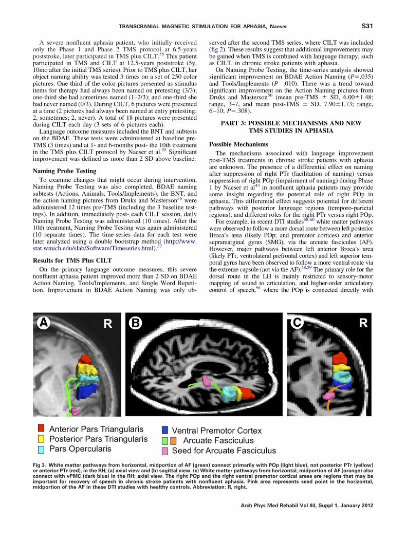
Fig 3. White matter pathways from horizontal, midportion of AF (gor anterior PTr (red), in the RH; (a) axial view and (b) sagittal view. (cconnect with vPMC (dark blue) in the RH; axial view. The right POp
important for recovery of speech in chronic stroke patients with nonflmidportion of the AF in these DTI studies with healthy controls. Abbrevserved after the second TMS series, where CILT was included(fig 2). These results suggest that additional improvements maybe gained when TMS is combined with language therapy, suchas CILT, in chronic stroke patients with aphasia.
On Naming Probe Testing, the time-series analysis showedsignificant improvement on BDAE Action Naming (P�.035)and Tools/Implements (P�.010). There was a trend towardsignificant improvement on the Action Naming pictures fromDruks and Masterson56 (mean pre-TMS � SD, 6.00�1.48;range, 3–7, and mean post-TMS � SD, 7.90�1.73; range,6–10; P�.308).
PART 3: POSSIBLE MECHANISMS AND NEWTMS STUDIES IN APHASIA
ossible MechanismsThe mechanisms associated with language improvement
ost-TMS treatments in chronic stroke patients with aphasiare unknown. The presence of a differential effect on namingfter suppression of right PTr (facilitation of naming) versusuppression of right POp (impairment of naming) during Phaseby Naeser et al43 in nonfluent aphasia patients may provide
some insight regarding the potential role of right POp inaphasia. This differential effect suggests potential for differentpathways with posterior language regions (temporo-parietalregions), and different roles for the right PTr versus right POp.
For example, in recent DTI studies58-60 white matter pathwaysere observed to follow a more dorsal route between left posteriorroca’s area (likely POp; and premotor cortices) and anterior
upramarginal gyrus (SMG), via the arcuate fasciculus (AF).owever, major pathways between left anterior Broca’s area
likely PTr, ventrolateral prefrontal cortex) and left superior tem-oral gyrus have been observed to follow a more ventral route viahe extreme capsule (not via the AF).58,59 The primary role for theorsal route in the LH is mainly restricted to sensory-motorapping of sound to articulation, and higher-order articulatory
ontrol of speech,58 where the POp is connected directly with
connect primarily with POp (light blue), not posterior PTr (yellow)te matter pathways from horizontal, midportion of AF (orange) alsothe right ventral premotor cortical areas are regions that may be
reen)) Whi
and
uent aphasia. Pink area represents seed point in the horizontal,iation: R, right.Arch Phys Med Rehabil Vol 93, Suppl 1, January 2012
r
pca
wrsdlT
i
K
1
1
1
1
1
1
1
S32 TRANSCRANIAL MAGNETIC STIMULATION FOR APHASIA, Naeser
premotor area 6 (involved with orofacial musculature).61,62 Theventral route in the LH likely performs linguistic processing ofsound to meaning, requiring temporo-frontal interaction and top-down regulation of linguistic processing such as verbal retrieval,62
and lexical/semantic aspects of language processing.59,62-67 Insupport of this, a dissociation between the roles of left PTr and leftPOp in semantic versus phonologic tasks has been supported byTMS application to these 2 areas in healthy controls, wheredifferential/opposite effects were observed.68
Kaplan et al69 recently studied white matter pathways for thePOp and PTr in the RH. Their study observed similar whitematter connections in the RH to those reported above in theLH. In most cases, there were no direct pathways between rightPTr and right AF, but in most cases, direct pathways werepresent between right POp and right AF (fig 3). The presenceof these different RH pathways may support the differentialeffect on naming after suppression of right PTr versus rightPOp in nonfluent aphasia cases. Suppression of right POp withrTMS may have had a direct, negative effect on the phonolog-ical aspects of naming and higher-order articulatory control ofspeech in the nonfluent aphasia patients.
Suppression of right PTr with 1-Hz rTMS may promote lessinhibition of right POp from right PTr via U-fibers. Bettermodulation of right POp may also indirectly support bettermodulation of right ventral premotor cortex (vPMC), located 1gyrus posterior to POp. A contributory role for the right POpand the right vPMC in aphasia recovery has been posited sincethe time of Barlow (1877).70 Current rTMS data support acontributory role for right POp in aphasia recovery becausesuppression of right POp impaired naming. In fact, Naeser etal33,36,37,43 have never observed the right POp to be a bestesponse ROI to improve naming.
Finally, it is worth considering that the POp and vPMC areart of the bilateral mirror neuron system.71 Mirror neurons areells that fire during both production and perception of similarctions. They are important in child language acquisition,72 and
they are thought to be present bilaterally. The right POp andright vPMC may have relevance in promoting recovery inaphasia (especially in phonological and motor aspects ofspeech), due to the presence of mirror neurons. This could helpto clarify why suppression of right POp in nonfluent aphasiapatients impaired naming, that is, there was possibly inter-rupted activation of some mirror neurons in this area; however,this is unknown and the unique role of right POp and vPMC inaphasia recovery requires further study.
New TMS Studies in AphasiaRecent research by Barwood et al73 used rTMS to suppress
the apical portion of right PTr for 2 weeks in chronic nonfluentaphasia patients, in a manner similar to the Phase 2 rTMStreatment protocol by Naeser et al.33,36,37 Similar results wereobserved. Significant improvements were observed in BDAEnaming Actions, BDAE naming Tools/Implements, BDAEoverall score, and BDAE picture description at 2-months post-real rTMS treatment. Importantly, these investigators found nosignificant improvements at 2-months post-sham rTMS.
Weiduschat et al74 obtained positron emission tomographyscans before and after rTMS combined with conventional speechtherapy in a variety of subacute stroke patients with aphasia.Language results showed a significant improvement in totalAachen Aphasia Test scores in cases who received rTMS tosuppress the right PTr, plus speech therapy, versus those whoreceived control rTMS to suppress the vertex, plus speech therapy.Positron emission tomography results showed that cases who hadreceived the control rTMS retained a right lateralization of brain
activation during verb generation; whereas, those cases who re-Arch Phys Med Rehabil Vol 93, Suppl 1, January 2012
ceived rTMS over right PTr, no longer retained right lateralizationand there was greater improvement in language.
CONCLUSIONSIn summary, the TMS studies by Naeser et al,33,36,37,49 as
ell as these new TMS studies,73,74 all suggest that use of 1-HzTMS for a series of at least 10 rTMS treatments results inignificant improvement in naming, and often in phrase lengthuring propositional speech. These improvements are long-asting, up to 2 months, or even as long as 2 years, post-MS.33,36,37,49,50 Functional imaging studies report significant
improvement in naming in those cases with new LH activa-tion50 or a shift to overall LH lateralization.74 When TMS iscombined with speech therapy, additional improvement hasbeen observed, beyond TMS alone.55,74 Additional TMS stud-es in aphasia are likely to replicate and expand these findings.
Acknowledgements: The authors would like to thank Annaharaz, BS, for data collection and literature searches.
References1. Barker AT, Jalinous R, Freeston IL. Non-invasive magnetic stim-
ulation of human motor cortex. Lancet 1985;1:1106-7.2. Lefaucheur JP. Stroke recovery can be enhanced by using repet-
itive transcranial magnetic stimulation (rTMS). Neurophysiol Clin2006;36:105-15.
3. Pascual-Leone A, Davey N, Wasserman EM, Rothwell J, Puri B,editors. Handbook of transcranial magnetic stimulation. London:Arnold Pr; 2002.
4. Wasserman EM, Epstein CM, Ziemann U, Walsh V, Paus T,Lisanby SH, editors. The Oxford handbook of transcranial stim-ulation. New York: Oxford Univ Pr; 2008.
5. Rothwell JC. Techniques and mechanisms of action of transcra-nial stimulation of the human motor cortex. J Neurosci Methods1997;74:113-22.
6. Pascual-Leone A, Tormos JM, Keenan J, Tarazona F, Canete C,Catala MD. Study and modulation of human cortical excitabilitywith transcranial magnetic stimulation. J Clin Neurophysiol 1998;15:333-43.
7. Chen R, Classen J, Gerloff C, et al. Depression of motor cortexexcitability by low-frequency transcranial magnetic stimulation.Neurology 1997;48:1398-403.
8. Maeda F, Keenan JP, Tormos JM, Topka H, Pascual-Leone A.Modulation of corticospinal excitability by repetitive transcranialmagnetic stimulation. Clin Neurophysiol 2000;111:800-5.
9. Pascual-Leone A, Valls-Sole J, Wassermann EM, Hallett M. Re-sponses to rapid-rate transcranial magnetic stimulation of thehuman motor cortex. Brain 1994;117:847-58.
0. Belin P, Van Eeckhout P, Zilbovicius M, et al. Recovery fromnonfluent aphasia after melodic intonation therapy: a PET study.Neurology 1996;47:1504-11.
1. Martin PI, Naeser MA, Doron KW, et al. Overt naming in aphasiastudied with a functional MRI hemodynamic delay design. Neu-roimage 2005;28:194-204.
2. Naeser MA, Martin PI, Baker EH, et al. Overt propositionalspeech in chronic nonfluent aphasia studied with the dynamicsusceptibility contrast fMRI method. Neuroimage 2004;22:29-41.
3. Perani D, Cappa SF, Tettamanti M, et al. A fMRI study of wordretrieval in aphasia. Brain Lang 2003;85:357-68.
4. Rosen HJ, Petersen SE, Linenweber MR, et al. Neural correlatesof recovery from aphasia after damage to left inferior frontalcortex. Neurology 2000;55:1883-94.
5. Price CJ, Crinion J. The latest on functional imaging studies ofaphasic stroke. Curr Opin Neurol 2005;18:429-34.
6. Karbe H, Thiel A, Weber-Luxenburger G, Herholz K, Kessler J,Heiss WD. Brain plasticity in poststroke aphasia: what is the
contribution of the right hemisphere? Brain Lang 1998;64:215-30.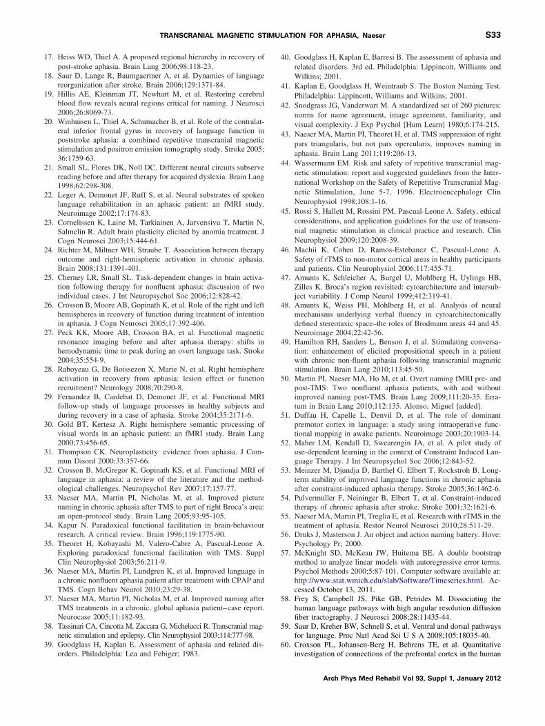
S33TRANSCRANIAL MAGNETIC STIMULATION FOR APHASIA, Naeser
17. Heiss WD, Thiel A. A proposed regional hierarchy in recovery ofpost-stroke aphasia. Brain Lang 2006;98:118-23.
18. Saur D, Lange R, Baumgaertner A, et al. Dynamics of languagereorganization after stroke. Brain 2006;129:1371-84.
19. Hillis AE, Kleinman JT, Newhart M, et al. Restoring cerebralblood flow reveals neural regions critical for naming. J Neurosci2006;26:8069-73.
20. Winhuisen L, Thiel A, Schumacher B, et al. Role of the contralat-eral inferior frontal gyrus in recovery of language function inpoststroke aphasia: a combined repetitive transcranial magneticstimulation and positron emission tomography study. Stroke 2005;36:1759-63.
21. Small SL, Flores DK, Noll DC. Different neural circuits subservereading before and after therapy for acquired dyslexia. Brain Lang1998;62:298-308.
22. Leger A, Demonet JF, Ruff S, et al. Neural substrates of spokenlanguage rehabilitation in an aphasic patient: an fMRI study.Neuroimage 2002;17:174-83.
23. Cornelissen K, Laine M, Tarkiainen A, Jarvensivu T, Martin N,Salmelin R. Adult brain plasticity elicited by anomia treatment. JCogn Neurosci 2003;15:444-61.
24. Richter M, Miltner WH, Straube T. Association between therapyoutcome and right-hemispheric activation in chronic aphasia.Brain 2008;131:1391-401.
25. Cherney LR, Small SL. Task-dependent changes in brain activa-tion following therapy for nonfluent aphasia: discussion of twoindividual cases. J Int Neuropsychol Soc 2006;12:828-42.
26. Crosson B, Moore AB, Gopinath K, et al. Role of the right and lefthemispheres in recovery of function during treatment of intentionin aphasia. J Cogn Neurosci 2005;17:392-406.
27. Peck KK, Moore AB, Crosson BA, et al. Functional magneticresonance imaging before and after aphasia therapy: shifts inhemodynamic time to peak during an overt language task. Stroke2004;35:554-9.
28. Raboyeau G, De Boissezon X, Marie N, et al. Right hemisphereactivation in recovery from aphasia: lesion effect or functionrecruitment? Neurology 2008;70:290-8.
29. Fernandez B, Cardebat D, Demonet JF, et al. Functional MRIfollow-up study of language processes in healthy subjects andduring recovery in a case of aphasia. Stroke 2004;35:2171-6.
30. Gold BT, Kertesz A. Right hemisphere semantic processing ofvisual words in an aphasic patient: an fMRI study. Brain Lang2000;73:456-65.
31. Thompson CK. Neuroplasticity: evidence from aphasia. J Com-mun Disord 2000;33:357-66.
32. Crosson B, McGregor K, Gopinath KS, et al. Functional MRI oflanguage in aphasia: a review of the literature and the method-ological challenges. Neuropsychol Rev 2007;17:157-77.
33. Naeser MA, Martin PI, Nicholas M, et al. Improved picturenaming in chronic aphasia after TMS to part of right Broca’s area:an open-protocol study. Brain Lang 2005;93:95-105.
34. Kapur N. Paradoxical functional facilitation in brain-behaviourresearch. A critical review. Brain 1996;119:1775-90.
35. Theoret H, Kobayashi M, Valero-Cabre A, Pascual-Leone A.Exploring paradoxical functional facilitation with TMS. SupplClin Neurophysiol 2003;56:211-9.
36. Naeser MA, Martin PI, Lundgren K, et al. Improved language ina chronic nonfluent aphasia patient after treatment with CPAP andTMS. Cogn Behav Neurol 2010;23:29-38.
37. Naeser MA, Martin PI, Nicholas M, et al. Improved naming afterTMS treatments in a chronic, global aphasia patient–case report.Neurocase 2005;11:182-93.
38. Tassinari CA, Cincotta M, Zaccara G, Michelucci R. Transcranial mag-netic stimulation and epilepsy. Clin Neurophysiol 2003;114:777-98.
39. Goodglass H, Kaplan E. Assessment of aphasia and related dis-
orders. Philadelphia: Lea and Febiger; 1983.40. Goodglass H, Kaplan E, Barresi B. The assessment of aphasia andrelated disorders. 3rd ed. Philadelphia: Lippincott, Williams andWilkins; 2001.
41. Kaplan E, Goodglass H, Weintraub S. The Boston Naming Test.Philadelphia: Lippincott, Williams and Wilkins; 2001.
42. Snodgrass JG, Vanderwart M. A standardized set of 260 pictures:norms for name agreement, image agreement, familiarity, andvisual complexity. J Exp Psychol [Hum Learn] 1980;6:174-215.
43. Naeser MA, Martin PI, Theoret H, et al. TMS suppression of rightpars triangularis, but not pars opercularis, improves naming inaphasia. Brain Lang 2011;119:206-13.
44. Wassermann EM. Risk and safety of repetitive transcranial mag-netic stimulation: report and suggested guidelines from the Inter-national Workshop on the Safety of Repetitive Transcranial Mag-netic Stimulation, June 5-7, 1996. Electroencephalogr ClinNeurophysiol 1998;108:1-16.
45. Rossi S, Hallett M, Rossini PM, Pascual-Leone A. Safety, ethicalconsiderations, and application guidelines for the use of transcra-nial magnetic stimulation in clinical practice and research. ClinNeurophysiol 2009;120:2008-39.
46. Machii K, Cohen D, Ramos-Estebanez C, Pascual-Leone A.Safety of rTMS to non-motor cortical areas in healthy participantsand patients. Clin Neurophysiol 2006;117:455-71.
47. Amunts K, Schleicher A, Burgel U, Mohlberg H, Uylings HB,Zilles K. Broca’s region revisited: cytoarchitecture and intersub-ject variability. J Comp Neurol 1999;412:319-41.
48. Amunts K, Weiss PH, Mohlberg H, et al. Analysis of neuralmechanisms underlying verbal fluency in cytoarchitectonicallydefined stereotaxic space–the roles of Brodmann areas 44 and 45.Neuroimage 2004;22:42-56.
49. Hamilton RH, Sanders L, Benson J, et al. Stimulating conversa-tion: enhancement of elicited propositional speech in a patientwith chronic non-fluent aphasia following transcranial magneticstimulation. Brain Lang 2010;113:45-50.
50. Martin PI, Naeser MA, Ho M, et al. Overt naming fMRI pre- andpost-TMS: Two nonfluent aphasia patients, with and withoutimproved naming post-TMS. Brain Lang 2009;111:20-35. Erra-tum in Brain Lang 2010;112:135. Alonso, Miguel [added].
51. Duffau H, Capelle L, Denvil D, et al. The role of dominantpremotor cortex in language: a study using intraoperative func-tional mapping in awake patients. Neuroimage 2003;20:1903-14.
52. Maher LM, Kendall D, Swearengin JA, et al. A pilot study ofuse-dependent learning in the context of Constraint Induced Lan-guage Therapy. J Int Neuropsychol Soc 2006;12:843-52.
53. Meinzer M, Djundja D, Barthel G, Elbert T, Rockstroh B. Long-term stability of improved language functions in chronic aphasiaafter constraint-induced aphasia therapy. Stroke 2005;36:1462-6.
54. Pulvermuller F, Neininger B, Elbert T, et al. Constraint-inducedtherapy of chronic aphasia after stroke. Stroke 2001;32:1621-6.
55. Naeser MA, Martin PI, Treglia E, et al. Research with rTMS in thetreatment of aphasia. Restor Neurol Neurosci 2010;28:511-29.
56. Druks J, Masterson J. An object and action naming battery. Hove:Psychology Pr; 2000.
57. McKnight SD, McKean JW, Huitema BE. A double bootstrapmethod to analyze linear models with autoregressive error terms.Psychol Methods 2000;5:87-101. Computer software available at:http://www.stat.wmich.edu/slab/Software/Timeseries.html. Ac-cessed October 13, 2011.
58. Frey S, Campbell JS, Pike GB, Petrides M. Dissociating thehuman language pathways with high angular resolution diffusionfiber tractography. J Neurosci 2008;28:11435-44.
59. Saur D, Kreher BW, Schnell S, et al. Ventral and dorsal pathwaysfor language. Proc Natl Acad Sci U S A 2008;105:18035-40.
60. Croxson PL, Johansen-Berg H, Behrens TE, et al. Quantitative
investigation of connections of the prefrontal cortex in the humanArch Phys Med Rehabil Vol 93, Suppl 1, January 2012

S34 TRANSCRANIAL MAGNETIC STIMULATION FOR APHASIA, Naeser
and macaque using probabilistic diffusion tractography.J Neurosci 2005;25:8854-66.
61. Petrides M, Cadoret G, Mackey S. Orofacial somatomotor re-sponses in the macaque monkey homologue of Broca’s area.Nature 2005;435:1235-8.
62. Petrides M. Broca’s area in the human and the non-human primatebrain. In: Grodzinsky Y, Amunts K, editors. Broca’s region. NewYork: Oxford Univ Pr; 2006. p 31-46.
63. Devlin JT, Matthews PM, Rushworth MF. Semantic processing inthe left inferior prefrontal cortex: a combined functional magneticresonance imaging and transcranial magnetic stimulation study. JCogn Neurosci 2003;15:71-84.
64. Gold BT, Buckner RL. Common prefrontal regions coactivatewith dissociable posterior regions during controlled semantic andphonological tasks. Neuron 2002;35:803-12.
65. Nixon P, Lazarova J, Hodinott-Hill I, Gough P, Passingham R.The inferior frontal gyrus and phonological processing: an inves-tigation using rTMS. J Cogn Neurosci 2004;16:289-300.
66. Poldrack RA, Wagner AD, Prull MW, Desmond JE, Glover GH,Gabrieli JD. Functional specialization for semantic and phonolog-ical processing in the left inferior prefrontal cortex. Neuroimage1999;10:15-35.
67. Price CJ, Wise RJ, Warburton EA, et al. Hearing and saying. Thefunctional neuro-anatomy of auditory word processing. Brain
1996;119:919-31.Arch Phys Med Rehabil Vol 93, Suppl 1, January 2012
68. Gough PM, Nobre AC, Devlin JT. Dissociating linguistic pro-cesses in the left inferior frontal cortex with transcranial magneticstimulation. J Neurosci 2005;25:8010-6.
69. Kaplan E, Naeser MA, Martin PI, et al. Horizontal portion ofarcuate fasciculus fibers track to pars opercularis, not pars trian-gularis, in right and left hemispheres: a DTI study. Neuroimage2010;52:436-44.
70. Barlow T. On a case of double hemiplegia, with cerebral sym-metrical lesions. Br Med J 1877;2:1034.
71. Iacoboni M. The role of premotor cortex in speech perception:evidence from fMRI and rTMS. J Physiol Paris 2008;102:31-4.
72. Rizzolatti G, Craighero L. The mirror-neuron system. Annu RevNeurosci 2004;27:169-92.
73. Barwood CH, Murdoch BE, Whelan BM, et al. Improved languageperformance subsequent to low-frequency rTMS in patients with chronicnon-fluent aphasia post-stroke. Eur J Neurol 2011;18:935-43.
74. Weiduschat N, Thiel A, Rubi-Fessen I, et al. Effects of repetitivetranscranial magnetic stimulation in aphasic stroke: a randomizedcontrolled pilot study. Stroke 2011;42:409-15.
Suppliers
a. The Magstim Company Ltd, Spring Gardens, Whitland, Car-marthenshire, Wales, UK SA34 0HR.
b. Brainsight; Rogue Research Inc., 206-4398 boul. St-Laurent, Mon-treal Quebec, H2W 1Z5 Canada.