Trachoma
22
Trachoma
-
Upload
sachin-patne -
Category
Education
-
view
907 -
download
0
Transcript of Trachoma

Trachoma

Definition Aetiology Clinical Features Sequelae Grading Management
Contents

Trachoma is the commonest infective cause of blindness in the world(15-20%)
Trachoma is spread worldwide but highly prevalent in North Africa, Middle East and certain Regions of South-East Asia.
Introduction
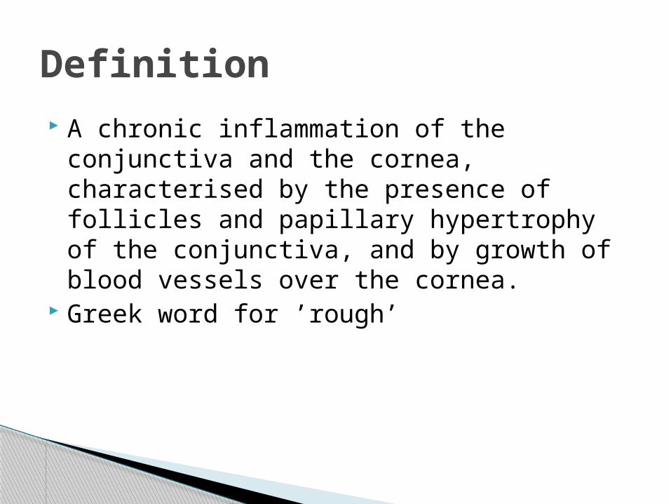
A chronic inflammation of the conjunctiva and the cornea, characterised by the presence of follicles and papillary hypertrophy of the conjunctiva, and by growth of blood vessels over the cornea.
Greek word for ’rough’
Definition

Causative agent: Chlamydia trachomatis Any age Dry, dirty and sandy weather Poor, unhygienic conditions “Eye-seeking” flies Use of kajal or surma from the same
container
Aetiology
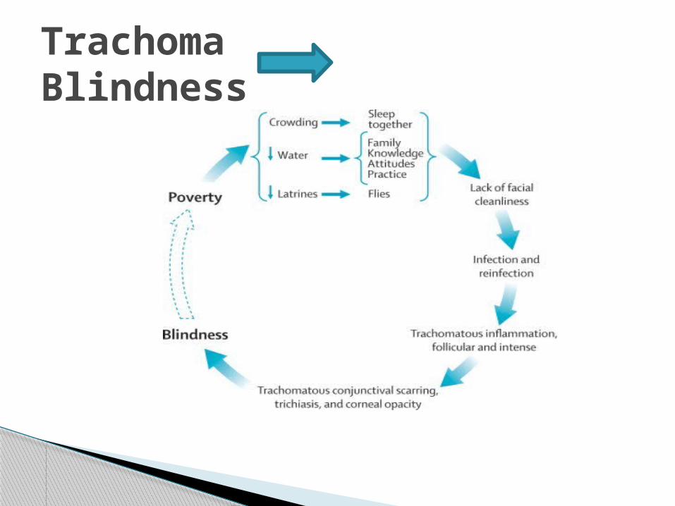
Trachoma Blindness

Foreign body sensation or grittiness Itching Watering, photophobia and redness Discharge is usually scanty, but may be
more due to secondary infections
Acute Trachoma- Secondary infection superimposed on a relatively mild trachoma
Symptoms

Bulbar congestion Velvety papillary hypertrophy Follicles-mostly seen in upper tarsal
conjunctiva or on the limbus or on the bulbar conjunctiva
Pannus: A Characteristic sign defined as fine sub-epithelial neovascularisation, arranged vertically with round cell infiltration, mainly seen at the upper limbus and cornea
Signs

Follicles (aggregation of lymphocytes and other cells in the adenoid layer) are most commonly seen in the upper tarsal conjunctiva and fornix.
Conjunctival Follicle

Pannus
Progressive Pannus Regressive Pannus
Infiltration of cornea is ahead of vessels
Vessels extend beyond the area of infiltration

Oval or circular pitted scars in the area of limbus, left after healing of herbert’s follicles.
Herbert’s Pit

1. Mac Callan’s classification2. Jone’s classification3. WHO classification
Classifications of Trachoma

Type Impression Diagnostic featureTF Follicles Active disease -needs
treatment5 or more follicles of at least 0.5mm diameter on the upper tarsal plate
TI Intense Severe Disease- Urgent treatment
Pronounced inflammatory thickening which obscures more than half of the normal deep tarsal vessels
TS Scarring Old, inactive infection Tarsal conjunctival scarring seen as white fibrous bands
TT Trichiasis Needs corrective surgery
Presence of at least one trichiatic eye lash
CO Opacities Corneal opacities from previous trachoma cause visual loss
Presence of corneal opacity over the pupil
Who classification of trachoma(FISTO)


Each case must have at least two of the following signs
1. Follicles at the upper tarsal conjunctiva2. Limbal Follicles or their sequelae,
Herbert’s pits3. Typical conjunctival Scar (stellate shaped)4. Vascular Pannus, mostly at the upper
limbus
Diagnostic criteria in field study
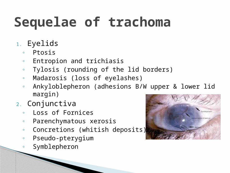
1. Eyelids◦ Ptosis◦ Entropion and trichiasis◦ Tylosis (rounding of the lid borders)◦ Madarosis (loss of eyelashes)◦ Ankyloblepheron (adhesions B/W upper & lower lid
margin)2. Conjunctiva
◦ Loss of Fornices◦ Parenchymatous xerosis◦ Concretions (whitish deposits)◦ Pseudo-pterygium◦ Symblepheron
Sequelae of trachoma

3. Cornea ◦ Herbert’s pits◦ Healed pannus leading to hazy cornea◦ Loss of sensation◦ Total corneal pannus (blinding)
4. Lacrimal sac◦ Chronic dacryocystitis
Sequelae of trachoma (continued)

1. Therapeutica) Topical- Tetracycline (1%) eye ointment Or
sulphacetamide(20% or 30%) eye drops- 4 times a day for 6 weeks
b) Systemic-Tetracycline or erythromycin 250mg orally, four times daily for 3-4 weeks Or Doxycycline 100mg orally twice daily for 3-4 weeks Or oral Azithromycin 250mg once daily for 4 days
Treatment

2. Prophylactic◦ Improvement of personal hygiene, and
environmental sanitaion◦ The use of common towel, handkerchief, surma
rods should be discouraged◦ Early treatment of conjunctivits◦ Blanket antibiotic therapy ( in Endemic areas)-
Intermittent treatment with tetracycline 1% eye ointment twice daily for 5 consecutive days in a month for 6 months
Treatment (continued)

3. Treatment of complications ◦ Trichiasis- epilation, electrolysis or cryolysis◦ Entropion – surgical correction ◦ Follicles - mechanically expressed by Roller
forceps, silver nitrate painting or diathermy◦ Concretions – removed with a hypodermic
needle◦ Pannus- Cryoapplication or peritomy◦ Xerosis – treated by artificial tears
Treatment (continued)

An Effective intervention aiming at total elimination of blindness due to trachoma by the year 2020
SAFE strategy of WHO

Thank you