Tracheobronchomegaly-the Mounier-Kuhn syndrome: report of … · in 1897; in 1932, Mounier-Kuhn...
4
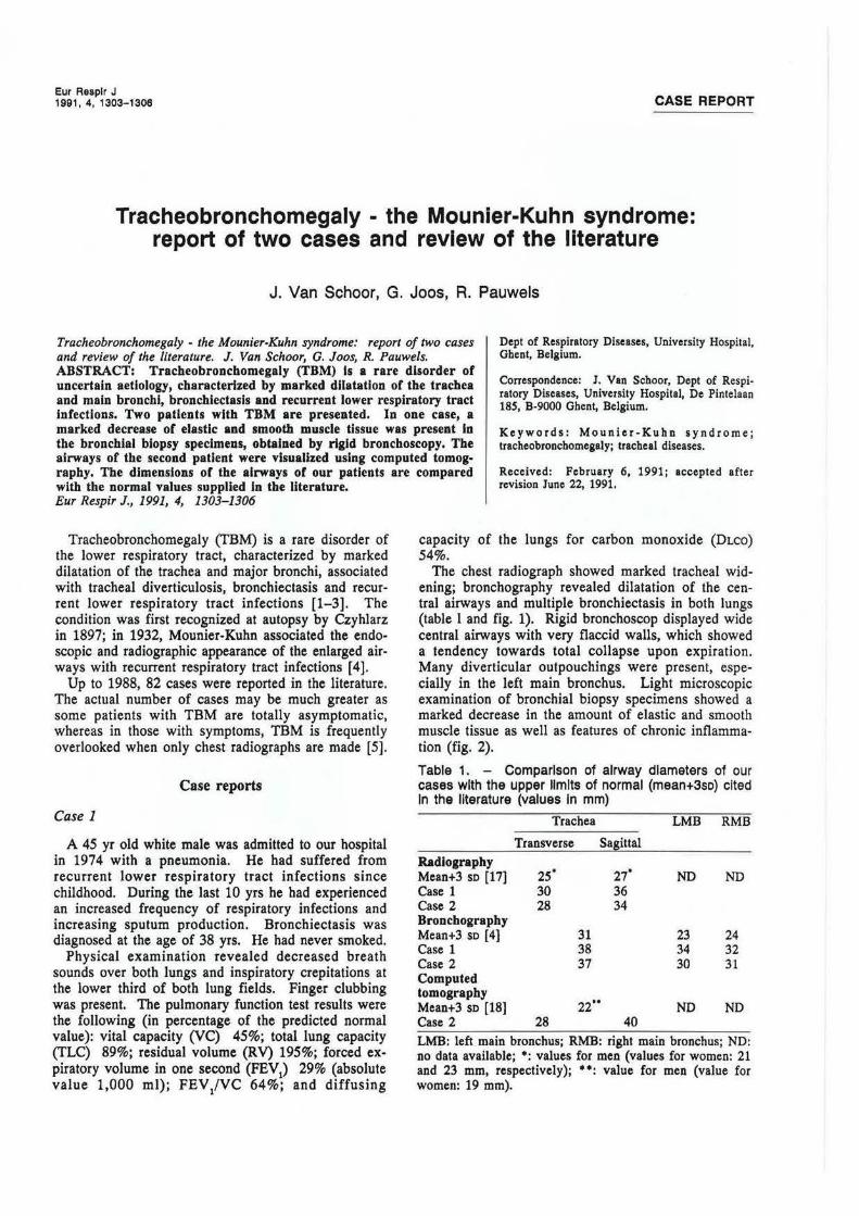
Eur Resplr J 1991, 4, 1303-1306 CASE REPORT Tracheobronchomegaly- the Mounier-Kuhn syndrome: report of two cases and review of the literature J. Van Schoor, G. Joos, R. Pauwels Tracheobronchomegaly - the Mounier-Kuhn syndrome: report of two cases and review of the literature. J. Van Schoor, G. Joos, R. Pauwels. ABSTRACT: Tracbeobronchomegaly (TBM) Is a rare disorder of uncertain aetiology, characterized by marked dilatation of the trachea and malo bronchi, bronchiectasis and recurrent lower respiratory tract infections. Two patients with TBM are presented. In one case, a marked decrease of elastic and smooth mu.scle tissue was present In the bronchial biopsy specimens, obtained by rigid bronchoscopy. The airways of the second patient were visualized using computed tomog· raphy. The dimensions or the airways or our patients are compared with the normal values supplied In the literature. Dept of Respiratory Diseases, University Hospital, Ghent, Belgium. Correspondence: J. Van Schoor, Dept of Respi- ratory Diseases, University Hospital, De Pintelaan 185, B-9000 Ghent, Belgium. Keywords: Mounier-Kuhn syndrome; lracheobronchomegaly; tracheal diseases. Received: February 6, 1991; accepted after revision June 22, 1991. Eur Respir J., 1991, 4, 1303-1306 Tracheobronchomegaly (TBM) is a rare disorder of the lower respiratory tract, characterized by marked dilatation of the trachea and major bronchi, associated with tracheal diverticulosis, bronchiectasis and recur- rent lower respiratory tract infections [1-3]. The condition was first recognized at autopsy by Czyhlarz in 1897; in 1932, Mounier-Kuhn associated the endo- scopic and radiographic appearance of the enlarged air- ways with recurrent respiratory tract infections [4]. Up to 1988, 82 cases were reported in the literature. The actual number of cases may be much greater as some patients with TBM are totally asymptomatic, whereas in those with symptoms, TBM is frequently overlooked when only chest radiographs are made (5). Case reports Case 1 A 45 yr old white male was admitted to our hospital in 1974 with a pneumonia. He had suffered from recurrent lower respiratory tract infections since childhood. During the last 10 yrs he had experienced an increased frequency of respiratory infections and increasing sputum production. Bronchiectasis was diagnosed at the age of 38 yrs. He had never smoked. Physical examination revealed decreased breath sounds over both lungs and inspiratory crepitations at the lower third of both lung fields. Finger clubbing was present. The pulmonary function test results were the following (in percentage of the predicted normal value): vital capacity (VC) 45%; total lung capacity (TLC) 89%; residual volume (RV) 195%; forced ex- piratory volume in one second (FEV 1 ) 29% (absolute value 1,000 ml); FEV.JVC 64%; and diffusing capacity of the lungs for carbon monoxide (DLco) 54%. The chest radiograph showed marked tracheal wid- ening; bronchography revealed dilatation of the cen- tral airways and multiple bronchiectasis in both lungs (table I and fig. 1). Rigid bronchoscop displayed wide central airways with very flaccid walls, which showed a tendency towards total collapse upon expiration. Many diverticular outpouchings were present, espe- cially in the left main bronchus. Light microscopic examination of bronchial biopsy specimens showed a marked decrease in the amount of elastic and smooth muscle tissue as well as features of chronic inflamma- tion (fig. 2). Table 1. - Comparison of airway diameters of our cases with the upper limits of normal (mean+3so) cited In the literature (values In mm) Trachea LMB RMB Transverse Sagittal Radiography 25. 27. Mean+3 so [17] ND ND Case 1 30 36 Case 2 28 34 Bronchography Mean+3 so [4] 31 23 24 Case 1 38 34 32 Case 2 37 30 31 Computed tomography 22 .. Mean+3 so [18] ND ND Case 2 28 40 LMB: left main bronchus; RMB: right main bronchus; ND: no data available; •: values for men (values for women: 21 and 23 mm, respectively); .. : value for men (value for women: 19 mm).
Transcript of Tracheobronchomegaly-the Mounier-Kuhn syndrome: report of … · in 1897; in 1932, Mounier-Kuhn...
Eur Resplr J 1991, 4, 1303-1306 CASE REPORT
Tracheobronchomegaly- the Mounier-Kuhn syndrome: report of two cases and review of the literature
J. Van Schoor, G. Joos, R. Pauwels
Tracheobronchomegaly - the Mounier-Kuhn syndrome: report of two cases and review of the literature. J. Van Schoor, G. Joos, R. Pauwels. ABSTRACT: Tracbeobronchomegaly (TBM) Is a rare disorder of uncertain aetiology, characterized by marked dilatation of the trachea and malo bronchi, bronchiectasis and recurrent lower respiratory tract infections. Two patients with TBM are presented. In one case, a marked decrease of elastic and smooth mu.scle tissue was present In the bronchial biopsy specimens, obtained by rigid bronchoscopy. The airways of the second patient were visualized using computed tomog· raphy. The dimensions or the airways or our patients are compared with the normal values supplied In the literature.
Dept of Respiratory Diseases, University Hospital, Ghent, Belgium.
Correspondence: J. Van Schoor, Dept of Respiratory Diseases, University Hospital, De Pintelaan 185, B-9000 Ghent, Belgium.
Keywords: Mounier-Kuhn syndrome; lracheobronchomegaly; tracheal diseases.
Received: February 6, 1991; accepted after revision June 22, 1991.
Eur Respir J., 1991, 4, 1303-1306
Tracheobronchomegaly (TBM) is a rare disorder of the lower respiratory tract, characterized by marked dilatation of the trachea and major bronchi, associated with tracheal diverticulosis, bronchiectasis and recurrent lower respiratory tract infections [1-3]. The condition was first recognized at autopsy by Czyhlarz in 1897; in 1932, Mounier-Kuhn associated the endoscopic and radiographic appearance of the enlarged airways with recurrent respiratory tract infections [4].
Up to 1988, 82 cases were reported in the literature. The actual number of cases may be much greater as some patients with TBM are totally asymptomatic, whereas in those with symptoms, TBM is frequently overlooked when only chest radiographs are made (5).
Case reports
Case 1
A 45 yr old white male was admitted to our hospital in 1974 with a pneumonia. He had suffered from recurrent lower respiratory tract infections since childhood. During the last 10 yrs he had experienced an increased frequency of respiratory infections and increasing sputum production. Bronchiectasis was diagnosed at the age of 38 yrs. He had never smoked.
Physical examination revealed decreased breath sounds over both lungs and inspiratory crepitations at the lower third of both lung fields. Finger clubbing was present. The pulmonary function test results were the following (in percentage of the predicted normal value): vital capacity (VC) 45%; total lung capacity (TLC) 89%; residual volume (RV) 195%; forced expiratory volume in one second (FEV
1) 29% (absolute
value 1,000 ml); FEV.JVC 64%; and diffusing
capacity of the lungs for carbon monoxide (DLco) 54%.
The chest radiograph showed marked tracheal widening; bronchography revealed dilatation of the central airways and multiple bronchiectasis in both lungs (table I and fig. 1). Rigid bronchoscop displayed wide central airways with very flaccid walls, which showed a tendency towards total collapse upon expiration. Many diverticular outpouchings were present, especially in the left main bronchus. Light microscopic examination of bronchial biopsy specimens showed a marked decrease in the amount of elastic and smooth muscle tissue as well as features of chronic inflammation (fig. 2).
Table 1. - Comparison of airway diameters of our cases with the upper limits of normal (mean+3so) cited In the literature (values In mm)
Trachea LMB RMB
Transverse Sagittal
Radiography 25. 27. Mean+3 so [17] ND ND
Case 1 30 36 Case 2 28 34 Bronchography Mean+3 so [4] 31 23 24 Case 1 38 34 32 Case 2 37 30 31 Computed tomography
22 .. Mean+3 so [18] ND ND Case 2 28 40 LMB: left main bronchus; RMB: right main bronchus; ND: no data available; •: values for men (values for women: 21 and 23 mm, respectively); .. : value for men (value for women: 19 mm).

1304 J. VAN SCHOOR, G. JOOS, R. PAUWELS
Fig. 1. - Bronchography (case 1): gross dilatation of the right main bronchus and multiple bronchiectatic sacculations in the more distal airways.
Fig. 2. - Photomicrography of bronchial biopsy (case 1): marked reduction of the smooth muscle layer; an island of remaining smooth muscle tissue is seen at the bottom of the figure (arrow). Features of chronic inflammation include a thickened basement membrane and a lymphocytic infiltration of the lamina propria. (Haematoxylin and eosin stain. Scale bar • 100 1-1m).
The patient was treated with bronchodilating agents and chest physiotherapy. His family members were in good health; their chest radiographs (in 1974) were normal.
Case 2
A 61 yr old white male had suffered from recurrent respiratory tract infections since 1950. He experienced progressive dyspnoea and chronic expectoration of large amounts of mucoid sputum, which became purulent during infectious exacerbations. He stopped smoking in 1955 (cumulative use: 10 pack yrs). The diagnosis of "tracheobronchomegaly" was made in 1976 on chest radiograph. This showed enlarged tracheal dimensions and multiple bullous deformations at both lung fields.
A
B
Fig. 3. - A) Postero·anterior chest X-ray (case 2): tracheomegaly (arrowheads) and multiple cystic deformations in both lungs. B) Lateral chest X-ray (case 2): marked tracheomegaly (arrowheads) with mucosal folds at the posterior tracheal wall.
TRACHEOBRONCHOMEGALY 1305
The patient was seen again at our centre in 1988, when he was admitted with a left-sided bronchopneumonia. Auscultation of the lungs revealed inspiratory crepitations at both lung bases and a diffuse prolonged expiration with expiratory wheezes bilaterall y. The pulmonary function test results were as follows: VC 56%; TLC 90%; RV 163%; FEVb48% (absolute value 1,240 ml); FEY/VC 60% and LCO 58%.
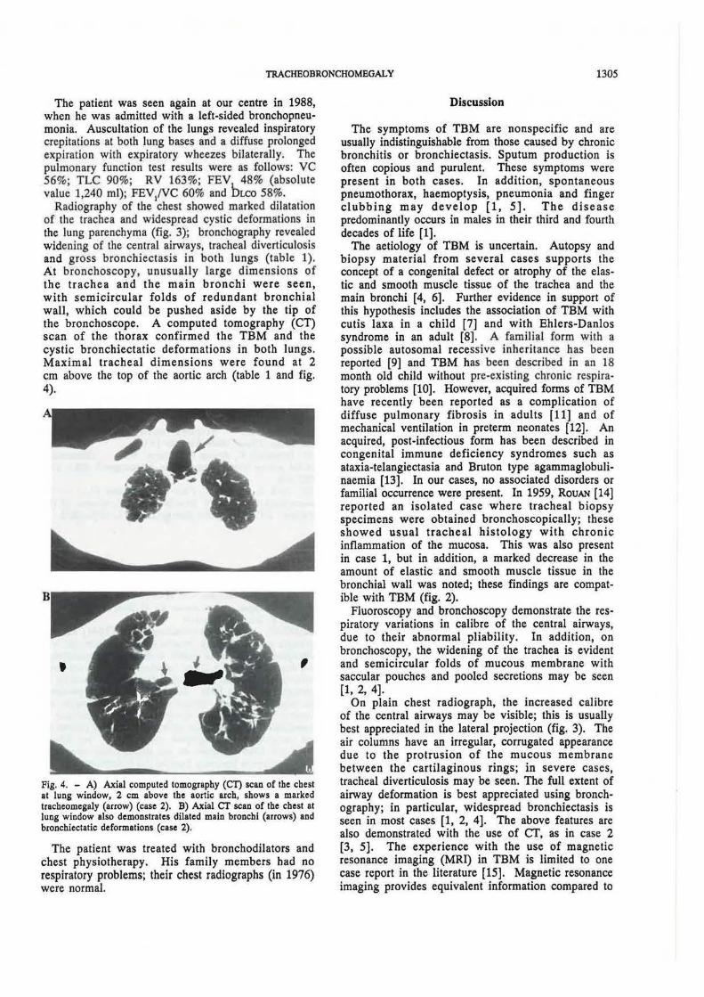
Radiography of the chest showed marked dilatation of the trachea and widespread cystic deformations in the lung parenchyma (fig. 3); bronchography revealed widening of the central airways, tracheal diverticulosis and gross bronchiectasis in both lungs (table 1). At bronchoscopy, unusually large dimensions of the trachea and the main bronchi were seen, with semicircular folds of redundant bronchial wall, which could be pushed aside by the tip of the bronchoscope. A computed tomography (CT) scan of the thorax confirmed the TBM and the cystic bronchiectatic deformations in both lungs. Maximal tracheal dimensions were found at 2 cm above the top of the aortic arch (table 1 and fig. 4).
Fig. 4. - A) Axial computed tomography (CT) scan of the chest at lung window, 2 cm above the aortic arch, shows a marked tracheomegaly (arrow) (case 2). B) Axial er scan of the chest at lung window also demonstrates dilated main bronchi (arrows) and bronchiectatic deformations (case 2).
The patient was treated with bronchodilators and chest physiotherapy. His family members had no respiratory problems; their chest radiographs (in 1976) were normal.
Discussion
The symptoms of TBM are nonspecific and are usually indistinguishable from those caused by chronic bronchitis or bronchiectasis. Sputum production is often copious and purulent. These symptoms were present in both cases. In addition, spontaneous pneumothorax, haemoptysis, pneumonia and finger clubbing may develop [1, 5] . The disease predominantly occurs in males in their third and fourth decades of life [1].
The aetiology of TBM is uncertain. Autopsy and biopsy material from several cases supports the concept of a congenital defect or atrophy of the elastic and smooth muscle tissue of the trachea and the main bronchi (4, 6]. Further evidence in support of this hypothesis includes the association of TBM with cutis laxa in a child [7] and with Ehlers-Danlos syndrome in an adult [8] . A familial form with a possible autosomal recessive inheritance has been reported (9] and TBM has been described in an 18 month old child without pre-existing chronic respiratory problems [10]. However, acquired forms of TBM have recently been reported as a complication of diffuse pulmonary fibrosis in adults [11] and of mechanical ventilation in preterm neonates (12]. An acquired, post-infectious form has been described in congenital immune deficiency syndromes such as ataxia-telangiectasia and Bruton type agammaglobulinaemia [13]. In our cases, no associated disorders or familial occurrence were present. In 1959, RouAN [14) reported an isolated case where tracheal biopsy specimens were obtained bronchoscopically; these showed usual tracheal histology with chronic inflammation of the mucosa. This was also present in case 1, but in addition, a marked decrease in the amount of elastic and smooth muscle tissue in the bronchial wall was noted; these findings are compatible with TBM (fig. 2).
Fluoroscopy and bronchoscopy demonstrate the respiratory variations in calibre of the central airways, due to their abnormal pliability. In addition, on bronchoscopy, the widening of the trachea is evident and semicircular folds of mucous membrane with saccular pouches and pooled secretions may be seen [1, 2, 4].
On plain chest radiograph, the increased calibre of the central airways may be visible; this is usually best appreciated in the lateral projection (fig. 3). The air colutnns have an irregular, corrugated appearance due to the protrusion of the mucous membrane between the cartilaginous rings; in severe cases, tracheal diverticulosis may be seen. The full extent of airway deformation is best appreciated using bronchography; in particular, widespread bronchiectasis is seen in most cases [1, 2, 4]. The above features are also demonstrated with the use of CT, as in case 2 (3, 5]. The experience with the use of magnetic resonance imaging (MRI) in TBM is limited to one case report in the literature [15]. Magnetic resonance imaging provides equivalent information compared to
1306 J. VAN SCHOOR, 0. JOOS, R. PAUWELS
CT in evaluating major airway dimensions [16]. Computed tomography, therefore, remains the most useful technique in these circumstances.
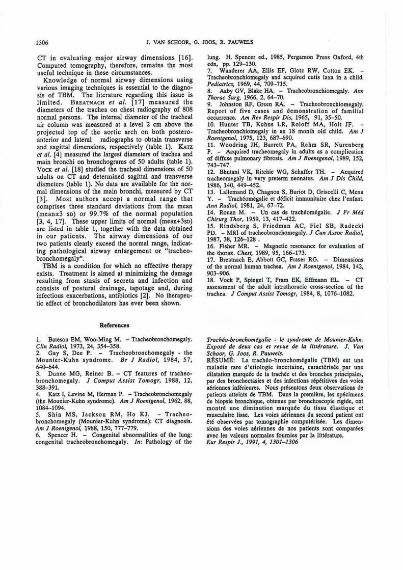
Knowledge of normal airway dimensions using various imaging techniques is essential to the diagnosis of TBM. The literature regarding this issue is limited. BRBATNACH et al. [ 17] measured the diameters of the trachea on chest radiography of 808 normal persons. The internal diameter of the tracheal air column was measured at a level 2 cm above the projected top of the aortic arch on both posteroanterior and lateral radiographs to obtain transverse and sagittal dimensions, respectively (table 1). KATZ et al. [ 4] measured the largest diameters of trachea and main bronchi on bronchograms of 50 adults (table 1). VocK et al. [18] studied the tracheal dimensions of 50 adults on CT and determined sagittal and transverse diameters (table 1). No data are available for the normal dimensions of the main bronchi, measured by CT [3]. Most authors accept a normal range that comprises three standard deviations from the mean (mean:d so) or 99 .7% of the normal population [3, 4, 17]. These upper limits of normal (mean+3so) are listed in table 1, together with the data obtained in our patients. The airway dimensions of our two patients clearly exceed the normal range, indicating pathological airway enlargement or "tracheobronchomegaly".
TBM is a condition for which no effective therapy exists. Treatment is aimed at minimizing the damage resulting from stasis of secreta and infection and consists of postural drainage, tapotage and, during infectious exacerbations, antibiotics [2). No therapeutic effect of bronchodilators has ever been shown.
References
1. Bateson EM, Woo-Ming M. - Tracheobronchomegaly. Clin Radio/, 1973, 24, 354-358. 2. Gay S, Dee P. - Tracheobronchomegaly - the Mounier-Kuhn syndrome. Br J Radio/, 1984, 57, 640-644. 3. Dunne MG, Reiner B. - CT features of tracheobronchomegaly. J Comput Assist Tomogr, 1988, 12, 388-391. 4. Katz I, Levine M, Herman P. - Tracheobro~chomegaly (the Mounier-Kuhn syndrome). Am J Roentgenol, 1962, 88, 1084-1094. 5. Shin MS, Jackson RM, Ho KJ. - Tracheobronchomegaly (Mounier-Kubn syndrome): CT diagnosis. Am J Roentgenol, 1988, 150, 777-779. 6. Spencer H. - Congenital abnormalities of the lung: congenital tracheobronchomegaly. In: Pathology of the
lung. H. Spencer ed., 1985, Pergamon Press Oxford, 4th edn, pp. 129-130. 7. Wanderer AA, Ellis EF, Olotz RW, Cotton EK. -Tracheobronchiomegaly and acquired cutis laxa in a child. Pediatrics, 1969, 44, 709-715. 8. Aaby OV, Blake HA. - Tracheobronchiomegaly. Ann Thorac Surg, 1966, 2, 64-70. 9. Johnston RF, Green RA. - Tracheobronchiomegaly. Report of five cases and demonstration of familial occurrence. Am Rev Respir Dis, 1965, 91, 35-50. 10. Hunter TB, Kuhns LR, Roloff MA, Holt JF. Tracheobronchiomegaly in an 18 month old child. Am J Roentgenol, 1975, 123, 687-690. 11. Woodring JH, Barrett PA, Rehm SR, Nurenberg P. - Acquired tracheomegaly in adults as a complication of diffuse pulmonary fibrosis. Am J Roentgenol, 1989, 152, 743-747. 12. Bhutani VK, Ritcbie WO, Schaffer TH. - Acquired tracheomegaly in very preterm neonates. Am J Dis Child, 1986, 140, 449-452. 13. Lallemand D, Chagnon S, Buriot D, Oriscelli C, Menu Y. - Tracbeom6galie et deficit immunitaire chez !'enfant. Ann Radio/, 1981, 24, 67-72. 14. Rouan M. - Un cas de tracbeomegalie. J Fr Med Chirurg Thor, 1959, 13, 417-422. 15. Rindsberg S, Friedman AC, Fiel SB, Radecki PD. - MRI of tracheobronchomegaly. J Can Assoc Radio/, 1987, 38, 126-128 . 16. Fisher MR. - Magnetic resonance for evaluation of the thorax .. Chest, 1989, 95, 166-173. 17. Breatnach E, Abbott GC, Fraser RG. - Dimensions of the normal human trachea. Am J Roentgenol, 1984, 142, 903-906. 18. Vock P, Spiegel T, Fram EK, Effmann EL. - Cf assessment of the adult intrathoracic cross-section of the trachea. J Comput Assist Tomogr, 1984, 8, 1076-1082.
Tracheo-bronchomegalie - le syndrome de Mounier·Kuhn. Expose de deux cas et revue de la litltrature. J. Van Schoor, G. Joos, R. Pauwels. RESUME: La tracheo-bronchomegalie (TBM) est une maladie rare d'etiologie incertaine, caracterisee par une dilatation marquee de la tracbee et des brooches principales, par des bronchectasies et des infections repetitives des voies aeriennes inferieures. Nous presentons deux observations de patients atteints de TBM. Dans la premiere, les specimens de biopsie bronchique, obtenus par bronchoscopie rigide, ont montre une diminution marquee du tissu elastique et musculaire lisse. Les voies aeriennes du second patient ont ete observees par tomographic computerisee. Les dimensions des voies aeriennes de nos patients soot comparees avec les valeurs normales fournies par la litterature. Eur Respir J., 199.1, 4, 1301-1306