Topics in Fetal Cardiology: 2015 - Home - NCUSncus.org/files/fall2015/cotton.pdf · How does fetal...
64
Topics in Fetal Cardiology: 2015 Dr. John Cotton Professor of Pediatrics Director, Fetal Cardiology Program Division of Pediatric Cardiology UNC Chapel Hill School of Medicine
Transcript of Topics in Fetal Cardiology: 2015 - Home - NCUSncus.org/files/fall2015/cotton.pdf · How does fetal...
Topics in Fetal Cardiology:
2015
Dr. John Cotton
Professor of Pediatrics
Director, Fetal Cardiology Program
Division of Pediatric Cardiology
UNC Chapel Hill School of Medicine
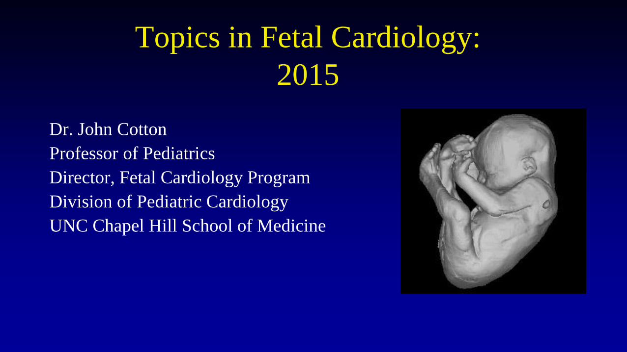
How does fetal heart disease progress?
• Progressive AV or semilunar valve insufficiency/obstruction
• Progressive vessel/chamber hypoplasia due to reduced blood
flow
• Development of cardiomyopathy/myocarditis
• Development/progression of cardiac tumors
• Development/progression/resolution of fetal arrhythmias
• Premature constriction of the ductus arteriosus
• Restriction of the foramen ovale
• Progressive cardiomegaly in high output states
Why Fetal Cardiology?
• Natural history of structural heart disease
• New imaging technology allows improved
visualization of structure and function
• New insights into fetal cardiovascular
physiology change treatment paradigms
• In-utero intervention may alter outcomes
Diagnosis and Treatment of Fetal Cardiac DiseaseA Scientific Statement From the American Heart Association
Endorsed by the American Society of Echocardiography and Pediatric and Congenital Electrophysiology
Society The American Institute of Ultrasound in Medicine supports the value and findings of the statement.*
The Society of Maternal Fetal Medicine supports the statement’s review of the subject matter and believe it is
consistent with its existing clinical guidelines.†
Mary T. Donofrio, MD, Chair; Anita J. Moon-Grady, MD; Lisa K. Hornberger, MD; Joshua A. Copel, MD; Mark S.
Sklansky, MD; Alfred Abuhamad, MD; Bettina F. Cuneo, MD;
James C. Huhta, MD; Richard A. Jonas, MD; Anita Krishnan, MD; Stephanie Lacey, DO; Wesley Lee, MD;
Erik C. Michelfelder, Sr, MD; Gwen R. Rempel, RN;
Norman H. Silverman, MD, DSc, FAHA; Thomas L. Spray, MD, FAHA; Janette F. Strasburger, MD; Wayne Tworetzky, MD;
Jack Rychik MD; on behalf of the American Heart Association Adults With Congenital Heart Disease Joint Committee of the
Council on Cardiovascular Disease in the Young and Council on Clinical Cardiology, Council on Cardiovascular Surgery and
Anesthesia, and Council on Cardiovascular and Stroke Nursing
Circulation 2014:129:2183
Topics
• Indications for referral for fetal echocardiography
• Components of the fetal echocardiogram including
advanced techniques
• Extracardiac assessment of the fetus with CHD
• Prenatal counseling and parental stress
• Fetal therapy for cardiovascular conditions before birth
Rhythm issues
In utero structural interventions- catheter and surgical
• Perinatal management and outcomes
Indications with a high risk profile >2%
• Maternal pregestional diabetes
• Diabetes mellitus diagnosed in the first trimester
• Maternal PKU - uncontrolled
• Maternal autoantibodies (SSA/SSB)
• Maternal medications
ACE inhibitors
Retinoic acid
NSAIDs in third trimester
• Maternal rubella in first trimester
• Maternal infection with risk of myocarditis
• Assisted reproductive technology
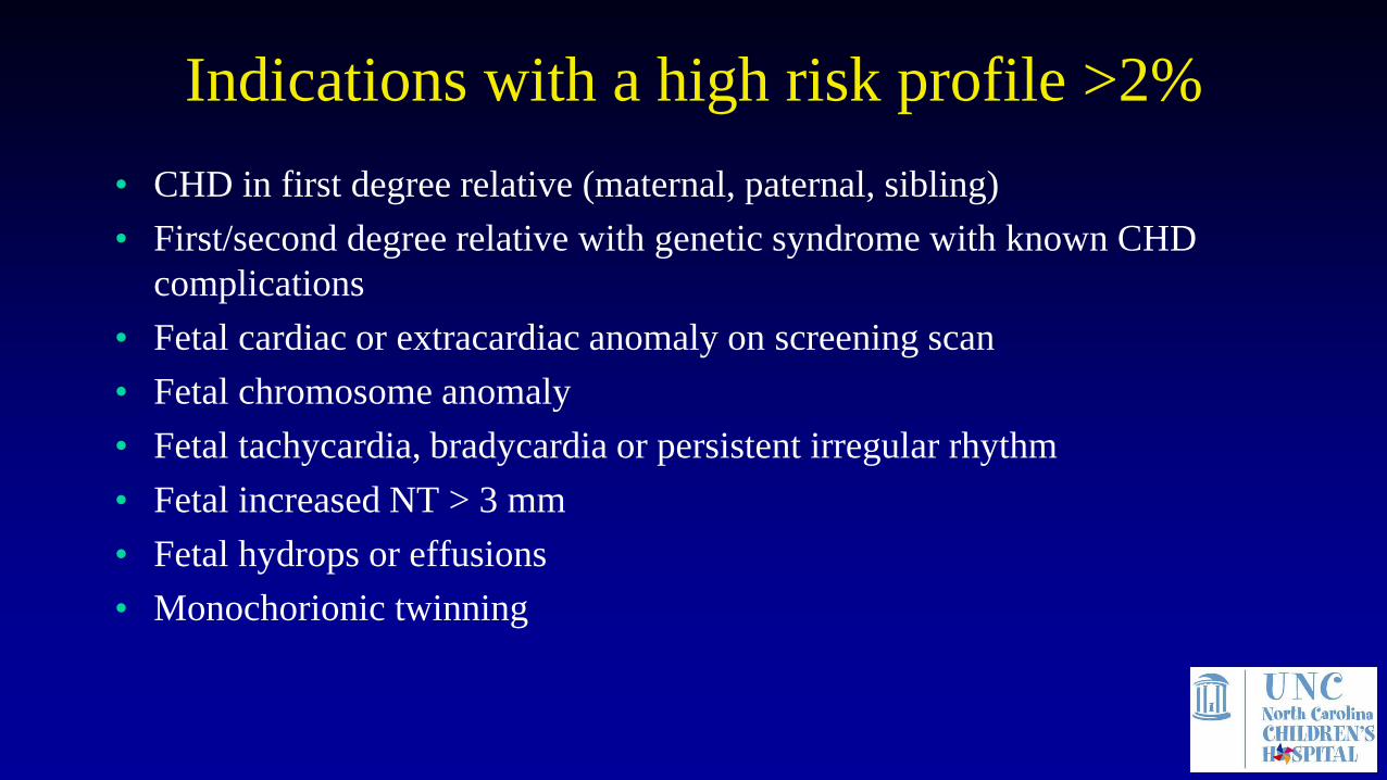
Indications with a high risk profile >2%
• CHD in first degree relative (maternal, paternal, sibling)
• First/second degree relative with genetic syndrome with known CHD
complications
• Fetal cardiac or extracardiac anomaly on screening scan
• Fetal chromosome anomaly
• Fetal tachycardia, bradycardia or persistent irregular rhythm
• Fetal increased NT > 3 mm
• Fetal hydrops or effusions
• Monochorionic twinning
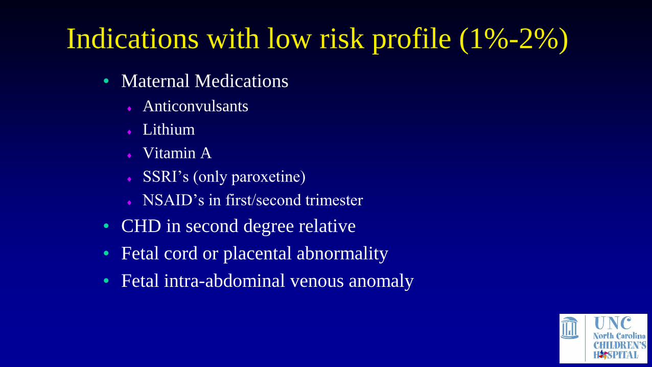
Indications with low risk profile (1%-2%)
• Maternal Medications
Anticonvulsants
Lithium
Vitamin A
SSRI’s (only paroxetine)
NSAID’s in first/second trimester
• CHD in second degree relative
• Fetal cord or placental abnormality
• Fetal intra-abdominal venous anomaly
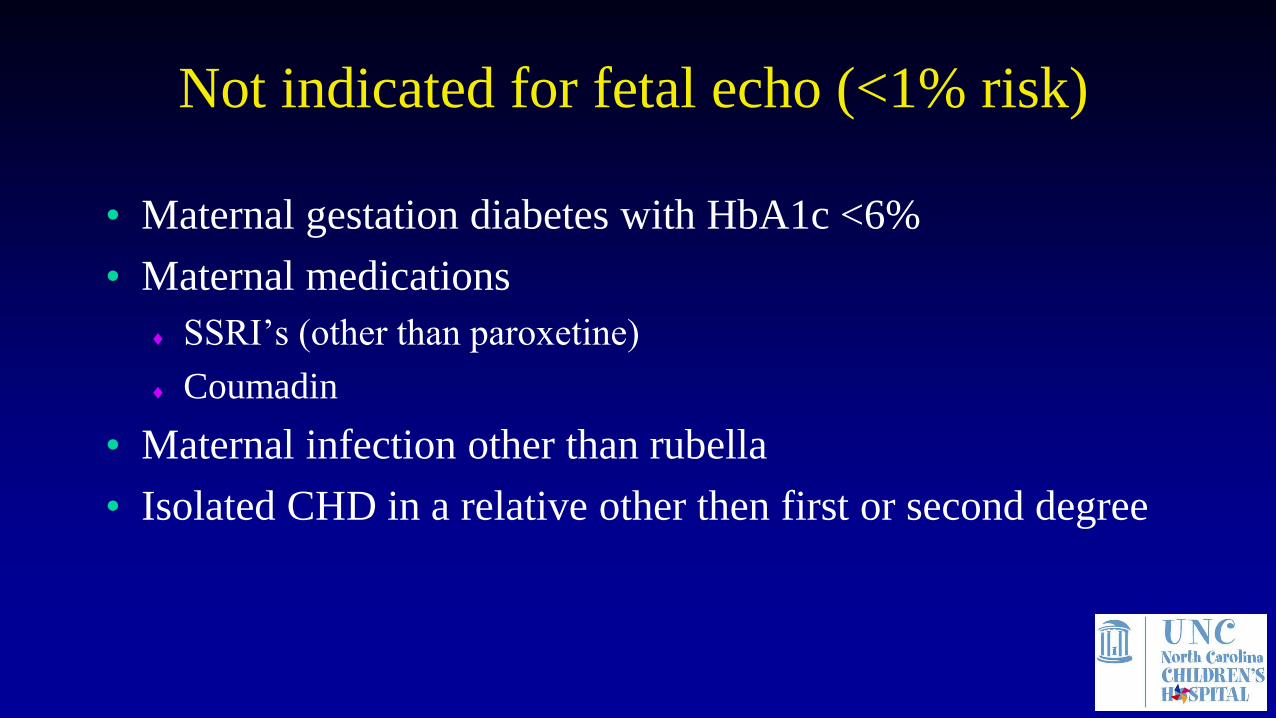
Not indicated for fetal echo (<1% risk)
• Maternal gestation diabetes with HbA1c <6%
• Maternal medications
SSRI’s (other than paroxetine)
Coumadin
• Maternal infection other than rubella
• Isolated CHD in a relative other then first or second degree
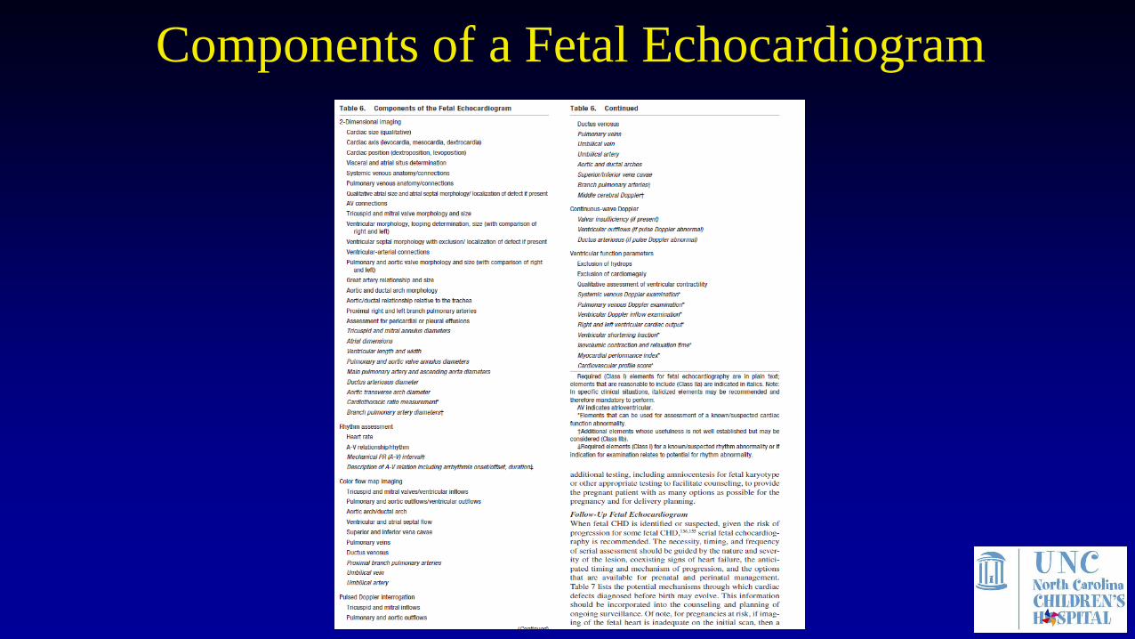
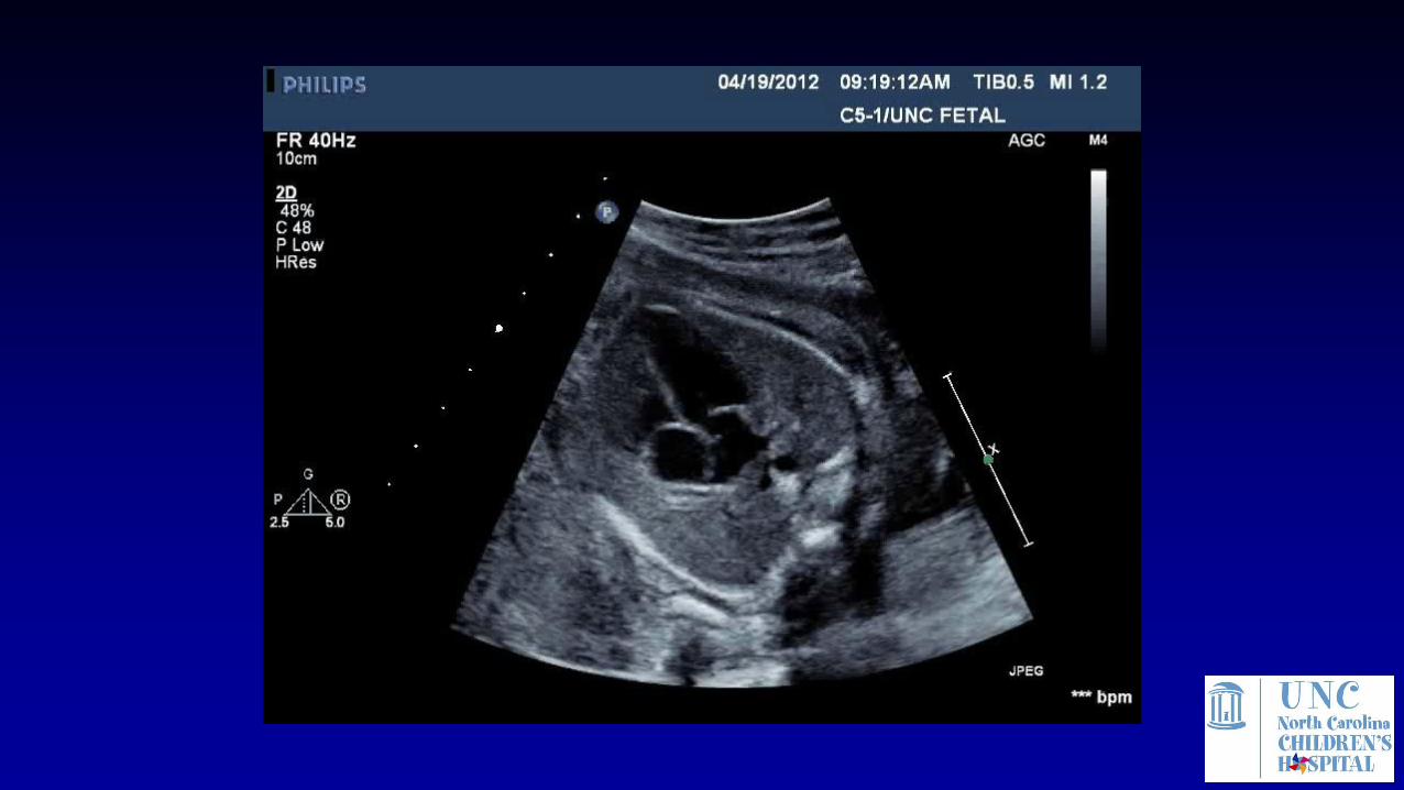
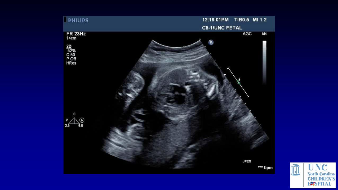
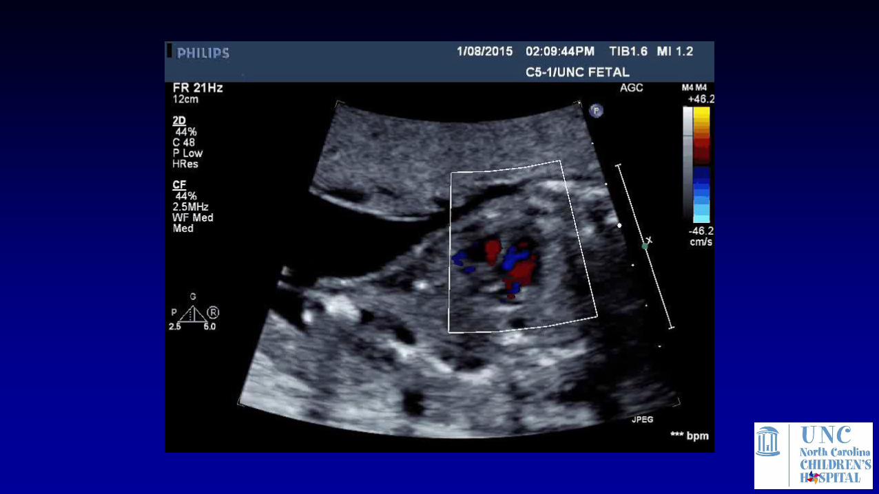
Components of a Fetal Echocardiogram
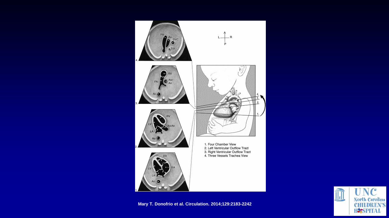
Mary T. Donofrio et al. Circulation. 2014;129:2183-2242

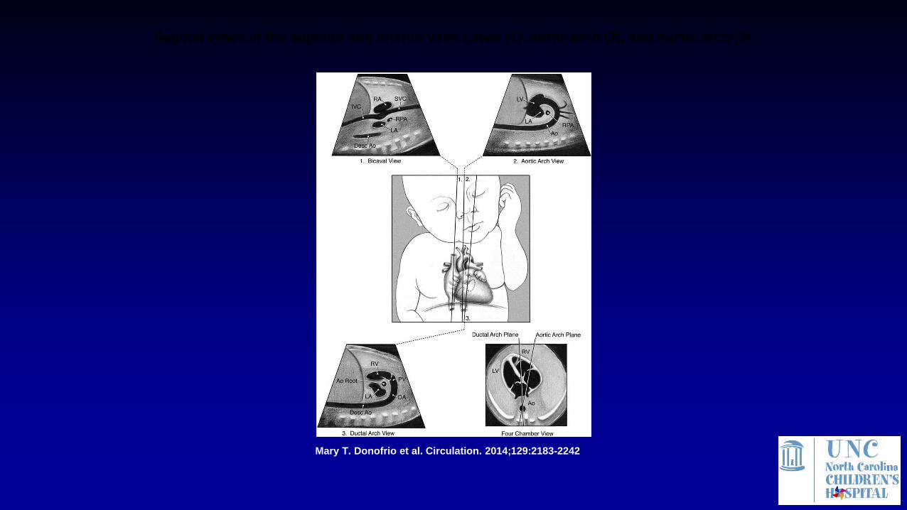
Sagittal views of the superior and inferior vena cavae (1), aortic arch (2), and ductal arch (3).
Mary T. Donofrio et al. Circulation. 2014;129:2183-2242
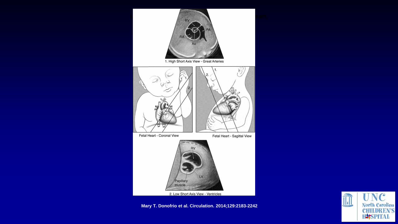
Low and high short-axis views of the fetal heart.
Mary T. Donofrio et al. Circulation. 2014;129:2183-2242
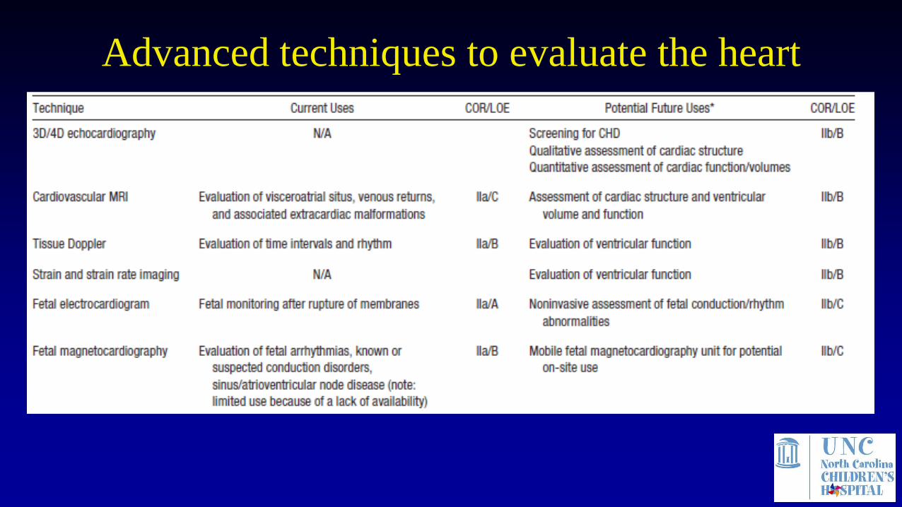
Advanced techniques to evaluate the heart
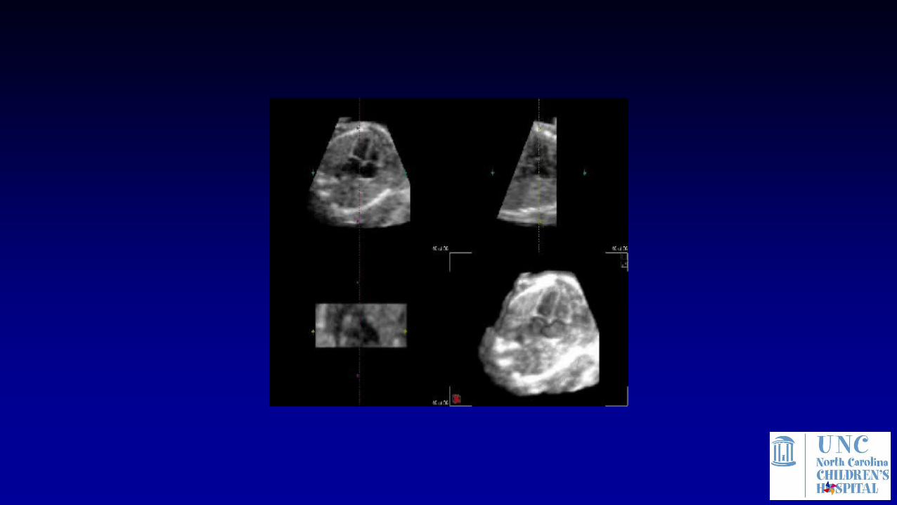
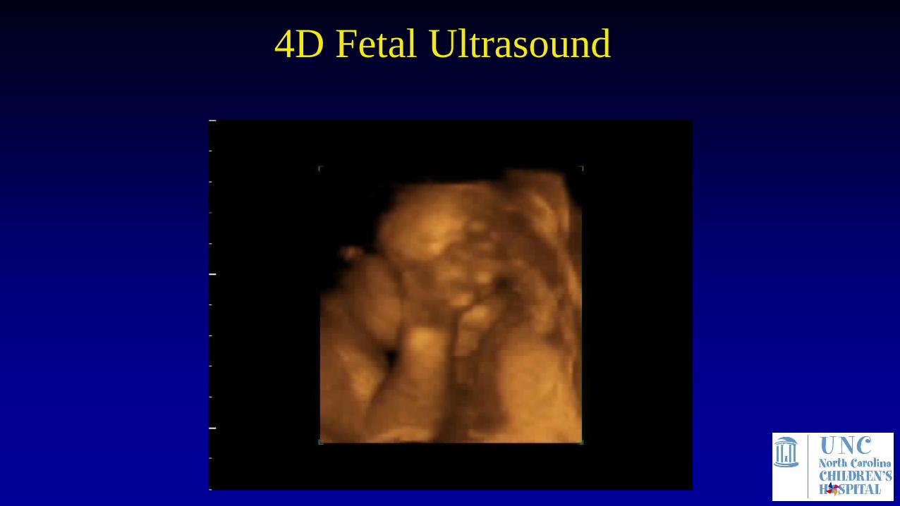
4D Fetal Ultrasound
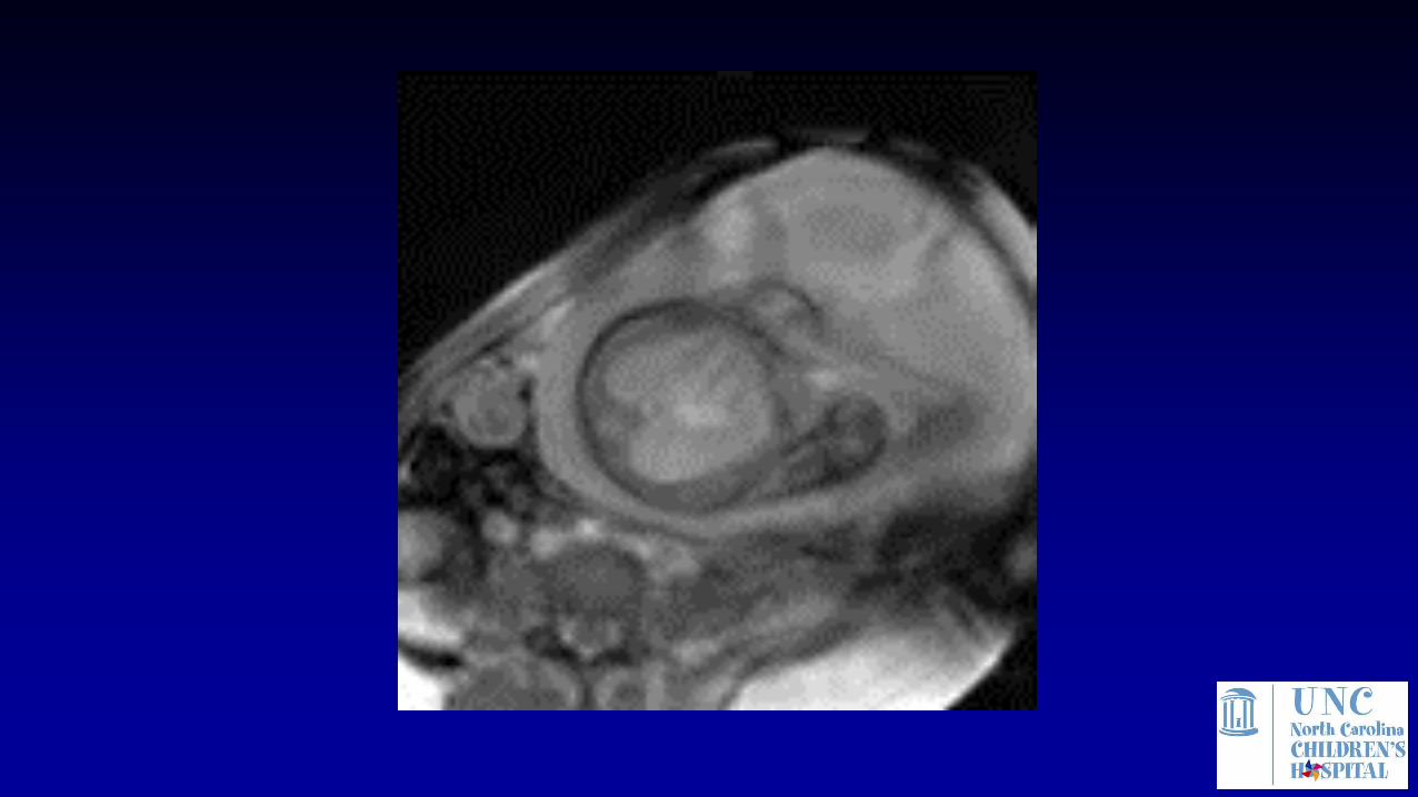
Fetal CT/MRI
• Lower doses of radiation
• Smaller slice thickness
• Faster scan times
• Volumetric rendering
• Contrast angiography
Fetal Magnetocardiography (fMCG)
• Maternal ECG signal is 10-100x stronger than fetus
• As cardiac tissue depolarizes, currents are generated and a
magnetic field is generated
• Strength is about one millionth the strength of the earths
magnetic field
• Maternal signal 50 pT, fetus 0.5-10 pT
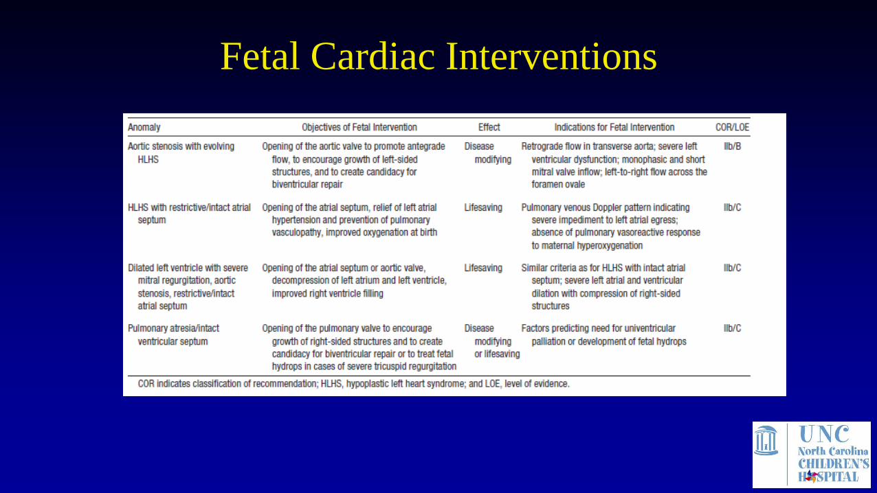
Fetal Cardiac Interventions
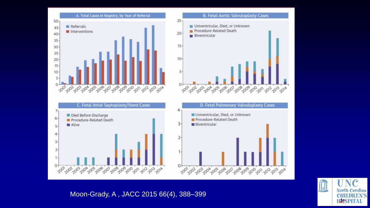
Moon-Grady, A , JACC 2015 66(4), 388–399
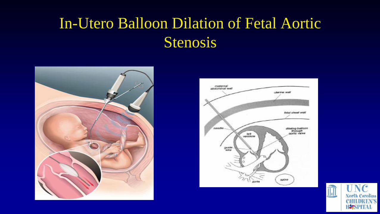
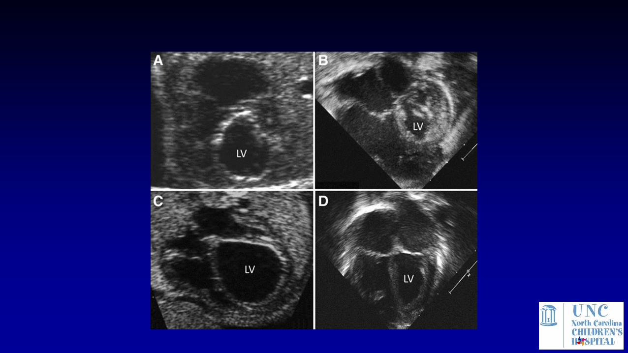
In-Utero Balloon Dilation of Fetal Aortic
Stenosis
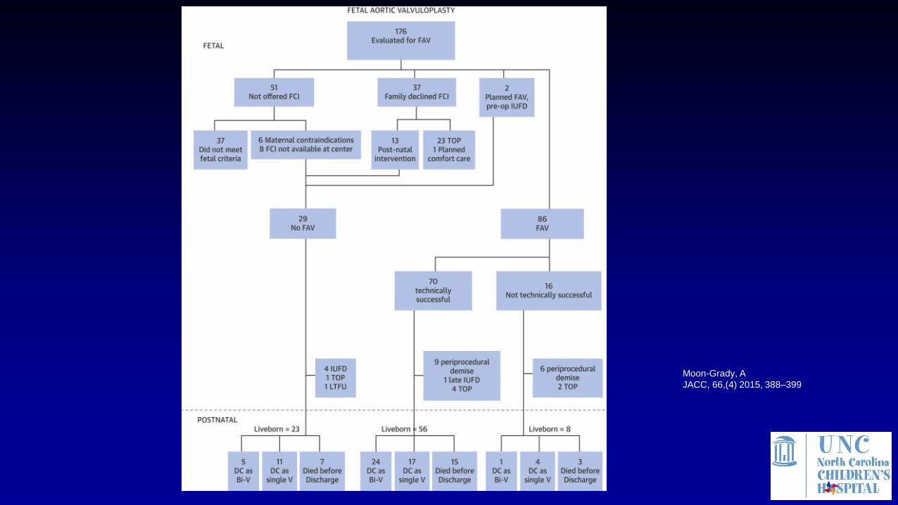
Moon-Grady, A
JACC, 66,(4) 2015, 388–399
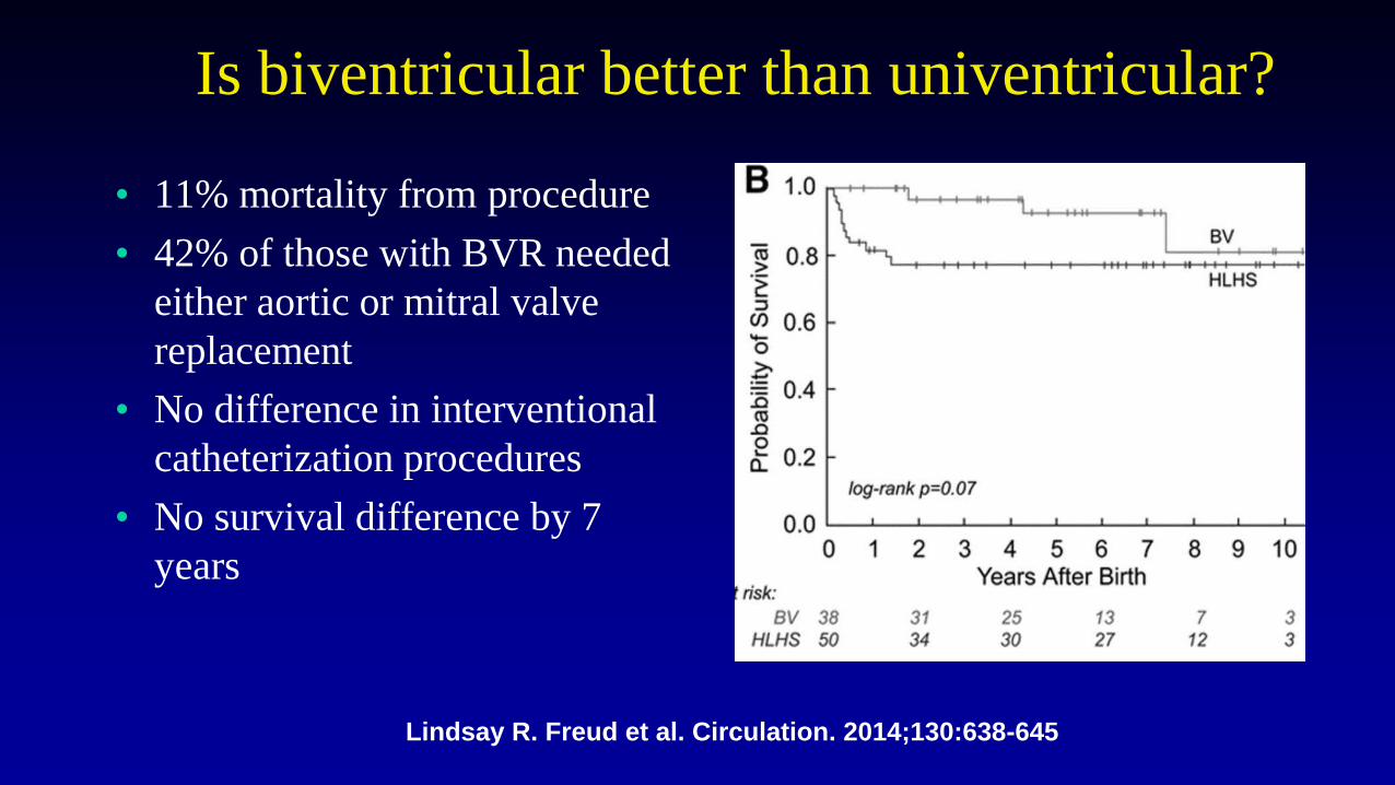
Is biventricular better than univentricular?
• 11% mortality from procedure
• 42% of those with BVR needed
either aortic or mitral valve
replacement
• No difference in interventional
catheterization procedures
• No survival difference by 7
years
Lindsay R. Freud et al. Circulation. 2014;130:638-645
Bleeding edge
• Maternal hyperoxygenation
• Deliberate ductal constriction
• Micro Pacemakers
• Gene therapy
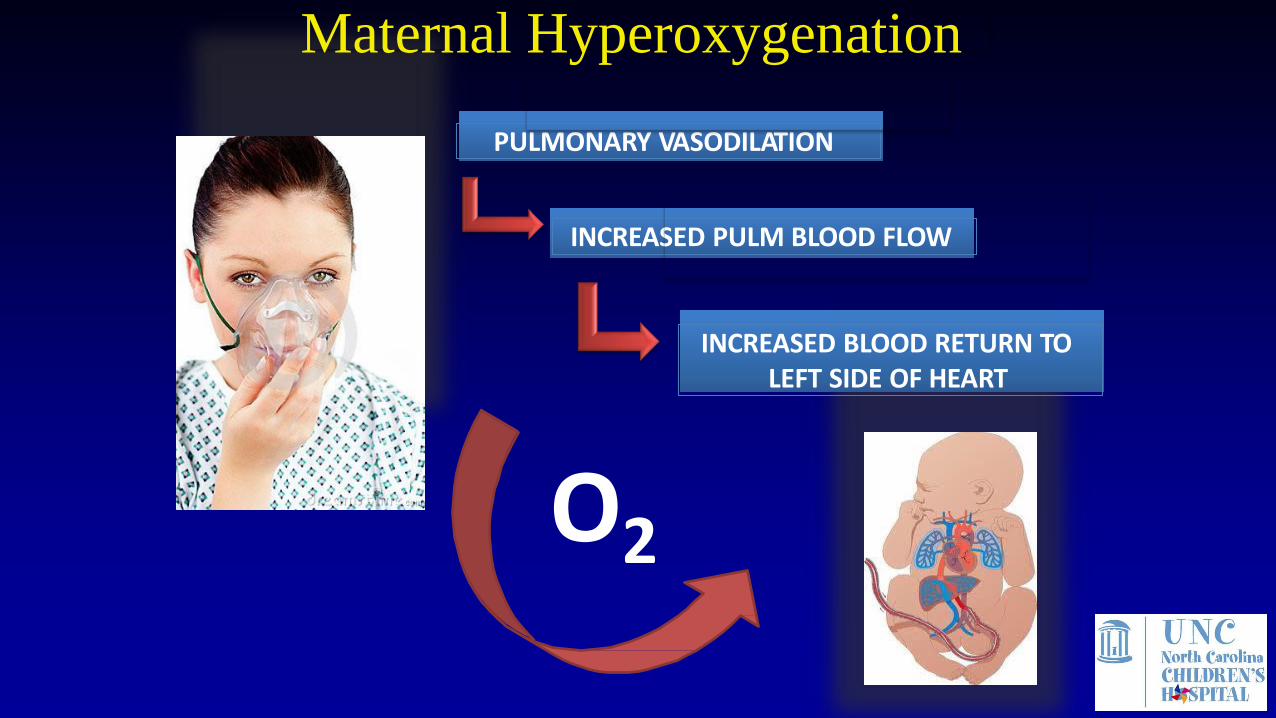
O2
Maternal Hyperoxygenation
PULMONARY VASODILATION
INCREASED PULM BLOOD FLOW
INCREASED BLOOD RETURN TOLEFT SIDE OF HEART
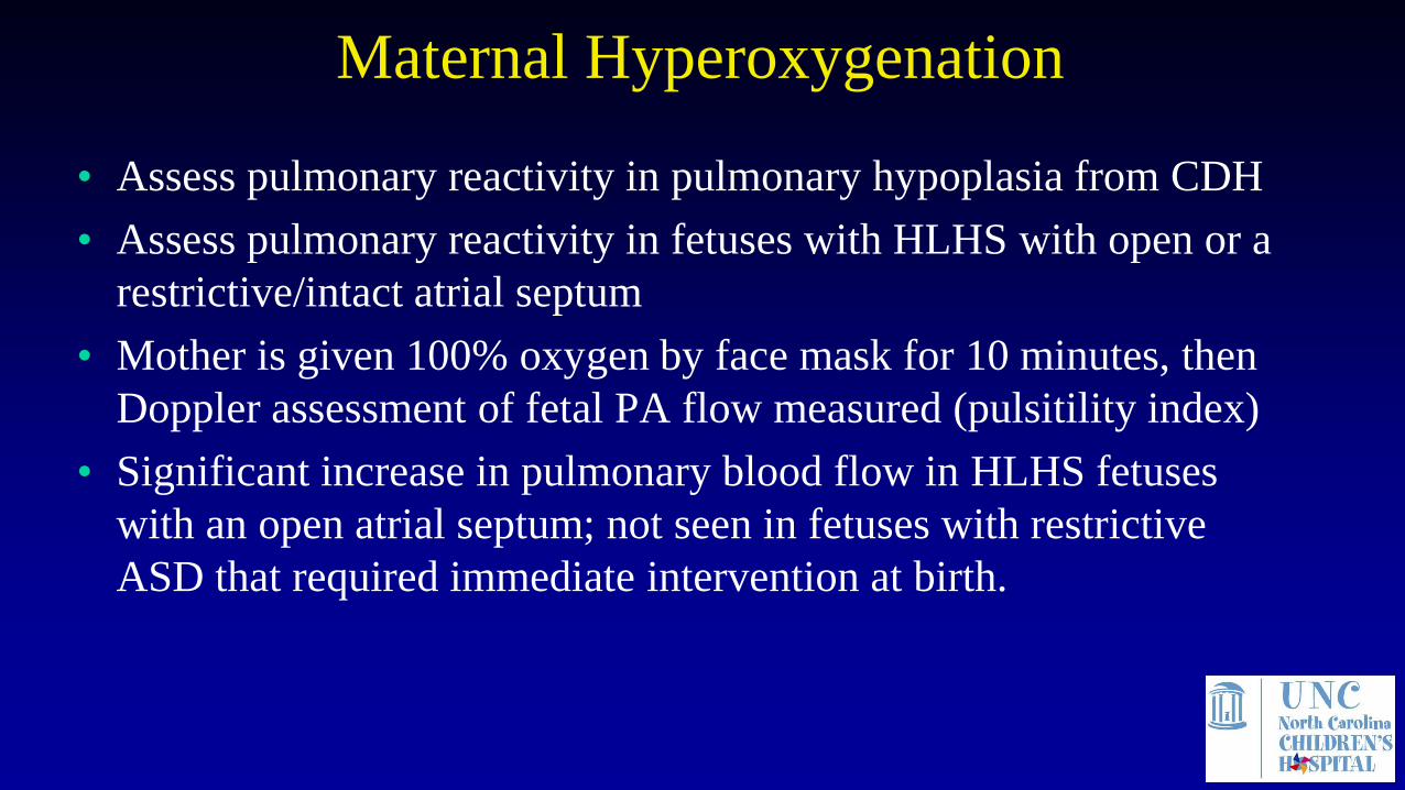
Maternal Hyperoxygenation
• Assess pulmonary reactivity in pulmonary hypoplasia from CDH
• Assess pulmonary reactivity in fetuses with HLHS with open or a
restrictive/intact atrial septum
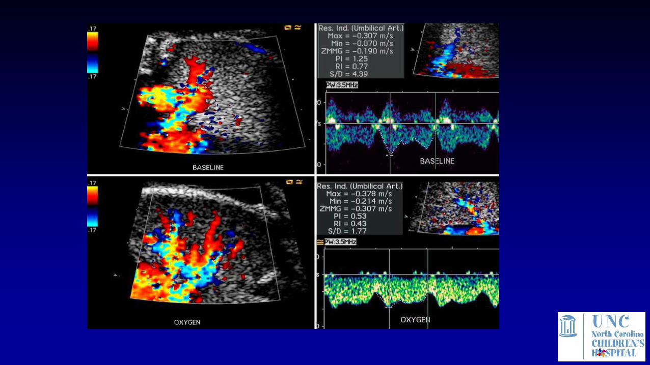
• Mother is given 100% oxygen by face mask for 10 minutes, then
Doppler assessment of fetal PA flow measured (pulsitility index)
• Significant increase in pulmonary blood flow in HLHS fetuses
with an open atrial septum; not seen in fetuses with restrictive
ASD that required immediate intervention at birth.
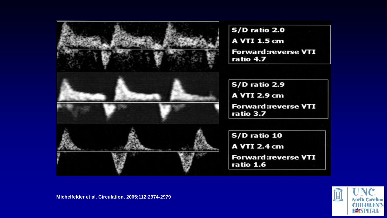
Michelfelder et al. Circulation. 2005;112:2974-2979

0.76
0.530.6
0.4
0.2
0
ASE
0.8
1
Pre O2 During O2
Atrial Septal Excursion with maternal hyperoxygenation
RA LA
ASE Evaluation
Channing et al. Ultrasound Obstet Gynecol 2014
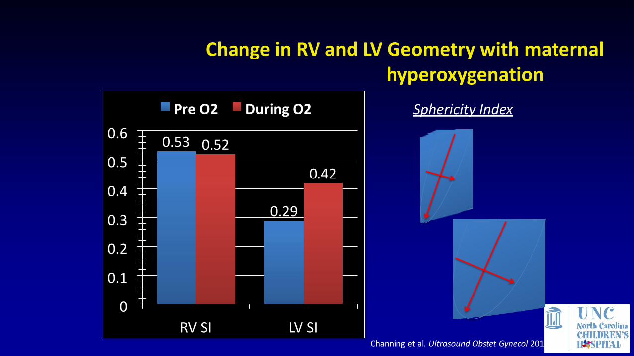
0.53
0.29
0.52
0.42
0.6
0.5
0.4
0.3
0.2
0.1
0
RV SI LV SI
Pre O2 During O2
Change in RV and LV Geometry with maternal hyperoxygenation
Sphericity Index
Channing et al. Ultrasound Obstet Gynecol 2014
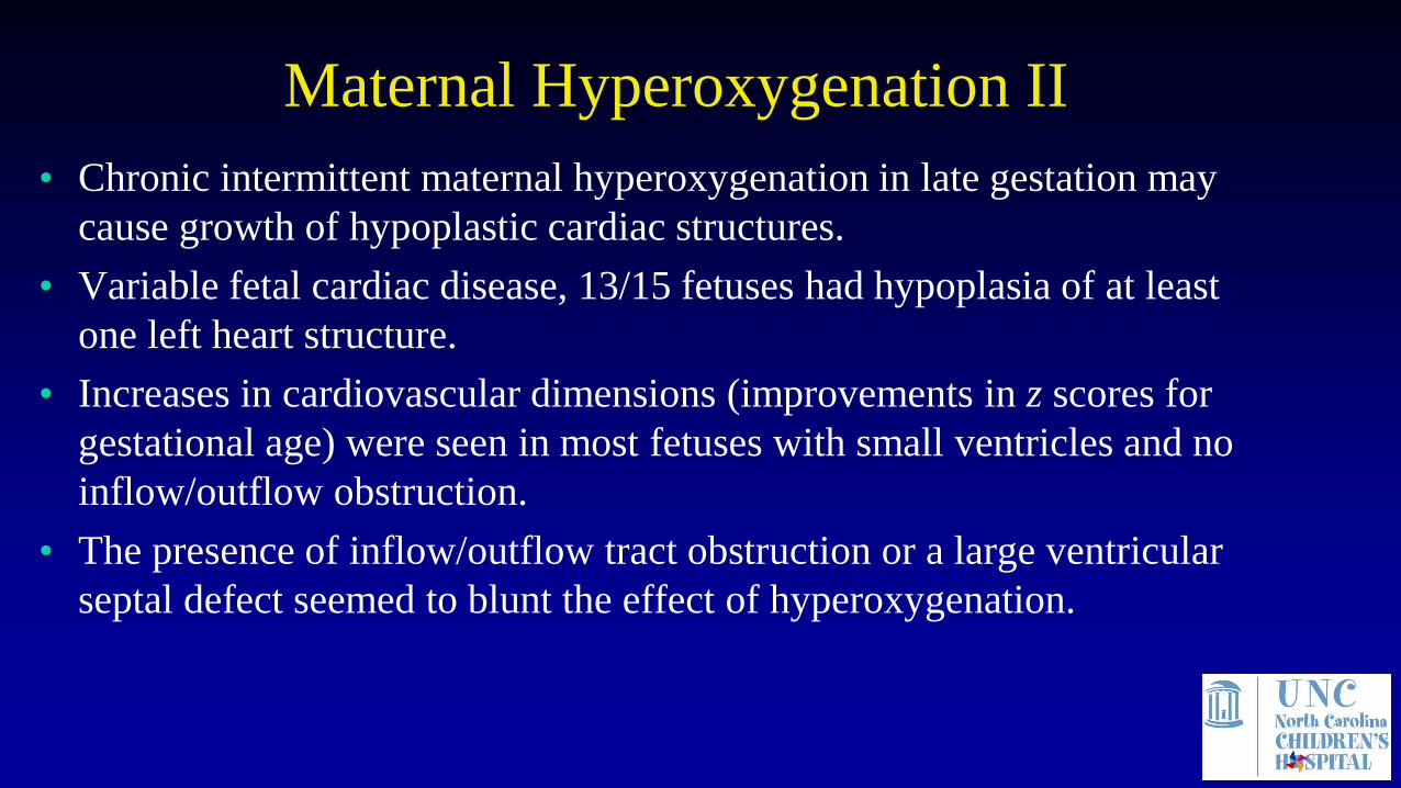
Maternal Hyperoxygenation II
• Chronic intermittent maternal hyperoxygenation in late gestation may
cause growth of hypoplastic cardiac structures.
• Variable fetal cardiac disease, 13/15 fetuses had hypoplasia of at least
one left heart structure.
• Increases in cardiovascular dimensions (improvements in z scores for
gestational age) were seen in most fetuses with small ventricles and no
inflow/outflow obstruction.
• The presence of inflow/outflow tract obstruction or a large ventricular
septal defect seemed to blunt the effect of hyperoxygenation.
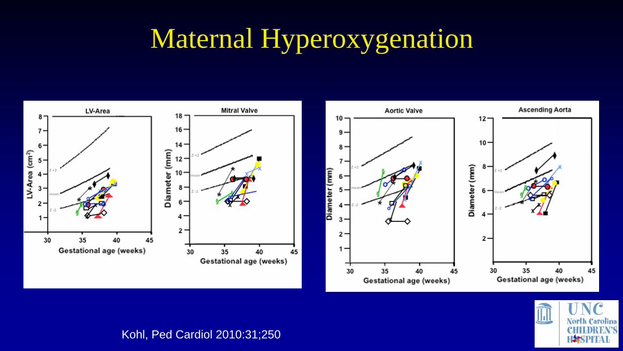
Maternal Hyperoxygenation
Kohl, Ped Cardiol 2010:31;250
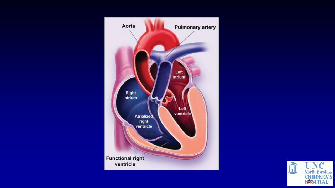
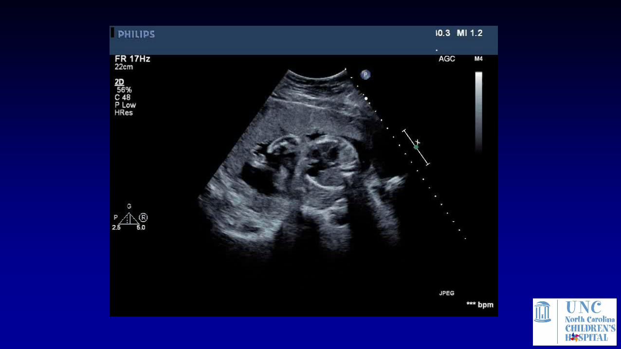
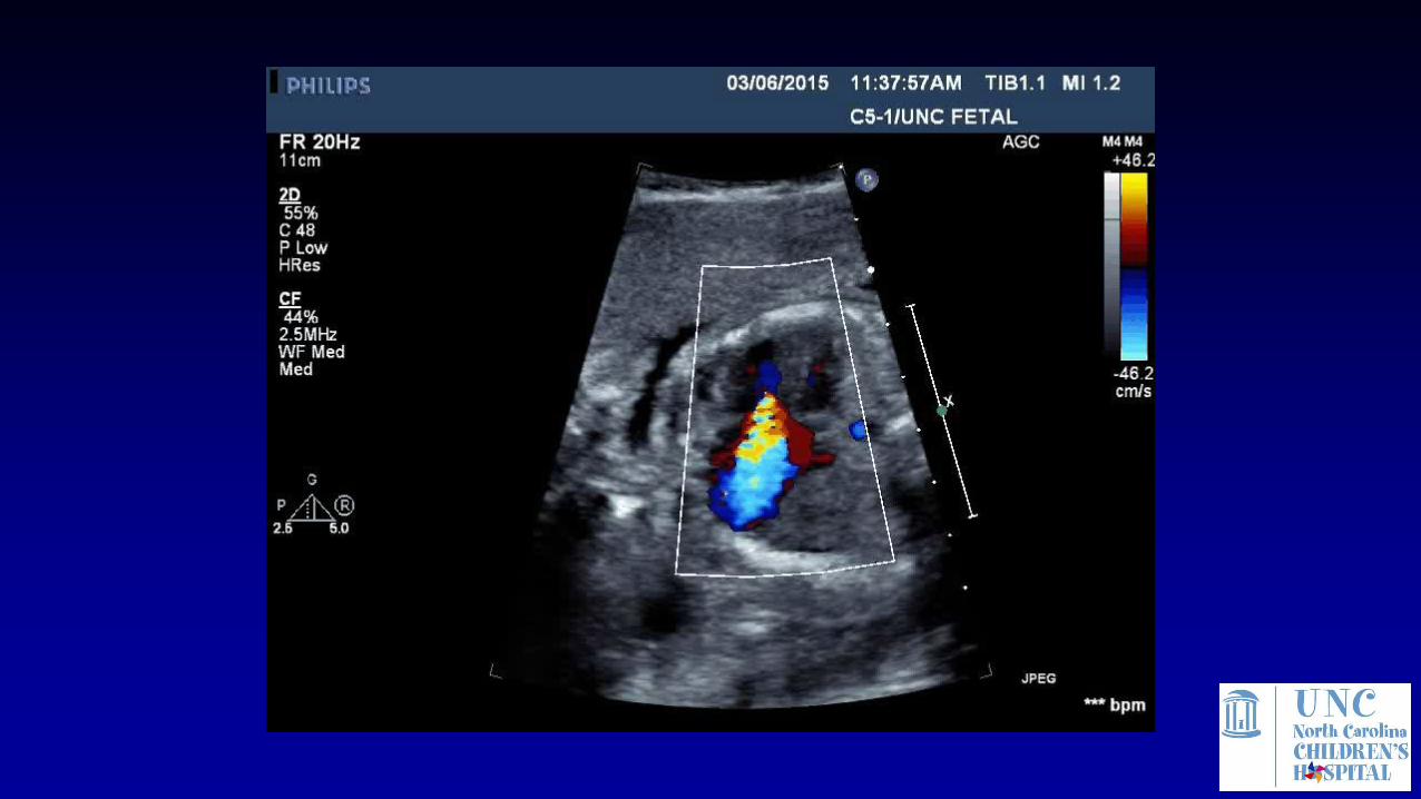
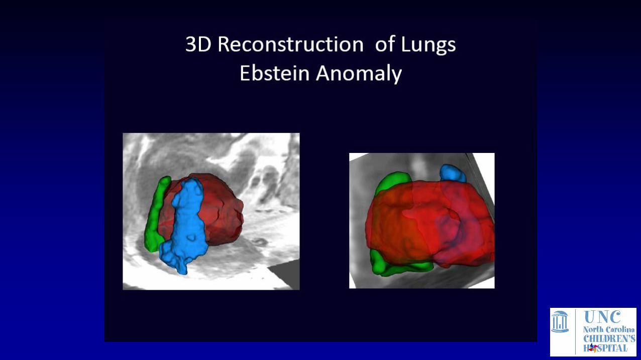
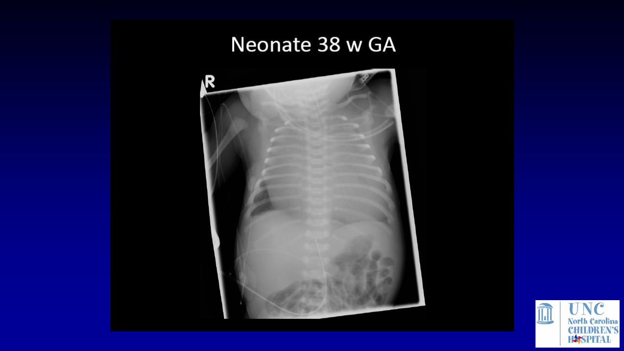
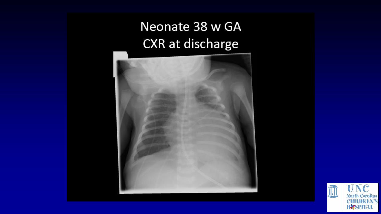
Ebstein Anomaly of the Tricuspid Valve
• Ebstein anomaly is a challenging lesion with high mortality
• Rarely has associated anomalies
• Potential for biventricular circulation vs single LV
• The earlier the presentation, the worse the disease
• Current perinatal survival for severe disease is ~ 50%
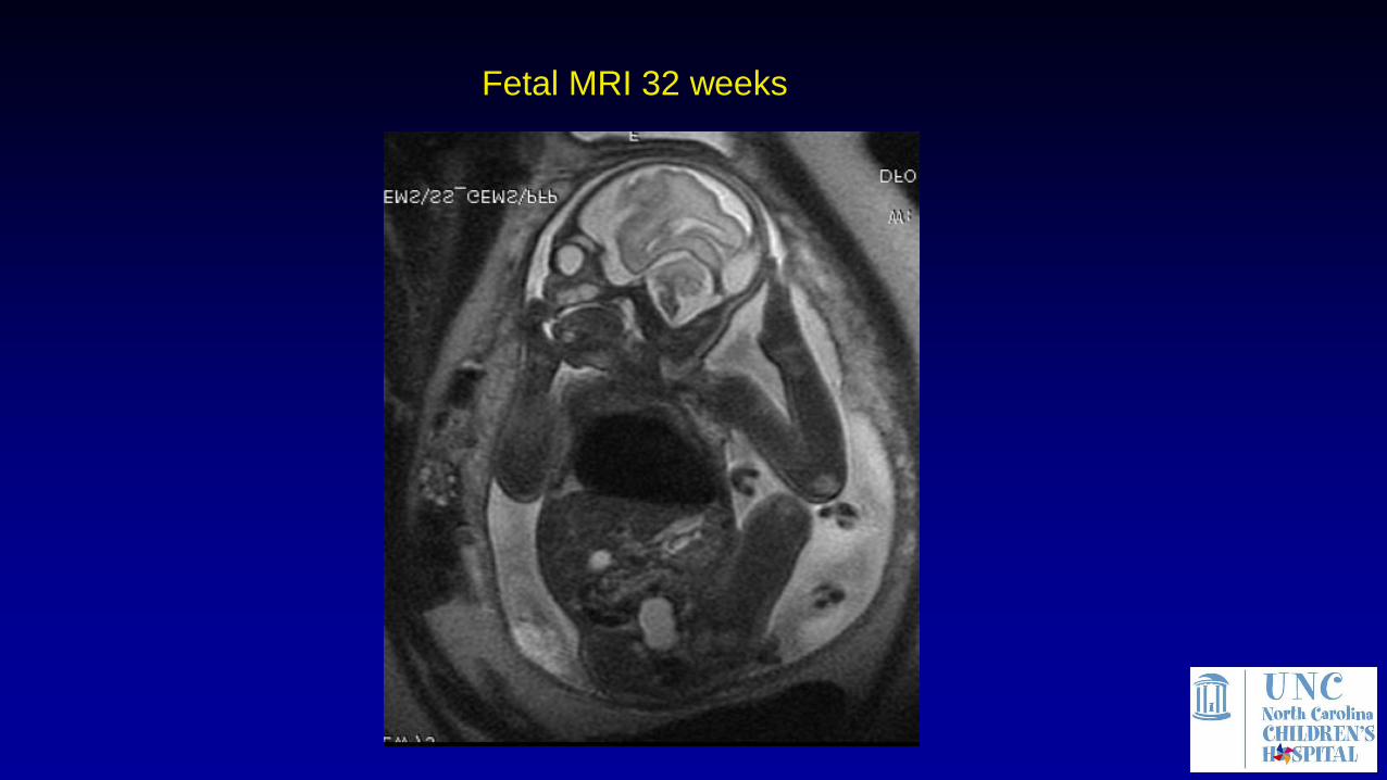
Fetal MRI 32 weeks
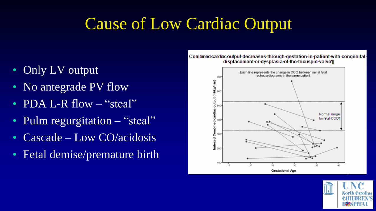
Cause of Low Cardiac Output
• Only LV output
• No antegrade PV flow
• PDA L-R flow – “steal”
• Pulm regurgitation – “steal”
• Cascade – Low CO/acidosis
• Fetal demise/premature birth
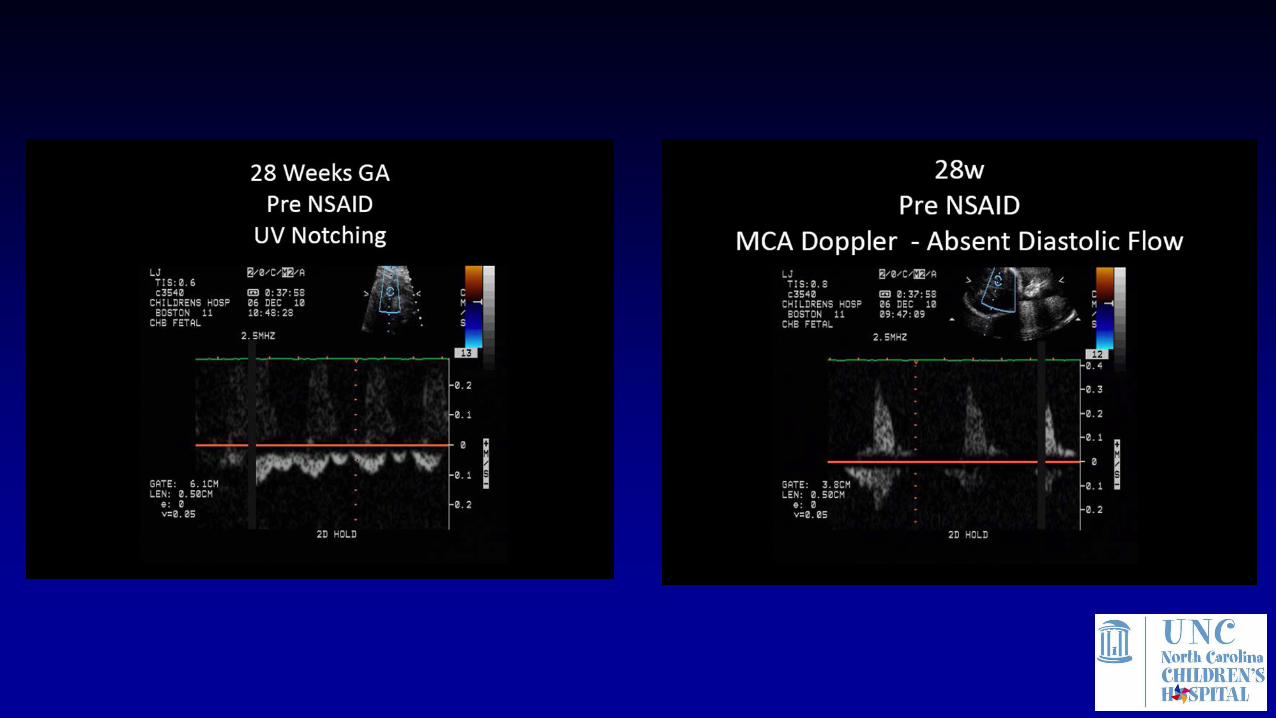
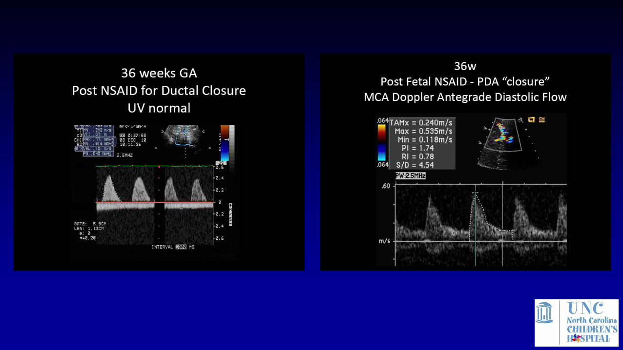
Can a “side effect” be theraputic?
• Ductal constriction is bad for the fetus (NSAID)
• Except…
• What about PDA L-R and PR in severe Ebstein anomaly?
• Could ductal constriction/closure improve hemodynamics?
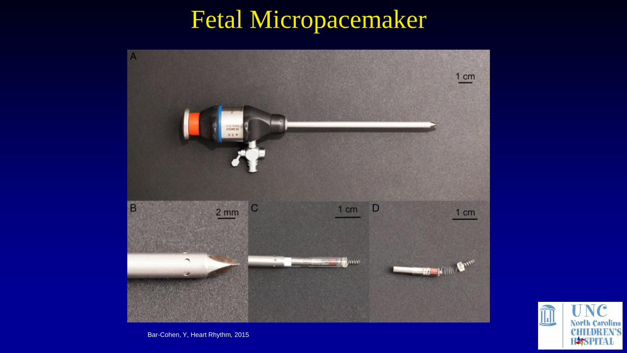
Bar-Cohen, Y, Heart Rhythm, 2015
Fetal Micropacemaker
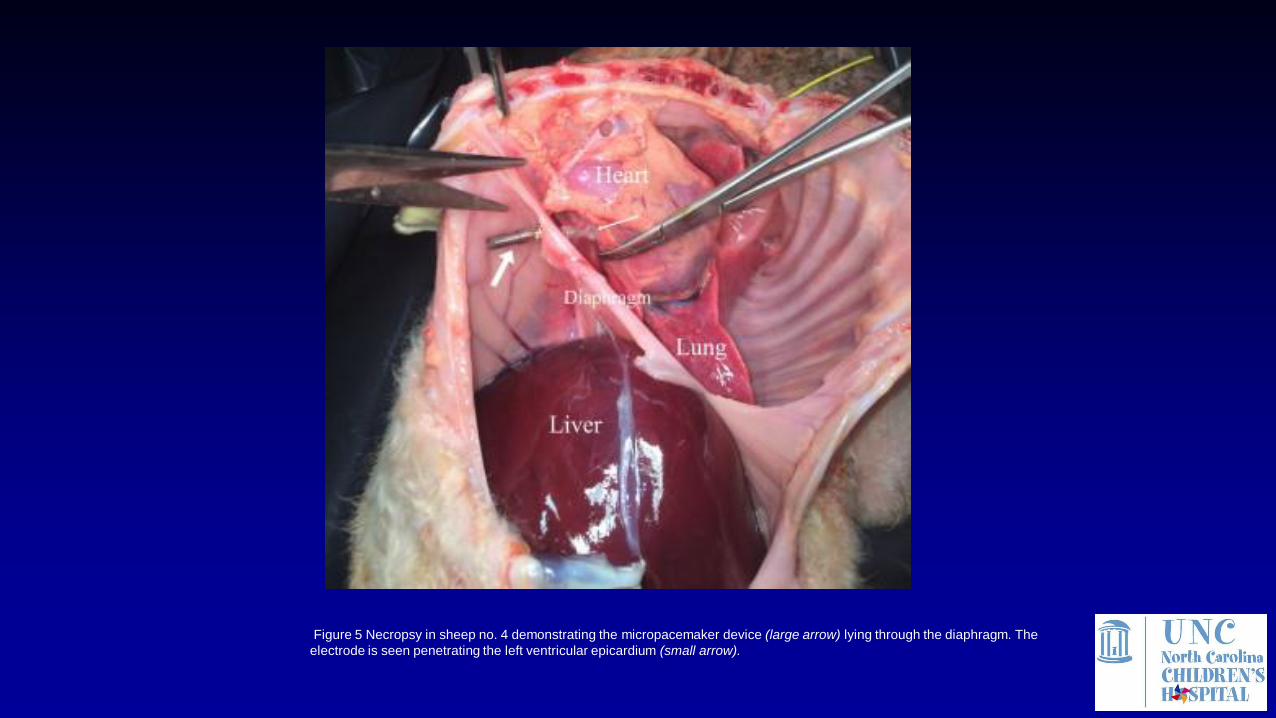
Figure 5 Necropsy in sheep no. 4 demonstrating the micropacemaker device (large arrow) lying through the diaphragm. The
electrode is seen penetrating the left ventricular epicardium (small arrow).
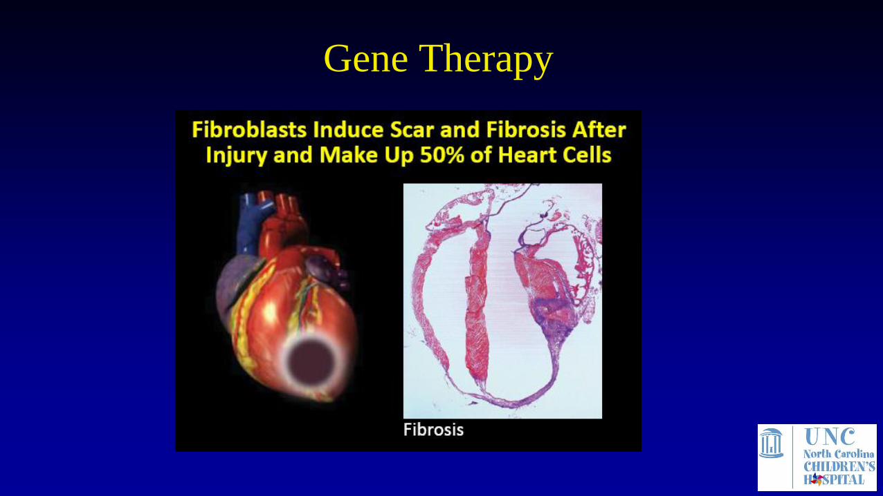
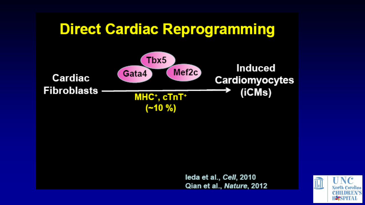
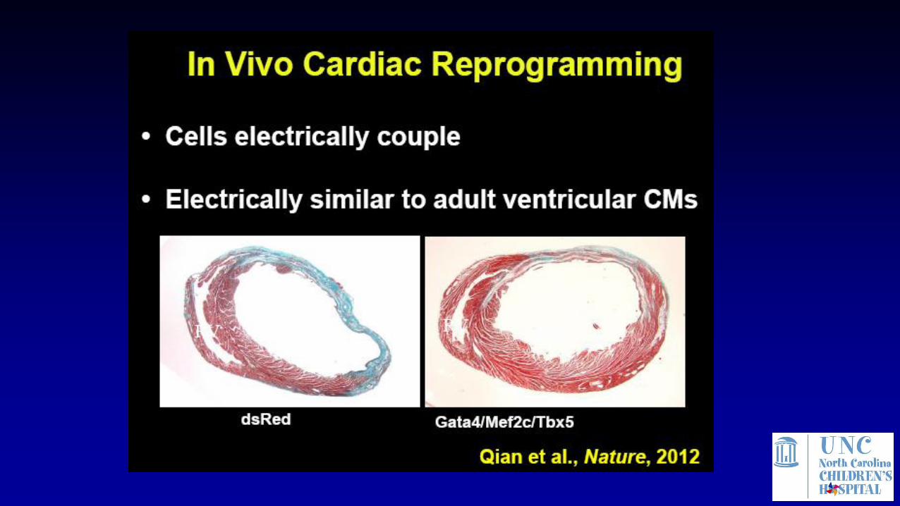
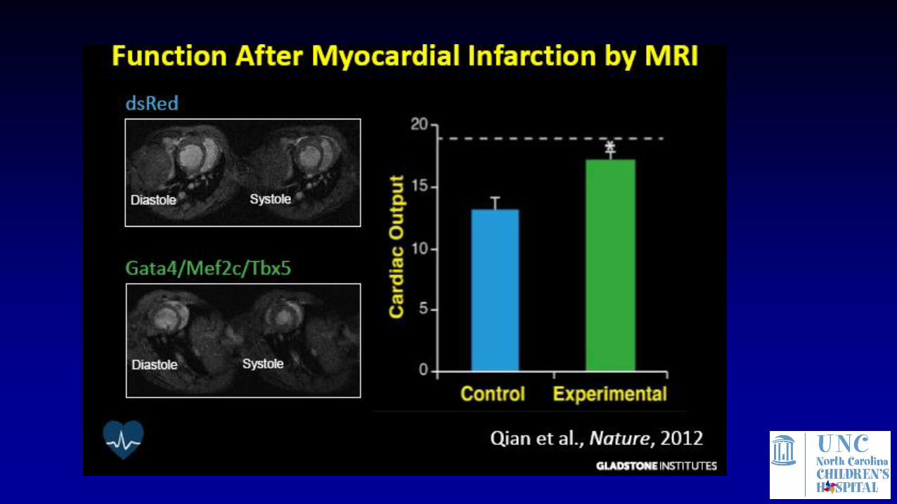
Gene Therapy
The Future of Medicine
• Regeneration of damaged hearts
Personalized stem cell transplants
Harnessing organ’s own cells for regeneration
• Drug Discovery
Drug discovery on human relevant cells
Clinical trials in a dish
• Personalized Medicine – drugs tested on your cells