Topical Membrane Therapy for Chronic Rhinosinusitis
24
6 Topical Membrane Therapy for Chronic Rhinosinusitis Alan Shikani and Konstantinos Kourelis Union Memorial Hospital, Department of Rhinology Baltimore, United States 1. Introduction Chronic Rhinosinusitis (CRS) constitutes a longstanding disease process and a significant health hazard. Its pathophysiology may entail inherent epithelial irregularities, infectious insults, antigenic fermentations, and anatomic abnormalities, acting separately or in cooperation. Hence, various state-of-the-art treatment modalities have evolved, focusing on the surgical restoration of sinus homeostasis: endoscopic approach and visualization, fine surgical tools, power-instrumentation, precise imaging, combination of intranasal and external accesses, and navigation techniques. Despite the impressive technological advances in operative interventions, the medical aspects of CRS have not been investigated to the same extent, and the relevant remedies have changed very little over the years. Topical therapy in CRS is a relatively novel methodology, which relies on the local pharmacological management of sinus inflammatory status, and aims to supplement the existing treatment options. Topically applied medications have been used successfully for decades in dermatology, ophthalmology and urology. This chapter reviews the philosophy of topical therapy for CRS, its applications and effectiveness, as well as our institution’s experience and findings regarding a complete local treatment protocol utilized for the management of refractory CRS. 2. Refractoriness of CRS and the rationale for topical therapy CRS is one of the commonest chronic diseases, affecting 14.2% of the United States population (Lethbridge-Cejku et al., 2004). It places a substantial cost burden on the health care system and is responsible for a considerable portion of sick leaves and decreased productivity(Gliklich and Metson, 1998). The modern opinion points towards a multifactorial etiology which includes fungi, bacterial superantigens, allergy, aspirin sensitivity, exposure to environmental irritants, and lately, bacterial biofilms (Chiu et al., 2008). Moreover, conditions impairing the mucociliary function, such as primary ciliary dyskinesia and cystic fibrosis(Armengot et al., 1994) have also been implicated. The resulting chronic inflammation of the sinus mucosa leads to defense reactions and alterations, i.e. edema, high mucus secretion, cilia loss, and particularly, polyp formation (Meltzer et al., 2004). Surgery to remove the diseased mucosa and open the sinus ostia in order to restore the physiological mucociliary clearance, in combination with systemic antibiotics, has been the www.intechopen.com
Transcript of Topical Membrane Therapy for Chronic Rhinosinusitis

6
Topical Membrane Therapy for Chronic Rhinosinusitis
Alan Shikani and Konstantinos Kourelis Union Memorial Hospital, Department of Rhinology
Baltimore, United States
1. Introduction
Chronic Rhinosinusitis (CRS) constitutes a longstanding disease process and a significant health hazard. Its pathophysiology may entail inherent epithelial irregularities, infectious insults, antigenic fermentations, and anatomic abnormalities, acting separately or in cooperation. Hence, various state-of-the-art treatment modalities have evolved, focusing on the surgical restoration of sinus homeostasis: endoscopic approach and visualization, fine surgical tools, power-instrumentation, precise imaging, combination of intranasal and external accesses, and navigation techniques. Despite the impressive technological advances in operative interventions, the medical aspects of CRS have not been investigated to the same extent, and the relevant remedies have changed very little over the years. Topical therapy in CRS is a relatively novel methodology, which relies on the local pharmacological management of sinus inflammatory status, and aims to supplement the existing treatment options. Topically applied medications have been used successfully for decades in dermatology, ophthalmology and urology. This chapter reviews the philosophy of topical therapy for CRS, its applications and effectiveness, as well as our institution’s experience and findings regarding a complete local treatment protocol utilized for the management of refractory CRS.
2. Refractoriness of CRS and the rationale for topical therapy
CRS is one of the commonest chronic diseases, affecting 14.2% of the United States population
(Lethbridge-Cejku et al., 2004). It places a substantial cost burden on the health care system
and is responsible for a considerable portion of sick leaves and decreased productivity(Gliklich
and Metson, 1998). The modern opinion points towards a multifactorial etiology which
includes fungi, bacterial superantigens, allergy, aspirin sensitivity, exposure to environmental
irritants, and lately, bacterial biofilms (Chiu et al., 2008). Moreover, conditions impairing the
mucociliary function, such as primary ciliary dyskinesia and cystic fibrosis(Armengot et al.,
1994) have also been implicated. The resulting chronic inflammation of the sinus mucosa leads
to defense reactions and alterations, i.e. edema, high mucus secretion, cilia loss, and
particularly, polyp formation (Meltzer et al., 2004).
Surgery to remove the diseased mucosa and open the sinus ostia in order to restore the physiological mucociliary clearance, in combination with systemic antibiotics, has been the
www.intechopen.com

Peculiar Aspects of Rhinosinusitis
92
mainstay of treatment for the past decades(Gosepath and Mann, 2005). The long-term success rate of endoscopic sinus surgery is reported as high as 76%. In the remaining patients, either no improvement is noted, or the CRS recurs soon after treatment. Interestingly, in the majority of failures, the post-operative sinus anatomy demonstrates ostium patency and wide-open ethmoid cavities, abundantly ventilated(Levine, 1990). Specifically, Kennedy has reported that 15% of patients who undergo endoscopic surgery, show mild to no clinical improvement, despite the “optimal” surgical outcome(Kennedy, 1992). These difficult-to-treat patients sometimes demonstrate inflammatory or idiosyncratic features, such as eosinophilia, history of asthma, allergic fungal sinusitis, nasal polyps, and aspirin sensitivity(Zadeh et al., 2002). The common denominator of the above conditions, is an intrinsic pro-inflammatory state of the sinus mucosa which predisposes to clinicopathological exacerbations, in the absence of substantial external irritation.
In addition to the aberrations of the end-organ, that is, the sinus epithelium, an unusual issue of resistance of ordinary bacteria to potent antimicrobials has emerged. This notable finding has been associated with the concept of biofilms, which cover the surface epithelium of paranasal cavities. The common bacterial species H. influenzae, S. pneumoniae, and S. aureus have been identified in biofilms, and their capacity to produce this organic matrix correlates with the refractoriness of CRS. Microorganisms colonizing the biofilms are much less vulnerable to systemic antibiotics which reach the standard tissue Minimally Inhibitory Concentration (MIC). Both the physical and chemical protection imposed by the organic layer on the microbial colonies, call for higher local concentrations of the antibacterial agents (Bendouah et al., 2006).
The principle of the local therapy is prolonged delivery of a highly concentrated drastic substance, whether pharmacological or not, to the sinus cavities, so as to exert its maximal effect on the desired anatomical site, without significant systemic toxicity. Oftentimes, the existing antibiotics and anti-inflammatory medications produce a temporary relief from CRS, combined with mild to moderate side effects, depending on the comorbidities of the patient.
3. Parameters affecting the efficacy of local treatment
3.1 Macro-anatomy
By definition, topical therapy should address thoroughly the target-organ, and reach all the subregions of the diseased paranasal cavities. Several patient- or drug-related factors influence the macro-delivery of medications, but the role of sinus surgery simply cannot be overstated enough.
The paranasal sinuses, have limited communication with the nasal cavity proper, and this is even more evident in the disease state, when the edema and mucociliary impairment further restrict the access to the inflamed regions. This situation changes dramatically after a successful endoscopic surgery. Even if, because of the aforementioned idiosyncratic factors, the CRS persists, creation of wide, readily-accessible surgical cavities is critical for the efficient local application of the therapeutic agents (Fig. 1). The frontal and sphenoid sinuses practically cannot be reached by intranasal administration, while a minimal diameter of 4mm is required for a slightly accessible maxillary ostium(Harvey and Schlosser, 2009). During an endoscopic procedure, the maxillary entrance can be opened as widely as 2.5cm,
www.intechopen.com

Topical Membrane Therapy for Chronic Rhinosinusitis
93
and the entire anterior wall of the sphenoid sinus may be removed. Regardless of the technique used for drug administration, the penetration in unoperated sinuses does not exceed 3% of the total volume placed intranasally(Hyo et al., 1989). On the other hand, radical surgical dissection allows contact of the drug with up to 96% of total sinus internal surface(Miller et al., 2004). Exactly how much improvement is provided by sinus surgery is difficult to assess though, as the various operative techniques are different in terms of intervention, and range broadly from minimally invasive (e.g. balloon sinuplasty) to extremely aggressive (e.g. modified endoscopic Lothrop procedure). Apart from the apparent gain in the total sinus surface contacted directly by the topical agents, clinical studies document as well that steroid sprays, when used by patients who had sinus surgery, produce more significant improvement of symptoms, endoscopical and histopathological findings, than in CRS sufferers having not being treated surgically(Lavigne et al., 2002).
Fig. 1. In the unoperated patient (Left), the lateral nasal wall is an anatomical barrier to the delivery of topical medications, whereas the post-surgery paranasal cavities (Right) are accessible through wide windows.
Individual anatomical details, further modify the pharmacological penetration into the
paranasal cavities. Inferior turbinate hypertrophy limits the intranasal flow, whereas in case
of uncorrected nasal septal deviation, accumulation of the local agent immediately anterior
to the spur is noted. Moreover, the variations taking place during the nasal cycle influence
the temporal pattern of drug dissemination. Altogether, a patent nasal passageway, not
narrowed by all of the above anatomical factors, permits 90% penetration rate of the locally
applied agents(Unno et al., 1983).
A much debatable issue related to the delivery of local treatment is the patient’s optimal
positioning during irrigations or nebulizations. The traditional “Mecca” position with the
head brought forward is becoming now less popular than placing the head backward.
Lateral positions have also been proposed as more appropriate for delivery to the frontal
sinus(Karagama et al., 2001).
A third consideration regarding the distribution of locally applied drugs is the configuration of sinuses in relation to gravity. Whereas the roof of the ethmoid and the frontal recess are dependent areas, so that the pharmacological deposits drain rapidly downwards, the
www.intechopen.com

Peculiar Aspects of Rhinosinusitis
94
maxillary antrum with its highly-situated ostium retains most of its contents, until they are cleared by the mucociliary mechanism.
3.2 Micro-anatomy
Even when the local agent enters the sinuses in sufficient quantities, its efficacy is not guaranteed, as it needs to reach its pharmacological target, and stay in contact for an adequate amount of time. This becomes particularly important in the sinonasal cavities, where the microenvironment is structured specifically to eliminate foreign particles, including medications, using several clearance mechanisms.
All sinus surfaces are covered by a mucus layer with the purpose to entrap foreign particles and filter the inhaled air. The production rate of the mucus fluctuates greatly, depending on the inflammatory status of the epithelium. Understandably, CRS is a condition predisposing to mucosal irritability and subsequent hypersecretion of thick mucus(Harvey and Schlosser, 2009). The thickness of the viscous gel phase of the mucus layer, which overlaps the respiratory cell cilia, varies from 7 μm in healthy mucosa, to 200 μm in high-grade inflammatory states(Tarran et al., 2001). Mechanical removal of the viscous mucus blanket, by high-volume sinus rinses results in more potent effect of locally administrated steroids(Daviskas and Anderson, 2006).
The mucus contents are as important as its physical dimensions and texture. Besides water, organic salts, enzymes, and immunoglobulins, the mucins constitute the basic ingredient of this supra-epithelial blanket. These are glycoproteins responsible for the viscous consistency of the mucus. In detail, mucins form fibers which bind to each other via cross-linking attachments, to make up a web that serves as the skeletal component of the three-dimensional layer. The mucin tangle contributes to the support of air particles, but also presents a network of hydrophobic sites, that act as receptors for macromolecules with similar physicochemical properties. Hydrophobic molecules are retained in the mucus and exert a prolonged pharmacologic effect(Ugwoke et al., 2005). On the basis of this finding, conjugation of topical medications with mucoadhesive gels, aiming to achieve sustained drug release, has been proposed(Nakamura et al., 1999).
In contrast with all the other Head and Neck sites, the sinonasal cavities are covered by respiratory-type epithelium, instead of the standard squamous-cell epithelial layer. Material that is captured in the mucus is gradually propelled outside the sinus cavities and carried to the nasopharynx, by means of constant ciliary beating. An intact mucociliary mechanism can clear the entire maxillary sinus of foreign particles in less than twenty minutes(Harvey and Schlosser, 2009). In CRS though, the chronic infection impairs the ciliary function, in favor of the prolonged residence of topically delivered medications. On the other hand, it is suggested that the active transfer of medications to the choanae might actually enhance their distribution to remote mucosal subsites(Goh and Goode, 2000).
3.3 Major delivery techniques
In the history of rhinosinusitis topical therapy, several methods for the delivery of the drastic agent have been utilized: Fluid irrigation, spray pumps, drops/powder/gel instillation, nebulization, and regional injection, aim to provide optimal spatiotemporal conditions of contact between the medication and its target.
www.intechopen.com
Topical Membrane Therapy for Chronic Rhinosinusitis
95
Fluid irrigations remain a traditional, simple, and well-tested technique for conveying treatment formulas directly to the sinonasal surface epithelium. It is well-established that commercial nasal sprays, do not penetrate the frontal and sphenoid sinuses. On the contrary, a high volume of liquid solution (over 100mL) ensures access into these unapproachable sinuses. In post-operative cases, irrigations with a bulb syringe are superior to every other delivery methods, in terms of access to anatomical subsites. Yet, up to 30mL of solution pour out immediately from the nasal cavities, so that a considerable irrigation volume is wasted(Miller et al., 2004). Given a specific volume of solution, the pressure of irrigations can be modified by the device used. Low-pressure lavage using commercial pots, seem to be suitable for unoperated sinuses, whereas high-pressure douches delivered by squeeze-bottles are proper in case of surgically created open cavities(Harvey and Schlosser, 2009).
Nasal sprays have been classically used to provide local application of drugs in rhinosinusitis. Among the various devices developed over the years (spray bottles, aqueous pumps, dry powder atomizers), aqueous spray pumps are most accepted. Such pumps contain a medication-containing solution, which is released in the form of droplets. Smaller, lighter droplets demonstrate a broad distribution across the mucosal surface, as they travel a longer distance from the nostril. The viscosity of the solution is an additional factor, as thicker liquids project in a narrower cone and do not reach the peripheral intranasal regions(Kundoor and Dalby, 2010). Despite the refinements of spraying pumps, the droplets barely penetrate the sinuses in unoperated patients, and their effect is essentially restricted to the nasal cavities only. The maximal concentration of the sprayed agent is detected in the anterior nasal cavity, due to the obstructive mass of the inferior turbinate. Half of the dose does not approach the ostiomeatal complex, an anatomical structure central to the pathogenesis of CRS(Merkus et al., 2006).
Nebulized medications are a novel topical approach to rhinosinusitis, and have been used for the past decade in clinical practice in Europe. In Japan, they were adopted in 1950, and in the United States, nebulizers and nebulized medications are covered by most medical insurances(Vaughan and Carvalho, 2002). Nebulization devices provide an aerosolized mist which is created by a mechanical pulse. The latter is produced either by a high-pressure jet, or a vibrating mesh. The earliest devices emitted an aerosolized stream of particles larger than 10μm, and the penetration of medications into the sinuses was limited as most of the particles are filtered by macro- or micro-anatomical barriers. Innovative technologies are now capable of generating airflow consisting of particles with a diameter less than 3μm, and accumulation on sinus mucosa is much more extensive. The main advantage of nebulizers, in comparison with the traditional spray pumps, is the deposition of pharmacological agents in the posterior nasal cavity. Moreover, sprayed formulations are undetected in the sinus cavities of patients who have not had surgery, whereas 8% of intranasally placed aerosols remain in the sinuses(Moller et al., 2010).
4. Systemic absorption
The advantage of topical therapy is the accumulation of very high concentrations of medications directly at the target site. Equivalent doses would not be possible to be administered systemically, due to unacceptable toxicity. Nonetheless, the systemic absorption of locally applied drugs should be always kept in mind, so that potential side-
www.intechopen.com
Peculiar Aspects of Rhinosinusitis
96
effects are avoided. The nasal mucosa incorporates a rich capillary network, and certain substances applied onto the broad epithelial cover of the sinus cavities may reach high concentrations in plasma. The significance of drug absorption by the nasal mucosa is evident from the strong interest in the design of intranasally administered systemic treatments for miscellaneous diseases, e.g. diabetes (insulin), migraine (propranolol, sumatriptan), smoking cessation (nicotine), osteoporosis (calcitonin), and acromegaly (octreotide)(Ranade, 2001).
Orally administered medications undergo first-pass hepatic metabolism, and therefore a portion of the dose does not reach the systemic circulation. This is not the case in topical sinus medications, which are absorbed directly by the surface respiratory epithelium, and thus by-pass liver metabolism. However, the epithelial target cells of the nasal mucosa, contain an array of drastic enzymes, which also metabolize the pharmacological deposits. The levels of Cytochrome P-450, which participates in hepatic metabolism, are extremely high in the nasal mucosa, too. Phase II enzymes, like glutathione-transferase, which transfer micromolecular groups to the metabolized medications, are also prevalent. The nasal mucosa is deficient in proteases, though. Consequently, proteins are not lysed topically, and their absorption rate is substantial(Chien and Chang, 1987).
5. Agents used in topical therapy
5.1 Saline
Prior to the local application of therapeutic antimicrobial or anti-inflammatory agents, mechanical cleansing of the sinuses with saline irrigations has been one of the oldest and most widely used methods for the management of CRS. Mucopurulent secretions filling up the infected cavities are a frequent finding in CRS exacerbations. Furthermore, during the post-operative period following endoscopic sinus surgery for chronic rhinosinusitis, a collection of old blood, crusts, necrotic debris, or allergic fungal mucin, is accumulating periodically and regular meticulous cleansing is as important as the surgical procedure itself(Palmer and Kennedy, 2003). Office debridement, with the help of curved suctions is the optimal way to maintain sinus health. However, it is impractical and uncomfortable for the patients to visit the rhinologist too often for debridements as the only means of removing the “toxic” material. Our own policy is performing this debridement on a weekly basis, until the sinus cavities are clean. Frequent, as needed saline irrigations may be performed easily at home and are the simplest and least expensive form of topical therapy.
The appropriate saline concentration for the sinus lavage is controversial. Iso-, hypo-, and hyper-tonic salive, as well as Ringer’s Lactate solution, have all been tested for their efficacy and side effects. Isotonic saline is the basic irrigation solution, as it provides only mechanical cleansing, without creating an osmotic gradient between the sinonasal cavities and the surface epithelial cells. It is suggested though that the isotonic concentration also modifies the rheological properties of the mucus, making the secretions less viscous. On the other hand, hypertonic solutions have been introduced subsequently into CRS management, as they decrease mucosal edema by creating an efflux of water from the intercellular space. Not only that, but it is documented that hypertonic irrigations improve the mucociliary function, in comparison to isotonic saline. High salt concentration in the sinus cavities is postulated to promote intracellular calcium release, which sets off the biochemical cascade resulting to cilia movement(Daviskas et al., 1996). Hypertonic sinus lavage also has an effect on allergic
www.intechopen.com
Topical Membrane Therapy for Chronic Rhinosinusitis
97
rhinitis, a major component of the CRS pathogenesis. Its main drawback however, is the discomfort often reported by the patients.
Apart from their chemical composition and salt concentration, a second characteristic of saline douches, unique in topical therapy, is the high volume of solution used in each irrigation. The importance of volume and pressure parameters has already been described.
Altogether, the benefit from saline irrigations to the management of CRS includes the
mechanical removal of infectious/irritating/allergenic material, decrease of mucosal edema,
improvement of the mucociliary function, and thinning of mucus secretions. Usually, saline
rinses are prescribed in combination with other means of topical therapy, and there is
evidence that they enhance the bio-supply of the primary medications(Papsin and
McTavish, 2003).
5.2 Corticosteroids
Intranasal steroids have been initially the mainstay of topical therapy for allergic rhinitis.
Due to their potent anti-inflammatory action, especially the deceleration of late-phase
response, they diminish the manifestations of nasal allergy (congestion, rhinorrhea,
pruritus). Their anti-decongestant properties in allergic rhinitis had been appreciated, and
local steroids were subsequently introduced to the treatment of acute bacterial sinusitis, as
adjuncts to systemic antibiotics. Interestingly, the infectious edema of acute sinusitis
requires higher doses of steroids than those administered in allergic rhinitis(Moller et al.,
2010). In CRS, local steroids were at first prescribed cautiously, and only in case of
exacerbations, which resemble the pathophysiology of acute rhinosinusitis. It was the
beneficial effect of steroids on nasal polyposis and hyperplastic sinusitis, that indicated their
prolonged use in CRS. When steroids are administered orally, they have a clearly superior
effect on nasal polyps (“medical polypectomy”), than any form of topical treatment(Palmer
and Kennedy, 2003). Naturally, the advantage of steroid sprays is their capacity for long-
term use with minimal side-effects. Perhaps the effective way to get the most out of
corticosteroid therapy, is a combination of “induction” systemic administration to reduce
the severe edema, along with a “maintenance” schedule of intranasal spaying, to control the
continuous, low-grade inflammation of CRS(Wahl and Otsuji, 2003).
Local steroids are administered in several forms, in order to achieve greater efficacy.
Intranasal placement of fluticasone drops has demonstrated a distinct benefit in patients
with hyperplastic CRS, precluding the need for endoscopic surgery in half of the cases. The
authors suggest that nasal drops are more successful than sprays, as they reach easier the
middle meatus(Aukema et al., 2005). A modification of drops administration involves direct
instillation in the office, utilizing a soft catheter, under endoscopic vision. This method
accomplishes focused application in difficult-to-approach regions, such as the frontal
recess(Palmer and Kennedy, 2003). A more invasive technique, combining the efficacy of
systemic treatment with the low side-effect pharmacological profile of local drops, utilizes
injection of steroids into the polyp mass. Although this procedure has been performed
enthusiastically in the 1950s, reports of visual loss emphasized the need for
cautiousness(Mabry, 1981). Embolization or spasm of the central retinal artery, have been
hypothesized as the mechanism of blindness. Placing the injection into the center of the
www.intechopen.com

Peculiar Aspects of Rhinosinusitis
98
polyp lessens the risk of intra-arterial administration. Possibly, intra-polyp steroid injections
do not have a role in the routine treatment of common CRS, but their efficacy could be
useful in recalcitrant cases, not responding to oral or instilled corticosteroids(Antunes and
Becker, 2010).
The chronic topical therapy with steroids has raised concerns of absorption into the
circulation, and their well-known systemic effects: growth inhibition due to hypothalamic
suppression, loss of bone density, hypertension, diabetes, and psychosis(Demoly, 2008).
From each sprayed dose, 30% of the medication stays within the sinonasal cavities, and
undergoes metabolism on the nasal mucosa, whereas the remaining 70% follows the oral
route and is subject to hepatic metabolism. Altogether, the absorption into the circulation
depends largely on the steroid compound, and ranges from 49%(flunisolide) to less than
0.1%(mometasone). A multitude of clinical studies has investigated the safety profile of
intranasal steroids and no significant systemic side-effect was reported, either in adult or
pediatric patients. Specifically for the latter, one-year duration of administration did not
impede growth(Schenkel et al., 2000). Local adverse effects (dry rhinitis, epistaxis),
sometimes causing considerable discomfort, have been documented, though(Giger et al.,
2003). Interestingly, even in the case of intranasal injection, there is no clinical or biochemical
evidence of adrenal suppression, although raised plasma concentration of the steroid has
been noted(Mabry, 1981).
5.3 Antimicrobials
Unquestionably, microorganisms have a fundamental role in the pathogenesis of CRS, either
by maintaining prolonged infectious processes, or by generating toxic allergic reactions.
Antibiotics have been persistently used for acute and chronic rhinosinusitis, in oral or
intravenous form. The rationale for topical administration derives from the concept of
biofilm, which is clearly an “epi-mucosal” phenomenon(Lim et al., 2008). Among the
various advantages that the microenvironment of biofilms provides to microorganisms is its
poor penetration by systemically administrated antimicrobial agents(Stewart and Costerton,
2001). Moreover, the bacterial species found in biofilms, are no different from those
commonly identified with conventional cultures in CRS (Al-Mutairi and Kilty, 2011).
Interestingly, the minimal antibiotic concentration for the eradication of microorganisms
residing in biofilms, can be as high as 1000 times their Minimal Inhibition Concentration
(MIC) in the cultures from the same bacteria(Ceri et al., 1999). Therefore, chronic sinus
infections refractory to culture-directed oral antibiotics was considered an indication for an
alternative approach, which could overcome resistance by delivering high concentrations of
medications in direct contact with the colonized epithelial coating.
The choice of antibiotic should be based on endoscopically-guided culturing of sinus
secretions, keeping in mind the multi-pathogen etiology of CRS. When it comes to selection
among drugs to which microbes demonstrate equal sensitivity, antibiotics that kill bacteria
once they reach a critical concentration (concentration-dependent), like quinolones or
aminoglycosides, may exert a more potent bactericidal effect, in comparison with time-
dependent antibacterial medications. The latter, although effective in lower levels, require
prolonged action at the target site. As already mentioned, the constant beating of the cilia
www.intechopen.com

Topical Membrane Therapy for Chronic Rhinosinusitis
99
propels the mucosal coating out of the cavities in less than twenty minutes. Thus, antibiotics
such as penicillins, cephalosporins, or macrolides, which are first-choice systemic treatment
options for sinusitis, cannot be considered ideal for topical therapy, due to their rapid
clearance(Palmer and Kennedy, 2003). An additional factor influencing the selection of
topical agents is their differential metabolic processing locally and systemically. Drugs
which are deactivated promptly in plasma, but remain intact in the sinus secretions, are both
effective and safe. Mupirocin belongs to this class, and is currently the only FDA-approved
medication for intranasal use(Uren et al., 2008). With regard to non-bacterial causes of CRS,
fungi are considered a prominent etiologic factor, in up to 90% of cases in several
studies(Ponikau et al., 1999). Therefore, antimycotic agents, like amphotericin B and
itraconazole, are promising local agents, since their chronic systemic administration
produces serious adverse effects.
A few delivery methods have been tried for local antimicrobial therapy. Spraying of
solutions with the use of atomizers, which is an efficient technique in the case of nasal
steroids, has produced the poorest results. Small mucosal surface of initial application, along
with the slow mucocliliary clearance which commonly accompanies CRS, possibly result in
a limited area of drug deposition(Sykes et al., 1986). In contrast, sinus lavage with
antibacterial solutions is more popular, and seems to be more effective as well. Frequent
bottle irrigations with 300ml of ceftazidime, an antibiotic not available in oral form, are
successful in eradicating Pseudomonas from patients with recalcitrant sinusitis(Leonard and
Bolger, 1999). However, their efficacy may not be dependent solely on the proper delivery of
the antibiotic, but also on the effects of the lavage itself, that is, the mechanical cleansing,
dilution of mucus, and decrease of mucosal edema(Lim et al., 2008). A more advanced
irrigation technique, employing endoscopic catheterization of the middle meatus, has
achieved the resolution of CRS in the morbid context of cystic fibrosis. Yet, this modification
of sinus rinsing requires frequent office visits, and interferes with patient compliance(Moss
and King, 1995). Nebulization of antibiotics has emerged as both an effective and convenient
delivery method. When the size of aerosolized particles is optimized to less than 5μm, this
form of topical antimicrobial therapy is superior even to IV mode of administration. Similar
to the case of antibiotic irrigations, an additional, non anti-infectious, beneficial mechanism
of the aerosolized stream was postulated. Possibly, nebulization into the sinonasal cavities
promotes anti-inflammatory and anti-edematous effects(Lim et al., 2008).
The findings from studies investigating the efficacy of topical antimicrobial therapy, are
quite encouraging. Up to 88% of patients experience significant improvement, good quality
of life, and few local side effects (rhinitis), after four weeks of treatment(Vaughan and
Carvalho, 2002). This symptomatic relief is concurrent with reversal of the endoscopic
findings. Refractory infections by resistant strains of Staphylococcus aureus in particular,
respond dramatically to irrigations with mupirocin solution(Uren et al., 2008). On the other
hand, topical application of antifungal agents did not produce a distinct therapeutic result in
the management of CRS(Weschta et al., 2004). This finding is not in agreement with the
hypothesis that fungal infection accounts for the majority of chronic sinusitis. Since fungi are
ubiquitous in the environment and the sinus cavities, they seem impossible to be eradicated
simply by local administration of antimycotic drugs. In the case of CRS induced by fungi,
the pathogenesis entails an immune host reaction against fungal antigens, and as a result,
www.intechopen.com

Peculiar Aspects of Rhinosinusitis
100
inflammatory modifiers might be more appropriate than any anti-infectious remedy(Lim et
al., 2008).
5.4 Mucoactive agents
As stated previously, biofilms may alter significantly the pathophysiology of CRS and protect the pathogens from systemic or local treatments, thus perpetuating the infection. Not only that, but it is suggested that this bioorganic coating acts as a reservoir of bacteria, and releases microorganisms in the conventional “planktonik” form, into the sinus cavity(Al-Mutairi and Kilty, 2011). Obviously, novel approaches to refractory sinusitis cases, focus on the elimination of this enigmatic entity. Being a distinct structure, analogous to a foreign body, biofilm is an ideal target for topical therapy, as the regional vasculature might not deliver sufficient amounts of systemic medications to the interface between the epithelium and the colonized structure.
Surfactants are ampthipathic compounds, that is, they possess both hydrophilic and
lipophilic properties. Consequently, they are soluble both to water and organic substances.
The well-known pulmonary surfactant decreases the adhesiveness of sputum to the lung
respiratory epithelium, and facilitates the removal of mucus from the lung parenchyma. It is
hypothesized that in a similar fashion, intranasally administered surfactants interfere with
the adherence of the biofilm layer to the underlying sinus epithelium(Suh et al., 2010). This
would result in biofilm peeling off the sinus walls, and the transition of recalcitrant CRS to a
less complicated form, amenable to treatment. This scenario has been clinically tested in
patients irrigating with baby shampoo, an inexpensive, nontoxic mixture of various
surfactants. Almost half of the patients reported a marked improvement in their “mucus-
related” symptoms, i.e. thick nasal discharge and post-nasal drip(Chiu et al., 2008).
Secondary effects of surfactants have also been postulated, such as destabilization of
bacterial membranes with leaking of electrolytes.
A promising local agent, due to its safety profile and low cost, is honey. In vitro testing
documented eradication of Staphylococcus aureus and Pseudomonas aeruginosa in biofilm
colonies, after treatment with several types of honey. Notably, honey was effective even
against Methicillin Resistant Staphylococcus Aureus (MRSA), which is considered a plague
of our time(Alandejani et al., 2009). The exact antimicrobial and mucoactive properties of
honey are yet to be discovered.
5.5 Decongestants
Intranasal decongestants are the most frequently local agents used by sinusitis patients
(16%) in the United States, more often even than local steroids. Perhaps the high prevalence
of use can be explained by their availability over the counter, as well as the rapid, almost
immediate, relief they provide from nasal congestion. Physicians usually prescribe a short-
term course of decongestant sprays only in cases of severe, acute exacerbations of
CRS(Sharp et al., 2007). Local decongestants are sympathomimetic agonists, which stimulate
alpha-adrenergic receptors on the smooth-muscle fibers of the vessels beneath the nasal
respiratory epithelium. As a result, brisk, potent vasoconstriction ensues. Interestingly, local
sympathomimetics and steroids produce decongestion via different mechanisms, so that an
www.intechopen.com

Topical Membrane Therapy for Chronic Rhinosinusitis
101
additive effect could be accomplished by concurrent use of the two medications(Yoo et al.,
1997). Although vasoactive decongestants reverse fast and effectively the mucosal edema,
their local complications (rebound congestion, dry rhinitis, epistaxis) preclude their chronic
use(Eccles et al., 2008).
5.6 Antihistamines
Antihistamines do not treat rhinosinusitis per se, but they alleviate the mucosal
inflammation of allergic rhinitis. Although allergic rhinitis reasonably seems a predisposing
factor for CRS, a direct etiologic relationship has not been demonstrated. Nonetheless, the
incidence of CRS is higher in patients with allergic rhinitis or atopy. Furthermore, chronic
sinusitis in the setting of allergic rhinitis is more resistant either to medical or surgical
treatment, than the CRS variant of the non-allergic population(Krouse, 2000). Therefore,
antihistamines could be incorporated into the management plan of selected CRS cases.
Systemic antihistamines have been traditionally used in allergic rhinitis, with certain central
nervous side-effects, such as sedation, poor attention, and impaired school or work
performance.
Aiming to achieve maximum therapeutic action, as well as a lower rate of adverse effects,
intranasally sprayed antihistamines are now included in the treatment armamentarium.
Topical agents (azelastine, olopatadine) have shown indeed superior efficacy and safer
profile than oral antihistamines. Moreover, they have the fastest onset of action (15 minutes
for azelastine), among all the drugs administered for rhinitis, whether systemically or
topically(Horak and Zieglmayer, 2009). Olopatadine, is well tolerated in children without
causing somnolence, or compromising school performance(Berger et al., 2009). Interestingly,
azelastine, besides blocking H1-receptors, exerts a few anti-inflammatory effects, e.g. mast-
cell stabilization, inhibition of Tumor Necrosis Factor-alpha, and reduction of pro-
inflammatory cytokines. Thus, it is effective in both allergic and non-allergic rhinitis(Horak
and Zieglmayer, 2009).
6. Our experience in topical CRS therapy
6.1 The rhinotopic protocol: Why we do it
As already mentioned, chronic rhinosinusitis is persistent and symptomatic even after
optimal medical or surgical management, in 5-25% of cases. In spite of widely open sinus
cavities that are ventilated and drain readily to the nasal cavity, the mucosa is still inflamed
and edematous, often with gross polyposis. We suspect that this variant of CRS is a medical
disease, and the element of surgical obstruction is not the key pathogenetic factor. The sinus
mucosa itself may be inherently predisposed to sustained inflammation, and in that case, it
should be the target of pharmacological interventions. Even when these patients receive
maximal standard medical treatment, sinus inflammation responds poorly or temporarily,
and relapses are very common.
Consequently, patients undergo multiple surgical procedures, essentially for polyp
debulking only, receive high doses of systemic steroids or potent antibiotics, and follow
long courses of immunotherapy or desensitization therapy to address the allergic
www.intechopen.com

Peculiar Aspects of Rhinosinusitis
102
component of rhinosinusitis. Chronic or recurrent severe symptoms, impose a considerable
cost burden due to multiple ineffective treatment attempts, but also impair dreadfully the
quality of life. The frustration that patients naturally experience, introduces the “psycho-
sinus” component to the natural history of refractory CRS. Typically, patients are fatigued,
depressed and express their hopelessness. They confront the rhinologist with their feelings
of disappointment and questions about new promising therapies, whereas they soon
become non-compliant with the physician’s instructions. The emotional distress of CRS
sufferers is more debilitating than that of more severe, life-threatening chronic illnesses,
such as congestive heart failure and chronic obstructive pulmonary disease(Gliklich and
Metson, 1995).
The rhinotopic protocol is a comprehensive form of topical therapy for CRS unresponsive to standard regimens. It focuses on sinus membrane therapy, and aims to provide long-term alleviation of symptoms, with minimal discomfort and adverse effects.
6.2 The rhinotopic protocol: How we do it
The rhinotopic protocol originated in our rhinology practice, in a teaching community
hospital. Moreover, patients follow a thorough treatment regimen at home(Shikani et al.,
2010).
6.2.1 Patients
Inclusion criteria:
1. Previous endoscopic sinus surgery. 2. Recurrent or chronic sinusitis symptoms. 3. Endoscopic and radiologic evidence of sinus mucosal thickening or polyps. 4. Endoscopic and radiologic evidence of patent sinus ostia. 5. Trial of at least 2 courses of oral antimicrobial treatment without significant
improvement. 6. Prolonged use of standard local treatments (saline irrigations, intranasal steroid sprays,
intranasal decongestants)
Exclusion criteria:
1. Minor (<18 years of age). 2. Patient above 80 years of age. 3. Pregnant and breastfeeding women. 4. Allergy to specific antibiotics. 5. Patient currently taking oral corticosteroids. 6. Patient currently taking oral antibiotics.
6.2.2 Protocol design
The rhinotopic protocol is a strictly local form of CRS therapy, and does not involve administration of any systemic medications at any point. Two weeks prior to the beginning of treatment, a swab aerobic & anaerobic culture is taken endoscopically from the middle meatus to determine the dominant microorganism(s). Under local anesthesia, a piece of
www.intechopen.com

Topical Membrane Therapy for Chronic Rhinosinusitis
103
diseased mucosa is excised for histopathological assessment, as well as identification and quantification of the supra-epithelial biofilm layer.
The patients follow a 6-week regimen at home, consisting of saline irrigations twice a day, followed by intranasal aerosolization (Fig. 2) of mometasone and an antibiotic chosen based on the pre-treatment naso-endoscopic guided swab culture. These drugs are FDA approved and already being used clinically both in pill form and liquid (injectable) formulations for the treatment of infections.
Mometasone is among the most potent intranasally used steroids, in terms of its affinity with the glucocorticoid receptor(Derendorf and Meltzer, 2008). The culture-directed antibiotic is selected among several concentration-dependent agents, with minimal systemic absorption: levofloxacin, tobramycin, mupirocin, and vancomycin. The steroid/antibiotic aerosolized mixture is self-administered using a vibrating mesh nebulizer, which creates an aerosol mist by a rapidly vibrating mesh with hundreds of 4 to 8 μm holes, and allows a fast and uniform delivery of small aerosolized medication particles to the sinus walls.
In addition, endoscopic nasal toilet, with careful removal of biofilm and crusts from the sinuses, will be performed weekly by the treating rhinologist after the application of numbing spray in the nasal cavity. Following nasal toilet, topical mometasone and culture-directed antibiotic preparations are instilled inside the sinus cavities using a curved suction tip (Fig. 2). The drugs are introduced into the sinuses in a gel form (Fig. 3), which is prepared by a pharmacy, specifically for the needs of the rhinotopic protocol. Three mL of gel are instilled in each side, and distributed evenly in the maxillary sinus cavity, along the opened sphenoid and ethmoid cells, and towards the frontal recess. This drug-containing hydrophilic gel contains the non-ionic ether hydroxyethyl cellulose, which is a mucoadhesive agent. Upon its placement into the paranasal sinuses, it forms a mucoadherent film, resistant to erosion, which remains in contact with the respiratory epithelium. The prolonged attachment of the polymer matrix to the diseased mucosa, facilitates effective drug release onto the pharmacological target, and negates the mucociliary clearance of therapeutic agents(Ugwoke et al., 2005). Patients are advised to refrain from sinus rinsing or drug nebulization for the next 24 hours subsequently to gel placement.
Fig. 2. The NasoNeb™ nebulizer (Left) is used for drug aerosolization in the rhinotopic protocol, and the hydroxyethyl cellulose gel (Right) is instilled through a curved suction tip.
www.intechopen.com

Peculiar Aspects of Rhinosinusitis
104
Fig. 3. The steroid & antibiotic releasing gel is introduced into the left maxillary and ethmoid cavities with a curved suction.
The duration of the protocol is six weeks, and one month later, the swab culture and the biopsy of the mucosa are repeated, to document the effect of treatment on the bacteriology, pathology, and biofilm formation.
On each patient encounter after treatment beginning (weekly debridement visits, one and two months post treatment), the clinical response to the protocol is monitored (Fig. 4). The evaluation outcome is quantified using the Lund-Kennedy (LK) symptoms score and the endoscopic appearance score(Lund and Kennedy, 1997).
Fig. 4. Graphic of the protocol time-plan (numbers represent days).
6.2.3 Risks
The most serious possible side effects of the topical treatment protocol may include:
Early and/or late recurrence of sinus symptoms. Allergic reaction to the antibiotic. If the patient has ever had any unusual or allergic reaction to any of the medications that are to be instilled, then these will be avoided, and another medication will be chosen. Subjects will be inconvenienced only by the need to undergo pre- and post-treatment biopsies of sinus mucosa. Bleeding, pain and infection may occur as a result of biopsies. Since there is minimal systemic absorption, we do not expect any immunosuppressive effect of the corticosteroids or any adrenal suppression effect. As mentioned previously, mometasone is the intranasally used steroid with the lowest systemic absorption rate (<0.1%).
www.intechopen.com

Topical Membrane Therapy for Chronic Rhinosinusitis
105
6.2.4 Patient monitoring
The response to treatment is assessed by pre- and post-therapeutic evaluation of four clinicopathological parameters:
LK symptom (Table 1) and endoscopic scores (Table 2).
Symptom Score
Nasal blockage or congestion 0-10 Headache 0-10 Facial Pain 0-10 Hyposmia 0-10 Nasal discharge or post-nasal drip 0-10 Sneezing 0-10
Total Symptom Score 0-60
Table 1. Lund-Kennedy symptom scale: The patient is interviewed with regard to the severity of his symptoms over the past week, and provides a score of 0 (absent symptom) to 10 (maximum possible symptom).
Endoscopic feature Score
R L
Edema 0-2 0-2 Polyps 0-2 0-2 Discharge 0-2 0-2 Crusting 0-2 0-2 Adhesions 0-2 0-2
Total Endoscopic Score 0-20
Table 2. Lund-Kennedy endoscopic appearance scale: The rhinologist assesses five endoscopic parameters on each side, providing a score of 0, 1, or 2, as follows: Edema, adhesions, and discharge: 0-absent, 1-mild, 2-severe. Polyps: 0-absent, 1-polyps only in middle meatus, 2-polyps extending beyond middle meatus. Discharge: 0-clear, 1-thin secretions, 2-thick, purulent secretions.
Swab culture from the middle meatus.
Histopathological diagnosis and grading of the mucosal inflammation. In detail, characteristics of chronic inflammation (epithelial necrosis, sub-mucosal edema, polypoid degeneration, lymphocyte infiltration) are identified on Hematoxylin & Eosin tissue sections.
Biofilm quantification. A piece of the mucosa specimen is subjected to a colony forming units - assay, which provides an estimate of the biofilm’s bacterial burden.
6.2.5 Rhinotopic study
In order to assess the efficacy of the rhinotopic protocol, we conducted a prospective study
with the participation of 20 patients. All subjects suffered from refractory CRS, and fulfilled
the inclusion criteria for receiving the rhinotopic therapy. This study tested the hypothesis
that direct intra-sinus administration of antibiotics and steroids in a gel & aerosol media, in
www.intechopen.com
Peculiar Aspects of Rhinosinusitis
106
addition to frequent sinus cleansing, restores the health of the mucosa, prevents adhesion
formation, reduces polyp recurrence, and eradicates sinus pathogens that are otherwise
resistant to other types of treatment. We did not use a control group, because every
candidate for rhinotopic therapy had been unsuccessfully treated in the past with a standard
regimen of oral antibiotics, so that all controls by definition represent treatment failures.
The study population included 12 women (60%) and 8 men (40%), with age range from 13 to
76 years (mean age: 48 years). Four patients (20%) were tested positive in allergy workup
(allergen-specific IgE measurement) upon enrolment, and received immunotherapy. Two
patients (10%) presented with the Samter’s triad of symptoms, and leukotriene inhibitors
were prescribed. The most commonly cultured aerobic bacteria were Staphylococcus aureus
(8 cases, 40%), and Pseudomonas aeruginosa (6 patients, 30%). Anaerobe growth was not
documented. The culture-directed antibiotics that were used in the study included
tobramycin in 14 cases (70%), vancomycin and levofloxacin.
Outcome measures of the sudy were the differences between pre- and post-treatment LK
symptom/endoscopic scores, swab culture results, histological gravity of chronic
inflammation, as well as the bacterial density of the supra-epithelial biofilm. There was a
statistically significant improvement between the mean pre- and post-treatment LK
symptom and endoscopic appearance scores (student’s t-test, P<0.001). All six LK symptoms
were individually improved as well. The post-treatment culture results showed no growth
in 65% of the cases, normal respiratory flora in 25%, and infection by the original pathogenic
organism in 10%. Comparison of histopathological findings in the pre- and post-treatment
specimens, revealed a substantial reversal of almost all indices of chronic inflammation (Fig.
5). With regard to the bacterial density of biofilm, the mean number of CFUs/ml has
decreased by 98.7%, one month after the completion of the rhinotopic protocol. This sharp
drop clearly indicates the elimination of viable microorganisms within the biofilm matrix.
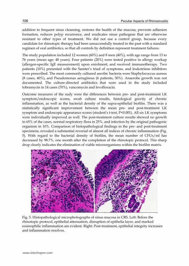
Fig. 5. Histopathological microphotographs of sinus mucosa in CRS. Left: Before the rhinotopic protocol, epithelial attenuation, disruption of epithelia layer, and marked eosinophilic inflammation are evident. Right: Post-treatment, epithelial integrity increases and inflammation resolves.
www.intechopen.com

Topical Membrane Therapy for Chronic Rhinosinusitis
107
None of the patients reported any systemic or local adverse reactions. Careful endoscopy during the follow-up visits, did not reveal severe irritation, crusting, or signs of recent epistaxis.
6.2.6 The rhinotopic protocol: How it works
Refractory chronic sinusitis is a multifactorial disease, and its chronicity relies on constant debris accumulation, unremitting inflammation, and insidious infection. The optimal management needs to be multifactorial as well, and address all three components concurrently. The rhinotopic protocol is a comprehensive, strictly topical, approach to this difficult-to-treat entity. It is not applied routinely to any CRS case, but it is rather indicated for selected patients, who had previously received high-quality surgical and medical treatment, but continue to experience prolonged symptoms of moderate to severe intensity.
Mechanical cleansing by means of frequent high-pressure saline irrigations and weekly office debridements, although simple, ensures the efficacy of the pharmacological interventions. Crusts, mucopurulent secretions, and exudates, are toxic to the underlying epithelium, and perpetuate the inflammation. Meticulous removal of debris is the sine qua non of every topical therapy, in the same way it is essential for the normal healing process post-sinus surgery(Palmer and Kennedy, 2003).
The anti-inflammatory effect of the treatment is achieved by the sustained action of mometasone, one of the most potent commercially available steroids. The steroid is applied locally via a combination of two advanced delivery techniques, i.e., nebulization of small aerosolized particles, and endoscopically-guided instillation of a mucoadhesive gel. Remarkable edema reduction, and down-regulation of the eosinophilic infiltration, are among the proven consequences of systemic steroid use. Even though, this clinical improvement may not be very long-lasting. According to our data, resolution of inflammation-related symptoms, such as congestion, nasal discharge, and facial tenderness, is still documented one month after the topical protocol’s completion.
Antimicrobial agents are administered simultaneously with the steroids, via the same two delivery methods. The role of infection in chronic sinusitis is unclear, and it is common belief that CRS exacerbations are pathophysiologically analogous to acute sinusitis, and should be treated as such. Typically, culture-directed systemic antibiotics may temporarily suppress the infection, but recurrence caused by the same pathogen is frequently noted(Lim et al., 2008). The sustained, highly concentrated application of antimicrobial agents directly onto the diseased membrane, according to the rhinotopic protocol, aims to eradicate the etiologic microorganism from the sinus mucosa. It is suggested that a key factor for the successful elimination of infection is overcoming the resistance of bacteria within the biofilm shelter. The antibiotic-releasing mucoadherent gel is specifically attached to this surface-organized community, and places a dense concentration of bactericidal agents at the infection site. Our findings show an impressive decline in the population of viable bacteria residing in biofilms, as assayed by Colony Forming Units cultures, following the rhinotopic therapy. This suggests that one of the mechanisms responsible for the protocol’s efficacy is the disruption of biofilms. A contributing factor to the biofilm extirpation, may be the high-pressure hydrotherapy performed by the patient alternately with the antibiotic administration. Saline irrigations possibly wash out panktonik bacteria before they become fixed to sinus walls and recolonize the organic matrix(Suh et al., 2010).
www.intechopen.com

Peculiar Aspects of Rhinosinusitis
108
7. Conclusion
Chronic rhinosinusitis is evidently a unique disease process, and far more complex than what we commonly describe as an “infection”. CRS refractory to standard treatment is not an exception to the rule, but rather an increasingly occurring phenomenon. In such a chronic illness, the side-effects of systemic medications underlie the necessity of topical therapy. The latter, as this chapter has showed, is not merely restricted to placement of a drug locally, but has been developed with the help of technology into a dynamic approach, tailored to the disease’s pathophysiology. Advances in endoscopy and particularly in sinus surgery, have made the paranasal cavities accessible to application of a variety of pharmacological agents.
Our proposal is an integrated topical protocol, for the restoration of the sinus mucosa homeostasis. Preliminary results are promising, and the ultimate goal of this approach is to establish a long-term effect after treatment completion, rather than transient symptom relief. Longer follow-up of patients, and modifications of the protocol guided by ongoing findings, would be the next step.
8. References
Al-Mutairi D & Kilty SJ. (2011). Bacterial biofilms and the pathophysiology of chronic rhinosinusitis. Curr Opin Allergy Clin Immunol, Vol. 11, No. 1, pp. 18-23, ISSN 1473-6322
Alandejani T, Marsan J, Ferris W, Slinger R, & Chan F. (2009). Effectiveness of honey on Staphylococcus aureus and Pseudomonas aeruginosa biofilms. Otolaryngol Head Neck Surg, Vol. 141, No. 1, pp. 114-118, ISSN 0194-5998
Antunes MB & Becker SS. (2010). The role of local steroid injection for nasal polyposis. Curr Allergy Asthma Rep, Vol. 10, No. 3, pp. 175-180, ISSN 1534-6315
Armengot M, Juan G, Barona R, Garin L, & Basterra J. (1994). Immotile cilia syndrome: nasal mucociliary function and nasal ciliary abnormalities. Rhinology, Vol. 32, No. 3, pp. 109-111, ISSN 0300-0729
Aukema AA, Mulder PG, & Fokkens WJ. (2005). Treatment of nasal polyposis and chronic rhinosinusitis with fluticasone propionate nasal drops reduces need for sinus surgery. J Allergy Clin Immunol, Vol. 115, No. 5, pp. 1017-1023, ISSN 0091-6749
Bendouah Z, Barbeau J, Hamad WA, & Desrosiers M. (2006). Biofilm formation by Staphylococcus aureus and Pseudomonas aeruginosa is associated with an unfavorable evolution after surgery for chronic sinusitis and nasal polyposis. Otolaryngol Head Neck Surg, Vol. 134, No. 6, pp. 991-996, ISSN 0194-5998
Berger WE, Ratner PH, Casale TB, Meltzer EO, & Wall GM. (2009). Safety and efficacy of olopatadine hydrochloride nasal spray 0.6% in pediatric subjects with allergic rhinitis. Allergy Asthma Proc, Vol. 30, No. 6, pp. 612-623, ISSN 1539-6304
Ceri H, et al. (1999). The Calgary Biofilm Device: new technology for rapid determination of antibiotic susceptibilities of bacterial biofilms. J Clin Microbiol, Vol. 37, No. 6, pp. 1771-1776, ISSN 0095-1137
Chien YW & Chang SF. (1987). Intranasal drug delivery for systemic medications. Crit Rev Ther Drug Carrier Syst, Vol. 4, No. 2, pp. 67-194, ISSN 0743-4863
www.intechopen.com

Topical Membrane Therapy for Chronic Rhinosinusitis
109
Chiu AG, et al. (2008). Baby shampoo nasal irrigations for the symptomatic post-functional endoscopic sinus surgery patient. Am J Rhinol, Vol. 22, No. 1, pp. 34-37, ISSN 1050-6586
Daviskas E, et al. (1996). Inhalation of hypertonic saline aerosol enhances mucociliary clearance in asthmatic and healthy subjects. Eur Respir J, Vol. 9, No. 4, pp. 725-732, ISSN 0903-1936
Daviskas E & Anderson SD. (2006). Hyperosmolar agents and clearance of mucus in the diseased airway. J Aerosol Med, Vol. 19, No. 1, pp. 100-109, ISSN 0894-2684
Demoly P. (2008). Safety of intranasal corticosteroids in acute rhinosinusitis. Am J Otolaryngol, Vol. 29, No. 6, pp. 403-413, ISSN 1532-818X
Derendorf H & Meltzer EO. (2008). Molecular and clinical pharmacology of intranasal corticosteroids: clinical and therapeutic implications. Allergy, Vol. 63, No. 10, pp. 1292-1300, ISSN 1398-9995
Eccles R, Eriksson M, Garreffa S, & Chen SC. (2008). The nasal decongestant effect of xylometazoline in the common cold. Am J Rhinol, Vol. 22, No. 5, pp. 491-496, ISSN 1050-6586
Giger R, et al. (2003). Comparison of once- versus twice-daily use of beclomethasone dipropionate aqueous nasal spray in the treatment of allergic and non-allergic chronic rhinosinusitis. Eur Arch Otorhinolaryngol, Vol. 260, No. 3, pp. 135-140, ISSN 0937-4477
Gliklich RE & Metson R. (1995). The health impact of chronic sinusitis in patients seeking otolaryngologic care. Otolaryngol Head Neck Surg, Vol. 113, No. 1, pp. 104-109, ISSN 0194-5998
Gliklich RE & Metson R. (1998). Economic implications of chronic sinusitis. Otolaryngol Head Neck Surg, Vol. 118, No. 3 Pt 1, pp. 344-349, ISSN 0194-5998
Goh YH & Goode RL. (2000). Current status of topical nasal antimicrobial agents. Laryngoscope, Vol. 110, No. 6, pp. 875-880, ISSN 0023-852X
Gosepath J & Mann WJ. (2005). Current concepts in therapy of chronic rhinosinusitis and nasal polyposis. ORL J Otorhinolaryngol Relat Spec, Vol. 67, No. 3, pp. 125-136, ISSN 0301-1569
Harvey RJ & Schlosser RJ. (2009). Local drug delivery. Otolaryngol Clin North Am, Vol. 42, No. 5, pp. 829-845, ix, ISSN 1557-8259
Horak F & Zieglmayer UP. (2009). Azelastine nasal spray for the treatment of allergic and nonallergic rhinitis. Expert Rev Clin Immunol, Vol. 5, No. 6, pp. 659-669, ISSN 1744-8409
Hyo N, Takano H, & Hyo Y. (1989). Particle deposition efficiency of therapeutic aerosols in the human maxillary sinus. Rhinology, Vol. 27, No. 1, pp. 17-26, ISSN 0300-0729
Karagama YG, Lancaster JL, Karkanevatos A, & O'Sullivan G. (2001). Delivery of nasal drops to the middle meatus: which is the best head position? Rhinology, Vol. 39, No. 4, pp. 226-229, ISSN 0300-0729
Kennedy DW. (1992). Prognostic factors, outcomes and staging in ethmoid sinus surgery. Laryngoscope, Vol. 102, No. 12 Pt 2 Suppl 57, pp. 1-18, ISSN 0023-852X
Krouse JH. (2000). Computed tomography stage, allergy testing, and quality of life in patients with sinusitis. Otolaryngol Head Neck Surg, Vol. 123, No. 4, pp. 389-392, ISSN 0194-5998
www.intechopen.com
Peculiar Aspects of Rhinosinusitis
110
Kundoor V & Dalby RN. (2010). Assessment of nasal spray deposition pattern in a silicone human nose model using a color-based method. Pharm Res, Vol. 27, No. 1, pp. 30-36, ISSN 1573-904X
Lavigne F, et al. (2002). Intrasinus administration of topical budesonide to allergic patients with chronic rhinosinusitis following surgery. Laryngoscope, Vol. 112, No. 5, pp. 858-864, ISSN 0023-852X
Leonard DW & Bolger WE. (1999). Topical antibiotic therapy for recalcitrant sinusitis. Laryngoscope, Vol. 109, No. 4, pp. 668-670, ISSN 0023-852X
Lethbridge-Cejku M, Schiller JS, & Bernadel L. (2004). Summary health statistics for U.S. adults: National Health Interview Survey, 2002. Vital Health Stat 10, Vol., No. 222, pp. 1-151, ISSN 0083-1972
Levine HL. (1990). Functional endoscopic sinus surgery: evaluation, surgery, and follow-up of 250 patients. Laryngoscope, Vol. 100, No. 1, pp. 79-84, ISSN 0023-852X
Lim M, Citardi MJ, & Leong JL. (2008). Topical antimicrobials in the management of chronic rhinosinusitis: a systematic review. Am J Rhinol, Vol. 22, No. 4, pp. 381-389, ISSN 1050-6586
Lund VJ & Kennedy DW. (1997). Staging for rhinosinusitis. Otolaryngol Head Neck Surg, Vol. 117, No. 3 Pt 2, pp. S35-40, ISSN 0194-5998
Mabry RL. (1981). Visual loss after intranasal corticosteroid injection. Incidence, causes, and prevention. Arch Otolaryngol, Vol. 107, No. 8, pp. 484-486, ISSN 0003-9977
Mabry RL. (1981). Evaluation of systemic absorption of intraturbinally injected triamcinolone. Otolaryngol Head Neck Surg, Vol. 89, No. 2, pp. 268-270, ISSN 0194-5998
Meltzer EO, et al. (2004). Rhinosinusitis: establishing definitions for clinical research and patient care. J Allergy Clin Immunol, Vol. 114, No. 6 Suppl, pp. 155-212, ISSN 0091-6749
Merkus P, Ebbens FA, Muller B, & Fokkens WJ. (2006). The 'best method' of topical nasal drug delivery: comparison of seven techniques. Rhinology, Vol. 44, No. 2, pp. 102-107, ISSN 0300-0729
Miller TR, Muntz HR, Gilbert ME, & Orlandi RR. (2004). Comparison of topical medication delivery systems after sinus surgery. Laryngoscope, Vol. 114, No. 2, pp. 201-204, ISSN 0023-852X
Moller W, Munzing W, & Canis M. (2010). Clinical potential of pulsating aerosol for sinus drug delivery. Expert Opin Drug Deliv, Vol. 7, No. 11, pp. 1239-1245, ISSN 1744-7593
Moss RB & King VV. (1995). Management of sinusitis in cystic fibrosis by endoscopic surgery and serial antimicrobial lavage. Reduction in recurrence requiring surgery. Arch Otolaryngol Head Neck Surg, Vol. 121, No. 5, pp. 566-572, ISSN 0886-4470
Nakamura K, et al. (1999). Uptake and release of budesonide from mucoadhesive, pH-sensitive copolymers and their application to nasal delivery. J Control Release, Vol. 61, No. 3, pp. 329-335, ISSN 0168-3659
Palmer JN & Kennedy DW. (2003). Medical management in functional endoscopic sinus surgery failures. Curr Opin Otolaryngol Head Neck Surg, Vol. 11, No. 1, pp. 6-12, ISSN 1068-9508
www.intechopen.com

Topical Membrane Therapy for Chronic Rhinosinusitis
111
Papsin B & McTavish A. (2003). Saline nasal irrigation: Its role as an adjunct treatment. Can Fam Physician, Vol. 49, No., pp. 168-173, ISSN 0008-350X
Ponikau JU, et al. (1999). The diagnosis and incidence of allergic fungal sinusitis. Mayo Clin Proc, Vol. 74, No. 9, pp. 877-884, ISSN 0025-6196
Ranade VV. (2001). Inhalation therapy: new delivery systems. Am J Ther, Vol. 8, No. 5, pp. 367-381, ISSN 1075-2765
Schenkel EJ, et al. (2000). Absence of growth retardation in children with perennial allergic rhinitis after one year of treatment with mometasone furoate aqueous nasal spray. Pediatrics, Vol. 105, No. 2, pp. E22, ISSN 1098-4275
Sharp HJ, Denman D, Puumala S, & Leopold DA. (2007). Treatment of acute and chronic rhinosinusitis in the United States, 1999-2002. Arch Otolaryngol Head Neck Surg, Vol. 133, No. 3, pp. 260-265, ISSN 0886-4470
Shikani AH, Chahine KA, & Alqudah MA. (2010). The rhinotopic protocol for chronic refractory rhinosinusitis: how we do it. Clin Otolaryngol, Vol. 35, No. 4, pp. 329-332, ISSN 1749-4486
Stewart PS & Costerton JW. (2001). Antibiotic resistance of bacteria in biofilms. Lancet, Vol. 358, No. 9276, pp. 135-138, ISSN 0140-6736
Suh JD, Ramakrishnan V, & Palmer JN. (2010). Biofilms. Otolaryngol Clin North Am, Vol. 43, No. 3, pp. 521-530, viii, ISSN 1557-8259
Sykes DA, Wilson R, Chan KL, Mackay IS, & Cole PJ. (1986). Relative importance of antibiotic and improved clearance in topical treatment of chronic mucopurulent rhinosinusitis. A controlled study. Lancet, Vol. 2, No. 8503, pp. 359-360, ISSN 0140-6736
Tarran R, Grubb BR, Gatzy JT, Davis CW, & Boucher RC. (2001). The relative roles of passive surface forces and active ion transport in the modulation of airway surface liquid volume and composition. J Gen Physiol, Vol. 118, No. 2, pp. 223-236, ISSN 0022-1295
Ugwoke MI, Agu RU, Verbeke N, & Kinget R. (2005). Nasal mucoadhesive drug delivery: background, applications, trends and future perspectives. Adv Drug Deliv Rev, Vol. 57, No. 11, pp. 1640-1665, ISSN 0169-409X
Unno T, Hokunan K, Yanai O, & Onodera S. (1983). Deposition of sprayed particles in the nasal cavity. Auris Nasus Larynx, Vol. 10, No. 2, pp. 109-116, ISSN 0385-8146
Uren B, Psaltis A, & Wormald PJ. (2008). Nasal lavage with mupirocin for the treatment of surgically recalcitrant chronic rhinosinusitis. Laryngoscope, Vol. 118, No. 9, pp. 1677-1680, ISSN 1531-4995
Vaughan WC & Carvalho G. (2002). Use of nebulized antibiotics for acute infections in chronic sinusitis. Otolaryngol Head Neck Surg, Vol. 127, No. 6, pp. 558-568, ISSN 0194-5998
Wahl KJ & Otsuji A. (2003). New medical management techniques for acute exacerbations of chronic rhinosinusitis. Curr Opin Otolaryngol Head Neck Surg, Vol. 11, No. 1, pp. 27-32, ISSN 1068-9508
Weschta M, et al. (2004). Topical antifungal treatment of chronic rhinosinusitis with nasal polyps: a randomized, double-blind clinical trial. J Allergy Clin Immunol, Vol. 113, No. 6, pp. 1122-1128, ISSN 0091-6749
Yoo JK, Seikaly H, & Calhoun KH. (1997). Extended use of topical nasal decongestants. Laryngoscope, Vol. 107, No. 1, pp. 40-43, ISSN 0023-852X
www.intechopen.com

Peculiar Aspects of Rhinosinusitis
112
Zadeh MH, Banthia V, Anand VK, & Huang C. (2002). Significance of eosinophilia in chronic rhinosinusitis. Am J Rhinol, Vol. 16, No. 6, pp. 313-317, ISSN 1050-6586
www.intechopen.com

Peculiar Aspects of RhinosinusitisEdited by Dr. Gian Luigi Marseglia
ISBN 978-953-307-763-5Hard cover, 112 pagesPublisher InTechPublished online 23, November, 2011Published in print edition November, 2011
InTech EuropeUniversity Campus STeP Ri Slavka Krautzeka 83/A 51000 Rijeka, Croatia Phone: +385 (51) 770 447 Fax: +385 (51) 686 166www.intechopen.com
InTech ChinaUnit 405, Office Block, Hotel Equatorial Shanghai No.65, Yan An Road (West), Shanghai, 200040, China
Phone: +86-21-62489820 Fax: +86-21-62489821
Rhinosinusitis has both a great practical interest and a broad significance due to the scientific complexity of thepathogenetic problems related to the disease, not yet completely resolved, and their implications for clinicaltreatment. This book highlights certain specific topics that usually are not clarified in other resources. The firstchapter is devoted to the impoverished quality of life experienced by patients suffering from rhinosinusitis. Thesecond chapter focuses on the microbiological aspects of rhinosinusitis, while the two subsequent chaptersexplain the peculiar aspects of chronic rhinosinusitis and of recurrent chronic rhinosinusitis. The first chapter ofthe second section of the book is dedicated to the imaging techniques used to visualize the nasal sinuses andthe other to a medical topical type of treatment.
How to referenceIn order to correctly reference this scholarly work, feel free to copy and paste the following:
Alan Shikani and Konstantinos Kourelis (2011). Topical Membrane Therapy for Chronic Rhinosinusitis,Peculiar Aspects of Rhinosinusitis, Dr. Gian Luigi Marseglia (Ed.), ISBN: 978-953-307-763-5, InTech, Availablefrom: http://www.intechopen.com/books/peculiar-aspects-of-rhinosinusitis/topical-membrane-therapy-for-chronic-rhinosinusitis

© 2011 The Author(s). Licensee IntechOpen. This is an open access articledistributed under the terms of the Creative Commons Attribution 3.0License, which permits unrestricted use, distribution, and reproduction inany medium, provided the original work is properly cited.