An Update of Rhinosinusitis
20
ISSN:2250-0359 Volume 4 Issue 1 2014 An Update of Rhinosinusitis. *Vishwambhar Singh *Krishna Murari Tiwari Rajendra Institute Of Medical Sciences, Ranchi. Abstract: Rhinosinusitis is one of most common disease worldwide, affecting the quality of life of the person afflicted by it. Its causes and pathophysiology has been well delineated so far, but its treatment is changing fast due to change in organisms causing it and also emergence of resistance to antimicrobial agents, thus making the management challenging. This article highlights the changing microbial pattern, classification of the disease, its socio-economic burden, medical treatment and ever evolving surgical treatment, with its indications. Keywords: Rhinosinusitis, Acute Bacterial Rhinosinusitis, Chronic Rhino sinusitis, Paranasal sinus, URTI. INTRODUCTION: - Rhinosinusitis (RS) is such a common entity although given the wide range of clinical presentation. The multiple potential types & varied etiological & associated factors. This diagnosis often includes several patho – physiologic conditions. In order to better define the incidence & prevalence, the more specific criteria should be applied. The prevalence rate has also been found to very substantially depending on the type of practitioner & method of diagnosis used. Prevalence in children in Primary Care setting with symptoms of on acute URTI is 9to 17% in various studies. The number increases 30% for patients presenting to a general medical clinic & this further increases to 83% of patients presenting with acute URTI having ABRS in an otolaryngology clinic[1,2 ]. The improper diagnosis & management of RS has given way to emergence of
-
Upload
anonymous-so6znlkyw -
Category
Documents
-
view
213 -
download
0
Transcript of An Update of Rhinosinusitis
ISSN:2250-0359 Volume 4 Issue 1 2014
An Update of Rhinosinusitis.
*Vishwambhar Singh *Krishna Murari Tiwari
Rajendra Institute Of Medical Sciences, Ranchi.
Abstract: Rhinosinusitis is one of most common disease worldwide, affecting the qualityof life of the person afflicted by it. Its causes and pathophysiology has been welldelineated so far, but its treatment is changing fast due to change in organisms causing itand also emergence of resistance to antimicrobial agents, thus making the managementchallenging. This article highlights the changing microbial pattern, classification of thedisease, its socio-economic burden, medical treatment and ever evolving surgicaltreatment, with its indications.
Keywords: Rhinosinusitis, Acute Bacterial Rhinosinusitis, Chronic Rhino sinusitis,Paranasal sinus, URTI.
INTRODUCTION: - Rhinosinusitis (RS) is such a common entity although given thewide range of clinical presentation. The multiple potential types & varied etiological &associated factors. This diagnosis often includes several patho – physiologic conditions.In order to better define the incidence & prevalence, the more specific criteria should beapplied. The prevalence rate has also been found to very substantially depending on thetype of practitioner & method of diagnosis used. Prevalence in children in Primary Caresetting with symptoms of on acute URTI is 9to 17% in various studies. The numberincreases 30% for patients presenting to a general medical clinic & this further increasesto 83% of patients presenting with acute URTI having ABRS in an otolaryngologyclinic[1,2 ]. The improper diagnosis & management of RS has given way to emergence of
Vikrams1
Highlight
Vikrams1
Highlight
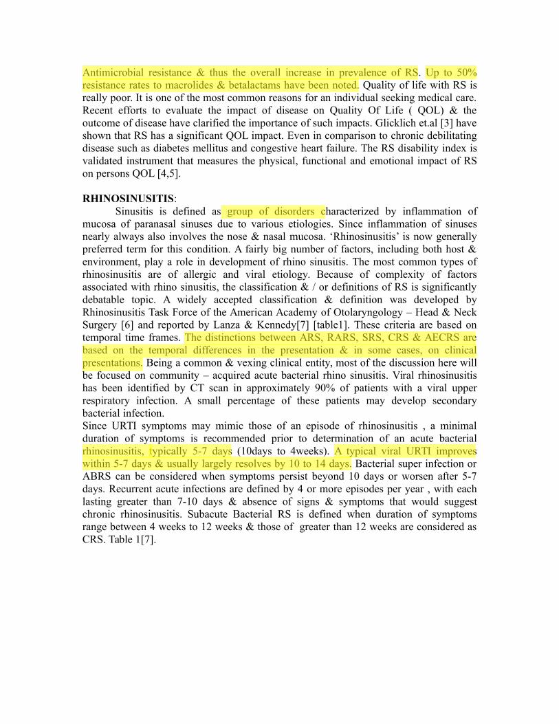
Antimicrobial resistance & thus the overall increase in prevalence of RS. Up to 50%resistance rates to macrolides & betalactams have been noted. Quality of life with RS isreally poor. It is one of the most common reasons for an individual seeking medical care.Recent efforts to evaluate the impact of disease on Quality Of Life ( QOL) & theoutcome of disease have clarified the importance of such impacts. Glicklich et.al [3] haveshown that RS has a significant QOL impact. Even in comparison to chronic debilitatingdisease such as diabetes mellitus and congestive heart failure. The RS disability index isvalidated instrument that measures the physical, functional and emotional impact of RSon persons QOL [4,5].
RHINOSINUSITIS: Sinusitis is defined as group of disorders characterized by inflammation of
mucosa of paranasal sinuses due to various etiologies. Since inflammation of sinusesnearly always also involves the nose & nasal mucosa. ‘Rhinosinusitis’ is now generallypreferred term for this condition. A fairly big number of factors, including both host &environment, play a role in development of rhino sinusitis. The most common types ofrhinosinusitis are of allergic and viral etiology. Because of complexity of factorsassociated with rhino sinusitis, the classification & / or definitions of RS is significantlydebatable topic. A widely accepted classification & definition was developed byRhinosinusitis Task Force of the American Academy of Otolaryngology – Head & NeckSurgery [6] and reported by Lanza & Kennedy[7] [table1]. These criteria are based ontemporal time frames. The distinctions between ARS, RARS, SRS, CRS & AECRS arebased on the temporal differences in the presentation & in some cases, on clinicalpresentations. Being a common & vexing clinical entity, most of the discussion here willbe focused on community – acquired acute bacterial rhino sinusitis. Viral rhinosinusitishas been identified by CT scan in approximately 90% of patients with a viral upperrespiratory infection. A small percentage of these patients may develop secondarybacterial infection. Since URTI symptoms may mimic those of an episode of rhinosinusitis , a minimalduration of symptoms is recommended prior to determination of an acute bacterialrhinosinusitis, typically 5-7 days (10days to 4weeks). A typical viral URTI improveswithin 5-7 days & usually largely resolves by 10 to 14 days. Bacterial super infection orABRS can be considered when symptoms persist beyond 10 days or worsen after 5-7days. Recurrent acute infections are defined by 4 or more episodes per year , with eachlasting greater than 7-10 days & absence of signs & symptoms that would suggestchronic rhinosinusitis. Subacute Bacterial RS is defined when duration of symptomsrange between 4 weeks to 12 weeks & those of greater than 12 weeks are considered asCRS. Table 1[7].
Vikrams1
Highlight
Vikrams1
Highlight
Vikrams1
Highlight
Vikrams1
Highlight
Vikrams1
Highlight
Vikrams1
Highlight
Vikrams1
Highlight
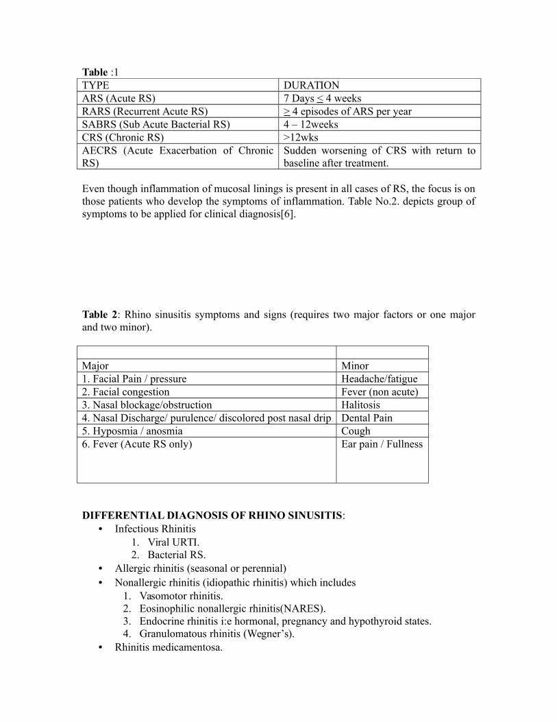
Table :1TYPE DURATIONARS (Acute RS) 7 Days < 4 weeksRARS (Recurrent Acute RS) > 4 episodes of ARS per yearSABRS (Sub Acute Bacterial RS) 4 – 12weeksCRS (Chronic RS) >12wksAECRS (Acute Exacerbation of ChronicRS)
Sudden worsening of CRS with return tobaseline after treatment.
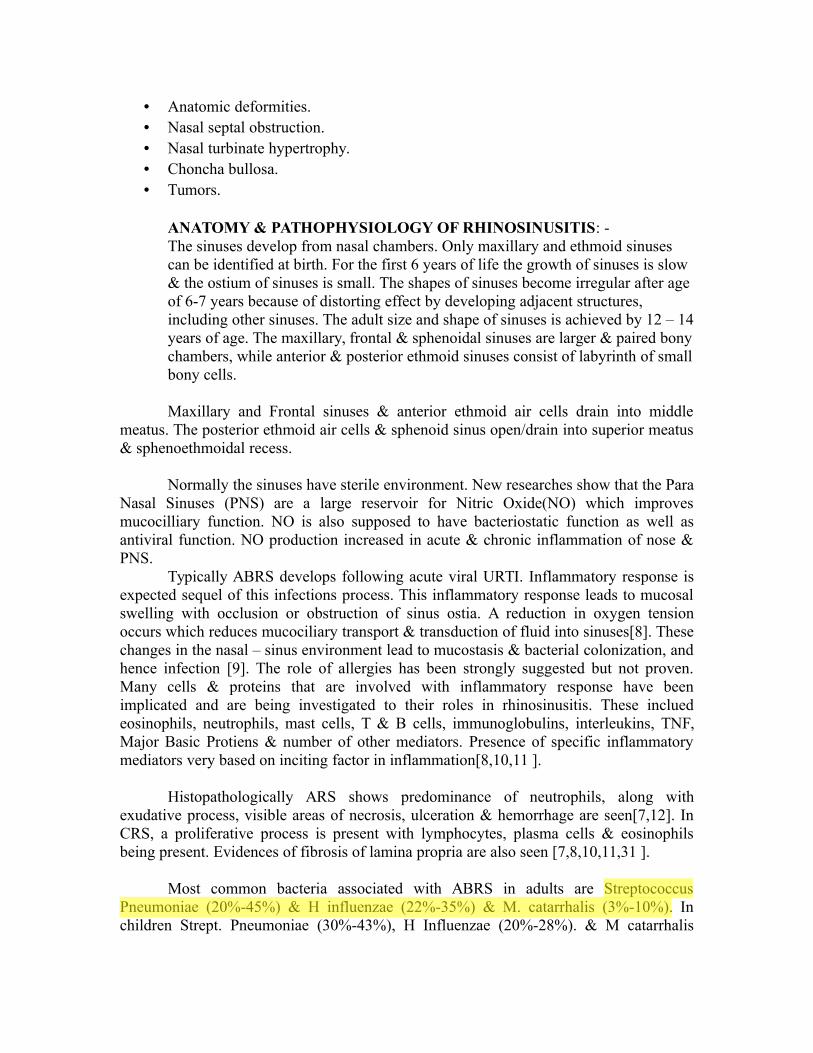
Even though inflammation of mucosal linings is present in all cases of RS, the focus is onthose patients who develop the symptoms of inflammation. Table No.2. depicts group ofsymptoms to be applied for clinical diagnosis[6].
Table 2: Rhino sinusitis symptoms and signs (requires two major factors or one majorand two minor).
Major Minor1. Facial Pain / pressure Headache/fatigue2. Facial congestion Fever (non acute)3. Nasal blockage/obstruction Halitosis4. Nasal Discharge/ purulence/ discolored post nasal drip Dental Pain5. Hyposmia / anosmia Cough6. Fever (Acute RS only) Ear pain / Fullness
DIFFERENTIAL DIAGNOSIS OF RHINO SINUSITIS:• Infectious Rhinitis
1. Viral URTI.2. Bacterial RS.
• Allergic rhinitis (seasonal or perennial)• Nonallergic rhinitis (idiopathic rhinitis) which includes
1. Vasomotor rhinitis.2. Eosinophilic nonallergic rhinitis(NARES).3. Endocrine rhinitis i:e hormonal, pregnancy and hypothyroid states.4. Granulomatous rhinitis (Wegner’s).
• Rhinitis medicamentosa.
• Anatomic deformities.• Nasal septal obstruction.• Nasal turbinate hypertrophy.• Choncha bullosa.• Tumors.
ANATOMY & PATHOPHYSIOLOGY OF RHINOSINUSITIS: -The sinuses develop from nasal chambers. Only maxillary and ethmoid sinuses can be identified at birth. For the first 6 years of life the growth of sinuses is slow & the ostium of sinuses is small. The shapes of sinuses become irregular after age of 6-7 years because of distorting effect by developing adjacent structures, including other sinuses. The adult size and shape of sinuses is achieved by 12 – 14years of age. The maxillary, frontal & sphenoidal sinuses are larger & paired bonychambers, while anterior & posterior ethmoid sinuses consist of labyrinth of smallbony cells.
Maxillary and Frontal sinuses & anterior ethmoid air cells drain into middlemeatus. The posterior ethmoid air cells & sphenoid sinus open/drain into superior meatus& sphenoethmoidal recess.
Normally the sinuses have sterile environment. New researches show that the ParaNasal Sinuses (PNS) are a large reservoir for Nitric Oxide(NO) which improvesmucocilliary function. NO is also supposed to have bacteriostatic function as well asantiviral function. NO production increased in acute & chronic inflammation of nose &PNS.
Typically ABRS develops following acute viral URTI. Inflammatory response isexpected sequel of this infections process. This inflammatory response leads to mucosalswelling with occlusion or obstruction of sinus ostia. A reduction in oxygen tensionoccurs which reduces mucociliary transport & transduction of fluid into sinuses[8]. Thesechanges in the nasal – sinus environment lead to mucostasis & bacterial colonization, andhence infection [9]. The role of allergies has been strongly suggested but not proven.Many cells & proteins that are involved with inflammatory response have beenimplicated and are being investigated to their roles in rhinosinusitis. These incluedeosinophils, neutrophils, mast cells, T & B cells, immunoglobulins, interleukins, TNF,Major Basic Protiens & number of other mediators. Presence of specific inflammatorymediators very based on inciting factor in inflammation[8,10,11 ].
Histopathologically ARS shows predominance of neutrophils, along withexudative process, visible areas of necrosis, ulceration & hemorrhage are seen[7,12]. InCRS, a proliferative process is present with lymphocytes, plasma cells & eosinophilsbeing present. Evidences of fibrosis of lamina propria are also seen [7,8,10,11,31 ].
Most common bacteria associated with ABRS in adults are StreptococcusPneumoniae (20%-45%) & H influenzae (22%-35%) & M. catarrhalis (3%-10%). Inchildren Strept. Pneumoniae (30%-43%), H Influenzae (20%-28%). & M catarrhalis
Vikrams1
Highlight
(20%-28%) are most commonly associated bacteria. Out of these M catarrhalis is largelya self limited pathogen[7,13,14 ].
As far as Pathogenesis of CRS is concerned it is thought to be an inflammatorydisease & it may or may not involve pathogenic microbes. In those patients with CRS,who do have potential pathogenic bacteria, the most common organisms are staph species(55%), staph aureus (20%)[7,14]. Prevalence of Enterobacteriaceae organisms [15,16].Anaerobes [17] & fungi [11] also been seen.
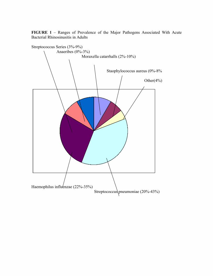
FIGURE 1 – Ranges of Prevalence of the Major Pathogens Associated With AcuteBacterial Rhinosinusitis in Adults
Streptococcus Series (3%-9%)Anaeribes (0%-3%)
Moraxella catarrhalls (2%-10%)
Staophylococcus aureus (0%-8%
Other(4%)
Haemophilus influenzae (22%-35%)Streptococcus pneumoniae (20%-43%)
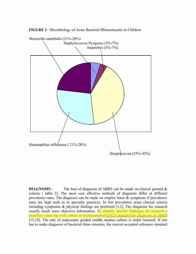
FIGURE 2– Microbiology of Acute Bacterial Rhinosinusitis in Children
Moraxella catarrhalis (21%-28%)Staphylococcus Pyogenes (3%-7%)
Anaerobes (3%-7%)
Haemophilus influenzae ( 21%-28%)
Streptococcus (35%-42%)
DIAGNOSIS: - The best of diagnosis of ABRS can be made on clinical ground &criteria ( table 2). The most cost effective methods of diagnosis differ at differentprevalence rates. The diagnosis can be made on empiric basis & symptoms if prevalencerates are high such as in specialty practices. In low prevalence areas clinical criteriaincluding symptoms & physical findings are preferred [1,2]. The diagnosis for researchusually needs more objective information. To identify specific Pathogen for research amaxillary sinus tap with culture is recommended(GOLD standard for diagnosis of ABRS)[15,18]. The role of endoscopic guided middle meatus culture is under research. If onehas to make diagnosis of bacterial rhino sinusitis, the current accepted reference standard
Vikrams1
Highlight
for culture is more than 1X 10⁴ CFU/ ml in sinus aspirate[19]. Lower counts potentiallyrepresent early infection.
In CRS & SRS definitive methods for diagnosis has yet not been determined. Therecommended time frame for greater than 12week for CRS & between 4-12 weeks forSRS have been commonly adopted. Thus appropriate history, proper physicalexamination & testing will confirm the diagnosis. A number of tests can be used toestablish the diagnosis of rhinosinusitis but these are more important in chronic ratherthan acute rhinosinusitis Nasal endoscopy is an important tool to assess the nasalanatomy, confirm drainage & evaluate treatment response. X-rays have been used for alltypes of rhinosinusitis but are not cost effective in diagnosis of ABRS[1,2]. At presenttime the preferred radiological test is sinus CT scan but it cannot distinguish betweeninflammation & infection .CT scan are not recommended in ABRS as they are not costeffective for this indication[1,2,20]. But are good for evaluation of suspected impendingcomplications such as orbital or intracranial involvement. CT scan is good for assessingthe severity of disease or response to treatment in CRS. Lund & Mackay grading (1993)of CT scan PNS is most commonly recommended as staging system to asses severity ofrhino sinusitis[21]. It has been recently recommended that either a CT scan or endoscopicevaluation of nose preferably with photo or video documentation should be part of anyprospective clinical trial in CRS[10].
TREATMENT:Medical management of RS can be simplified into three groups-antimicrobial,anti-inflammatory and mechanical. It is breakdown treatment from each group andcombine them when appropriate into a comprehensive treatment plan. Also, it isimportant to consider the side effect of each therapy, and weigh them with the patient’ssymptoms severity and other medical conditions. In general, medical management of RSshould include culture directed(or broad spectrum) antibiotics, a nasal steroid spray,saline irrigation and decongestant. Strong consideration should also be given for steaminhalation.
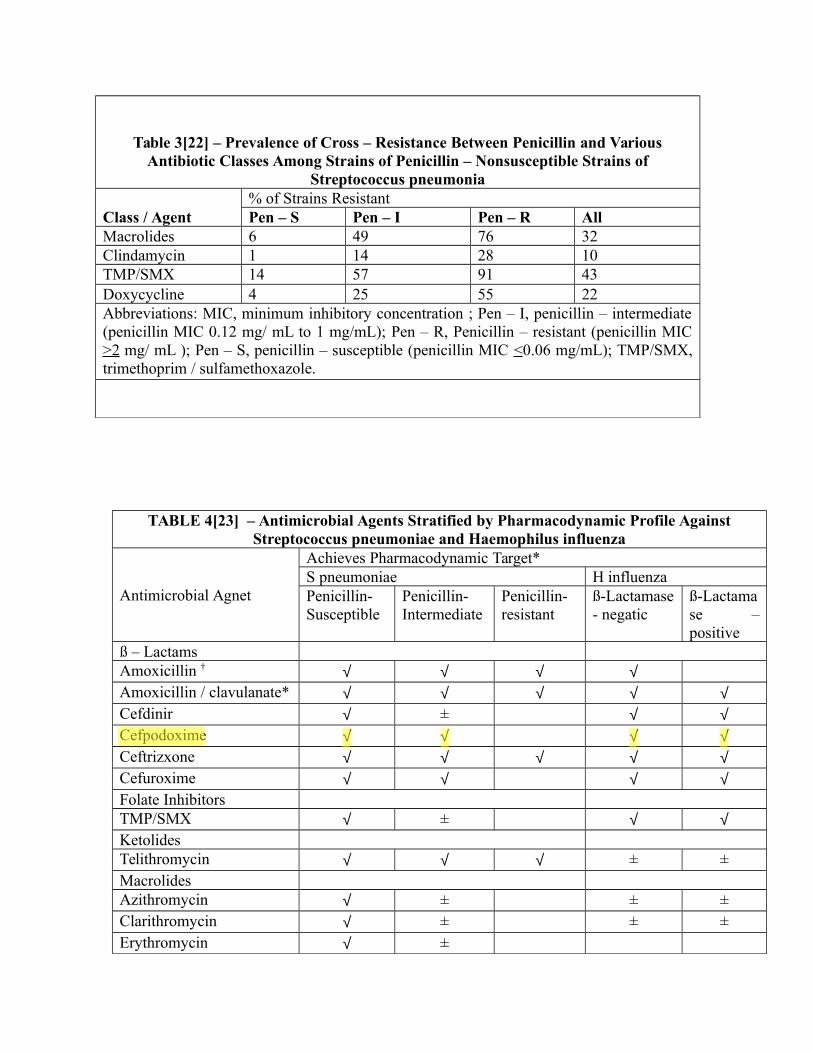
Table 3[22] – Prevalence of Cross – Resistance Between Penicillin and VariousAntibiotic Classes Among Strains of Penicillin – Nonsusceptible Strains of
Streptococcus pneumonia
Class / Agent% of Strains ResistantPen – S Pen – I Pen – R All
Macrolides 6 49 76 32Clindamycin 1 14 28 10TMP/SMX 14 57 91 43Doxycycline 4 25 55 22Abbreviations: MIC, minimum inhibitory concentration ; Pen – I, penicillin – intermediate(penicillin MIC 0.12 mg/ mL to 1 mg/mL); Pen – R, Penicillin – resistant (penicillin MIC>2 mg/ mL ); Pen – S, penicillin – susceptible (penicillin MIC <0.06 mg/mL); TMP/SMX,trimethoprim / sulfamethoxazole.
TABLE 4[23] – Antimicrobial Agents Stratified by Pharmacodynamic Profile AgainstStreptococcus pneumoniae and Haemophilus influenza
Antimicrobial Agnet
Achieves Pharmacodynamic Target*S pneumoniae H influenzaPenicillin-Susceptible
Penicillin-Intermediate
Penicillin-resistant
ß-Lactamase- negatic
ß-Lactamase –positive
ß – LactamsAmoxicillin † √ √ √ √Amoxicillin / clavulanate* √ √ √ √ √Cefdinir √ ± √ √Cefpodoxime √ √ √ √Ceftrizxone √ √ √ √ √Cefuroxime √ √ √ √Folate InhibitorsTMP/SMX √ ± √ √KetolidesTelithromycin √ √ √ ± ±MacrolidesAzithromycin √ ± ± ±Clarithromycin √ ± ± ±Erythromycin √ ±
Vikrams1
Highlight
Vikrams1
Highlight
Vikrams1
Highlight
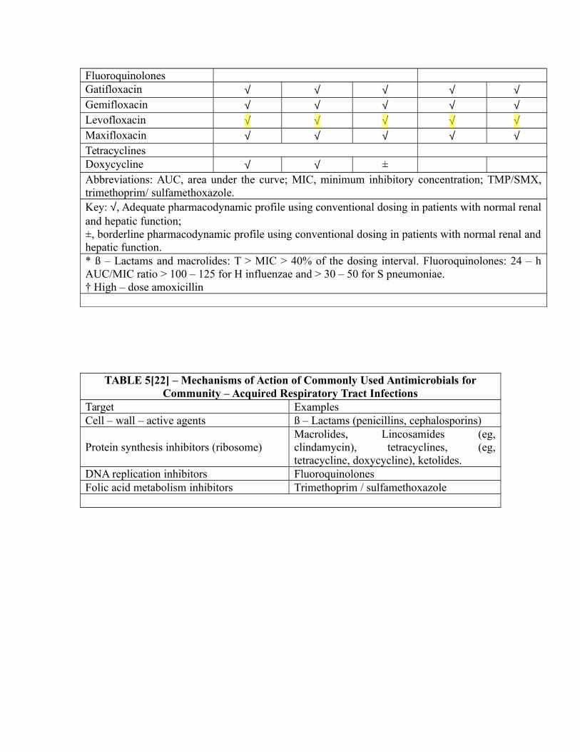
FluoroquinolonesGatifloxacin √ √ √ √ √Gemifloxacin √ √ √ √ √Levofloxacin √ √ √ √ √Maxifloxacin √ √ √ √ √TetracyclinesDoxycycline √ √ ±Abbreviations: AUC, area under the curve; MIC, minimum inhibitory concentration; TMP/SMX,trimethoprim/ sulfamethoxazole.Key: √, Adequate pharmacodynamic profile using conventional dosing in patients with normal renaland hepatic function; ±, borderline pharmacodynamic profile using conventional dosing in patients with normal renal andhepatic function.* ß – Lactams and macrolides: T > MIC > 40% of the dosing interval. Fluoroquinolones: 24 – hAUC/MIC ratio > 100 – 125 for H influenzae and > 30 – 50 for S pneumoniae.† High – dose amoxicillin
TABLE 5[22] – Mechanisms of Action of Commonly Used Antimicrobials forCommunity – Acquired Respiratory Tract Infections
Target ExamplesCell – wall – active agents ß – Lactams (penicillins, cephalosporins)
Protein synthesis inhibitors (ribosome)Macrolides, Lincosamides (eg,clindamycin), tetracyclines, (eg,tetracycline, doxycycline), ketolides.
DNA replication inhibitors FluoroquinolonesFolic acid metabolism inhibitors Trimethoprim / sulfamethoxazole
Vikrams1
Highlight
Vikrams1
Highlight
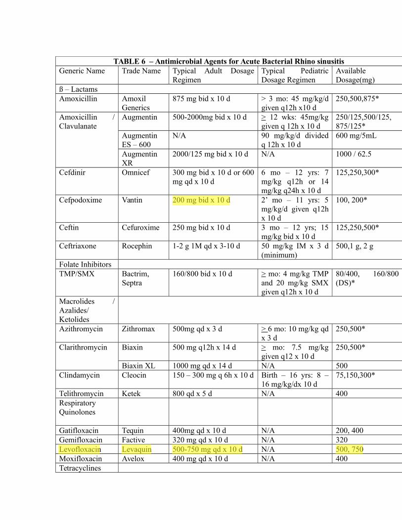
TABLE 6 – Antimicrobial Agents for Acute Bacterial Rhino sinusitisGeneric Name Trade Name Typical Adult Dosage
RegimenTypical PediatricDosage Regimen
AvailableDosage(mg)
ß – LactamsAmoxicillin Amoxil
Generics875 mg bid x 10 d > 3 mo: 45 mg/kg/d
given q12h x10 d250,500,875*
Amoxicillin /Clavulanate
Augmentin 500-2000mg bid x 10 d > 12 wks: 45mg/kggiven q 12h x 10 d
250/125,500/125,875/125*
AugmentinES – 600
N/A 90 mg/kg/d dividedq 12h x 10 d
600 mg/5mL
AugmentinXR
2000/125 mg bid x 10 d N/A 1000 / 62.5
Cefdinir Omnicef 300 mg bid x 10 d or 600mg qd x 10 d
6 mo – 12 yrs: 7mg/kg q12h or 14mg/kg q24h x 10 d
125,250,300*
Cefpodoxime Vantin 200 mg bid x 10 d 2’ mo – 11 yrs: 5mg/kg/d given q12hx 10 d
100, 200*
Ceftin Cefuroxime 250 mg bid x 10 d 3 mo – 12 yrs; 15mg/kg bid x 10 d
125,250,500*
Ceftriaxone Rocephin 1-2 g 1M qd x 3-10 d 50 mg/kg IM x 3 d(minimum)
500,1 g, 2 g
Folate InhibitorsTMP/SMX Bactrim,
Septra160/800 bid x 10 d > mo: 4 mg/kg TMP
and 20 mg/kg SMXgiven q12h x 10 d
80/400, 160/800(DS)*
Macrolides /Azalides/KetolidesAzithromycin Zithromax 500mg qd x 3 d > 6 mo: 10 mg/kg qd
x 3 d 250,500*
Clarithromycin Biaxin 500 mg q12h x 14 d > mo: 7.5 mg/kggiven q12 x 10 d
250,500*
Biaxin XL 1000 mg qd x 14 d N/A 500Clindamycin Cleocin 150 – 300 mg q 6h x 10 d Birth – 16 yrs: 8 –
16 mg/kg/dx 10 d75,150,300*
Telithromycin Ketek 800 qd x 5 d N/A 400RespiratoryQuinolones
Gatifloxacin Tequin 400mg qd x 10 d N/A 200, 400Gemifloxacin Factive 320 mg qd x 10 d N/A 320Levofloxacin Levaquin 500-750 mg qd x 10 d N/A 500, 750Moxifloxacin Avelox 400 mg qd x 10 d N/A 400Tetracyclines
Vikrams1
Highlight
Vikrams1
Highlight
Vikrams1
Highlight
Doxycycline Vibramycin,others
100 mg qd given q12h x10 d
>8 yrs; < 100 lbs; 1mg/ lb bid on day 1,then 1 mg/lb or 0.5mg/lb bid x 10 d
50,75,100*
Abbreviations DS, double strength; N/A, not approved for use; TMP / SMX, trimethoprim/sulfamethoxazole.This table provides typical dosage regimen.* Some dosages are also available in suspension form.
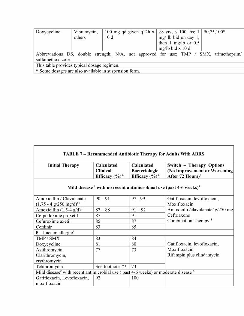
TABLE 7 – Recommended Antibiotic Therapy for Adults With ABRS
Initial Therapy Calculated Clinical Efficacy (%)*
Calculated Bacteriologic Efficacy (%)*
Switch – Therapy Options (No Improvement or WorseningAfter 72 Hours)†
Mild disease † with no recent antimicrobioal use (past 4-6 weeks)$
Amoxicillin / Clavulanate (1.75 - 4 g/250 mg/d)$II
90 – 91 97 - 99 Gatifloxacin, levofloxacin,MoxifloxacinAmoxicilli /clavulanate4g/250 mgCeftriaxoneCombination Therapy $
Amoxicillin (1.5-4 g/d)II 87 – 88 91 – 92Cefpodoxime proxetil 87 91Cefuroxime axetil 85 87Cefdinir 83 85ß – Lactam allergic#
TMP / SMX 83 84Gatifloxacin, levofloxacin,MoxifloxacinRifampin plus clindamycin
Doxycycline 81 80Azithromycin, Clarithromycin, erythromycin
77 73
Telithromycin See footnote. ** 73Mild disease# with recent antimicrobial use ( past 4-6 weeks) or moderate disease $
Gatifloxacin, Levofloxacin, moxifloxacin
92 100

Reevaluate patient $$Amoxicilli /clavulanate (/250 mg)
91 99
Ceftriaxone 91 99(Combination therapy)$ N/A N/Aß – Lactam allergic #
Gatifloxacin, levofloxacin, moxifloxacin
92 100Reevaluate patient $$
Clindamycin and rifampin†† N/A N/A
Abbreviations : ABRS, acute bacterial rhinosinusitis; CT, computed tomography; N/A, not available; TMP/SMX, trimethoprim / sulfamethoxazole.
• Clinical and bacterial efficacy (ie, linical and microbiologic adequacy) is represented by thecalculation from the poole Therapeutic Outcome Model using the mean values of twosurveillance data sets: the US component of the Alexander project (1998 to 2001) andSENTRY surveillance data. These values to not guarantee clinical success or failure. ThePoole Therapeutic outcome model has not been clinically validated.
† When a change in antibiotic therapy is made, the clinician should consider the limitations incoverage of the intitial antibiotic. The respiratory fluoroquinolones ( gatifloxacin,Levofloxacin, and moxifloxacin), ceftriaxone, and amoxicillin/ clavulanate (4 g/250 mg)currently have the best coverage for both Streptococcus pneumoniae and Haemophilusinfluenzae. The terms mild and moderate are designed to aid in selecting antibiotic therapy.
+ The difference in severity of disease does not imply the presence or absence of antimicrobialresistance. Rather, this terminology indicates the relative degree of acceptance of possibletherapeutic failure and the likelihood of achieving spontaneous resolution of symptoms. Thedetermination of disease severity lies with the clinician’s evaluation of the patient’s historyand clinical presentation. Severe, life-threatening infection, with or without complications, isnot addressed in these guidelines.
$ Prior antibiotic therapy within 4 to 6 weeks is a risk factor for infection with resistantorganisms. Antibiotic choices should be based on this and other risk factors.
II The total daily dose of amoxicillin and the amoxicillin component of amoxicillin / clavulanatecan vary from 1.5 to 4 g/day. Lower daily doses (1.5 g/day) are more appropriate in milddisease in patients with no risk factors for infection with a resistant pathogen (including recentantibiotic use). Higher daily doses (4 g/day) may be advantageous in areas with a highprevalence of penicillin – resistant S pneumoniae or drug – resistant S pneumoniae, forpatients with moderate disease, for patients who may need better H influenzae coverage or forpatients with risk factors for infection with a resistant pathogen. There is a greater potentialfor treatment failure or resistant pathogens is these patient groups.
II Based on in vitro spectrum of activity; combination therapy using appropriate gram – positiveand gram – negative coverage may be appropriate. Examples of combination therapyregimens include high – dose amoxicillin ( 4 g/day) or clindamycin plus cefixime, or high –
dose amoxicillin (4 g/day) or clindamycin, plus rifampin. There is no clinical evidence at thistime, however, of the safety or efficacy of these combinations.
# Cephalosporins should be considered initially for patients with penicillin intolerance / non–Type I hypersensitivity reactions (eg, rash). TMP/SMX, doxycycline, macrolides, azalides,and ketolides are not recommended unless the patient is ß – lactam allergic. Theireffectiveness against the major pathogens of ABRS is limited, and bacterial failure of 20% to25% is possible. A respiratory fluoroquinolone (eg, gatifloxacin, lovofloxacin, moxifloxacin)is recommended for patients who have allergies to ß – lactams or who have recently failedother regimens.
** Further Pharmacokinetic/ pharmacodynamic data on this compound is needed.
†† Rifampin is a well – known inducer of several cytochrome P450 isoenzymes and therefore hasa high potential for drug interactions.
++ Reevaluation is necessary because the antibiotics recommended for initial therapy provideexcellent activity against the predominant ABRS pathogens, including S pneumoniae or Hinfluenzae. Additional history, physical examination, cultures, and / or CT scan may beindicated, and the possibility of other less common pathogens considered.
Chronic Rhinosinusitis
Chronic rhinosinusitis (CRS) is an umbrella term for a number of pathologicconditions in which inflammation of the nose and paranasal sinuses is the final commonpathway. These include:
• Allergy• Immunodeficiency states• Neoplasm’s• Virus• Bacteria• Fungi• Genetic / congenital abnormalities• Mucociliary dysfunction• Noxious chemicals• Pollutants• Smoking.
Subgroups of these categories, such as super antigen formation, bacterial biofilms, andallergic fungal disease, have been identified.
The diagnosis of CRS has been defined by consensus in order to develop aframework of terminology that clinicians and researchers can utilize for standardized

communication (Table ). CRS is when symptoms have been present for 12 weeks andthere is physical evidence (usually by nasal endoscopy) of mucosal swelling, nasaldischarge, or polyps. Mucosal abnormalities of the middle meatus or bulla are alsoconcrete signs of inflammatory disease. Generalized or localized edema, erythema, orgranulation tissue can be caused by other rhinologic diseases, such as allergic rhinitis, andtherefore requires imaging confirmation.
TABLE 8 – Measures for Diagnosing Chronic Rhinosinusitis for Adult Clinical Care• Duration of disease is qualified by continuous symptoms for > 12 consecutive
weeks or > 12 weeks of physical findings.*• One of these signs of inflammation must be present and identified in association
with ongoing symptoms consistent with CRS.- Discolored nasal drainage arising from the nasal passage, nasal polyps, or
polypoid swelling as identified on physical examination with anteriorrhinoscopy or nasal endoscopy. Anterior rhinoscopy should be performedin the decongested state.
- Edema or erythema of the middle meatus or ethmoid bulla as identified bynasal endoscopy.
- Generalized or localized erythema, edema, or granulation tissue. If it doesnot involve the middle meatus or ethmoid bulla, radiologic imaging isrequired to confirm diagnosis #.
• Imaging modalities for confirming the diagnosis: - CT scan: demonstrating isolated or diffuse mucosal thickening, bone
changes, air-fluid level.- Plain sinus radiograph: Water’s view revealing mucous membrane
thickening of >5 mm or complete opacification of one or more sinuses. Anair – fluid level is more predictive of acute rhino sinusitis but may also beseen in CRS**.
- MRI is not recommended as an alterative to CT for routine diagnosis ofCRS because of its excessively high sensitivity and lack of specificity.
Abbreviations: CRS, chronic rhino sinusitis; CT, computed tomography; MRI, magneticresonance imaging.
*Signs consistent with CRS will support the symptom time duration#Other chronic rhinologic conditions such as allergic rhinitis can result in such findings, andtherefore they may not be associated with rhino sinusitis. It is recommended that a diagnosisof rhino sinusitis require radiologic confirmation under these circumstances **A plain sinus radiograph without the equivocal signs listed here is not considereddiagnostic. Aside from an air-fluid level, plain sinus radiographs have low sensitivity andspecificity.
Vikrams1
Highlight
Vikrams1
Highlight
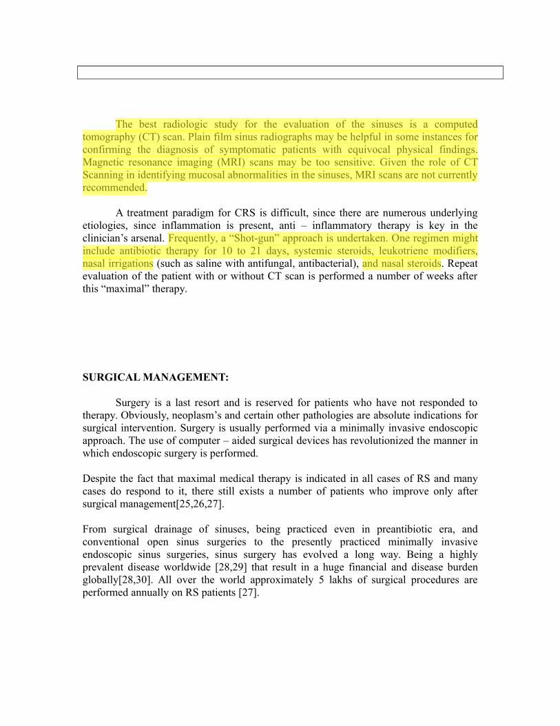
The best radiologic study for the evaluation of the sinuses is a computedtomography (CT) scan. Plain film sinus radiographs may be helpful in some instances forconfirming the diagnosis of symptomatic patients with equivocal physical findings.Magnetic resonance imaging (MRI) scans may be too sensitive. Given the role of CTScanning in identifying mucosal abnormalities in the sinuses, MRI scans are not currentlyrecommended.
A treatment paradigm for CRS is difficult, since there are numerous underlyingetiologies, since inflammation is present, anti – inflammatory therapy is key in theclinician’s arsenal. Frequently, a “Shot-gun” approach is undertaken. One regimen mightinclude antibiotic therapy for 10 to 21 days, systemic steroids, leukotriene modifiers,nasal irrigations (such as saline with antifungal, antibacterial), and nasal steroids. Repeatevaluation of the patient with or without CT scan is performed a number of weeks afterthis “maximal” therapy.
SURGICAL MANAGEMENT:
Surgery is a last resort and is reserved for patients who have not responded totherapy. Obviously, neoplasm’s and certain other pathologies are absolute indications forsurgical intervention. Surgery is usually performed via a minimally invasive endoscopicapproach. The use of computer – aided surgical devices has revolutionized the manner inwhich endoscopic surgery is performed.
Despite the fact that maximal medical therapy is indicated in all cases of RS and manycases do respond to it, there still exists a number of patients who improve only aftersurgical management[25,26,27].
From surgical drainage of sinuses, being practiced even in preantibiotic era, andconventional open sinus surgeries to the presently practiced minimally invasiveendoscopic sinus surgeries, sinus surgery has evolved a long way. Being a highlyprevalent disease worldwide [28,29] that result in a huge financial and disease burdenglobally[28,30]. All over the world approximately 5 lakhs of surgical procedures areperformed annually on RS patients [27].
Vikrams1
Highlight
Vikrams1
Highlight
Vikrams1
Highlight
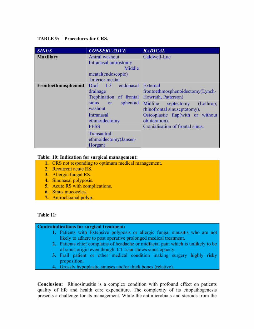
TABLE 9: Procedures for CRS.
SINUS CONSERVATIVE RADICALMaxillary Antral washout Caldwell-Luc
Intranasal antrostomy Middlemeatal(endoscopic) Inferior meatal
Frontoethmosphenoid Draf 1-3 endonasaldrainage
Externalfrontoethmosphenoidectomy(Lynch-Howrath, Patterson)Trephination of frontal
sinus or sphenoidwashout
Midline septectomy (Lothrop;rhinofrontal sinuseptotomy).
Intranasalethmoidectomy
Osteoplastic flap(with or withoutobliteration).
FESS Cranialisation of frontal sinus.
Transantralethmoidectomy(Jansen-Horgan)
Table: 10: Indication for surgical management:1. CRS not responding to optimum medical management.2. Recurrent acute RS.3. Allergic fungal RS.4. Sinonasal polyposis.5. Acute RS with complications.6. Sinus mucoceles.7. Antrochoanal polyp.
Table 11:
Contraindications for surgical treatment:1. Patients with Extensive polyposis or allergic fungal sinusitis who are not
likely to adhere to post operative prolonged medical treatment.2. Patients chief complains of headache or midfacial pain which is unlikely to be
of sinus origin even though CT scan shows sinus opacity.3. Frail patient or other medical condition making surgery highly risky
proposition.4. Grossly hypoplastic sinuses and/or thick bones.(relative).
Conclusion: Rhinosinusitis is a complex condition with profound effect on patientsquality of life and health care expenditure. The complexity of its etiopathogenesispresents a challenge for its management. While the antimicrobials and steroids from the
mainstay of treatment, topical therapy in form of improved delivery system is beingpreferred as of now. Potential development of microbial resistance remains a salutaryconcern in these patients due to repeated and prolonged use of antibiotics. Surgerycontinues to play an important role in management of recalcitrant disease, resulting inimproved quality of life. Immune modulators, such as anti-IgE and anti-IL5 antibodies,are promising newer agents today. Surgical modalities like balloon sinuplasty andstenting are under research. The complex and diverse nature of rhinosinusitis requires anindividualized approach to both medical and surgical management in a multidisciplinarysetting. References:
1. Agency on Health Care Policy and Research. Diagnosis and Treatment ofAcute Bacterial Rhinosinusitis. AHCPR Publications Clearinghouse,Publication No. 99–E016, 1999.
2. Benninger MS, Holzer SE, Lau J. Diagnosis and treatment of uncomplicatedacute bacterial rhinosinusitis : Summary of the Agency on Health Care Policyand Research Evidence Based Report. Otolaryngology and Head and NeckSurgery. 2000; 122: 1-7.
3. Glicklich RE, Metson R. The health imnpact of chronic sinusitis in patientsseeking otolayngologic care. Otolaryngology and Head and Neck Surgery.1995; 113: 104-9.
4. Benninger MS. Senior BA. The development of the rhinosinusitis disabilityindex. Archives of Otolaryngology and Head and Neck Surgery. 1997; 123:1175-9.
5. Senior BA, Benninger MS, Glaze C. The use of the Rhinosinusitis DisabilityIndex (RSDI) in rhinologic disease. American Journal of Rhinology. 2001; 15:21-5.
6. Report of the Rhinosinusitis Task Force Committee Meeting. Otolaryngologyand Head and Neck Surgery. 1997; 117: S1-68.
7. Lanza DC, Kennedy Dw. Adult rhinosinusitis defined. Otolaryngology andHead and Neck Surgery. 1997; 117: S1-7.
8. Benninger MS, Anon J, Mabry RL. The medical management ofrhinosinusitis. Otolaryngology and Head and Neck Surgery. 1997; 117: S41-9.
9. Kennedy DW, Zinreich SJ, Rosenbaum AE, Johns ME Functional endoscopicsinus surgedry: theory and diagnostic evaluation. Archives of Otolaryngology.1985; 111: 576-82
10. Benninger MS, Ferguson BJ, Hadley JA, Hamilos DJ, Jacobs M, KennedyDW et al. Adult chronic rhinosinusitis: Definitions, diagnosis, epidemiologyand pathophysiology. Otolaryngology and Head and Neck Surgery. 2003; 129:S1-32.
11. Ponikau JU, Sherris DA, Kern EB, Homburger HA, Frigas E, Gaffey TA et al.The diagnosis and incidence of allergic fungal sinusitis. Mayo ClinicProceedings. 1999;74:877-84.
12. Coltran RS, Kumar V, Robbins SL (eds). Robbins pathologic basis of disease,5th edn. Philadelphia: WB Saunders, 1994:55-83.
13. Sinus and Allergy Health Partnership. Antimicrobial treatment guidelines foracute bacterial rhinosinusitis, Otolaryngology and Head and Neck Surgery.2004; 130: S1-45.
14. Gwaltney JM, Scheld WM, Sande MA, Sydor A. The microbial etiology andantimicrobial therapy of adults with acute community-acquired sinusitis: afifteen-year experience at the University of Virginia and review of otherselected studies, Journal of Allergy and Clinical Immunology. 1992;90:457-62.
15. Benninger MS, Appelbaum PC, Denneny JC, Osguthorpe DJ, StankiewiczJA. Maxillary sinus puncture and culture in the diagnosis of acuterhinosinusitis: The case of pursuing alternative culture methods.Otolaryngology and Head and Neck Surgery. 2002; 127: 7-12.
16. Rombaux P, Gigi J, Hamoir M, Eloy P, Bertrand B. Bacteriology of chronicsinusitis: the bulla ethmoidalis content. Rhinology. 2002; 40: 18-23.
17. Erkan M, Aslan T, Ozcan M, Koc N. Bacteriology of antrum in adults withchronic maxillary sinusitis. Laryngoscope. 1994; 104: 321-4.
18. Benninger MS, Payne SC, Ferguson BJ, Ahmad N, Hadley J, Endoscopicallydirected middle meatal cultures versus maxillary sinus taps in acute bacterialrhinosinusitis: A meta-analysis. Otolaryngology and Head and Neck Surgery.2006; 134: 1-7.
19. Turner BW, cell WS, Hendley JD Haydon FG, Doyle WJ, Sorrentino JV.Physiologic abnormalities in the paranasal sinuses during experimentalrhinovirus colds. Journal of Allergy and Clinical Immunology 1992;90: 474-8.
20. Gwaltney JW, Phillips CD, Miller RD Riker DK. Computed tomography ofthe common cold. New England Journal of Medicine. 1994;330: 25-30.
21. Lund V, Mackay IS Staging in rhinosinusitis. Rhinology. 1993;107:183-4.22. Jacobs MR. Anon J, Appelbaum PC. Mechanisms of resistance among
respiratory tract pathogens. Clin Lab Med. 2004;24:419-453.23. Sinus and Allergy Health Partnership. Antimicrobial treatment guidelines for
acute bacterial rhinosinusitis. Sinus and Allergy Health Patnership:ExecutiveSummary. Otolaryngol Head Neck Surg. 2000;123(suppl): 5-31
24. Benninger MS. Ferguson BJ. Hadley JA, et al. Adult chronic rhinosinusitis:definitions. Diagnosis, epidemiology, and pathophysiology. OtolaryngologyHead Neck Surg.. 2003; 129 (suppl 3): S1-S32.
25. Gliklich RE, Metson R. Effect of sinus surgery on quality of life.Otolaryngology Head and Neck Surgery. 1997; 117: 12-7.
26. Hopkins C, Slach R, Lund V, et al. Long-term outcomes from the Englishnational comparative audit of surgery for nasal polyposis andchronicrhinosinusitis. Laryngoscope 2009;119:2459-65.
27. Anand VK. Epidemiology and endoscopic impact of rhinosinusitis. Ann OtolRhinol Laryngol Suppl 2004; National Centre for Health Statistics VitalHealth Stat 1998;13:25.
28. Anand VK. Epidemiology and endoscopic impact of rhinosinusitis. Ann OtolRhinol Laryngol Suppl 2004;193:3-5.
29. Zang L, Han D, et al. Prevalence of self reported allergic rhinitis in elevenmajor cities in China. Inta rch Allergy Immunol 2001;49:47-57.
30. Ray NF, Baroniuk JN, Thamer M, et al. Health care expenditure in 1996:contributions of asthma, rhinitis and other airway disorders. Allergy ClinImmunol 1993;103(pt1):408-14.
31. Meltzer EO, Hamilos DJ, Hadley JA, Lanza DC, Marple BF, Nicklas RA et al.Rhinosinustitis: Establishing definitions for clinical research and patient care.Otolaryngology and Head and Neck Surgery. 2004; 114: S1-62.