TOOTH STRUCTURE IN Rhizodus hibberti
11
TOOTH STRUCTURE IN Rhizodus hibberti AG., A RHIPIDISTIAN FISH by A. R. J. Cruickshank Department of Zoology, Edinburgh University, West Mains Road, Edinburgh, 9, Scotland Pr esent address: Bernard Price Inst itute for Palaeontological Research, Univers it y of the Witwatersrand, Jan Smuts Avenue, Johannesburg, South Africa ABSTRACT The structure of the large laniary teeth of the Lower Carboniferous rhipidistian fish Rhizodu hibberti Ag. is interpreted in terms of a new theory of tooth development. The struct ur e of these teeth is found to correspond almost exactly to that of the synchronomorial scale as defined by Orvig (195 1) and Stensio (1961 ; 1962). These labyrinthodont teeth are thus shown to be composed of many tooth primordia, and are not a single unit of dentine. Some isolated Rhizodus teeth are described in which the entire labyrinthodont structure is missing, leaving an empty space inside the tooth. From this, a non-mechanical tooth remova l mechanism is postulated. I TRODUCTION New int erpr etat ions of vertebrate hard tissues (bone, dentine and enamel) have been made by Orvig (1951) and Stensio ( 1961; 1962). The former reviewed the characteristics of the various types of dentine and intr od uced into his discussion a n ew theory on which to base descriptions of teeth and scales. This is termed the" Lepidol11orial Theory". The le pidol11 orial th eory propos es that each tooth or scale in any vertebrate is composed in general of severall epidol11oria or units of dentine. Lepidol11oria can be combined in two basic ways, to give what are known as either adesmic or monodesmic teeth or scales. Stensio (1961 ; 1962) elaborated th e lepidomorial theory and intr oduc ed the terms' cyclomorial' and 'synchronomorial' to describe th e state of affairs if mor e than one generation of lepidol11oria were involv ed in tooth or scale formation. In the adesmic or cyclo- morial state the lepidomoria lay down hard tissue before the papillae have time to fuse and in the monodesmic or synchronomorial scale the oppos it e holds good (figs. 1 & 2). 3
Transcript of TOOTH STRUCTURE IN Rhizodus hibberti

TOOTH STRUCTURE IN Rhizodus hibberti AG., A RHIPIDISTIAN FISH
by A. R. J. Cruickshank
Department of Zoology, Edinburgh University, West Mains Road, Edinburgh, 9, Scotland
Present address: Bernard Price Institute for Palaeontological Research,
University of the Witwatersrand, Jan Smuts A venue,
Johannesburg, South Africa
ABSTRACT
The structure of the large laniary teeth of the Lower Carboniferous rhipidistian fish Rhizodu hibberti Ag. is interpreted in terms of a new theory of tooth development. The structure of these teeth is found to correspond almost exactly to that of the synchronomorial scale as defined by Orvig (195 1) and Stensio (1961 ; 1962). These labyrinthodont teeth are thus shown to be composed of many tooth primordia, and are not a single unit of dentine. Some isolated Rhizodus teeth are described in which the entire labyrinthodont structure is missing, leaving an empty space inside the tooth. From this, a non-mechanical tooth remova l mechanism is postulated.
I TRODUCTION
New interpretations of vertebrate hard tissues (bone, dentine and enamel) have been made by Orvig (1951) and Stensio ( 1961; 1962). The former reviewed the characteristics of the various types of dentine and introduced into his discussion a new theory on which to base descriptions of teeth and scales. This is termed the" Lepidol11orial Theory". The lepidol11orial theory proposes that each tooth or scale in any vertebrate is composed in general of severallepidol11oria or units of dentine. Lepidol11oria can be combined in two basic ways, to give what are known as e ither adesmic or monodesmic teeth or scales. Stensio (1961 ; 1962) elaborated the lepidomorial theory and introduced the terms' cyclomorial' and 'synchronomorial' to describe the state of affairs if more than one generation of lepidol11oria were involved in tooth or scale formation. In the adesmic or cyclomorial state the lepidomoria lay down hard ti ssue before the papillae have time to fuse and in the monodesmic or synchronomorial scale the opposite holds good (figs. 1 & 2).
3

Synchronomorial scales thus contain at an early stage one large compound pulp cavity. It is surrounded by a thin layer of mantle dentine which bulges outward into vertical ridges, marking the position of the individuallepidomorial crowns. Between these ridges, the mantle dentine projects inwards into the pulp cavity as comparatively low intrapulpar crests. These crests represent the vestiges of the mantle dentine in the lepidomorial crowns where the latter fused together. During further development they grow inward and join, thus forming several partition walls which divide the embryonic monodesmic pulp cavity into secondary lepidomorial units. Circumvascular dentine is gradually developed on the partition walls so that the interior of the crown is almost completely filled with hard tissue (fig. 2c).
Running through the simple pulp cavities of the lepidomoria of Palaeozoic elasmobranchs was a vascular loop. It entered the scale by means of a neck canal and left through a basal canal (fig. 1). These canals persist in considerable numbers in the composite scales of these fish and must be correlated with the vascular canals of Williamson as seen in ganoid scales (fig. 4). Dentinal tubules in each lepidomorial unit or crown issue from the upper part of each ascending vascular canal of Williamson.
Orvig (1951) concludes that all hard tissues in the vertebrates are related and laid down by cells which, although differing in anyone animal, have a common ancestry. Thus bone, dentine and enamel are closely related tissues. Enamel is further differentiated into that belonging to the lower vertebrates and that belonging to the mammals. In the former it is of mesodermal origin and in the latter it is of ectodermal origin (Kvam 1946; Poole 1956a & b; Scott & Symons 1964). As opposed to many earlier authors (e.g. Widdowson 1928) Orvig recognises only three types of dentine in the lower vertebrates. These he calls osteodentine, tubular dentine and orthodentine.
Osteodentine is composed of dentinal osteons (modified primary osteons as opposed to Haversian systems proper) and an interstitial bony substance which mayor may not contain bone cells. The bony substance arises first as trabeculae in the pulp tissue and the dentinal osteons are then deposited on the trabeculae without any resorption of the hard tissue. Before the formation of the dentinal osteons the intervascular trabeculae are lined with cells, frequently osteocyte-like, which during the process of deposition of the dentinal osteons modify into true odontoblasts.
Tubular dentine, as defined by Moy-Thomas (1939b), is composed of dentinal osteons with an interstitial enamel-like substance. However, Poole (1956a & b) regards this sort of enamel as being similar to dentine and thus tubular dentine may be only a variety of orthodentine.
Orthodentine consists of an outer layer of pallial (mantle) dentine and an inner layer of circumvascular (circumpulpar) dentine. The latter consists of large dentiml osteons, either lining vascular cavities in the teeth or the whole pulp cavity. The mantic dentine is pierced by odontoblast processes and may be very thin. Vasodentine (Tomes) is regarded as a variety of orthodentine showing

secondary loss of odontoblasts and the incorporation of blood capillaries in the dentine.
It is not known which type of dentine is the oldest. Orvig (1951) considers that teeth originally consisted of an outer layer of mantle dentine with an inner mass of osteodentine. In the majority of cases the osteodentine has been lost and the teeth are thus formed from orthodentine. Tarlo (1963; 1964) and Orvig (1965) discuss the nature of "aspidin", an acellular bone-like tissue found in Ordovician ostracoderms. In support of his earlier work, Orvig considers it to be secondarily acellular, whereas Tarlo concludes that the condition is primitive, seeing that it is the oldest vertebrate hard tissue known.
Bystrow (1938; 1939; 1942) has described the structure of teeth and dermal bones of osteolepid and dipnoan fish and labyrinthodont amphibia. This present publication is the first to describe the well-known labyrinthine tooth structure of Rhipidistians in terms of the lepidomorial theory. The similarity of the labyrinthodont tooth to the synchronomorial scale is notable (figs. 2 & 3). In addition, tooth replacement phenomena are described here which agree with those illustrated by Bystrow, but which do not agree with the usually accepted patterns.
MATERIAL AND METHOD
The specimens used in the descriptions are from the unregistered collection of the Royal Scottish Museum, except for the tooth sectioned longitudinally (figs. 5 & 6). This latter specimen is from the Hugh Miller collection and has the number E. (Nat. Hist.) 352.
The techniques used in preparing the teeth for study were small variations of standard rock-section cutting. An attempt to make serial transverse sections of one tooth was only partially successful. The apparatus used for this attempt was a screw slot-cutter with a reinforcing washer. However it tended to shatter both the tooth and itself if the specimen was not held absolutely rigid.
DESCRIPTION OF MATERIAL
The fish from which the teeth came is classified by Romer (1966, p. 361) as follows: order Crossopterygii, sub-order Rhipidistia and family Rhizodontidae. Rhizodus is common in the Lower Carboniferous of both Europe and orth America. In general the fish of this family have slender bodies, paired fins with short obtuse lobes and cycloid scales. In some forms there is a heterocercal tail and the backbone has ring-like centra (Rhizodopsis), while in others the tail is diphycercal and the notochord is unconstricted (Moy-Thomas 1939a). Hugh Miller, quoted by Barkas (1875), estimated that some individuals of Rhizodus must have measured at least 40 feet overall although the only complete specimen recorded was 9 feet long (Stock 1880).
5
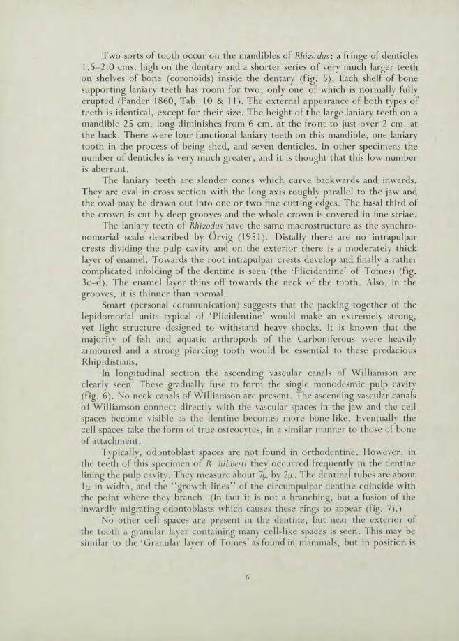
Two sorts of tooth occur on the mandibl es of Rhizo dus : a fringe of denticles 1.5-2.0 cms . high on the dentary and a shorter series of very much larger teeth on shelves of bone (coronoids) inside the dentary (fig. 5). Each shelf of bone supporting Jan iary teeth has room for two, only one of which is normally fully erupted (Pander 1860, Tab. 10 & II) . The external appearance of both types of teeth is identical, except for their size. The height of the large laniary teeth on a mandible 25 cm. long diminishes from 6 cm. at the fro nt to just over 2 cm . at the back. There were four functional laniary teeth on this mandibl e, one laniary tooth in the process of being shed, and seven denticl es . In other specimens the number of denticles is very much greater, and it is thought that this low number is aberrant.
The lan iary teeth are slender cones which curve backwards and inwards. They are oval in cross section with the long axis roughly parall e l to the jaw and the oval may be drawn out into one or two fine cutting edges. The basal third of the crown is cut by deep grooves and the whole c rown is covered in fine striae.
The lan iary teeth of Rhizodus have the same macrostructure as the synchronomorial scale described by Orvig ( 195] ). Distally there are no intrapulpar crests dividing the pulp cavi ty and on the ex terior there is a moderately thi ck layer of enamel. Towards the root intrapulpar crests develop and finally a rather compl icated infolding of the dentine is seen (the' Plicidentine' of Tomes) (f ig. 3c-d). The ename l layer thins off towards the neck of the tooth. Also, in the grooves, it is th inner than normal.
Smart (personal communication) suggests that the pack ing together of the lepidomorial units typical of 'Plicidentine' would l1'lake an ext reme ly strong, yet light structure designed to withstand heavy shocks. It is known that the majority of fish and aquat ic arthropods of thc Ca rbonifero us were heavily armoured and a strong piercing tooth would be essenti?1 to thcse predacious Rhipidistians.
In longitudi nal sect ion the ascending vascu lar ca nals of Will iamso n are c learly seen. These gradual ly fuse to form the single monodesmic pulp cavity (fig. 6). No neck canals of Williamson are present. The ascendi ng vascu lar canals 0 1 Williamson connect directly w ith the vascular spaces in the jaw and the ce ll spaces become visible as the dentine becomes more bone-like. Eventuall y the cell spaces take the fo rm of true osteocytes, in a simi lar manner to those of bone of attachment.
Typically, odontob last spaces are not found in orthodentine. However, in the teeth of this spec imen of R. hibberti they occurred frequently in the dentine lining the pu lp cav ity . They measure about 7fL by 2fL. The dentinal tubes are about lfL in width, and the "growth lines" of the circumpulpar dent ine coincide with the point where they branch. (In fact it is not a branching, but a fusion of the inwardly migrat ing odontob lasts whi ch causes these rings to appear (fi g. 7).)
No other ce ll spaces are present in the dentine, but near the exter ior of the tooth a granular layer containing many cell-li ke spaces is seen. This may be sim ilar to the 'G ranu lar layer of Tomes' as found in mammals, but in position is
6
more akin to the regions of globular dentine described by Bystrow in Osteolepis and other labyrin thodonts. It is however not near! y so ext ensi ve as in these other ani mals.
The teeth of Rhizodus are thecodont and are held in the jaw with a well developed bone of attachment. Basally and radially this bone of attachment passes into true bone with cell spaces, and is distinct from the dentine at all levels except the lowest. Bone of attachment is seen in the crevices on the exterior of the tooth. As the grooves widen towards the base of the crown, this bone of attachment tends to cover more and more of the outer surface until at the level of the jaw it surrounds the entire crown. Where enamel is present, bone of attachment always overlies it. In the lower basal sections there are some spaces between the trabeculae of bone of attachment rather bigger than normal. These spaces may mark regions where bone has not been formed, or, having been formed, is in the process of being resorbed prior to the tooth being shed (fig. 3d) (see also under Discussion).
Cell spaces do not occur in the bone of attachment except near the base of the tooth and where it merges into the bone of the jaw. At first these cell spaces are compact with few processes and measure approximately 30fL by 8fL. Their long axes are parallel to the trabeculae. Towards the jaw their shape gradually changes and more processes are seen. The cells here measure 40fL by 5fL (Roux 1942).
Bone of attachment presents rather higher birefringence in polarised light than does dentine and a more even colour than ordinary bone. This is possibly due to the presence of smaller crystallites more constantly orientated and coincides with the idea that bone of attachment was being constantly removed and relaid while the tooth erupted.
Resorption of tooth tissue is known to occur in most vertebrates from the outside initiated through mechanical stimulus . Whereas this probably occurred in Rhizodus, there is also evidence that dentine was resorbed through the pulp cavity. Transverse sections of an isolated tooth found in rock, and especially those sections from the base of this tooth, show a marked difference from the corresponding sections of teeth still attached to jaws (fig. 9a & b). In the latter all structures associated with the labyrinthodont pattern are seen, but in the former most of the dentine has been removed and all that remains is the outem10st layer. Bone of attachment shows signs of erosion, but enamel seems to be untouched. The complete jaw referred to on page 6 has a pair of laniary teeth with their roots contiguous. If, as is most likely, one of these teeth is replacing the other, then signs of dentine removal are to be expected on the side of the older tooth, nearest the replacing one. No evidence of this can be seen.
In figure 3d it will be noticed that alongside the fully functional tooth there lies remnants of another. The matter is discussed below.
DISCUSSION
.. The interpretation of the labyrinthodont tooth given in this paper, using Orvig's descriptions of the synchronomorial scale as a basis, gives an entirely new
7

orientation to one of the best known features of the Palaeozoic fish and amphibia. A consideration of the generalised synchronomorial scale shows that the equi valent of many generations of tooth material fuse to form a single tooth. This is the state of affairs seen in the labyrinthodont tooth of Rhizodus hibberti of the Lower Carboniferous.
Under Tomes' classification the dentine in labyrinthodont teeth was known as 'Plicidentine'. It can now be interpreted more accurately as a variety of orthodentine, with an outer layer of mantle dentine and an inner layer of circumvascular dentine (Orvig 1951, pp . 347-359) . Mantle dentine is characterised by the presence of odontoblast processes; it is of even structure and does not have the concentric lines similar to growth rings that circumvascular dentine has (figs. 7 & 8). Therefore it would appear to represent an initial rapid deposition of hard tissue and would be expected to predominate in the tip of the tooth. As far as can be ascertained this is so.
A granular layer similar to that in the dentine is seen in the enamel, becoming more marked towards the tip. At the same time the layer in the dentine becomes reduced (fig. 8). The presence of this layer in the enamel can be explained if this enamel is formed by the dentine organ and is subjected to similar influences as the dentine during development. Therefore the enamel is probably mesodermal in origin.
In postulating that the dentine of these teeth was removed under control of a non-mechanical system, rather than in the more normal way, the following facts must be examined. Firstly, each shelf of bone supporting the laniary teeth has room for only two, and, of which, it seems normal that only one was functional at anyone time. On the lower jaw of Rhizodus referred to on page 6, a pair of teeth lie side by side. There is no evidence of erosion where they are in contact, which would be expected if mechanical stimulus was the normal method of removing the older tooth. Secondly, transverse sections of adjacent tooth sockets show remnants of dentine with labyrinthodont structure lying round the edge of the empty socket and other sections of isolated (presumably shed) teeth identified as belonging to Rhizodus, reveal that all internal dentine is absent. These observations suggest that the dentine is removed from the interior before the tooth is .shed. Any mechanism responsible for dentine resorption could also trigger off the eruption of the other tooth of the pair.
It could be postulated that the isolated teeth with no labyrinthodont structures present were in fact newly erupted teeth broken away from the pair as a natural hazard of life. However in figure 9b it will be seen that the remnants of dentine and enamel are in general only slightly thinner than the walls of the tooth in figure 9a. The dissociated nature of the fragments can be explained only if the supporting structures in the centre of the tooth had been eroded from the inside.
ewly erupted teeth figured by Bystrow (1938) for Benthosuchus always have the labyrinthodont structures thinly developed, as are the tooth walls themselves. They are much thinner than the tooth wall in figure 9b.
Bystrow (1942) discusses the pattern of dermal bone growth in Dipterus
8
with reference to Westoll-lines. In sections of dermal bones he shows resorption spaces occurring within the hard tissues and, in other publications (1938; 1939), he shows similar resorption spaces in the bases of teeth of Benthosuchus, Glyptolepis, Ho10ptychius and Eusthenopteron. Regarding the Westoll-lines, Jarvik (194-8) agrees that they must represent growth stages, an0 Bystrow's analysis of their nature must lead to the conclusion that some cyclical mechanism of dentine resorption from within the tooth must have operated in these vertebrates.
In some mammals milk teeth are shed even if the permanent tooth-germ is excised (Oberstzyn 1963). Therefore it seems possible that replacement teeth do not need to erupt from directly below the functional tooth and that the basal dentine can be removed by some process other than mechanici'.l stimulus.
ACKNOWLEDGMENTS
The descriptions of the laniary teeth of Rhizodus hibberti in this paper were carried out as part of the requirem.ents for the degree of B.Sc. (Hons.) in Zoology in Edinburgh University. I am very grateful to Dr C. D. Waterson of the Royal Scottish Museum for the loan of the material, to Mr G. F. Friend for supervising the work, and the technical staff of both the Geology and Zoology Departments for the time and trouble taken helping to cut the sections and take the photographs. The Cambridgeshire Education Authority supplied a very generous grant during the course of the year. Dr R. Sprinz and Dr Alun Maddy kindly read this paper in manuscript and made many valuable suggestions for its improvement. Mr G. M. Burnett undertook the onerous task of translating the German texts, for which I am very grateful.
REFERENCES
BARKAS, W. J., 1875. On the microscopical structure of fossil teeth from the Northumberland Coal Measures. Mon. rev. dent. sura., 4, 393- 394-.
BYSTROW, A. P., 1938. Zahnstruktur der Labyrinthodonten. Acta zool. Stockh., 19, 357-4-25.
BYSTROW, A. P., 1939. Zahnstruktur der Crossopterygier. Acta zool. Stockh., 20, 283-338.
BYSTROW, A. P., 194-2. Deckknochen und Zahne der Osteolepis und Dipterlls. Acta zool. Stockh., 23, 263-289.
JARVIK, E., 194-8. On the morphology and taxonomy of the Middle Devonian osteolepid fishes of Scotland. K. Svenska Vetensk-Akad. Handl., 25, 1- 301.
KVAM, T., 194-6. Comparative study of the ontogenetic and phylogenetic development of dental enamel. Norske Tandelaeaiforen. Tid. (supplement). 56, 1-198.
Moy-THOMAS, J. A., 1939a. Palaeozoic fishes. London, Methuen. Moy-THOMAS, J. A., 1939b. Evolution of the elasmobranchs. Biol. Rev., 14, 1-26.
9

OBERSZTYN, A., 1963. Experimental investigations of factors causing resorption of deciduous teeth. J. dent. Res., 42, 660-674.
ORVIG, T., 1951. Histologic studies of placoderms and fossil elasmobranchs. I. The endoskeleton. Ark. Zo01., 2, 321-456.
ORVIG, T., 1965. Palaeohistological notes. 2. Ark. Zool., 16,551-556. PANDER, C. H., 1860. Uber die Saurodipterinen, DendrodOriten, Glypto1epiden und
Cheirolepiden des devonischen Systems. St Petersburg. POOLE, D. F. G., 1956a. Fine structure of scales and teeth of Raia clavata.
Q.)I. microsc. Sci., 97, 99-107. POOLE, D. F. G., 1956b. Structure of teeth in mammal-like reptiles. Q. )1.
microsc. Sci., 97, 303-312. ROMER, A. S., 1966. Vertebrate palaeontology. 3rd edition. UniverSity of Chicago
Press. Roux, G. H., 1942. Minute structures in the scales of Latimeria. S. Afr. J. med.
Sci., 7, 1-18. SCOTT, J. H. & SYMONS, N. B., 1964. Introduction to dental anatomy. Edinburgh
and London, E. & S. Livingston. STENSIO, E. A., 1961. Permian vertebrates in Geology ?Jthe Arctic. Ed. G. O. Raasch.
Toronto University Press. pp. 231-247. STENSIO, E. A., 1962. Origin et natur des ecailles placoids et des dents. Colloq.
into Cent. naw. Rech. scient., 104, 75-85. STOCK, T., 1880. Note on the discovery of an entire specimen of Rhizodus sp.
in the Wardie Shales. Trans. Edinb. geol. Soc., 4, 38. TARLO, L. B. H., 1963. Aspidin: the precursor of bone. Nature, London, 199,
46-48. TARLO, L. B. H., 1964. The origin ?Jbone in Proceedings ?J the first bone and tooth
symposium, Oiford. 1963. Oxford, Pergamon Press. WlDDOWSON, T. W., 1928. Dental anatomy and physiology and dental histology.
London, Bale, Sons & Danielson Ltd.
10
Fig. 1.
Fig. 2.
Fig. 3.
Fig. 4.
Fig. 5
Fig. 6.
Fig. 7.
Fig. 8.
Fig. 9.
EXPLANATION OF TEXT FIGURES The mode of formation of Cyclomorial scales. From brvig, 1951. (a) Vertical section of the primordial lepidomorium, consisting of a dentine crown and a bony base. (b) A more advanced stage of development. The hard tissues of the second lepidomorium have begun to form. (c) Two lepidomoria in the final stages of development. lpca-lepidomorial pulp cavity; cr-crown of primordial lepidomorium; ve-vessels entering the pulp cavity through both the neck and basal canals of Williamson; bp-basal plate of the primordiallepidomorium; can. W. n. - neck canal of Williamson; p. Idu' papilla of soft tissue of the first areal zone of growth; ldup-ldu'-crowns of the primordial and second lepidomorium respectively; bpa-basal plate of the Cyclomorial scale; can. W. b-basal canal of Williamson; bp'- basal plate of the second lepidomorium. The mode of formation of a Synchronomorial scale. From brvig, 1951. (a) Horizontal section of the crown at early stage of development. A thin layer of mantle dentine has formed round the embryonic synchronomorial pulp cavity. (b) More mantle dentine has formed, and the intrapulpar crests have grown out to form partition walls. (c) Formation of dentinal osteons within the secondary lepidomorial pulp cavities formed in stage two. pd-pallial (mantle) dentine; ipcr- intrapulpar crests; v. sp.-secondary lepidomorial pulp cavities; v. can-vascular canals; osd-dentinal osteons; empca-embryonic monodesmic pulp cavity. Transverse sections of laniary tooth of Rhizodus hibberti Ag. Unregistered collection Royal Scottish Museum. See also Fig. 5. (a) 2 cm. from apex. (b) 2.26 cm. from apex. (c) 2.46 cm. from apex. (d) 4.11 cm. from apex . Pc-main pulp cavity; ipcr- intrapulpar crests; ce-cutting edge of tooth; Sp-large spaces between tooth and jaw-bone; Od-dentine of functional tooth; Od' -dentine of alternate tooth; Ba-bone of attachment; Bj-bone of jaw. lines represent 1 cm. Vertical section of ganoid scale to show ascending canals of Williamson (can. W), but no neck canals. From brvig, 1951. Tooth of Rhizodus hibberri Ag. prior to being sectioned transversely . See also Fig. 3. Unregistered collection, Royal Scottish Museum. Bj- bone of jaw; Sh-shale in which tooth and jaw remnant is embedded. Line represents 1 cm . Longitudinal section of incomplete laniary tooth of R. hibberri. Registered number E (nat. hist.) 352. Hugh Miller collection, Royal Scottish Museum. Pc-main pulp cavity; Ba-bone of attachment; Bb- basal bony region of tooth; Bj- bone of jaw; can. W-ascending canal of Williamson; V. can . J-vascular canals of the jaw. Line is 1 cm. long. Orthodentine in laniary tooth of R. hibberti, showing mantle dentine (pd) and circumvascular dentine (Cpd). The latter with "growth rings" caused by synchronous fusion of inwardly migrating odontoblasts (Gr). Ba- bone of attachment. Line is 1 mm. long. High power view of figure 3b. to show granular layer in both dentine and enamel. e-enamel; gr. e-granular layer in enamel; gr. d-granular layer in dentine; pd-mantle dentine. Line is 50fL long. Transverse sections of Rhizodus teeth at equivalent levels. (a) Section of tooth immediately above level of jaw-bone. c.f. fig. 3c. (b) Basal section of isolated tooth with dentine missing from interior of tooth. Distortion of tooth caused by pressure of overlying sediment. Od-dentine.
11
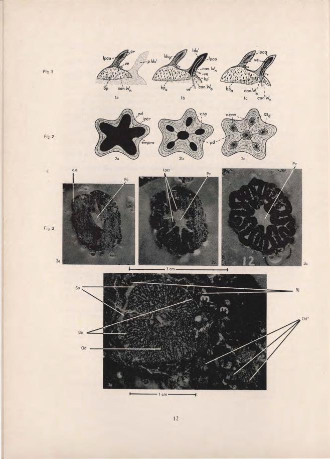
Fig. 1
i-.'·,X:·_ ... ::/
b'p
FiJ.3
~,.,,:-':~~~t:.,,:.~:~::;.~-.~.:,; . );' \. con·""n
1.
12
1b
3c
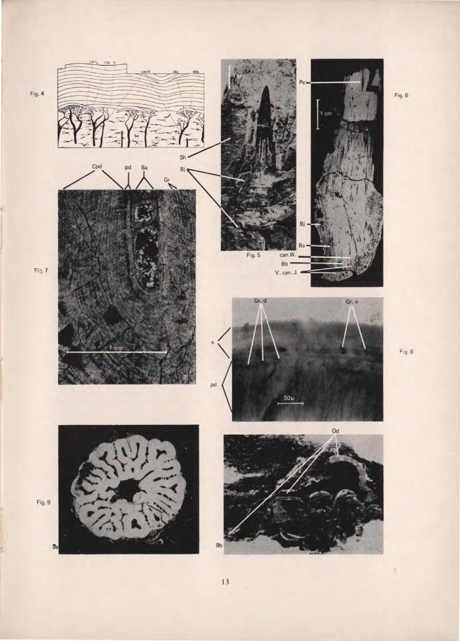
Fig.6
13