TMJ findings chart - FINAL
19
• Patient Questionnaire • Screening Exam Record • TMJ Findings Worksheet • Clinical Exam Guidelines: (found in the back pocket of this guide) ■ Muscle Palpation ■ Evaluating Joint Sounds ■ Load Testing with the Lucia Jig • TMJ Screening Results Table (found in the back pocket of this guide) Invest 45 minutes in reading this guide and you will be able to reliably and efficiently treat your patients with TMD issues. Patient Screening Guide Step-by-step (found in the back of this section)
Transcript of TMJ findings chart - FINAL
• Patient Questionnaire• Screening Exam Record• TMJ Findings Worksheet
• Clinical Exam Guidelines:(found in the back pocket of this guide)
n Muscle Palpationn Evaluating Joint Soundsn Load Testing with the Lucia Jig
• TMJ Screening Results Table(found in the back pocket of this guide) Invest 45 minutes
in reading this guide and you will be able
to reliably and efficientlytreat your patients with TMD issues.
PatientScreeningGuide
Step-by-step
(found in the back of this section)
We know that up to 40% of the patients currently in your practice today have occlusal pathology.Additionally, we know that 80% of these patients have muscle imbalances and can benefit from anocclusal splint.*
This guide is designed to help you treat this population efficiently and effectively.
The screening guide is part of a larger kit containing tools you will need to identify, diagnose, and treatyour TMD patients. In addition to the screening guide, the kit includes a tool to help you select theright splint; instructions on impression-taking, bite registration, and splint adjustment; information onmaximizing insurance benefits, accessing a splint specialist for technical support, as well as access to aDoctor Consult service.
Through Great Lakes Doctor Consult service, I can help you with TMD patient screening and diagnosticquestions, effective appliance management, and TMD treatment troubleshooting. Take a look at theflier included in the kit for more information.
I know that you want to provide the very best care for your patients with TMD. I also realize that yourpatients want you to handle as much of their care as possible. So this is really a winning situation forboth you and your patients.
With your expertise and Great Lakes technology and skills, you can confidently and successfully treatyour TMD patients.
Fred McIntyre, DDS, MSDoctor Consult Service
* Dr. Gordon J . Chr is tensen, Occlusal Spl in ts -Predic table , Frequent Use Pract ica l C l in ica l Course DVD
Dear Colleague,

The step-by-step patient screening guide will help you identify appropriate patients to treat as well asidentify those patients who should be referred out of the practice. The key to making this critical decision is communication with the patient and your clinical exam.
We have included tools that will help you, a patient questionnaire and a screening exam record.
The questionnaire and the exam record are featured in this guide with an explanation of when andhow to use them. They coordinate with two additional tools in the guide called, the TMJ FindingsWorksheet and the TMJ Screening Results Reference Table.
Also included in this guide are instructions (found in the back pocket) that coordinate with the screening exam record on muscle palpation, evaluating joint sounds, and load testing with a Lucia Jig.
Case ExamplesThe shaded boxes throughout the guide feature examples of two patients who have reported symptoms during a routine exam. For each step, the examples show the patient's responses to the questionnaire, screening exam results, and TMJ Findings Worksheet information. The last section discusses the findings and possible treatment plans for both patients.
H ow to Use This Guide
At any time during your review of this guide or throughout
evaluation or treatmentplanning, feel free to contact
our Splint Support Specialistat 1.800.828.7626 with any
questions or to access the Doctor Consult Service.
Customize patient-specific tools (found in the back of this section)
■ Patient Questionnaire ■ Screening Exam Record ■ TMJ Findings Worksheet
Customize with your practice name, address, and logo. Download the tools from our website at www.greatlakesortho.com.
The general dental practitioner is recognized as having ultimate responsibility for patient evaluation,diagnostic, treatment and/or referral decisions. The information contained in this guide is compiled fromtextbooks, articles, and courses available to the profession and is provided in an advisory capacity only.Great Lakes Orthodontics, Ltd, is not responsible for patient outcomes.
1

By incorporating a routine patient questionnaire and exam into patient visits, you will become increasingly more
aware of potential TMD symptoms and signs. Either the patient will talk about symptoms such as headaches or
muscle soreness or you may notice occlusal signs such as excessive wear or fractures.
The first step is the patient questionnaire which can be completed in just a few minutes by the patient. It's an ideal
tool to use chair-side to discuss symptoms in more detail with the patient.
The questionnaire is designed for simple 'yes' or 'no' responses. This makes it easy for the patient to respond and
allows you to immediately focus on any red flag responses such as 'injury to the jaw or face' or 'having been
previously treated by a TMJ specialist'.
The responses from the questionnaire will be transferred to the TMJ Findings Worksheet for further evaluation along
with the results of the screening exam.
Patient Questionnaire
2

An existing patient, Mary has been scheduled for aTMJ Screening visit. At her last routine exam, shecomplained of frequent headaches and youthought you saw some occlusal wear on the anterior teeth.
From her questionnaire, you can see that she didindicate headaches in the late afternoon, tenderjaw muscles, and some teeth that are sore, aching,and uncomfortable. Also note, Mary indicated thatshe has not received an injury to the face and hasnever been treated by a TMJ specialist before.
During the exam, special attention should be givento assessing occlusal stability, muscle palpation,and load testing.
These responses will be transferred to Mary's TMJFindings Worksheet (Page 7).
SMLP162Rev011508
TMJ Patient Questionnaire Patient Name:____________________________
Date:__________
Answer all that apply.
YES NO1) Do you have frequent or regular headaches?
Upon awakening
Late afternoon
2) Are your jaw muscles sore or tender?
3) Are your joints sore or tender when you eat or chew?
4) Have you ever received an injury to your jaw or face?
If yes: Describe:
5) Do your joints make any noise such as snapping, clicking, or popping?
6) Do your joints lock when you are trying to open or close?
7) Do you have any teeth that are sensitive, sore, aching, or uncomfortable?
8) Have you ever worn a splint or nightguard?
If yes: How many?_____
9) Are you taking or have you taken any medication for these symptoms?
If yes: Describe:
10) Have you ever seen a dentist or a TMJ specialist for treatment of any of the above symptoms?
If yes: How many?_______
A new patient, Bob has been scheduled for a TMJScreening visit. During his initial exam, he complained of frequent headaches and youthought you saw some occlusal wear on the anterior teeth.
From his questionnaire, you can see that he didindicate headaches upon awakening, tender jawmuscles, and some teeth that are sore, aching,and uncomfortable. Also note, Bob indicated thathe noticed some noise in his joints.
Chair-side, Bob mentioned that his wife could heara click in his right joint.
During the exam, special attention should be givento assessing occlusal stability, muscle palpation,evaluating joint sounds, and load testing.
These responses will be transferred to Bob's TMJFindings Worksheet (Page 7).
3
(Your Letterhead)
(Your Letterhead)
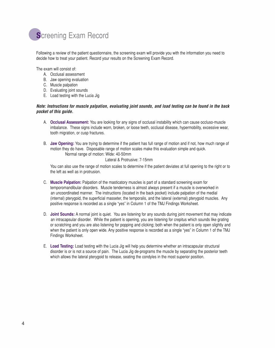
Following a review of the patient questionnaire, the screening exam will provide you with the information you need todecide how to treat your patient. Record your results on the Screening Exam Record.
The exam will consist of:A. Occlusal assessment B. Jaw opening evaluationC. Muscle palpationD. Evaluating joint soundsE. Load testing with the Lucia Jig
Note: Instructions for muscle palpation, evaluating joint sounds, and load testing can be found in the backpocket of this guide.
A. Occlusal Assessment: You are looking for any signs of occlusal instability which can cause occluso-muscle imbalance. These signs include worn, broken, or loose teeth, occlusal disease, hypermobility, excessive wear, tooth migration, or cusp fractures.
B. Jaw Opening: You are trying to determine if the patient has full range of motion and if not, how much range of motion they do have. Disposable range of motion scales make this evaluation simple and quick.
Normal range of motion: Wide: 40-50mmLateral & Protrusive: 7-15mm
You can also use the range of motion scales to determine if the patient deviates at full opening to the right or to the left as well as in protrusion.
C. Muscle Palpation: Palpation of the masticatory muscles is part of a standard screening exam for temporomandibular disorders. Muscle tenderness is almost always present if a muscle is overworked in an uncoordinated manner. The instructions (located in the back pocket) include palpation of the medial (internal) pterygoid, the superficial masseter, the temporalis, and the lateral (external) pterygoid muscles. Any positive response is recorded as a single “yes” in Column 1 of the TMJ Findings Worksheet.
D. Joint Sounds: A normal joint is quiet. You are listening for any sounds during joint movement that may indicatean intracapsular disorder. While the patient is opening, you are listening for crepitus which sounds like grating or scratching and you are also listening for popping and clicking; both when the patient is only open slightly and when the patient is only open wide. Any positive response is recorded as a single “yes” in Column 1 of the TMJ Findings Worksheet.
E. Load Testing: Load testing with the Lucia Jig will help you determine whether an intracapsular structural disorder is or is not a source of pain. The Lucia Jig de-programs the muscle by separating the posterior teeth which allows the lateral pterygoid to release, seating the condyles in the most superior position.
Screening Exam Record
4
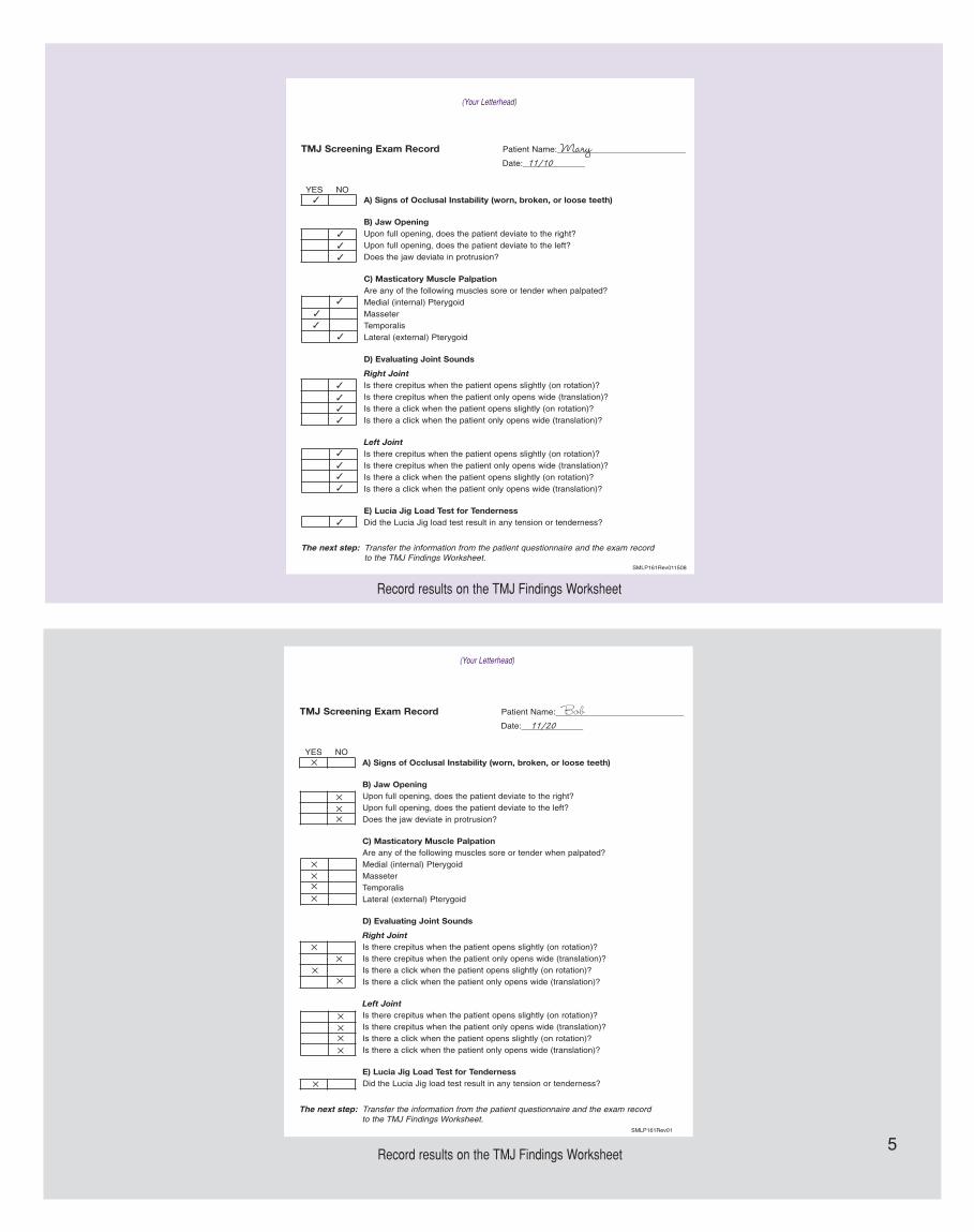
TMJ Screening Exam Record Patient Name:___________________________
YES NO
SMLP161Rev011508
The next step: Transfer the information from the patient questionnaire and the exam record to the TMJ Findings Worksheet.
A) Signs of Occlusal Instability (worn, broken, or loose teeth)
B) Jaw OpeningUpon full opening, does the patient deviate to the right?Upon full opening, does the patient deviate to the left?Does the jaw deviate in protrusion?
C) Masticatory Muscle PalpationAre any of the following muscles sore or tender when palpated?Medial (internal) PterygoidMasseterTemporalisLateral (external) Pterygoid
D) Evaluating Joint Sounds
Right JointIs there crepitus when the patient opens slightly (on rotation)?Is there crepitus when the patient only opens wide (translation)?Is there a click when the patient opens slightly (on rotation)?Is there a click when the patient only opens wide (translation)?
Left JointIs there crepitus when the patient opens slightly (on rotation)?Is there crepitus when the patient only opens wide (translation)?Is there a click when the patient opens slightly (on rotation)?Is there a click when the patient only opens wide (translation)?
E) Lucia Jig Load Test for TendernessDid the Lucia Jig load test result in any tension or tenderness?
Date:_____________
TMJ Screening Exam Record Patient Name:___________________________
YES NO
SMLP161Rev01
A) Signs of Occlusal Instability (worn, broken, or loose teeth)
B) Jaw OpeningUpon full opening, does the patient deviate to the right?Upon full opening, does the patient deviate to the left?Does the jaw deviate in protrusion?
C) Masticatory Muscle PalpationAre any of the following muscles sore or tender when palpated?Medial (internal) PterygoidMasseterTemporalisLateral (external) Pterygoid
D) Evaluating Joint Sounds
Right JointIs there crepitus when the patient opens slightly (on rotation)?Is there crepitus when the patient only opens wide (translation)?Is there a click when the patient opens slightly (on rotation)?Is there a click when the patient only opens wide (translation)?
Left JointIs there crepitus when the patient opens slightly (on rotation)?Is there crepitus when the patient only opens wide (translation)?Is there a click when the patient opens slightly (on rotation)?Is there a click when the patient only opens wide (translation)?
E) Lucia Jig Load Test for TendernessDid the Lucia Jig load test result in any tension or tenderness?
Date:_____________
The next step: Transfer the information from the patient questionnaire and the exam record to the TMJ Findings Worksheet.
Record results on the TMJ Findings Worksheet
Record results on the TMJ Findings Worksheet5
(Your Letterhead)
(Your Letterhead)

Once completed, the TMJ Findings Worksheet will reveal a specific course of action and you can begin to treatment planfor your patient. The table is easy to use.
Step 1: Record all "YES" responses in COLUMN 1 from the patient questionnaire and the screening exam worksheet.
Step 2: For all "YES" responses, place a check mark in the associated shaded box to the right in Columns 2-5. There's only one shaded box per row.
Interpreting the Table:
Columns 2 through 5 are organized from the least to the most treatment options. Read left to right, the first check markindicates the appropriate course of action.
Start with Column 2, if there is even one check mark in Column 2, consider referring the patient to a TMJ specialist.
Provided there are no check marks in Column 2, one check mark in Column 3 indicates a Full Contact splint w/anterior guidance.
If there are no check marks in Columns 2 or 3, but a check mark in Column 4, either a Full Contact or Flat Plane splint is indicated.
If there are no check marks in Columns 2, 3, or 4, but a check mark in Column 5, either a Full Contact, FlatPlane, or deprogrammer is indicated.
After you have determined the appropriate course of action to take, you can use the TMJ Screening Results ReferenceTable to further evaluate possible causes and learn more about appliance and treatment options. Refer to the SplintAppliance Selection Guide to choose the appropriate Full Contact splint with anterior guidance, Flat Plane splint, ordeprogrammer.
TMJ Findings Worksheet
-
COLUMN 1 COLUMN 2 COLUMN 3 COLUMN 4 COLUMN 5
Mark 'YES'
HereRefer
Full Contact with
anterior guidance
Full Contact w/ant.
guide or Flat Plane
Full Contact w/ant. guide,
Flat Plane, or
Deprogrammer
1 Awakening Headache
Afternoon Headache
2 Jaw Muscle Soreness
3 Joint Soreness
4 Injury
5 Joint Click
6 Locking Joints
7 Sensitive/Sore Teeth
8 Splint or Nightguard
9 Medication(s)
10 TMJ Specialist
A Occlusal Instability
B Jaw Opening-Right
Jaw Opening-Left
Jaw Opening-Protrusion
C Muscle Palpation
D Crepitus open slightly
Crepitus open wide
Click open slightly
Click open wide
E Pain on Load Testing
Screening Exam
Findings
Question
Patient
Questionnaire
Responses
Potential indication of occlusal problem
Indication of severit
yIndication of severit
y
6

Interpreting the results for MaryAfter transferring all the “YES” answers onto the worksheet and completing the table, you can now assess your results.
Reading left to right across the table, the first check mark to the left appears in Column 4 which indicates a Flat Plane splint or Full Contactsplint with anterior guidance. See Page 8 for more discussion about Mary's case.
Interpreting the results for BobAfter transferring all the “YES” answers onto the worksheet and completing the table, you can now assess your results.
Reading left to right across the table, the first check mark to the left appears in Column 2 which indicates that Bob should be referredto a specialist. See Page 8 for more discussion about Bob's case. 7
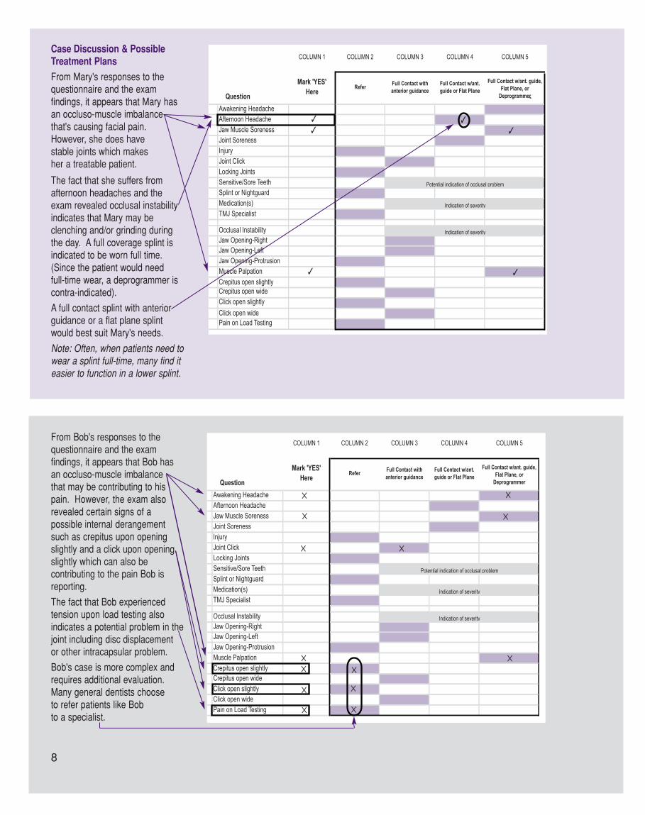
Case Discussion & PossibleTreatment Plans
From Mary's responses to the questionnaire and the exam findings, it appears that Mary hasan occluso-muscle imbalancethat's causing facial pain.However, she does have stable joints which makes her a treatable patient.
The fact that she suffers fromafternoon headaches and theexam revealed occlusal instabilityindicates that Mary may be clenching and/or grinding duringthe day. A full coverage splint isindicated to be worn full time.(Since the patient would need full-time wear, a deprogrammer is contra-indicated).
A full contact splint with anteriorguidance or a flat plane splintwould best suit Mary's needs.
Note: Often, when patients need towear a splint full-time, many find iteasier to function in a lower splint.
From Bob's responses to the questionnaire and the exam findings, it appears that Bob hasan occluso-muscle imbalance that may be contributing to hispain. However, the exam alsorevealed certain signs of a possible internal derangementsuch as crepitus upon openingslightly and a click upon openingslightly which can also be contributing to the pain Bob isreporting.
The fact that Bob experienced tension upon load testing also indicates a potential problem in thejoint including disc displacement or other intracapsular problem.
Bob's case is more complex andrequires additional evaluation.Many general dentists chooseto refer patients like Bob to a specialist.
8

n
Confirm your diagnosis or review treatment options with an experienced TMD practitioner...
Great Lakes offers a new Doctor Consult Service with Frederick M. McIntyre, DDS, MS.
Access the service by calling Great Lakes splint support specialist at 1.800.828.7626 to set up a telephone consultation.
The Doctor Consult service is available by appointmentonly. $35 for up to 1/2 hour.
Clinical Exam Guidelinesn Muscle Palpationn Evaluating Joint Soundsn Load Testing with the Lucia Jig
TMJ Screening Results Table
Want to Learn More?Contact Great Lakes for information on nationally-accredited courses offering training in:
Management of moreadvanced TMD issues
Long-term resolution toocclusal problems
Occlusion for aesthetics
1.800.828.7626716.871.1161
www.greatlakesortho.com
n
n
TMJ Diagnostic Materials & Equipment
For Jaw Opening Evaluation:Therabite Range of Motion Scales(100/pkg)
For Load Testing using the Lucia Jig:Lucia Jig Kit (18 standard and 6 Class II)Whale Tails (3/pkg)Acu-Flow™ Bite Registration MaterialAcu-Flow™ Dispensing Gun
For Evaluating Joint Sounds:Great Lakes TMJ Doppler™(Includes 5MHZ transducer, gel,carrying case, and manual)
For Patient Education:Great Lakes TMJ DemonstratorSkull with Masticatory Musculature
255-009
255-025255-027100-005100-010
250-041
255-012255-015
For more information or tocontact our Splint Specialist:
1.800.828.7626716.871.1161
www.greatlakesortho.com
Palpation is the examination of the soft tissues using the sense of touch. Palpation of themasticatory muscles will reveal tension or tenderness which can be related to hyperactivity ofthe muscle as a result of overworking it in an uncoordinated manner. A muscle is overworkedwhen it is required to constantly hold the jaw in an avoidance pattern during closure to maximum intercuspation.
When you begin palpation, explain to the patient that you will be applying a light pressure tothe muscle just as you would apply light pressure to their forearm. Demonstrate the amount ofpressure you will be using on the patient's forearm and establish a “no pain” point of reference. Tell the patient that he or she will need to let you know if there is any tenderness orpain greater than the “no pain” point of reference and if it is mild, moderate, or severe. Bealert to wincing and body language as some patients have a higher tolerance for discomfortthan others.
To palpate means to press lightly on the muscle. In a continuous movement, slide yourfinger(s) along the length and width of the muscle while asking the patient if he or she feelsany tension or tenderness as you apply pressure. You are also trying to feel for any abnormality, contraction, or enlargement of the muscle.
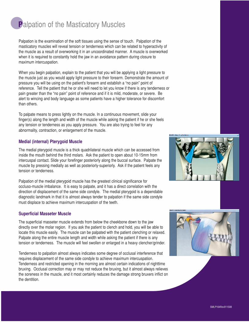
Medial (internal) Pterygoid Muscle
The medial pterygoid muscle is a thick quadrilateral muscle which can be accessed frominside the mouth behind the third molars. Ask the patient to open about 10-15mm from intercuspal contact. Slide your forefinger posteriorly along the buccal surface. Palpate themuscle by pressing medially as well as posteriorly-superiorly. Ask if the patient feels anytension or tenderness.
Palpation of the medial pterygoid muscle has the greatest clinical significance for occluso-muscle imbalance. It is easy to palpate, and it has a direct correlation with the direction of displacement of the same side condyle. The medial pterygoid is a dependablediagnostic landmark in that it is almost always tender to palpation if the same side condylemust displace to achieve maximum intercuspation of the teeth.
Superficial Masseter Muscle
The superficial masseter muscle extends from below the cheekbone down to the jaw directly over the molar region. If you ask the patient to clench and hold, you will be able tolocate this muscle easily. The muscle can be palpated with the patient clenching or relaxed.Palpate along the entire muscle length and width while asking the patient if there is any tension or tenderness. The muscle will feel swollen or enlarged in a heavy clencher/grinder.
Tenderness to palpation almost always indicates some degree of occlusal interference thatrequires displacement of the same side condyle to achieve maximum intercuspation.Tenderness and restricted opening in the morning are almost certain indications of nighttimebruxing. Occlusal correction may or may not reduce the bruxing, but it almost always relievesthe soreness in the muscle, and it most certainly reduces the damage strong bruxers inflict onthe dentition.
Palpation of the Masticatory Muscles
SMLP164Rev011508
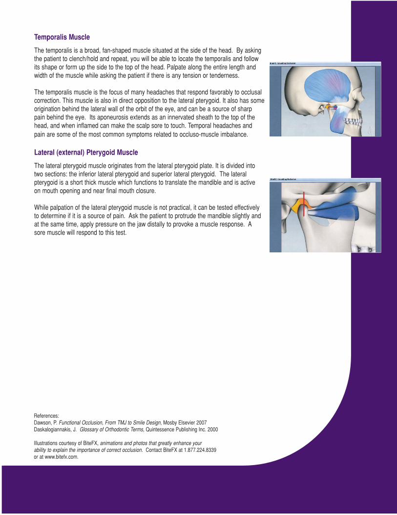
Temporalis Muscle
The temporalis is a broad, fan-shaped muscle situated at the side of the head. By asking the patient to clench/hold and repeat, you will be able to locate the temporalis and follow its shape or form up the side to the top of the head. Palpate along the entire length andwidth of the muscle while asking the patient if there is any tension or tenderness.
The temporalis muscle is the focus of many headaches that respond favorably to occlusal correction. This muscle is also in direct opposition to the lateral pterygoid. It also has some origination behind the lateral wall of the orbit of the eye, and can be a source of sharp pain behind the eye. Its aponeurosis extends as an innervated sheath to the top of thehead, and when inflamed can make the scalp sore to touch. Temporal headaches andpain are some of the most common symptoms related to occluso-muscle imbalance.
Lateral (external) Pterygoid Muscle
The lateral pterygoid muscle originates from the lateral pterygoid plate. It is divided into two sections: the inferior lateral pterygoid and superior lateral pterygoid. The lateralpterygoid is a short thick muscle which functions to translate the mandible and is activeon mouth opening and near final mouth closure.
While palpation of the lateral pterygoid muscle is not practical, it can be tested effectivelyto determine if it is a source of pain. Ask the patient to protrude the mandible slightly andat the same time, apply pressure on the jaw distally to provoke a muscle response. Asore muscle will respond to this test.
References:Dawson, P. Functional Occlusion, From TMJ to Smile Design, Mosby Elsevier 2007Daskalogiannakis, J. Glossary of Orthodontic Terms, Quintessence Publishing Inc. 2000
Illustrations courtesy of BiteFX, animations and photos that greatly enhance yourability to explain the importance of correct occlusion. Contact BiteFX at 1.877.224.8339 or at www.bitefx.com.
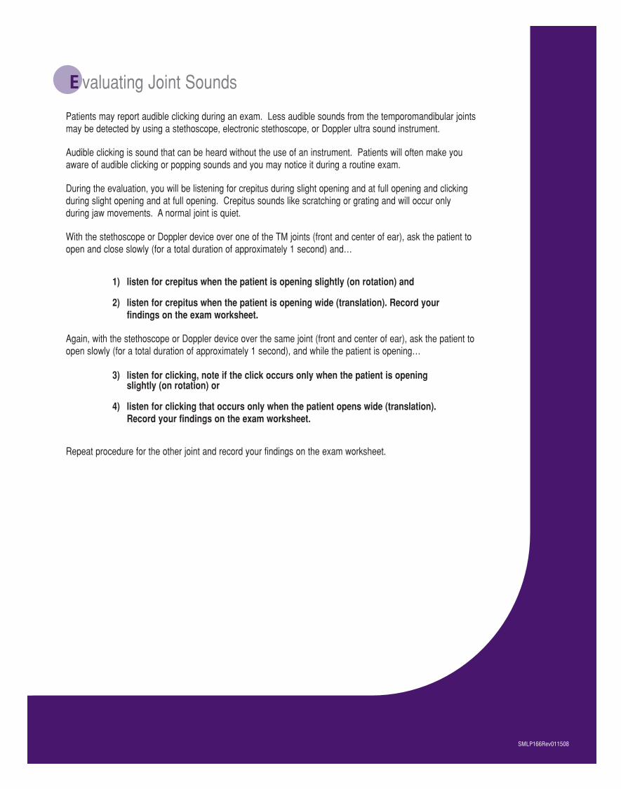
Patients may report audible clicking during an exam. Less audible sounds from the temporomandibular jointsmay be detected by using a stethoscope, electronic stethoscope, or Doppler ultra sound instrument.
Audible clicking is sound that can be heard without the use of an instrument. Patients will often make you aware of audible clicking or popping sounds and you may notice it during a routine exam.
During the evaluation, you will be listening for crepitus during slight opening and at full opening and clickingduring slight opening and at full opening. Crepitus sounds like scratching or grating and will occur onlyduring jaw movements. A normal joint is quiet.
With the stethoscope or Doppler device over one of the TM joints (front and center of ear), ask the patient toopen and close slowly (for a total duration of approximately 1 second) and…
1) listen for crepitus when the patient is opening slightly (on rotation) and
2) listen for crepitus when the patient is opening wide (translation). Record your findings on the exam worksheet.
Again, with the stethoscope or Doppler device over the same joint (front and center of ear), ask the patient toopen slowly (for a total duration of approximately 1 second), and while the patient is opening…
3) listen for clicking, note if the click occurs only when the patient is opening slightly (on rotation) or
4) listen for clicking that occurs only when the patient opens wide (translation). Record your findings on the exam worksheet.
Repeat procedure for the other joint and record your findings on the exam worksheet.
E valuating Joint Sounds
SMLP166Rev011508

One of the most practical uses for load testing is that it is a fast, simple, and safe procedurefor determining whether an intracapsular structural disorder is or is not a source of orofacialpain.*
The Lucia Jig is ideal to load test because it de-programs the muscles which allows thecondyles to seat in the most superior position. The Lucia Jig separates the posterior teethwhich allows the lateral pterygoid to release, and when the lateral pterygoid releases, thecondyle seats.
For this procedure, ACU-Flow™ is used. It is a syringeable poly vinyl siloxane material with a15 second working time, 45 second intra-oral setting time, and a maximum total cure time of 1minute.
Selecting the right Lucia Jig is based on the vertical opening of the posterior teeth. The vastmajority of patients will use the Class I jig.
Use the standard (Class I) jig to determine the vertical opening. The Class II jig is used ifthere are several millimeters (more than 2.5 mm) between the posterior teeth to reduce thevertical opening.
For patients with very irregular mandibular incisors, use the Lucia Jig on the lower incisors. Itwill function in the same way against the maxillary incisors.
Step 1: Paint the curved surface of the jig with a small amount of silicone tray adhesive.
Step 2: Dispense Acu-Flow material into the curved portion of the jig.
Step 3: Place the jig on the upper centrals and place the Whale Tail directly beneath the jig. Ask the patient to bite down and hold. The Whale Tail levels and orients the jig to the occlusal plane. It parallels the jig with the occlusal plane anteriorly andposteriorly and in a right and left direction as well. Allow to set - approximately 45 seconds.
L oad Testing Using a Lucia Jig
SMLP165Rev011508
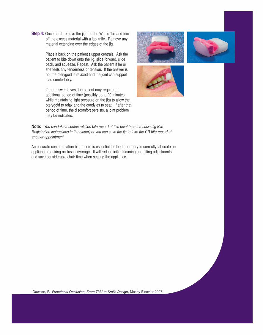
Step 4: Once hard, remove the jig and the Whale Tail and trim off the excess material with a lab knife. Remove any material extending over the edges of the jig.
Place it back on the patient's upper centrals. Ask the patient to bite down onto the jig, slide forward, slide back, and squeeze. Repeat. Ask the patient if he or she feels any tenderness or tension. If the answer is no, the pterygoid is relaxed and the joint can support load comfortably.
If the answer is yes, the patient may require an additional period of time (possibly up to 20 minutes while maintaining light pressure on the jig) to allow thepterygoid to relax and the condyles to seat. If after thatperiod of time, the discomfort persists, a joint problem may be indicated.
Note: You can take a centric relation bite record at this point (see the Lucia Jig BiteRegistration instructions in the binder) or you can save the jig to take the CR bite record atanother appointment.
An accurate centric relation bite record is essential for the Laboratory to correctly fabricate anappliance requiring occlusal coverage. It will reduce initial trimming and fitting adjustmentsand save considerable chair-time when seating the appliance.
*Dawson, P. Functional Occlusion, From TMJ to Smile Design, Mosby Elsevier 2007
TMJ Screening Results Reference Table
SMLP163Rev011508
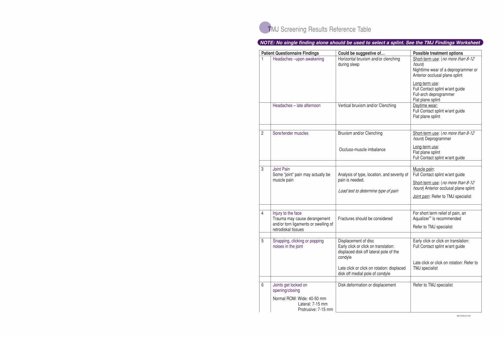
Patient Questionnaire Findings Could be suggestive of… Possible treatment options 1 Headaches –upon awakening Horizontal bruxism and/or clenching
during sleepShort-term use: (no more than 8-12 hours)Nighttime wear of a deprogrammer or Anterior occlusal plane splint
Long-term use:Full Contact splint w/ant guideFull-arch deprogrammer Flat plane splint
Headaches – late afternoon Vertical bruxism and/or Clenching Daytime wear:Full Contact splint w/ant guideFlat plane splint
2 Sore/tender muscles Bruxism and/or Clenching
Occluso-muscle imbalance
Short-term use: (no more than 8-12 hours) Deprogrammer
Long-term use:Flat plane splint Full Contact splint w/ant guide
3 Joint Pain Some “joint” pain may actually be muscle pain
Analysis of type, location, and severity of pain is needed.
Load test to determine type of pain
Muscle pain:Full Contact splint w/ant guide
Short-term use: (no more than 8-12 hours) Anterior occlusal plane splint
Joint pain: Refer to TMJ specialist
4 Injury to the face Trauma may cause derangement and/or torn ligaments or swelling of retrodiskal tissues
Fractures should be considered For short term relief of pain, an Aqualizer is recommended
Refer to TMJ specialist
5 Snapping, clicking or popping noises in the joint
Displacement of disc Early click or click on translation: displaced disk off lateral pole of the condyle
Late click or click on rotation: displaced disk off medial pole of condyle
Early click or click on translation:Full Contact splint w/ant guide
Late click or click on rotation: Refer to TMJ specialist
6 Joints get locked on opening/closing
Normal ROM: Wide: 40-50 mm Lateral: 7-15 mm Protrusive: 7-15 mm
Disk deformation or displacement Refer to TMJ specialist
NOTE: No single finding alone should be used to select a splint. See the TMJ Findings Worksheet
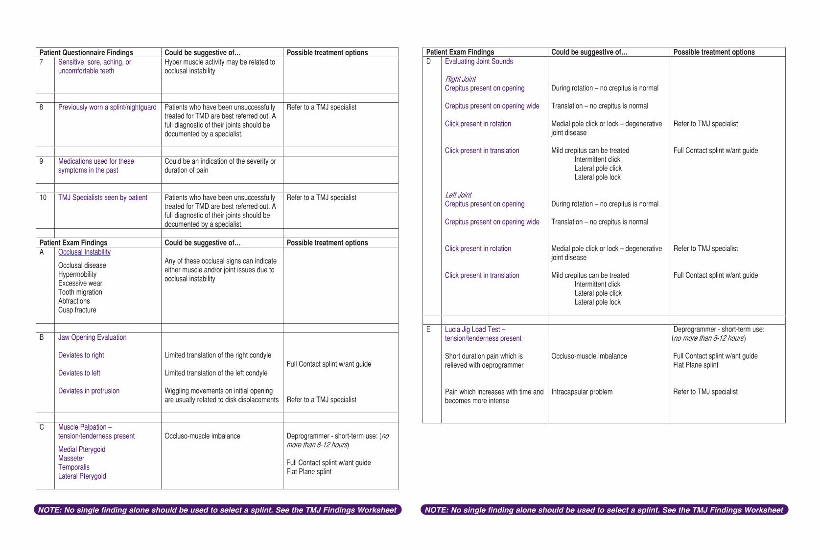
Patient Questionnaire Findings Could be suggestive of… Possible treatment options 7 Sensitive, sore, aching, or
uncomfortable teeth Hyper muscle activity may be related to occlusal instability
8 Previously worn a splint/nightguard Patients who have been unsuccessfully treated for TMD are best referred out. A full diagnostic of their joints should be documented by a specialist.
Refer to a TMJ specialist
9 Medications used for these symptoms in the past
Could be an indication of the severity or duration of pain
10 TMJ Specialists seen by patient Patients who have been unsuccessfully treated for TMD are best referred out. A full diagnostic of their joints should be documented by a specialist.
Refer to a TMJ specialist
Patient Exam Findings Could be suggestive of… Possible treatment options A Occlusal Instability
Occlusal disease HypermobilityExcessive wear Tooth migration AbfractionsCusp fracture
Any of these occlusal signs can indicate either muscle and/or joint issues due to occlusal instability
B Jaw Opening Evaluation
Deviates to right
Deviates to left
Deviates in protrusion
Limited translation of the right condyle
Limited translation of the left condyle
Wiggling movements on initial opening are usually related to disk displacements
Full Contact splint w/ant guide
Refer to a TMJ specialist
C Muscle Palpation – tension/tenderness present
Medial Pterygoid MasseterTemporalisLateral Pterygoid
Occluso-muscle imbalance Deprogrammer - short-term use: (nomore than 8-12 hours)
Full Contact splint w/ant guideFlat Plane splint
Patient Exam Findings Could be suggestive of… Possible treatment options D Evaluating Joint Sounds
Right JointCrepitus present on opening
Crepitus present on opening wide
Click present in rotation
Click present in translation
Left Joint Crepitus present on opening
Crepitus present on opening wide
Click present in rotation
Click present in translation
During rotation – no crepitus is normal
Translation – no crepitus is normal
Medial pole click or lock – degenerative joint disease
Mild crepitus can be treated Intermittent click Lateral pole click Lateral pole lock
During rotation – no crepitus is normal
Translation – no crepitus is normal
Medial pole click or lock – degenerative joint disease
Mild crepitus can be treated Intermittent click Lateral pole click Lateral pole lock
Refer to TMJ specialist
Full Contact splint w/ant guide
Refer to TMJ specialist
Full Contact splint w/ant guide
E Lucia Jig Load Test – tension/tenderness present
Short duration pain which is relieved with deprogrammer
Pain which increases with time and becomes more intense
Occluso-muscle imbalance
Intracapsular problem
Deprogrammer - short-term use:no more than 8-12 hours)
Full Contact splint w/ant guideFlat Plane splint
Refer to TMJ specialist
)
NOTE: No single finding alone should be used to select a splint. See the TMJ Findings Worksheet NOTE: No single finding alone should be used to select a splint. See the TMJ Findings Worksheet