Title Page: Going beyond common drug metabolizing...
35
Title Page: Going beyond common drug metabolizing enzymes: Case studies of biotransformation involving aldehyde oxidase, gamma-glutamyl transpeptidase, cathepsin B, flavin-containing monooxygenase, and ADP-ribosyltransferase Authors: Peter W. Fan, Donglu Zhang, Jason S. Halladay, James P. Driscoll, S. Cyrus Khojasteh Department of Drug Metabolism and Pharmacokinetics (P.W.F., D.Z., S.C.K.), Genentech, Inc., 1 DNA Way, South San Francisco, CA 94080, USA; Anacor Pharmaceuticals, Inc. (J.S.H.), 1020 E Meadow Cir, Palo Alto, CA 94303, USA; Myocardia Inc. (J.P.D.), 333 Allerton Ave., South San Francisco, CA 94080, USA DMD #70169 This article has not been copyedited and formatted. The final version may differ from this version. DMD Fast Forward. Published on April 26, 2016 as DOI: 10.1124/dmd.116.070169 at ASPET Journals on August 25, 2018 dmd.aspetjournals.org Downloaded from
-
Upload
duongnguyet -
Category
Documents
-
view
219 -
download
0
Transcript of Title Page: Going beyond common drug metabolizing...
1
Title Page:
Going beyond common drug metabolizing enzymes: Case studies of biotransformation involving
aldehyde oxidase, gamma-glutamyl transpeptidase, cathepsin B, flavin-containing monooxygenase,
and ADP-ribosyltransferase
Authors: Peter W. Fan, Donglu Zhang, Jason S. Halladay, James P. Driscoll, S. Cyrus Khojasteh
Department of Drug Metabolism and Pharmacokinetics (P.W.F., D.Z., S.C.K.), Genentech, Inc., 1 DNA Way,
South San Francisco, CA 94080, USA; Anacor Pharmaceuticals, Inc. (J.S.H.), 1020 E Meadow Cir, Palo Alto,
CA 94303, USA; Myocardia Inc. (J.P.D.), 333 Allerton Ave., South San Francisco, CA 94080, USA
DMD #70169This article has not been copyedited and formatted. The final version may differ from this version.
DMD Fast Forward. Published on April 26, 2016 as DOI: 10.1124/dmd.116.070169 at A
SPET
Journals on August 25, 2018
dmd.aspetjournals.org
Dow
nloaded from

2
Running Title Page: Non-P450 drug metabolizing enzymes
Corresponding Author: Cyrus Khojasteh, Genentech, Inc., 1 DNA Way, MS 412a, South San Francisco, CA
94080. USA. Tel.: +1 650 225-6094; e-mail address: [email protected]
Text Pages 22
Tables 1
Figures 8
References 55
Words in Abstract 252
Words in Discussion 4204
ABBREVIATIONS: ADC, antibody drug conjugate; ADR, adenine dinucleotide ribose; AO, aldehyde oxidase;
ART, ADP-ribosyltransferase, BNPP, bis-(p-nitrophenyl) phosphate; BTK, Bruton’s tyrosine kinase; CES,
carboxylesterase; DCPIP, 2,6-dichlorophenolindophenol; FAD, flavin adenine dinucleotide; FMO,
flavin-containing monooxygenase; GGT, gamma-glutamyl transpeptidase; GSH, glutathione; MoCo,
molybdenum cofactor; NAC, N-acetylcysteine; NAD+/H, nicotinamide adenine dinucleotide and its reduced form,
NADP+/H, nicotinamide adenine dinucleotide phosphate and its reduced form, NMN, nicotinamide
mononucleotide; PAB, para-aminobenzyl; Val-Cit, valine-citrulline.
DMD #70169This article has not been copyedited and formatted. The final version may differ from this version.
DMD Fast Forward. Published on April 26, 2016 as DOI: 10.1124/dmd.116.070169 at A
SPET
Journals on August 25, 2018
dmd.aspetjournals.org
Dow
nloaded from
3
Abstract
The significant roles that cytochrome P450 (P450) and UDP-glucuronosyl transferase (UGT) enzymes play in
drug discovery cannot be ignored, and these enzyme systems are commonly examined during drug optimization
using liver microsomes or hepatocytes. At the same time, other drug metabolizing enzymes have a role in the
metabolism of drugs and can lead to challenges in drug optimization that could be mitigated if the contributions
of these enzymes were better understood. This paper presents examples (mostly from Genentech) of five
different non-P450 and non-UGT enzymes that contribute to the metabolic clearance or bioactivation of drugs
and drug candidates. Aldehyde oxidase mediates a unique amide hydrolysis of GDC-0834, leading to high
clearance of the drug. Likewise, the rodent-specific ribose conjugation by ADP-ribosyltransferase leads to high
clearance of an interleukin-2-inducible T-cell kinase inhibitor. Metabolic reactions by flavin-containing
monooxygenases (FMO) are easily mistaken for P450-mediated metabolism such as oxidative defluorination of
4-fluoro-N-methylaniline by FMO. Gamma-glutamyl transpeptidase is involved in the initial hydrolysis of
glutathione metabolites, leading to formation of proximate toxins and nephrotoxicity, as is observed with cisplatin
in the clinic, or renal toxicity, as is observed with efavirenz in rodents. Finally, cathepsin B is a lysosomal
enzyme that is highly expressed in human tumors and has been targeted to release potent cytotoxins, as in the case
of brentuximab vedotin. These examples of non-P450- and non-UGT-mediated metabolism show that a more
complete understanding of drug metabolizing enzymes allows for better insight into the fate of drugs and
improved design strategies of molecules in drug discovery.
DMD #70169This article has not been copyedited and formatted. The final version may differ from this version.
DMD Fast Forward. Published on April 26, 2016 as DOI: 10.1124/dmd.116.070169 at A
SPET
Journals on August 25, 2018
dmd.aspetjournals.org
Dow
nloaded from
4
Introduction
The drug metabolism and pharmacokinetic disciplines play central roles in our understanding of the fate of most
drugs in the body. Since most marketed drugs are cleared by metabolism (Williams, 2004), it is not surprising
that a clear understanding of metabolism and metabolic enzymes and pathways are critical in drug discovery in
order to optimize metabolic rates. The roles of cytochrome P450 (P450) and UDP-glucuronosyl transferase
(UGT) enzymes are well established and considered the most important enzymes for metabolism of most drugs.
While we do a good job with P450-mediated metabolism and predicting human metabolism via this pathway,
other enzymes are less studied and tools are not readily available. Because the need for more creative drug
design for more difficult to treat diseases, the broad metabolic stability screening and metabolite identification in
early discovery using microsomal fractions or hepatocytes for oxidative and glucuronic acid-conjugative
metabolism in the pharmaceutical industries has selected compounds as non-substrates of cytochrome P450s or
UGTs as new drug candidates. This strategy would ultimately lead to discovery of the new chemical entities
(NCE) falling in chemical spaces which are substrates of non-P450 or non-UGT enzymes for their
biotransformation and disposition. The role of non-P450 oxidation enzymes in drug biotransformation has been
recognized in recent years. This review will use selected examples mainly from Genentech to illustrate
metabolism catalyzed by some of these enzymes, namely, aldehyde oxidase and flavin-containing
monooxygenase as non-P450 oxidation enzymes, ADP-ribosyltransferase as a non-UGT conjugation enzyme, and
gamma-glutamyl transpeptidase, and cathepsin B as hydrolysis enzymes. The two hydrolysis enzymes were
selected to reflect the increased importance of prodrugs and antibody drug conjugates as expanded new chemical
spaces. Although hydrolysis-mediated bioactivation was intended for drug release from pro-drugs and antibody
drug conjugates (ADC) to support their pharmacological activities, detailed understanding on hydrolytic enzyme
kinetics and substrate specificity as well as non-target tissue expression is an important area of research for
optimizations of prodrugs and next generation of ADCs.
DMD #70169This article has not been copyedited and formatted. The final version may differ from this version.
DMD Fast Forward. Published on April 26, 2016 as DOI: 10.1124/dmd.116.070169 at A
SPET
Journals on August 25, 2018
dmd.aspetjournals.org
Dow
nloaded from
5
Aldehyde oxidase (AO)
AO is a molybdo-flavoenzyme that is widely studied for its role in phase I metabolism of a number of xenobiotics
in humans (Kitamura et al., 2006; Garattini and Terao, 2012). This enzyme in humans has only one isoform
(AOX1) and is known to be involved in both oxidation and reduction metabolic reactions (Pryde DC et al., 2010).
Recent studies have discovered that AO is also capable of catalyzing amide hydrolysis in human liver cytosol
(Sodhi et al., 2015).
This discovery began with exploratory clinical studies that were conducted on the small molecule, GDC-0834, a
selective reversible ATP-competitive, Bruton’s tyrosine kinase (BTK) inhibitor. A single dose clinical study
was designed to determine the compound’s pharmacokinetic liability and was preceded by a single dose
preclinical toxicology study. The compound was deemed a high-risk candidate for achieving adequate exposure
in the clinic due to the high rate of amide hydrolysis found in in vitro human liver fractions; however, the enzyme
involved in this hydrolysis was not known (Liu et al., 2011). Despite the potential ADME liabilities, the
molecule was advanced to the clinic for three main reasons: (1) the limited experience and lack of understanding
in the translatability of in vitro CLint due to hydrolysis to in vivo clearance, (2) the limited amide hydrolysis
observed in preclinical species, and (3) the high plasma protein binding (>99%) that could potentially protect the
compound from rapid hydrolysis. GDC-0834 failed in the exploratory clinical studies with limited exposure in
circulation (<1 ng/ml) after oral administration to healthy volunteers and only an inactive metabolite, M1, was
detected in circulation (Fig. 1). Although this compound failed in the clinic, the findings allowed the project team
to concentrate their efforts on removing this liability in subsequent compounds and advance candidates with
optimized pharmacokinetic properties (Young et al., 2016).
The discovery that AO is the enzyme responsible for the amide hydrolysis of GDC-0834 came several years
following the clinical study. At the time of the clinical study, from a preclinical, drug discovery point of view, it
was sufficient to measure the rates of metabolism in hepatocytes or liver cytosol and ultimately remove this
metabolic liability without knowing the specific enzyme(s) involved (Young et al., 2016). Selective chemical
DMD #70169This article has not been copyedited and formatted. The final version may differ from this version.
DMD Fast Forward. Published on April 26, 2016 as DOI: 10.1124/dmd.116.070169 at A
SPET
Journals on August 25, 2018
dmd.aspetjournals.org
Dow
nloaded from

6
inhibitors and recombinant enzymes are commonly used for the purpose of reaction phenotyping for a handful of
enzymes, mainly P450, FMO, UGT, and AO. Although AO is on this list, at the time it was not considered to be
involved in the hydrolysis of GDC-0834. Since then, we have been working on developing tools to characterize
a variety of drug-metabolizing enzymes by complementing separation techniques with proteomic approaches (see
below). As we continued to develop protocols for the characterization of these enzymes, identification of the
enzyme(s) involved in the metabolism of GDC-0834 became particularly interesting, in part because the
phenotyping of hydrolytic enzymes is quite challenging due to the diversity of enzymes.
The amide hydrolysis of GDC-0834 was originally observed in human liver microsomes in the presence and
absence of reduced nicotinamide adenine dinucleotide phosphate (NADPH), but following further investigations,
the source of hydrolysis in liver microsomes was determined to be due to the contamination of cytosolic enzymes.
We, therefore, focused on developing the tools to identify the cytosolic enzyme involved in this reaction. One
advantage of identifying cytosolic enzymes is that the separation of the soluble enzymes is possible while
retaining enzymatic activity, as opposed to microsomal enzymes that are membrane-bound. A size-exclusion
technique was deployed using phosphate-buffered saline, pH 7.4, as the mobile phase, and the activity for each
fraction was determined. For fractions that contained measurable amide hydrolytic activity, samples were
loaded on a SDS-PAGE and partially separated and subjected to in-gel trypsin digestion. The peptides were
analyzed by various high-resolution mass spectrometry techniques and by comparison of the identified peptide
sequence to human proteins. Finally, Pearson correlations were utilized for each protein using the spectral count
data relative to the metabolic activity observed across the series of fractions. This analytical technique led to
several potential hits with AO being one of the best matches with respect to activity profile and peptide spectral
data. Armed with this knowledge, incubations with chemical inhibitors were used to confirm the role of this
enzyme. AO inhibitors such as β-estradiol, 2,6-dichlorophenolindophenol (DCPIP), menadione, and raloxifene
showed potent inhibition of GDC-0834 hydrolysis with single digit micromolar IC50 values. The GDC-0834
inhibition potential against AO-mediated selective substrates was also examined. Six commonly used AO
DMD #70169This article has not been copyedited and formatted. The final version may differ from this version.
DMD Fast Forward. Published on April 26, 2016 as DOI: 10.1124/dmd.116.070169 at A
SPET
Journals on August 25, 2018
dmd.aspetjournals.org
Dow
nloaded from
7
substrates were used, and the IC50 range of GDC-0834 was determined to be 0.86–1.87 µM.
Interestingly, carboxylesterase (CES) was also among the potential proteins identified by the proteomic
technique. In inhibition studies, the CES inhibitors, bis(p-nitrophenyl)phosphate (BNPP) and loperamide (a
mixed inhibitor of both AO and CES), were also able to completely inhibit the amide hydrolysis reaction of
GDC-0834 in the micromolar range. The role of CES was further investigated in other species. In dog liver
cytosol, in which GDC-0834 was also hydrolyzed, only CES inhibitors, and not AO inhibitors, were capable of
inhibiting this reaction. This is consistent with the lack of AO activity in dog liver. Human plasma, which has
low levels of CES and no AO (Sharma et al., 2011), also had a limited enzymatic activity for hydrolyzing
GDC-0834, and only CES inhibitors were able to prevent the reaction from proceeding. Attempts to separate
AO and CES in human liver cytosol were unsuccessful. These results suggest a stark contrast between CES
activity in human plasma and human liver cytosol. The difference in these activities might be due to the
presence of both AO and CES in liver subcellular fractions, resulting in the dual contribution of these enzymes to
GDC-0834 hydrolysis in liver cytosol. Although both AO and CES were identified by mass
spectrometry-proteomics analysis of the enzymatically active fractions and the relative contributions of AO
compared to CES in the amide hydrolysis of GDC-0834 remains unanswered, the proteomic analysis of the
fractions and the enzyme activity profile of AO in the cytosolic fractions was a better correlation than that of
CES. The findings from these studies provide convincing evidence for the role that AO plays in the amide
hydrolysis of GDC-0834.
A mechanism of AO-mediated hydrolysis was postulated. AO is a homodimer with 150 kDa subunits and is
composed of three distinct domains: a flavin adenine dinucleotide (FAD) binding domain, two iron-thiol
containing cluster domains, and a molybdenum pyranopterin cofactor (MoCo). The MoVI center is considered
essential for enzymatic catalysis. Even though this enzyme is named for the oxidation of an aldehyde to a
carboxylic acid, its contribution to the oxidation of aromatic aza-heterocycles is considered to be more important
in drug metabolism. In a typical proposed reaction mechanism, a hydroxymolybdenum group with MoVI is
DMD #70169This article has not been copyedited and formatted. The final version may differ from this version.
DMD Fast Forward. Published on April 26, 2016 as DOI: 10.1124/dmd.116.070169 at A
SPET
Journals on August 25, 2018
dmd.aspetjournals.org
Dow
nloaded from

8
deprotonated by Glu1270 (Dastmalchi and Hamzeh-Mivehrod, 2005) and attacks a sp2 carbon center next to a
heteroatom (Fig. 2, Step 1A). With assistance from the heteroatom, the hydride on the same carbon atom is
heterolytically cleaved and forms a new thiol bond. The thiol consequently donates two electrons to the Mo
center, resulting in a reduction to MoIV (Step 2A). A water molecule then reacts with the carbon center to form a
tetrahedral intermediate (Steps 3A and 4A). This intermediate then collapses to separate the oxidized product
from the MoIV center (Step 5A). The MoIV donates two electrons through MoCo to the iron-thiol clusters and
finally to FAD, leading to the reformation of MoVI and to the reduction of oxygen gas to various reactive oxygen
species (ROS; Step 6A).
Although this mechanism is a reasonable for oxidation, it does not completely explain the amide hydrolysis
observed for GDC-0834. In the case of an amide bond, the sp2 carbon atom attached to two heteroatoms was
postulated to be attacked. In order to have the electron density necessary to attack the electrophilic carbon center
of the amide, the oxidation state of Mo is postulated to start at MoIV, rather than the more typically considered
MoVI. The proximity of the amide bond to MoCo was confirmed by a docking experiment that shows that the
amide bond is exposed to the hydroxyl-molybdenum without interference from the rest of the molecule. The
reaction mechanism begins with an attack of the hydroxyl group to the sp2 carbon center (Step 1B), resulting in a
tetrahedral intermediate that collapses to free the amine (M1 metabolite of GDC-0834; Step 2B). The newly
formed ester center is attacked by a water molecule (Step 3B) that frees the enzyme at the same oxidation state as
the starting material (i.e. MoIV). This mechanism is similar to that of the oxidation reaction in Fig. 2A except that
in the oxidation, the MoIV is postulated to be converted back to MoVI via formation of ROS at the FAD center.
In conclusion, until recently, the main role of AO in drug metabolism was thought to involve the oxidation of
aromatic aza-heterocycles and a few reduction reactions. In light of these new findings, the involvement of AO
should also be considered in the mediation of amide hydrolysis.
DMD #70169This article has not been copyedited and formatted. The final version may differ from this version.
DMD Fast Forward. Published on April 26, 2016 as DOI: 10.1124/dmd.116.070169 at A
SPET
Journals on August 25, 2018
dmd.aspetjournals.org
Dow
nloaded from
9
Flavin-containing monooxygenase (FMO)
The FMO family of membrane-bound enzymes is associated with the endoplasmic reticulum and is active in
microsomal subcellular fractions. Similar to P450s, FMOs require NADPH as a co-factor and typically catalyze
the addition of an oxygen atom to substrates that contains a nucleophilic heteroatom with a lone pair of electrons.
This type of oxidation mechanism is well known for FMOs (Cashman, 2008); however, reports on carbon
oxidation by FMOs are less common. Here we examine two FMO-mediated carbon oxidation reactions in detail.
Several P450 reactions are known to mimic FMO reactions, and therefore various techniques have been
developed to differentiate between the contributions of each of these enzymes. These techniques include the use
of FMO inhibitors (i.e., methimazole), recombinant enzymes, heat treatment of microsomes (50�C inactivates
FMO), and/or varying the pH (pH of 8.5–9.5 is optimum for FMOs) (Cashman, 2005). However, in the absence
of a shift in FMO activity by use of these techniques, the contribution of FMOs at times cannot be distinguished
from that of P450s. Due to the challenges associated with differentiating oxidations between P450 and
non-P450 enzymes, it is not surprising that initially we attributed the oxidative defluorination reaction to P450s.
Upon further studies using recombinant enzymes and selective P450 or FMO inhibitors, 1-aminobenzotriazole
(ABT) and methimazole, respectively, we determined that this reaction could actually be carried out by FMOs.
Since carbon oxidation via FMOs is uncommon, we decided to investigate this reaction using a simpler probe,
4-fluoro-N-methylaniline (1, Fig. 3), in order to gain insight into the reaction mechanism. Boersma et al. (1993)
first proposed that 1 is converted to 4-(methylamino)phenol (2) via FMOs by using rat purified FMO and
19F-NMR. 2 was further characterized using FMO inhibitors in liver microsomes. Investigations were
undertaken to elucidate this mechanism using trapping studies in addition to labeled water and oxygen gas to
determine the source of the oxygen atom in 2 (Driscoll et al., 2010). The origin of the incorporated oxygen in 2
was determined to be from O2, as is expected from an FMO-type reaction. In the proposed mechanism, the
lone-pair of electrons from the aniline nitrogen contribute to formation of an imine, which ultimately results in a
new bond between the carbon atom at the 4 position of 1 and the distal oxygen of FAD-OOH (Fig. 3). To
DMD #70169This article has not been copyedited and formatted. The final version may differ from this version.
DMD Fast Forward. Published on April 26, 2016 as DOI: 10.1124/dmd.116.070169 at A
SPET
Journals on August 25, 2018
dmd.aspetjournals.org
Dow
nloaded from

10
further support the proposed mechanism, ab initio calculations were performed and confirmed the donation of
electrons from the N-methylaniline nitrogen. The intermediate then collapses, resulting in the release of HF and
eventual formation of 2. If this mechanism were correct, a quinoneimine intermediate would need to form. To
test this hypothesis, incubations with 4-fluoro-N-methylaniline were conducted in rat liver microsomes fortified
with NADPH and the trapping agent, glutathione (GSH). No detectable levels of GSH conjugates were
observed. Interestingly, when N-acetylcysteine (NAC) was used as a trapping agent instead of GSH,
NAC-conjugates were detected. We attribute this difference in the formation of conjugates to the smaller size of
NAC that allows the compound to access the active site of FMO, while the larger GSH molecule could not. The
formation of NAC-conjugates indicates the formation of a quinoneimine intermediate.
Another important process involving carbon oxidation by FMO is the Baeyer-Villiger (BV) reaction. Several
reported examples of the BV reaction include salicylaldehyde (Chen et al., 1995) and 4-piperidinone-containing
compounds such as E7016 (Lai et al., 2011, Fig. 4B) and MRX-1 (Meng et al., 2015, Fig. 4C). In the reaction
with 4-piperidinone-containing compounds, an oxygen atom is inserted alpha to the carbonyl moiety, resulting in
the formation of an ester. The ester is then hydrolyzed to a ring opened alcohol and carboxylic acid. E7016
and MRX-1 illustrate specificity for FMO5 since the other isoforms did not lead to formation of a product. FMO5
is thought to have a deeper binding pocket than either FMO1 or FMO3 on the basis of data collected from long
chain hydrocarbons with terminal tertiary amines (Cashman, 2008). E7016 and MRX-1 could possibly be
employed as specific probes for FMO5 activity.
FMO5 typically does not get a lot of attention due to previous thoughts that it had very narrow substrate
specificity and low activity to typical FMO substrates (Cashman & Zhang, 2006). The enzyme is thought to
have a deeper binding pocket than either FMO1 or FMO3 on the basis of data collected from long chain
hydrocarbons with terminal tertiary amines (Cashman, 2008). Recent effort on the catalytic properties of FMO5
as a Bayer-Villiger mono-oxygenase was published (Fiorentini et al., 2016). This work took a set of cyclic and
non-cyclic ketones and aldehydes and performed in vitro experiments with hFMO5. The results show that this
DMD #70169This article has not been copyedited and formatted. The final version may differ from this version.
DMD Fast Forward. Published on April 26, 2016 as DOI: 10.1124/dmd.116.070169 at A
SPET
Journals on August 25, 2018
dmd.aspetjournals.org
Dow
nloaded from

11
enzyme can catalyze Bayer-Villiger reactions on a fairly wide set of substrates containing ketones and aldehydes.
FMO continues to be one of the oxidative enzymes that does not receive much exposure in the literature.
However, it is an important enzyme that may contribute to both heteroatom and carbon atom oxidation. Being
able to dissect the relative contribution of FMOs compared to P450s may allow for different strategies to be
deployed for better understanding and optimization during the drug discovery stage.
DMD #70169This article has not been copyedited and formatted. The final version may differ from this version.
DMD Fast Forward. Published on April 26, 2016 as DOI: 10.1124/dmd.116.070169 at A
SPET
Journals on August 25, 2018
dmd.aspetjournals.org
Dow
nloaded from

12
Gamma-Glutamyl Transpeptidase (GGT)
GGT is a membrane-bound glycoprotein consisting of two subunits of 51 and 22 kDa (Tate and Meister, 1976)
and is mainly present on the luminal surface of the proximal tubules in the kidney as well as in bile ducts of the
liver (Tate and Meister, 1981). The main function of GGT is to hydrolyze GSH conjugates or GSH at the
γ-linkage between the γ-carboxyl group of glutamate and the α-amino group of cysteine in GSH, leaving the
cysteinyl-glycine peptide susceptible to additional cleavage by aminopeptidases so that GSH can be hydrolyzed to
amino acids for renal reabsorption (Pompella et al., 2006). Studies with the GGT suicide inhibitor,
L-gamma-glutamyl-(O-carboxy)phenyl-hydrazine, resulted in elevated GSH levels (3,000-fold) in rat urine,
which provided direct evidence to support this role of GGT (Griffith and Meister, 1979). Hydrolysis by GGT is
considered the first step in the conversion of GSH conjugates of xenobiotics to cysteinyl-glycine that ultimately
generates mercapturic acids that are eliminated either in urine or bile (Lohr et al., 1998; Fig. 5A).
The role of GGT in drug discovery is highlighted in several cases in which GSH conjugates are further
metabolized to form a free thiol by β-lyase that, in some cases, are nephrotoxic metabolites. A couple examples
of this include cisplatin and efavirenz (Dekant, 2001). In the case of cisplatin, the dose limiting toxicity is
nephrotoxicity in cancer patients (Ries and Klastersky, 1986). The highly expressed GGT at the luminal surface
of proximal tubule cells hydrolyze the extracellular cisplatin-GSH complex in glomerular filtrate to produce a
cisplatin-cysteinyl-glycine intermediate that is further hydrolyzed by another cell surface membrane enzyme,
amino-dipeptidase. The resulting cysteine-cisplatin conjugate is then taken up by tubule cells and converted to a
highly toxic and reactive thiol by cysteine S-conjugate β-lyase (the intermediate in Figure 5C upper right corner)
(Zhang and Hanigan, 2003; Fig. 5B). In agreement with this mechanism, no nephrotoxicity was observed in
GGT knockout mice or pre-treatment with the GGT inhibitor acivicin in mice (Hanigan et al., 1994; 2001).
Another example of GGT-mediated nephrotoxicity is that of efavirenz, an HIV reverse transcriptase inhibitor.
Efavirenz is interesting in that it exhibits rat-specific nephrotoxicity that is not observed in either monkey or
human (Mutlib et al., 1999). The proximal toxic metabolite is formed only in rats through the formation and
DMD #70169This article has not been copyedited and formatted. The final version may differ from this version.
DMD Fast Forward. Published on April 26, 2016 as DOI: 10.1124/dmd.116.070169 at A
SPET
Journals on August 25, 2018
dmd.aspetjournals.org
Dow
nloaded from

13
subsequent processing of the GSH conjugate to a sulfate metabolite, which was postulated to be responsible for
the species-specific renal toxicity in rats (Mutlib et al., 2000). Metabolism studies showed that rodent-specific
glutathione S-transferase (GST) formed 3 (Fig. 5C) in rats and mice but not in monkey or human. Intermediate
3 is further hydrolyzed by GGT to the cysteinylglycine conjugate in the kidneys of rats, and it is this metabolite
that leads to the ultimate nephrotoxic metabolite in the kidney (Mutlib et al., 1999). This finding clearly
demonstrates the value of metabolic comparison studies in animal species to advance a drug discovery program.
In summary, GGT is the first upstream enzyme involved in the hydrolysis of GSH conjugates. The products of
this hydrolysis, mainly cysteinylglycine conjugates, do not play a role in toxicity in the kidney; however, several
cases exist in which these conjugates lead to nephrotoxicity due to formation of proximate toxic metabolites.
The drug-induced nephrotoxicity have been reported for a number of halogenated compounds such as
trichloroethene and hexachloro-1,3-butadiene. Similar mechanisms have been proposed to involve GGT-mediated
hydrolysis of glutathione adducts and subsequent processing to generate reactive and toxic thiol metabolites
(Dekant, 2001). Formation of glutathione adducts is considered as an important clearance pathway for recently
discovered targeted protein covalent inactivators (Singh et al., 2011).
DMD #70169This article has not been copyedited and formatted. The final version may differ from this version.
DMD Fast Forward. Published on April 26, 2016 as DOI: 10.1124/dmd.116.070169 at A
SPET
Journals on August 25, 2018
dmd.aspetjournals.org
Dow
nloaded from

14
Cathepsin B
Cathepsin B is a lysosomal enzyme whose enzymatic activity has been targeted for hydrolysis of prodrugs and
antibody-drug conjugates (ADCs) to introduce the active drug to specific tumors. Cathepsin B is both an
endopeptidase and a carboxypeptidase (Vasiljeva et al., 2007), and its physiological function is in the turnover of
proteins and in maintaining the normal metabolism of cells. Concentrations of cathepsin B can reach up to 1
mM in human tumors (Mohamed and Sloane, 2006) and, as is expected from lysosomal enzymes, its catalytic
activity is optimal under acidic conditions (Conus and Simon, 2010). The enzyme is composed of a dimer of
disulfide-linked heavy and light chains (~30 kDa) and belongs to the superfamily of papain-like cysteine
proteases.
The ADC is a relatively old concept that has come to fruition with the recent approval of two ADCs, brentuximab
vedotin (Adcetris®) and ado-trastuzumab emtansine (Kadcyla®, T-DM1) and the more than 40 ADCs in clinical
trials (Chari et al., 2014). Some ADCs are designed such that the linker is a substrate of cathepsin B and is
rapidly cleaved in the lysosomes of the tumor (Fig. 6). One such linker is valine-citrulline (Val-Cit), which is
hydrolyzed by cathepsin B. Cathepsin B is also targeted in prodrug approaches. Paclitaxel linked through the
tetrapeptide Gly-Phe-Leu-Gly has been used in the design of prodrugs that target cathepsin B in tumors (Satsangi
et al., 2014; Fig. 6A) This prodrug showed a higher cytotoxicity in cell lines with moderate to high expression
of cathepsin B than in those with low expression. The conjugate also showed a higher tumor size reduction than
paclitaxel in xenograft models. In developing therapy to treat bone cancers, doxorubicin was conjugated to
bisphosphonates that was highly accumulated in bone and bone metastases to enhance its effectiveness (Zhong et
al., 2013; Fig. 6B). The bisphosphonate, bone-seeking agents demonstrate an uptake in bone of up to 20–80%
of the administered doses, and, more importantly, the uptake in bone metastases might be 10–20-fold higher than
in healthy bone tissue (Hirabayashi et al., 2001). Such a design ensures an effective release of doxorubicin at
the site of action. Shao et al. (2012) also used a novel prodrug, acetyl-Phe-Lys-PABC-doxorubicin, to
demonstrate the utility of the cathepsin B cleavable linker (Fig. 6C). para-Aminobenzyl (PAB) undergoes
DMD #70169This article has not been copyedited and formatted. The final version may differ from this version.
DMD Fast Forward. Published on April 26, 2016 as DOI: 10.1124/dmd.116.070169 at A
SPET
Journals on August 25, 2018
dmd.aspetjournals.org
Dow
nloaded from
15
self-immolation to release to cytotoxic drug. This prodrug shows a low dose-dependent inhibitory effect on the
growth of the gastric cancer cell line SGC-7901 and significantly lowered bone marrow, kidney, liver and heart
toxicities in mice, thus making this an effective targeting drug to treat gastric cancer. The linking of doxorubicin
to N-(2-hydroxypropyl)methacrylamide copolymers (FCE 28068) via a cathepsin B-cleavable tetrapeptide spacer
(Duncan et al., 1983) enhanced permeability and the retention effect. FCE 28068 was also linked to
galactosamine, which binds to the hepatic asialoglycoprotein receptor to achieve liver-specific doxorubicin
delivery (Seymour et al., 2002). Val-Cit is linked to para-aminobenzyl (PAB) to self-immolate to release to
cytotoxic drug into the tumor. Such a design is used in brentuximab vedotin (Adcetris®) and is approved to
treat refractory Hodgkin’s lymphoma and anaplastic large cell lymphoma (Chari et al., 2014; Fig. 6D). After
internalization of the antigen-receptor complex, cytotoxic drugs such as calicheamycin, maytansine,
duocarmycin, auristatin, and irinotecan are released intracellularly after linker cleavage (Carter and Senter, 2013).
Cathepsin B is a targeted enzyme used to release an active drug at the site of action. This approach has been
used effectively in the ADC field as well as in prodrug design of small molecules and has opened doors to new
therapeutic targets. This is especially true in oncology. The result is a maximization of drug effect at the site
of action in tumors by taking advantage of the elevated expression of cathepsin B. Note that cathepsin B
cleavable peptidomimetics have also been shown tp be cleaved by other protease enzymes such as cathepsin C, F,
H, K, and L (Mohamed and Sloane, 2006).
DMD #70169This article has not been copyedited and formatted. The final version may differ from this version.
DMD Fast Forward. Published on April 26, 2016 as DOI: 10.1124/dmd.116.070169 at A
SPET
Journals on August 25, 2018
dmd.aspetjournals.org
Dow
nloaded from

16
ADP-ribosyltransferase (ART)
Ribose conjugation is another uncommon metabolic pathway that contributes to the formation of polar
metabolites, similarly to the more well-known carbohydrate conjugates, glucuronide and glucose. The enzyme
responsible for ribose conjugation is ART (Le et al., 2013). The ribose conjugation modifies azo-heterocycles
or hydroxyl-containing moieties (Kulkarni and Hodgson, 1980, Fig. 7). One of the members of the ART
superfamily, NAD+ hydrolase, also known as glycohydrolase, nucleosidase or NADase (EC 3.2.2.5), is the
enzyme responsible for the hydrolysis of NAD+ to form ADP-ribose plus nicotinamide and is also involved in
ribose conjugation. The reaction is proposed to proceed via either a direct transfer (pathway A, Fig. 8) or a
two-step process (pathway B) specifically for NADase: first, a highly reactive oxonium ion species is generated,
followed by SN1 nucleophilic attack of a nitrogen or oxygen (Oppenheimer, 1994) with nicotinamide acting as a
leaving group. Examples of substrates are rifampin, imatinib (Miao et al., 2012), otenabant, natural toxin
4-ipomeanol (Chen et al., 2006), nitrosamines, 4-(methylnitrosamino)-1-(3-pyridyl)-1-butanone (NNK) and
4-(methylnitrosamino)-1-(3-pyridyl)-1-butanol (NNAL) (Peterson et al., 1994) and
6-(1H-pyrazol-4-yl)-N-(1-(thiazol-4-ylmethyl)-1H-pyrazol-4-yl)-1H-indazole-3-carboxamide (4). Rifampin
ribose conjugation has been reported to lead to the rapid deactivation of its antibiotic activities towards bacteria
(Baysarowich et al., 2008).
Our interest in this metabolic pathway started with a series of interleukin-2-inducible T-cell kinase (ITK)
inhibitors that had demonstrated high clearance in rat hepatocyte metabolic stability assays. One such
compound was 4, which exclusively formed ribose conjugates in rodent hepatocytes (Le et al., 2013). In
cytosol, only the adenine dinucleotide ribose (ADR) conjugate was detected with NAD+ as the cofactor. Liver
cytosol only fortified with NADP+ failed to generate the corresponding conjugate in any significant amount. In
microsomes, however, either NAD+ or NADP+ served as an efficient ribose source for 4. Taken together, this
suggests that the enzyme responsible for ribosylation is different in these two matrices, with the enzyme in
microsomes being membrane-bound. Interestingly, nicotinamide mononucleotide (NMN) and reduced form of
DMD #70169This article has not been copyedited and formatted. The final version may differ from this version.
DMD Fast Forward. Published on April 26, 2016 as DOI: 10.1124/dmd.116.070169 at A
SPET
Journals on August 25, 2018
dmd.aspetjournals.org
Dow
nloaded from

17
NAD(P) performed efficiently well in our subcellular fractions, suggesting that the former cofactor and the
reduced form are also able to supply ribose. In hepatocytes, ribose conjugates were detected instead of the full
length adenine dinucleotide phosphate ribose (AD(P)R) conjugates. After addition of a phosphatase inhibitor
cocktail, the ADR conjugate was detected in hepatocytes, which suggests that plasma membrane phosphatase in
hepatocytes may play an important role in hydrolyzing the phosphate bond and releasing the ribose conjugate as
the final product. In silico estimation of binding for 4 were very useful to determine the specific binding of the 4
in the active site of ART. From a structural point of view, 4 contains tetrameric aromatic rings, but the terminal
pyrazole was identified as the likely active site for ART. Interestingly, replacing the distal thiazole group in 4 to
a phenylpropyl group (or
(S)-N-(1-(1-phenylpropyl)-1H-pyrazol-4-yl)-6-(1H-pyrazol-4-yl)-1H-indazole-3-carboxamide), 5, removed the
key interactions and eliminated the ribose conjugation liability.
ADP-ribosylation in proteins has been shown to be responsible for many post-translational modifications, such as
in apoptosis (Bricker et al., 2005), cell signaling (Ziegler et al., 1997), DNA repair (Althaus et al., 1982) and gene
regulation (Ryu et al., 2015). Such modification often results in the inactivation of target proteins (Haag and
Koch-Nolte, 1997). Conversely, NAD+ hydrolase is responsible for the reactivation of modified proteins by
hydrolyzing the conjugate.
In summary, ADP-ribosylation is highly species specific and is mainly observed in rodents (i.e., imatinib,
ipomeanol, NNK/NNAL and 4) but not in higher species such as dog, monkey and human. ADP conjugate,
specifically 4, is subject to rapid phosphatase hydrolysis to form ribose conjugates. Finally, the biological
importance of this biotransformation pathway requires more research.
DMD #70169This article has not been copyedited and formatted. The final version may differ from this version.
DMD Fast Forward. Published on April 26, 2016 as DOI: 10.1124/dmd.116.070169 at A
SPET
Journals on August 25, 2018
dmd.aspetjournals.org
Dow
nloaded from

18
Conclusion
Metabolism is considered the most important pathway for the elimination of drugs from the body, and the
significant roles of P450 and UGT enzymes in this process are well established. Here we present a variety of
examples that demonstrate the contributions of other Phase I and Phase II enzymes to the metabolism and
elimination of drugs. These enzymes are responsible for the high clearance of the human-specific amide
hydrolysis of GDC-0834 by AO and the rodent-specific ribose conjugation by ART. GGT is an enzyme that is
involved in the initial hydrolysis of glutathione metabolites that can lead to nephrotoxicity, as in the case of
human-specific cisplatin or rodent-specific efavirenz. Hydrolysis by cathepsin B has been targeted to release
potent cytotoxins, as in the case of several ADCs. Finally, carbon oxidation, which is usually attributed to P450
enzymes, can also be a result of FMO-mediated metabolism. All these examples show that a wide array of drug
metabolizing enzymes should be examined during the design and evaluation of molecules in drug discovery.
That is, understanding the nature of these enzymes and their mechanisms will allow rational design of better
prodrugs or drugs with improved safety or PK profiles. Species unique metabolism or bioactivation can also be
better evaluated based on the examples presented in this paper.
DMD #70169This article has not been copyedited and formatted. The final version may differ from this version.
DMD Fast Forward. Published on April 26, 2016 as DOI: 10.1124/dmd.116.070169 at A
SPET
Journals on August 25, 2018
dmd.aspetjournals.org
Dow
nloaded from
19
Acknowledgement
The authors greatly acknowledge the editorial support of Ronitte Libedinsky.
Authorship Contributions
Participated in research design:
Conducted experiments:
Contributed new reagents or analytic tools:
Performed data analysis:
Wrote or contributed to the writing of the manuscript: Fan, Zhang, Halladay, Driscoll, Khojasteh.
DMD #70169This article has not been copyedited and formatted. The final version may differ from this version.
DMD Fast Forward. Published on April 26, 2016 as DOI: 10.1124/dmd.116.070169 at A
SPET
Journals on August 25, 2018
dmd.aspetjournals.org
Dow
nloaded from

20
REFERENCES
Althaus FR, Lawrence SD, Sattler GL, and Pitot HC (1982) ADP-ribosyltransferase activity in cultured
hepatocytes. Interactions with DNA repair. J Biol Chem 257: 5528-5535.
Baysarowich J, Koteva K, Hughes DW, Ejim L, Griffiths E, Zhang K, Junop M, and Wright GD (2008)
Rifamycin antibiotic resistance by ADP-ribosylation: Structure and diversity of Arr. Proc Natl Acad Sci USA
105: 4886-4891.
Boersma, MG, Cnubben NH, van Berkel WJ, Blom M, Vervoort J, and Rietjens IM (1993) Role of cytochromes
P-450 and flavin-containing monooxygenase in the biotransformation of 4-fluoro-N-methylaniline. Drug Metab
Dispos 21: 218–230.
Bricker AL, Carey VJ, and Wessels MR (2005) Role of NADase in virulence in experimental invasive group A
streptococcal infection. Infect Immun 73: 6562-6566.
Carter PJ and Senter PD (2013) Antibody-drug conjugates for cancer therapy. Cancer J 14: 154–169.
Cashman JR (2005) Some distinctions between flavin-containing and cytochrome P450 monooxygenases.
Biochem Biophys Res Commun 338: 599–604.
Cashman JR (2008) Role of flavin-containing monooxygenase in drug development. Expert Opin Drug Metab
Toxicol 4: 1507–1521.
Cashman JR and Zhang J (2006). Human flavin-containing monooxygenases. Annu Rev Pharmacol Toxicol 46:
65–100.
Chen GP, Poulsen LL, and Ziegler DM (1995) Oxidation of aldehydes catalyzed by pig liver flavin-containing
monooxygenase. Drug Metab Dispos 23: 1390–1393.
Chen LJ, DeRose EF, and Burka LT (2006) Metabolism of furans in vitro: ipomeanine and 4-ipomeanol. Chem
Res Toxicol 19: 1320-1329.
Chari RVJ, Miller ML, and Widdison WC (2014) Antibody-drug conjugates: An emerging concept in cancer
therapy. Angew Chem Int Ed Engl 53: 3796-3827.
DMD #70169This article has not been copyedited and formatted. The final version may differ from this version.
DMD Fast Forward. Published on April 26, 2016 as DOI: 10.1124/dmd.116.070169 at A
SPET
Journals on August 25, 2018
dmd.aspetjournals.org
Dow
nloaded from

21
Conus S and Simon HU (2010) Cathepsins and their involvement in immune responses. Swiss Med Wkly 140:
1–8.
Dastmalchi S and Hamzeh-Mivehrod M (2005) Molecular modelling of human aldehyde oxidase and the
identification of the key interactions in the enzyme-substrate complex. DARU-J Fac Pharm 13: 82–93.
Dekant W (2001) Chemical-induced nephrotoxicity mediated by glutathione S-conjugate formation. Toxicol Lett
124: 21–36.
Driscoll JP, Aliagas I, Harris JJ, Halladay JS, Khatib-Shahidi S, Deese A, and Khojasteh, SC (2010) Formation of
a quinoneimine intermediate of 4-fluoro-N-methylaniline by FMO1: carbon oxidation plus defluorination.
Chem Research Toxicol 23: 861–863.
Duncan R, Kopecek J, Rejmanova P, Lloyd JBB (1983) Targeting of N-(2-hydroxypropyl)methacrylamide
copolymers to liver by incorporation of galactose residues. Biochim Biophys Acta 755: 518–521.
Fiorentini F, Geier M, Binda C, Winkler M, Faber K, Hall M, Mattevi A (2016). Biocatalytic Characterization of
Human FMO5: Unearthing Baeyer-Villiger Reactions in Humans. ACS Chem Biol. 11: 1039–1048
Garattini E and Terao M (2012) The role of aldehyde oxidase in drug metabolism. Expert Opin Drug Metab
Toxicol 8: 487-503.
Griffith OW and Meister A (1979) Translocation of intracellular glutathione to membrane-bound
gamma-glutamyl transpeptidase as a discrete step in the gamma-glutamyl cycle: glutathionuria after inhibition
of transpeptidase. Proc Natl Acad Sci USA 76: 268–272.
Haag F and Koch-Nolte F (1997) ADP-ribosylation in animal tissues: structure, function, and biology of mono
(ADP-ribosyl)transferases and related enzymes. Plenum Publishing, New York.
Hanigan MH, Gallagher BC, Taylor PT, and Large MK (1994) Inhibition of�gamma-glutamyl transpeptidase
activity by acivicin in vivo protects the kidney from cisplatin-induced toxicity. Cancer Res 54: 5925–5929.
Hanigan MH, Lykissa ED, Townsend DM, Ou CN, Barrios R, and Lieberman MW (2001) Gamma-glutamyl
transpeptidase-deficient mice are resistant to the nephrotoxic effects of cisplatin. Am J Pathol 159: 1889–1894.
DMD #70169This article has not been copyedited and formatted. The final version may differ from this version.
DMD Fast Forward. Published on April 26, 2016 as DOI: 10.1124/dmd.116.070169 at A
SPET
Journals on August 25, 2018
dmd.aspetjournals.org
Dow
nloaded from

22
Hirabayashi H, Sawamoto T, Fujisaki J, Tokunaga Y, Kimura S, and Hata T (2001) Relationship between
physicochemical and osteotropic properties of bisphosphonic derivatives: Rational design for Osteotropic Drug
Delivery System (ODDS). Pharm Res 18: 646–651.
Kulkarni AP and Hodgson E (1980) Comparative toxicology, in: Introduction to biochemical toxicology
(Hodgson E and Guthrie FE eds), pp 115, Elsevier, New York.
Kitamura S, Sugihara K, and Ohta S (2006) Drug-metabolizing ability of molybdenum hydroxylases. Drug Metab
Pharmacokinet 21: 83-98.
Lai WG, Farah N, Moniz GA, and Wong YN (2011) A Baeyer-Villiger oxidation specifically catalyzed by human
flavin-containing monooxygenase 5. Drug Metab Dispos 39: 61–70.
Le H, Ford KA, Khojasteh SC, and Fan PW (2013) Elucidation of the mechanism of ribose conjugation in a
pyrazole-containing compound in rodent liver. Xenobiotica 43: 236-245.
Liu L, Halladay JS, Shin Y, Wong S, Coraggio M, La H, Baumgardner M, Le H, Gopaul S, Boggs J, Kuebler P,
Davis JC Jr, Liao XC, Lubach JW, Deese A, Sowell CG, Currie KS, Young WB, Khojasteh SC, Hop CE, and
Wong H (2011) Significant species difference in amide hydrolysis of GDC-0834, a novel potent and selective
Bruton's tyrosine kinase inhibitor. Drug Metab Dispos 39: 1840-1849.
Lohr JW, Willsky GR, and Acara M (1998) Renal drug metabolism. Pharmacol Rev 50: 107–141.
Meng J, Zhong D, Li L, Yuan Z, Yuan H, Xie C, Zhou J, Li C, Gordeev MF, Liu J, Chen X (2015). Metabolism
of MRX-I, a Novel Antibacterial Oxazolidinone, in Humans: The Oxidative Ring Opening of
2,3-Dihydropyridin-4-One Catalyzed by Non-P450 Enzymes. Drug Metab Dispos 43: 646–659.
Miao Z, Sun H, Liras J, and Prakash C (2012) Excretion, metabolism, and pharmacokinetics of
1-(8-(2-chlorophenyl)-9-(4-chlorophenyl)-9H-purin-6-yl)-4-(ethylamino)piperidine- 4-carboxamide, a selective
cannabinoid receptor antagonist, in healthy male volunteers. Drug Metab Dispos 40: 568-578.
Mohamed MM and Sloane BF (2006) Cysteine cathepsins: multifunctional enzymes in cancer. Nat Rev Cancer 6:
764–775.
DMD #70169This article has not been copyedited and formatted. The final version may differ from this version.
DMD Fast Forward. Published on April 26, 2016 as DOI: 10.1124/dmd.116.070169 at A
SPET
Journals on August 25, 2018
dmd.aspetjournals.org
Dow
nloaded from
23
Mutlib E, Chen H, Nemeth G, Gan LS, and Christ DD (1999) Liquid chromatography/mass spectrometry and
high-field nuclear magnetic resonance characterization of novel mixed diconjugates of the non-nucleoside
human immunodeficiency virus-1 reverse transcriptase inhibitor, efavirenz. Drug Metab Dispos 27:
1045–1056.
Mutlib E, Gerson RJ, Meunier PC, Haley PJ, Chen H, Gan LS, Davies MH, Gemzik B, Christ DD, Krahn DF,
Markwalder J, Seitz SP, Robertson RT, and Miwa GT (2000) The species-dependent metabolism of efavirenz
produces a nephrotoxic glutathione conjugate in rats. Toxicol Appl Pharmacol 169: 102–113.
Oppenheimer NJ (1994) NAD hydrolysis: chemical and enzymatic mechanisms. Mol Cell Biochem 138: 245-251.
Peterson LA, Ng DK, Stearns RA, and Hecht SS (1994) Formation of NADP(H) analogs of tobacco-specific
nitrosamines in rat liver and pancreatic microsomes. Chem Res Toxicol 7: 599-608.
Pompella A, De Tata V, Paolicchi A, and Zunino F (2006) Expression of gamma-glutamyltransferase in cancer
cells and its significance in drug resistance. Biochem Pharmacol 71: 231–238.
Pryde DC, Dalvie D, Hu Q, Jones P, Obach RS, and Tran TD (2010) Aldehyde oxidase: an enzyme of emerging
importance in drug discovery. J Med Chem 53: 8441-8460.
Ries F and Klastersky J (1986) Nephrotoxicity induced by cancer chemotherapy with special emphasis on
cisplatin toxicity. Am J Kidney Dis 8: 368–379.
Ryu KW, Kim DS, and Kraus WL (2015) New facets in the regulation of gene expression by ADP-ribosylation
and poly(ADP-ribose) polymerases. Chem Rev 115: 2453-2481.
Satsangi A, Roy SS, Satsangi RK, Vadlamudi RK, and Ong JL (2014) Design of a paclitaxel prodrug conjugate
for active targeting of an enzyme upregulated in breast cancer cells. Mol Pharm 11: 1906–1918.
Seymour LW, Ferry DR, Anderson D, Hesslewood S, Julyan PJ, Poyner R, Doran J, Young AM, Burtles S, Kerr
DJ, and Committee for the CRCPICT (2002) Hepatic drug targeting: phase I evaluation of polymer-bound
doxorubicin. J Clin Oncol 20: 1668–1676.
Shao LH, Liu SP, Hou JX, Zhang YH, Peng CW, Zhong YJ, Liu X, Liu XL, Hong YP, Firestone R, and Li Y
DMD #70169This article has not been copyedited and formatted. The final version may differ from this version.
DMD Fast Forward. Published on April 26, 2016 as DOI: 10.1124/dmd.116.070169 at A
SPET
Journals on August 25, 2018
dmd.aspetjournals.org
Dow
nloaded from

24
(2012) Cathepsin B cleavable novel prodrug Ac-Phe-Lys-PABC-ADM enhances efficacy at reduced toxicity in
treating gastric cancer peritoneal carcinomatosis: an experimental study. Cancer 118: 2986–2996.
Sharma R, Eng H, Walker GS, Barreiro G, Stepan AF, McClure KF, Wolford A, Bonin PD, Cornelius P, and
Kalgutkar AS (2011) Oxidative metabolism of a quinoxaline derivative by xanthine oxidase in rodent plasma.
Chem Res Toxicol 24: 2207–2216.
Singh J, Petty RC, Baillie TA, Whitty A (2011) The Resurgence of covalent drugs. Nat Rev Drug Discov 10:
307-317.
Sodhi JK, Wong S, Kirkpatrick DS, Liu L, Khojasteh SC, Hop CE, Barr JT, Jones JP, and Halladay JS (2015) A
novel reaction mediated by human aldehyde oxidase: amide hydrolysis of GDC-0834. Drug Metab Dispos 43:
908-915.
Tate SS and Meister A (1976) Subunit structure and isozymic forms of gamma-glutamyl transpeptidase. Proc
Natl Acad Sci USA 73: 2599–2603.
Tate SS and Meister A (1981) gamma-Glutamyl transpeptidase: catalytic, structural and functional aspects. Mol
Cell Biochem 39:357–368.
Vasiljeva O, Reinheckel T, Peters C, Turk D, Turk V, and Turk B (2007) Emerging roles of cysteine cathepsins in
disease and their potential as drug targets. Curr Pharm Des 13: 387–403.
Williams JA, Hyland R, Jones BC, Smith DA, Hurst S, Goosen TC, Peterkin V, Koup JR, and Ball SE (2004)
Drug-drug interactions for UDP-glucuronosyltransferase substrates: a pharmacokinetic explanation for typically
observed low exposure (AUCi/AUC) ratios. Drug Metab Dispos 32: 1201-1208.
Young WB, Barbosa J, Blomgren P, Bremer MC, Crawford JJ, Dambach D, Eigenbrot C, Gallion S, Johnson AR,
Kropf JE, Lee SH, Liu L, Lubach JW, Macaluso J, Maciejewski P, Mitchell SA, Ortwine DF, Di Paolo J, Reif
K, Scheerens H, Schmitt A, Wang X, Wong H, Xiong JM, Xu J, Yu C, Zhao Z, and Currie KS (2016)
Discovery of highly potent and selective Bruton's tyrosine kinase inhibitors: Pyridazinone analogs with
improved metabolic stability. Bioorg Med Chem Lett 26: 575-579.
DMD #70169This article has not been copyedited and formatted. The final version may differ from this version.
DMD Fast Forward. Published on April 26, 2016 as DOI: 10.1124/dmd.116.070169 at A
SPET
Journals on August 25, 2018
dmd.aspetjournals.org
Dow
nloaded from

25
Zhang L and Hanigan MH (2003) Role of cysteine S-conjugate beta-lyase in the metabolism of cisplatin. J
Pharmacol Exp Ther 306: 988–994.
Zhong YJ, Shao LH, and Li Y (2013) Cathepsin B-cleavable doxorubicin prodrugs for targeted cancer therapy
(Review). Int J Oncol 42: 373–383.
Ziegler M, Jorcke D, and Schweiger M (1997) Metabolism of cyclic ADP-ribose: a new role for NAD+
glycohydrolases. Rev Physiol Biochem Pharmacol 131: 89-126.
DMD #70169This article has not been copyedited and formatted. The final version may differ from this version.
DMD Fast Forward. Published on April 26, 2016 as DOI: 10.1124/dmd.116.070169 at A
SPET
Journals on August 25, 2018
dmd.aspetjournals.org
Dow
nloaded from

26
Legends for Figures:
Fig. 1. Enzymatic hydrolysis of GDC-0834 in human liver cytosol to form M1 and M2.
Fig. 2. (A) The mechanism of aldehyde or aromatic aza-heterocyclic oxidation by aldehyde oxidase (AO). When
X is an oxygen atom, R2 is an electron lone pair and when X is a nitrogen atom, R1 and R2 are
substituents. (B) The proposed mechanism for amide hydrolysis by AO.
Fig. 3. Mechanism of oxidative defluorination of 4-fluoro-N-methylaniline (1) by rat FMO and formation of
4-(methylamino)phenol (2). NAC is N-acetylcysteine.
Fig. 4. (A) Overall mechanism of the Baeyer-Villiger reaction by FMO and (B) and (C) are specific examples of
such reactions catalyzed by FMO5.
Fig. 5. (A) Glutathione (GSH) conjugates are first hydrolyzed by gamma-glutamyl transpeptidase (GGT) to
cysteinylglycine conjugates, which are subsequently further hydrolyzed by amino dipeptidase to cysteine
conjugates. These conjugates can be either conjugated by N-acetyl transferase to form mercapturic
acids or further cleaved by β-lyase to form possible thiol conjugates (Drug-S-). Cisplatin (B) and
efavirenz (C) are two examples in which these reactions are observed. GST is glutathione S-transferase
and SULT is sulfotransferase.
Fig. 6. Cathepsin B-cleavable linkers and associated with ADC and prodrugs. The arrows show the site of
hydrolysis by this enzyme system. (A) is dendrimer-Gly-Phe-Leu-Gly-paclitaxel, (B) is
bisphosphonate-Val-Ala-PABC-doxorubicin, (C) is acetyl-Phe-Lys-PABC-doxorubicin, and (D) is
brentuximab vedotin.
Fig. 7. Examples of small molecule ADP-ribosylation. The arrow shows the site of ribose conjugation. 4 is
6-(1H-pyrazol-4-yl)-N-(1-(thiazol-4-ylmethyl)-1H-pyrazol-4-yl)-1H-indazole-3-carboxamide, 5 is
(S)-N-(1-(1-phenylpropyl)-1H-pyrazol-4-yl)-6-(1H-pyrazol-4-yl)-1H-indazole-3-carboxamide, NNK is
DMD #70169This article has not been copyedited and formatted. The final version may differ from this version.
DMD Fast Forward. Published on April 26, 2016 as DOI: 10.1124/dmd.116.070169 at A
SPET
Journals on August 25, 2018
dmd.aspetjournals.org
Dow
nloaded from

27
4-(methylnitrosamino)-1-(3-pyridyl)-1-butanone, NNAL is
4-(methylnitrosamino)-1-(3-pyridyl)-1-butanol.
Fig. 8. Enzymes and cofactors responsible for ribose conjugation of nitrogen and oxygen-containing xenobiotics:
The first step is the transfer of ADP from either NAD/P or NMN to the accepting xenobiotic followed by
phosphatase hydrolysis (proposed pathway A or B; pathway A is direct transfer of cofactor to the
acceptor nucleophile; pathway B was proposed by Oppenheimer (1994) for NADase in a two-step
process. ADP-ribose conjugates have been reported in rodent liver microsomes, cytosol and pig brain
homogenate, whereas ribose conjugates have been reported in rodent hepatocytes and in vivo excreta.
NAD = nicotinamide adenine dinucleotide and its reduced form, NADP = nicotinamide adenine
dinucleotide phosphate and its reduced form, NMN = nicotinamide mononucleotide.
DMD #70169This article has not been copyedited and formatted. The final version may differ from this version.
DMD Fast Forward. Published on April 26, 2016 as DOI: 10.1124/dmd.116.070169 at A
SPET
Journals on August 25, 2018
dmd.aspetjournals.org
Dow
nloaded from

Fig. 1.
This article has not been copyedited and formatted. The final version may differ from this version.DMD Fast Forward. Published on April 26, 2016 as DOI: 10.1124/dmd.116.070169
at ASPE
T Journals on A
ugust 25, 2018dm
d.aspetjournals.orgD
ownloaded from
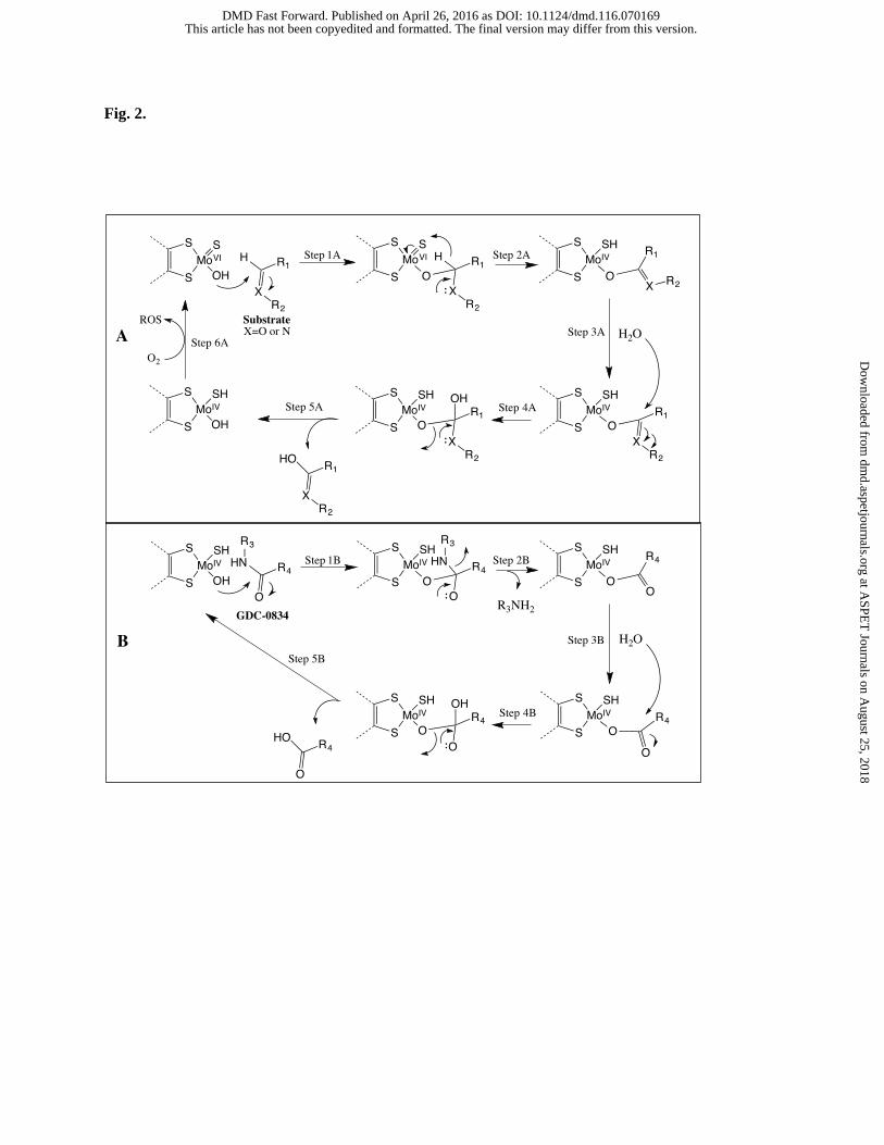
Fig. 2.
This article has not been copyedited and formatted. The final version may differ from this version.DMD Fast Forward. Published on April 26, 2016 as DOI: 10.1124/dmd.116.070169
at ASPE
T Journals on A
ugust 25, 2018dm
d.aspetjournals.orgD
ownloaded from

Fig. 3.
This article has not been copyedited and formatted. The final version may differ from this version.DMD Fast Forward. Published on April 26, 2016 as DOI: 10.1124/dmd.116.070169
at ASPE
T Journals on A
ugust 25, 2018dm
d.aspetjournals.orgD
ownloaded from
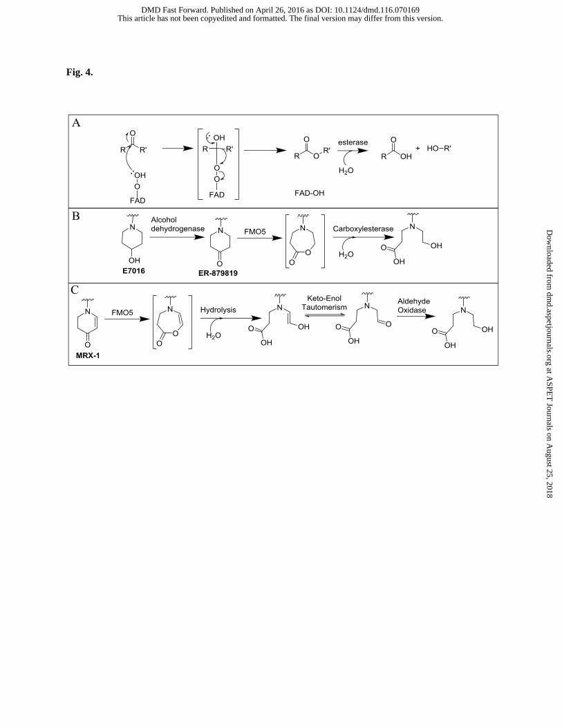
Fig. 4.
This article has not been copyedited and formatted. The final version may differ from this version.DMD Fast Forward. Published on April 26, 2016 as DOI: 10.1124/dmd.116.070169
at ASPE
T Journals on A
ugust 25, 2018dm
d.aspetjournals.orgD
ownloaded from

Fig. 5.
This article has not been copyedited and formatted. The final version may differ from this version.DMD Fast Forward. Published on April 26, 2016 as DOI: 10.1124/dmd.116.070169
at ASPE
T Journals on A
ugust 25, 2018dm
d.aspetjournals.orgD
ownloaded from

Fig 6.
A
B
C
D
This article has not been copyedited and formatted. The final version may differ from this version.DMD Fast Forward. Published on April 26, 2016 as DOI: 10.1124/dmd.116.070169
at ASPE
T Journals on A
ugust 25, 2018dm
d.aspetjournals.orgD
ownloaded from

Fig. 7.
Rifampin 4 Imatinib 4-Ipomeanol NNK/NNAL
5 (not an ART substrate)
This article has not been copyedited and formatted. The final version may differ from this version.DMD Fast Forward. Published on April 26, 2016 as DOI: 10.1124/dmd.116.070169
at ASPE
T Journals on A
ugust 25, 2018dm
d.aspetjournals.orgD
ownloaded from

Fig. 8.
This article has not been copyedited and formatted. The final version may differ from this version.DMD Fast Forward. Published on April 26, 2016 as DOI: 10.1124/dmd.116.070169
at ASPE
T Journals on A
ugust 25, 2018dm
d.aspetjournals.orgD
ownloaded from