TissueNet: Detect Lesions in Cervical Biopsies
17
TissueNet: Detect Lesions in Cervical Biopsies Kirill Brodt
Transcript of TissueNet: Detect Lesions in Cervical Biopsies

TissueNet: Detect Lesions
in Cervical BiopsiesKirill Brodt
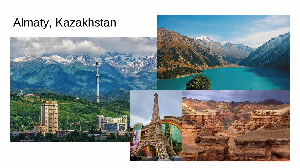
Almaty, Kazakhstan
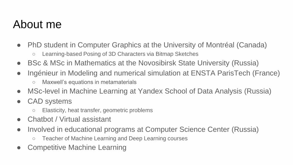
About me
● PhD student in Computer Graphics at the University of Montréal (Canada)○ Learning-based Posing of 3D Characters via Bitmap Sketches
● BSc & MSc in Mathematics at the Novosibirsk State University (Russia)
● Ingénieur in Modeling and numerical simulation at ENSTA ParisTech (France)○ Maxwell’s equations in metamaterials
● MSc-level in Machine Learning at Yandex School of Data Analysis (Russia)
● CAD systems○ Elasticity, heat transfer, geometric problems
● Chatbot / Virtual assistant
● Involved in educational programs at Computer Science Center (Russia)○ Teacher of Machine Learning and Deep Learning courses
● Competitive Machine Learning

Task description
● Image classification task
● ~1k train images with extremely high resolution
○ ~150,000 x 85,000 pixels (~30 Gb RAM)
● 4 ordinal classes
● Weighted Class Score
● https://www.drivendata.org/competitions/67/competition-
cervical-biopsy/
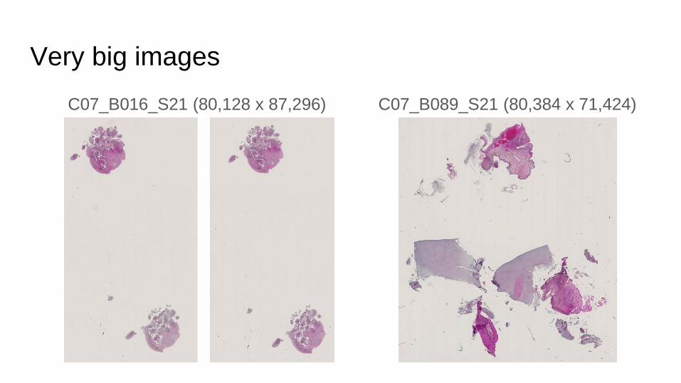
Very big images
C07_B016_S21 (80,128 x 87,296) C07_B089_S21 (80,384 x 71,424)
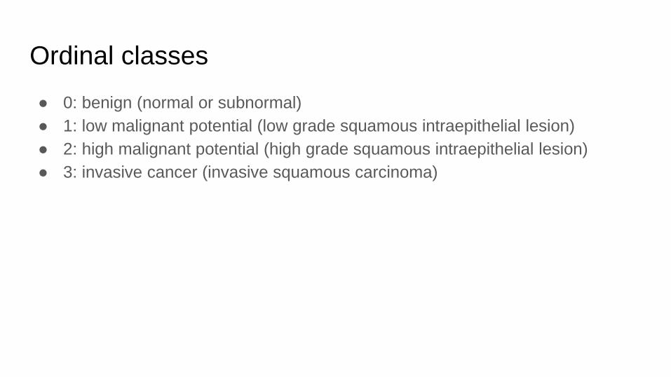
Ordinal classes
● 0: benign (normal or subnormal)
● 1: low malignant potential (low grade squamous intraepithelial lesion)
● 2: high malignant potential (high grade squamous intraepithelial lesion)
● 3: invasive cancer (invasive squamous carcinoma)

Performance metric
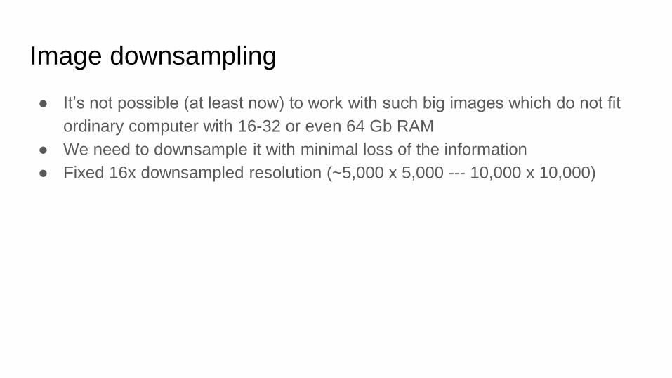
Image downsampling
● It’s not possible (at least now) to work with such big images which do not fit
ordinary computer with 16-32 or even 64 Gb RAM
● We need to downsample it with minimal loss of the information
● Fixed 16x downsampled resolution (~5,000 x 5,000 --- 10,000 x 10,000)
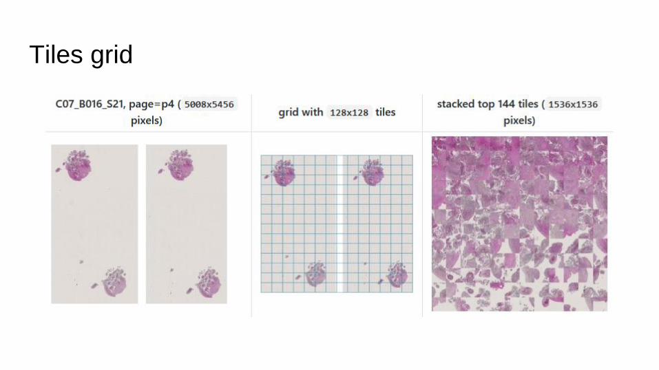
Tiles grid

Tiles grid
● 36 tiles with 256 x 256 size
● 64 tiles with 192 x 192 size
● 144 tiles with 128 x 128 size
In all cases we have image with 1,536 x 1,536 input size

Insane huge sample
C13_B054_S11
with a lot of white pixels
294,144 x 272,128
Issue: Huge image with white pixels
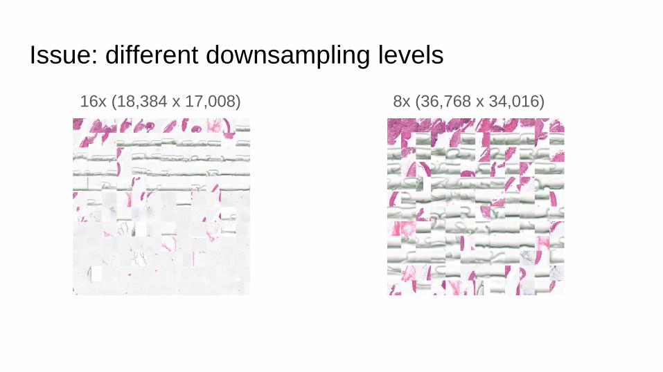
Issue: different downsampling levels
16x (18,384 x 17,008) 8x (36,768 x 34,016)
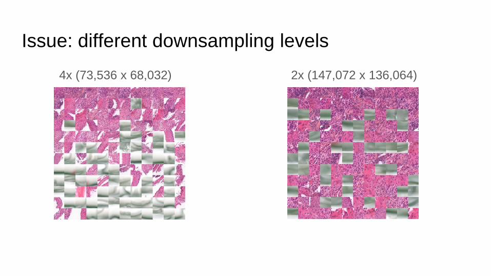
Issue: different downsampling levels
4x (73,536 x 68,032) 2x (147,072 x 136,064)
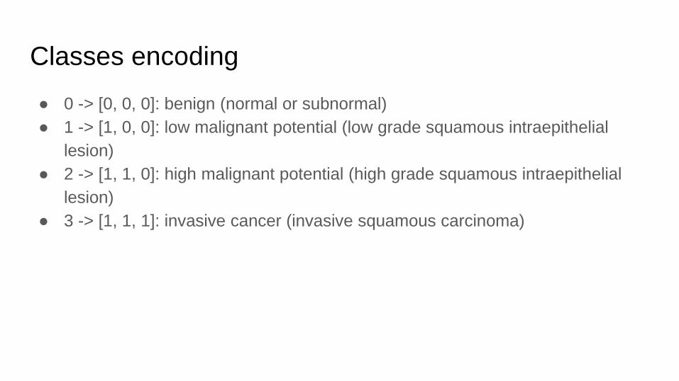
Classes encoding
● 0 -> [0, 0, 0]: benign (normal or subnormal)
● 1 -> [1, 0, 0]: low malignant potential (low grade squamous intraepithelial
lesion)
● 2 -> [1, 1, 0]: high malignant potential (high grade squamous intraepithelial
lesion)
● 3 -> [1, 1, 1]: invasive cancer (invasive squamous carcinoma)
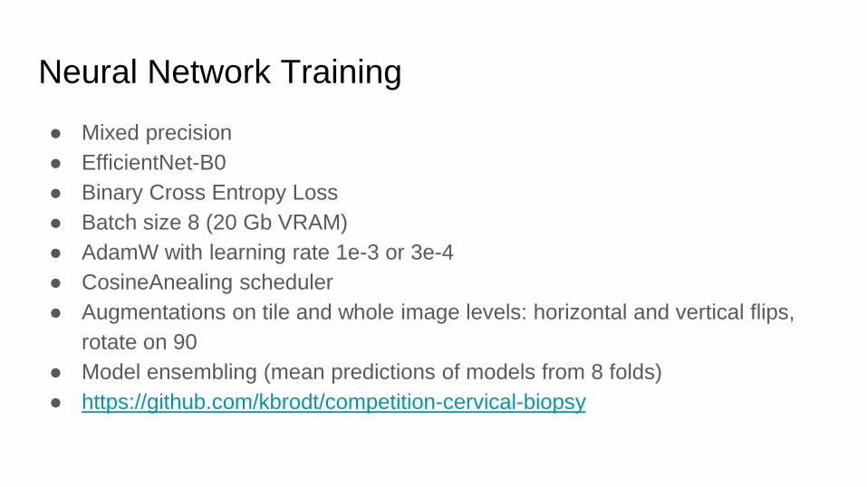
Neural Network Training
● Mixed precision
● EfficientNet-B0
● Binary Cross Entropy Loss
● Batch size 8 (20 Gb VRAM)
● AdamW with learning rate 1e-3 or 3e-4
● CosineAnealing scheduler
● Augmentations on tile and whole image levels: horizontal and vertical flips,
rotate on 90
● Model ensembling (mean predictions of models from 8 folds)
● https://github.com/kbrodt/competition-cervical-biopsy

Learning curve
~24H Nvidia V100 32GB
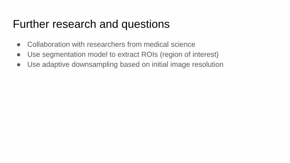
Further research and questions
● Collaboration with researchers from medical science
● Use segmentation model to extract ROIs (region of interest)
● Use adaptive downsampling based on initial image resolution