Treatment of noncarious cervical lesions: when, why, and how
27
16 | The International Journal of Esthetic Dentistry | Volume 15 | Number 1 | Spring 2020 CLINICAL RESEARCH Treatment of noncarious cervical lesions: when, why, and how Marleen Peumans, DMD, PhD Associate Professor, Oral Health Sciences, BIOMAT, Catholic University Leuven, Belgium Gianfranco Politano, DMD Private Practice, Rome, Italy Bart Van Meerbeek, DMD, PhD Full Professor, Oral Health Sciences, BIOMAT, Catholic University Leuven, Belgium Correspondence to: Prof Marleen Peumans Oral Health Sciences, BIOMAT, Catholic University A A Leuven, Kapucijnenvoer 33, Leuven, 3000, Belgium; Tel: +3216332744; Email: marleen.p[email protected]
Transcript of Treatment of noncarious cervical lesions: when, why, and how

16 | The International Journal of Esthetic Dentistry | Volume 15 | Number 1 | Spring 2020
CLINICAL RESEARCH
Treatment of noncarious cervical
lesions: when, why, and how
Marleen Peumans, DMD, PhD
Associate Professor, Oral Health Sciences, BIOMAT, Catholic University Leuven, Belgium
Gianfranco Politano, DMD
Private Practice, Rome, Italy
Bart Van Meerbeek, DMD, PhD
Full Professor, Oral Health Sciences, BIOMAT, Catholic University Leuven, Belgium
Correspondence to: Prof Marleen Peumans
Oral Health Sciences, BIOMAT, Catholic University AA Leuven, Kapucijnenvoer 33, Leuven, 3000, Belgium;
Tel: +3216332744; Email: [email protected]

PEUMANS ET AL
1717
Abstract
Noncarious cervical lesions (NCCLs) involve the loss
of hard tissue from the cervical areas of teeth through
processes unrelated to caries. NCCLs are nowadays a
common pathology caused by changes in lifestyle
and diet. The prevalence and severity of cervical wear
increase with age. It is generally accepted that the le-
sions are not generated by a single factor but result
from a combination of factors. Among the factors
proposed to be related to the formation and progres-
sion of NCCLs are biocorrosion (erosion), friction
(abrasion), and possibly occlusal stress (abfraction).
The clinical appearance of NCCLs can vary depending
on the type and severity of the etiologic factors in-
volved. Practitioners should follow a checklist to
achieve an accurate diagnosis of the etiology of multi-
factorial NCCLs.
The successful prevention and management of NCCLs
require an understanding of the etiology and risk fac-
tors, including how these change over time in individ-
ual patients. The decision to monitor NCCLs rather
than intervene should be based on the progression of
the lesions and how they compromise tooth vitality,
function, and esthetics. Treatment options include
techniques to alleviate dentin hypersensitivity and the
placement of an adhesive restoration, eventually in
combination with a root coverage surgical procedure.
An adhesive restoration is considered the last treat-
ment option for NCCLs.
Based on their excellent esthetic properties and good
clinical performance, there is a general indication to
place composite restorations for NCCLs. The clinical
performance of these restorations is highly prod-
uct-dependent, particularly regarding the adhesive
system used. The type of composite material seems
to have no significant influence on the clinical perfor-
mance of NCCL restorations in clinical trials. It is much
more important that the operator carries out the clin-
ical procedure correctly.
Marginal degradation is frequently seen during aging.
Yearly maintenance with the eventual repolishing of
the restoration margins will lengthen the lifespan of
the restorations.
(Int J Esthet Dent 2020;15:16–42)
17The International Journal of Esthetic Dentistry | Volume 15 | Number 1 | Spring 2020 |

CLINICAL RESEARCH
18 | The International Journal of Esthetic Dentistry | Volume 15 | Number 1 | Spring 2020
Introduction
NCCLs are the result of a pathological con-
dition characterized by the loss of tooth
structure at the cementoenamel junction
(CEJ) that is unrelated to dental caries.1 The
occurrence of NCCLs is an increasingly
common finding in dental clinical practice.
This article aims to provide an up-to-date
summary of the prevalence, appearance,
and etiology of NCCLs. A checklist is pre-
sented to analyze the etiology of NCCLs.
Also discussed are treatment possibilities
based on current knowledge: prevention,
monitoring, alleviation of dentin hypersensi-
tivity, and treatment with an adhesive restor-
ation.
Prevalence
The prevalence of NCCLs has been report-
ed to vary between 5% and 85%.2,3 This large
variation can be attributed to the wide age
range of participants and the inclusion of
both genders in study populations as well as
the diverse criteria used to distinguish le-
sions caused by one precise etiologic fac-
tor.
A general finding is that prevalence, se-
verity, and progression of NCCLs increase
with age. This can be explained by extended
exposure to etiologic factors, increased oc-
currence of gingival recession and bone
loss with more root surface exposure (which
raises the risk of cervical lesions), diminished
quantity and quality of saliva, and composi-
tional and microstructural changes of
enamel and dentin.2,4,5
NCCLs are almost exclusively situated
on the facial surfaces of teeth; they seldom
occur on the lingual surfaces and rarely on
the proximal ones.
Fig 1 Extracted teeth showing different forms of NCCLs. (a) Cervical wear lesion
on a mandibular central incisor mainly caused by erosion. Notice the poorly
defined margins and adjacent smooth enamel surface. The lesion is most visible
at the buccal side but is also present at the proximal and lingual sides. (b) Very
deep NCCL with sharply defined margins on a maxillary premolar caused by a
combination of abfraction (wedge shaped) and abrasion (notice the horizontal
scratches in the depth of the lesion created by a toothbrush). The pulp is
retracted and the dentin in the depth of the lesion is brown, discolored, and
sclerotic. (c) U-shaped cervical wear lesion on a mandibular premolar caused by
a combination of erosion and abrasion. The scratch in the depth of the lesion,
created by the bristles of a toothbrush, is slightly smoothened by erosion.
(d) Abfraction wedge-shaped lesion on retaining primary canine with sharp
internal and external line angles and an apical extent relative to the CEJ. Notice
the severe occlusal wear facet, which suggests abnormal occlusal loading.
a
b
c
d

PEUMANS ET AL
19The International Journal of Esthetic Dentistry | Volume 15 | Number 1 | Spring 2020 |
Any tooth (incisor, canine, premolar or
molar) may exhibit an NCCL.3,5 Several epi-
demiological studies have reported that
premolars show the highest frequency of
lesions.6-9 In some studies, the most com-
monly affected teeth were mandibular pre-
molars,6,7 which also have the highest per-
centage of severe lesions.7
Appearance
Generally, NCCLs vary from shallow grooves
or lesions with poorly defined margins to
large wedge-shaped defects with sharp line
angles (Fig 1a to d).3,4 A link between the
morphological characteristics of lesions
and their main etiologic factor has been
suspected.
■ Disk-shaped broad and shallow lesions
with poorly defined margins and adja-
cent smooth enamel margins are con-
sidered to be the best predictive criteria
for the diagnosis of erosion as well as a
pathognomonic sign of erosive tooth
wear (Fig 1a). In young patients with cer-
vical erosive lesions, an enamel border is
often noticed at the gingival margin
(Fig 2a and b).
■ Lesions caused by abrasive forces such
as improper tooth brushing techniques
generally exhibit sharply defined margins
and a hard surface with traces of scratch-
ing (Figs 1b and 3).
■ Lesions caused by a combination of abra-
sion and erosion often have a U shape
(Figs 1c and 4).
■ Lesions caused by abfraction due to ab-
normal occlusal loading are typically
wedge shaped, with sharp internal and
external line angles, and an apical extent
relative to the CEJ (Figs 1d and 5).
Fig 2 (a and b) Generalized tooth wear in a 21-year-old bruxing patient who
drinks 3 liters of coca cola per day. Noncarious cervical active erosive lesions are
present on almost all of the buccal surfaces. The lesions are disk-shaped, broad,
and shallow, with poorly defined margins and adjacent smooth enamel areas.
Notice the defined enamel border at the gingival margins. All the teeth have
become shorter with time. The incisal/occlusal and palatal wear was caused by a
combination of erosion and bruxism, which can be destructive for the dentition.
All teeth showed hypersensitivity.
a
b
Fig 3 Cervical lesions on mandibular anterior incisors caused by an improper
tooth brushing technique. These typical abrasive lesions exhibit sharply defined
margins and a hard surface with traces of scratching. The mandibular incisors do
not make contact during habitual occlusion. Abfraction could have played a role
in the formation of the cervical wear lesions on the mandibular left canine and
first premolar.

CLINICAL RESEARCH
20 | The International Journal of Esthetic Dentistry | Volume 15 | Number 1 | Spring 2020
As there is overwhelming evidence that the
etiology of NCCLs is multifactorial, a combi-
nation of these different geometric forms
often presents (Figs 1b, 6, and 7).2-5
In general, the development of NCCLs
tends to be a slow process occurring over
an extended period of time, with pulpal re-
traction, sclerosis, and lack of dentin sensi-
tivity among the consequences (Figs 1b, 6,
and 7).10-12
Fig 4 NCCLs on teeth 32, 33, 34, and 36. The quite large lesions on teeth 33
and 34 are caused by a combination of abrasion (notice the presence of grooves
and gingival recession) and erosion (notice no sharp boundary of the lesion at the
enamel side). These lesions often have a U shape.
Fig 5 Full ceramic crowns were placed on the four incisors 20 years previously.
The patient is missing several posterior teeth. Notice the overeruption of teeth 11
and 12, which has resulted in abnormal occlusal loading on these teeth. Abfrac-
tion lesions are visible on teeth 11, 12, and 21. These lesions are typically wedge
shaped, with sharp internal and external line angles and an apical extent relative
to the CEJ.
A common complaint of patients with
erosive lesions is tooth hypersensitivity, an
unavoidable problem if the corrosive chal-
lenge continues. In these lesions, dentin
tubule exposure and enamel rod corrosion
are widely reported.11
Etiology
The development of a particular NCCL is
usually the consequence of a synergistic ac-
tion of two or three of the etiologic mech-
anisms unique to that individual case: bio-
corrosion (erosion), friction (abrasion), and
stress (abfraction) (Figs 6 and 7). In addition,
several risk factors can have an influence on
the formation of NCCLs: saliva, tooth form,
composition, microstructure, mobility, pos-
itional prominence, presence of restor-
ations, magnitude, direction, frequency,
site, and duration of the applied forces.4,13
Biocorrosion (erosion)
Biocorrosion of teeth can occur due to ex-
trinsic acids (acidic foods and drinks, acidic
mouth rinse, acidic medication) and/or in-
trinsic acids (gastric acid). In addition, pro-
teolytic enzymes present in the crevicular
fluid14 and proteolytic enzymes from the
stomach (pepsin) and pancreas (trypsin)15
released during vomiting can degrade the
demineralized dentinal organic matrix.
Risk factors are the composition and fre-
quency of the intake of acids, the position
and form of teeth in the dental arch, and the
presence of gingival recession. Saliva (pH,
viscosity, flow, composition, buffer capaci-
ty) is an important risk factor in the develop-
ment of NCCLs. The ions in saliva are able
to induce remineralization of demineralized
tooth structure and can thus inhibit the for-
mation of NCCLs.
NCCLs are more common on facial sur-
faces of teeth than on lingual ones. A possi-
ble explanation is the difference in chemis-
PEUMANS ET AL
21The International Journal of Esthetic Dentistry | Volume 15 | Number 1 | Spring 2020 |
try and character of saliva in lingual areas
(more serous saliva – higher buffer capacity)
and facial areas (mucous saliva – lower buf-
fer capacity), which accounts for the differ-
ences in remineralization of the tooth struc-
ture and the dilution of buffering acids.16
Xerostomia and dehydration from perspira-
tion with physical activity create impaired
salivary flow and inhibit the buffering of
acids in the oral cavity.
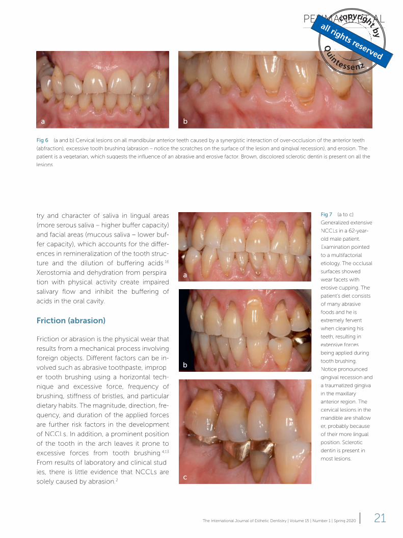
Friction (abrasion)
Friction or abrasion is the physical wear that
results from a mechanical process involving
foreign objects. Different factors can be in-
volved such as abrasive toothpaste, improp-
er tooth brushing using a horizontal tech-
nique and excessive force, frequency of
brushing, stiffness of bristles, and particular
dietary habits. The magnitude, direction, fre-
quency, and duration of the applied forces
are further risk factors in the development
of NCCLs. In addition, a prominent position
of the tooth in the arch leaves it prone to
excessive forces from tooth brushing.4,13
From results of laboratory and clinical stud-
ies, there is little evidence that NCCLs are
solely caused by abrasion.2
a b
Fig 6 (a and b) Cervical lesions on all mandibular anterior teeth caused by a synergistic interaction of over-occlusion of the anterior teeth
(abfraction), excessive tooth brushing (abrasion – notice the scratches on the surface of the lesion and gingival recession), and erosion. The
patient is a vegetarian, which suggests the influence of an abrasive and erosive factor. Brown, discolored sclerotic dentin is present on all the
lesions.
Fig 7 (a to c)
Generalized extensive
NCCLs in a 62-year-
old male patient.
Examination pointed
to a multifactorial
etiology. The occlusal
surfaces showed
wear facets with
erosive cupping. The
patient’s diet consists
of many abrasive
foods and he is
extremely fervent
when cleaning his
teeth, resulting in
extensive forces
being applied during
tooth brushing.
Notice pronounced
gingival recession and
a traumatized gingiva
in the maxillary
anterior region. The
cervical lesions in the
mandible are shallow-
er, probably because
of their more lingual
position. Sclerotic
dentin is present in
most lesions.
a
b
c

CLINICAL RESEARCH
22 | The International Journal of Esthetic Dentistry | Volume 15 | Number 1 | Spring 2020
Stress (abfraction)
The abfraction theory is based on a bio-
mechanical concept in which the cervical
area of a tooth becomes a fulcrum during
occlusal function, bruxing, and parafunc-
tional activity, causing tensile stresses in the
area where NCCLs occur. These stresses
are thought to disrupt the crystalline struc-
ture of the locally thin enamel and underly-
ing dentin by cyclic fatigue, leading to
cracks. Ultimately, the enamel breaks away
at the cervical margin and progressively ex-
poses the dentin, where the process contin-
ues.13,17 This theory is quite controversial.
Several finite element analysis (FEA) studies
have shown a clear correlation between oc-
clusion and NCCLs.18-22 In addition, several
clinical trials also report strong evidence of
an association between occlusal factors
and NCCLs (or their progression);23-26 never-
theless, two systematic reviews were unable
to show a clear association.27,28 The large
heterogeneity among methodologies, the
lack of standardization, and differences in
the diagnoses of NCCLs could contribute to
the weak evidence in these two reviews,
both of which concluded that prospective
clinical studies with a qualitatively and quan-
titatively strong study design are needed to
prove a clear association between occlu-
sion and NCCLs.
According to Grippo et al,13 some risk
factors can play a role in the development
of abfraction lesions. First, resultant stresses
within the teeth are dependent on the mag-
nitude, direction, frequency, site of applica-
tion, and duration of a force, in addition to
its orientation with respect to the principal
axes of the teeth and their form, composi-
tion, and stability. Second, the cushioning
effect of the periodontal ligament is another
modifying factor, as there is a negative cor-
relation between mobility and NCCLs. Third,
the weakening effect of an occlusal restor-
ation may contribute to the development of
an abfraction lesion. Finally, the occlusal
positional prominence of the teeth is anoth-
er important factor in determining possible
overstress and trauma (Fig 8).
Most lesions are caused by an interac-
tion of two or three causes that result in in-
creased cervical tooth wear, ie, the com-
bined effect of erosion and abrasion is
greater than the effect of either force oper-
ating on its own.29,30 It also appears that li-
quid acid, frequently found in modern diets,
may be necessary in order for occlusal load-
ing to be a factor in the formation of cervi-
cal lesions.31 In addition, it is suggested that
biocorrosion plays a very important role in
the formation of NCCLs.
Clinicians should consider all etiologic
and risk factors before completing the diag-
nosis or initiating treatment, if indicated.
Practitioners should follow a checklist to de-
termine the etiology of each particular lesion
in order to make as precise a diagnosis as
possible. Table 1 shows a simplified version
of the schema proposed by Grippo et al.13
Treatment options
To date, there is no conclusive evidence for
reliable, predictable, and successful treat-
Fig 8 This patient showed several NCCLs. The maxillary first molar extends
beyond the occlusal plane. Occlusal overstress resulted in additional cervical
wear due to abfraction in the mandibular second premolar and the maxillary first
molar.
PEUMANS ET AL
23The International Journal of Esthetic Dentistry | Volume 15 | Number 1 | Spring 2020 |
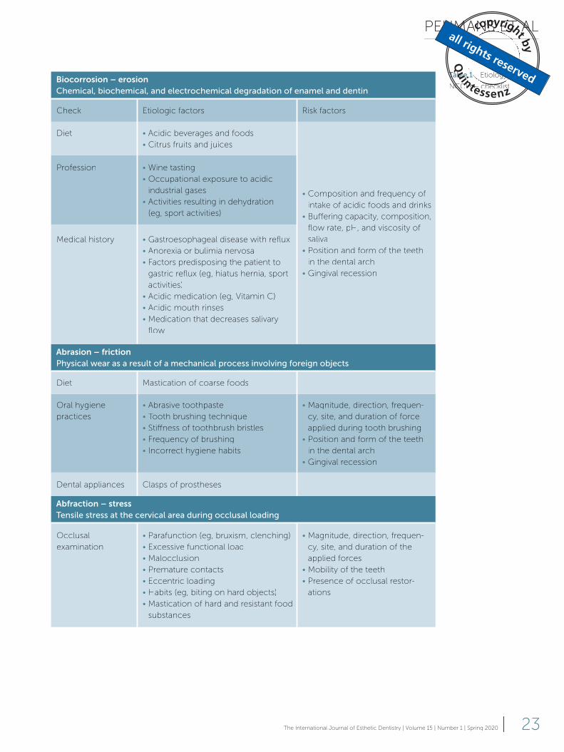
Biocorrosion – erosion
Chemical, biochemical, and electrochemical degradation of enamel and dentin
Check Etiologic factors Risk factors
Diet • Acidic beverages and foods
• Citrus fruits and juices
• Composition and frequency of
intake of acidic foods and drinks
• Buffering capacity, composition,
flow rate, pH, and viscosity of
saliva
• Position and form of the teeth
in the dental arch
• Gingival recession
Profession • Wine tasting
• Occupational exposure to acidic
industrial gases
• Activities resulting in dehydration
(eg, sport activities)
Medical history • Gastroesophageal disease with reflux
• Anorexia or bulimia nervosa
• Factors predisposing the patient to
gastric reflux (eg, hiatus hernia, sport
activities)
• Acidic medication (eg, Vitamin C)
• Acidic mouth rinses
• Medication that decreases salivary
flow
Abrasion – friction
Physical wear as a result of a mechanical process involving foreign objects
Diet Mastication of coarse foods
Oral hygiene
practices
• Abrasive toothpaste
• Tooth brushing technique
• Stiffness of toothbrush bristles
• Frequency of brushing
• Incorrect hygiene habits
• Magnitude, direction, frequen-
cy, site, and duration of force
applied during tooth brushing
• Position and form of the teeth
in the dental arch
• Gingival recession
Dental appliances Clasps of prostheses
Abfraction – stress
Tensile stress at the cervical area during occlusal loading
Occlusal
examination
• Parafunction (eg, bruxism, clenching)
• Excessive functional load
• Malocclusion
• Premature contacts
• Eccentric loading
• Habits (eg, biting on hard objects)
• Mastication of hard and resistant food
substances
• Magnitude, direction, frequen-
cy, site, and duration of the
applied forces
• Mobility of the teeth
• Presence of occlusal restor-
ations
Table 1 Etiology of
NCCLs – checklist

CLINICAL RESEARCH
24 | The International Journal of Esthetic Dentistry | Volume 15 | Number 1 | Spring 2020
ment regimens for NCCLs. The current
knowledge and available treatment strat-
egies for NCCLs are discussed below, with a
focus on prevention, monitoring, treatment
of dentin hypersensitivity, restorative treat-
ment, and root coverage surgical proced-
ures with or without restorative treatment.
Prevention
The objectives of preventive therapy are to
prevent the progression of existing lesions
or the development of new ones and to as-
sure the longevity of restorations in restored
lesions. Preventive interventions include
counseling for changes in the patient’s be-
havior depending on the etiology (abrasion,
erosion, and/or abfraction).4,13,32
Cervical abrasions are generally thought
to be a consequence of tooth brushing fac-
tors such as frequent or forceful brushing,
faulty or vigorous techniques, filament stiff-
ness or design, dominant hand dexterity, or
abrasive dentifrices. Patients should be in-
formed about these factors and, where nec-
essary, advised to alter their brushing mater-
ials or habits. In addition, patients should be
instructed to avoid brushing immediately
after an erosive challenge.
Dental erosion can also be effectively
modified and should be correctly diagnosed.
Treatment success depends on the patient’s
collaboration.4,13 When erosion is caused by
eating disorders or gastroesophageal reflux
disease, the treatment may require the par-
ticipation of a physician. The extrinsic etiolo-
gy is more easily treatable. During the diag-
nostic phase, the checklist (Table 1) must be
followed to see which external erosive fac-
tors exist. Removing or altering harmful
habit/s provides consistent results.
For NCCLs with an abfraction etiology,
there is no consensus on preventive treat-
ment strategies. On the one hand, there is a
weak association between NCCLs and oc-
clusal factors (interference in excursive
movements, force, premature contacts,
type of guidance, and slide of centric occlu-
sion to maximum intercuspation). In two
systematic reviews,27,28 several authors ad-
vise that any decision to carry out a destruc-
tive, irreversible treatment such as occlusal
adjustment should be considered very care-
fully.1,4,5,28 If abfraction is suspected to be a
dominant factor in the etiology of NCCLs,
the most conservative treatment option
suggested is an occlusal splint as this re-
duces the amount of nocturnal bruxism and
nonaxial tooth forces,4,5 although in this
case too there is no scientific evidence to
support the use of an occlusal splint. On the
other hand, Soares and Grippo33 state that
preventive management strategies should
consider occlusal therapy in patients with
parafunctional habits and occlusal prema-
turities. According to these authors, occlu-
sal therapy can mitigate the development of
NCCLs by correcting occlusal imbalance
and removing occlusal prematurities. They
emphasize that occlusal therapy must be
carried out with a thorough knowledge of
the occlusion.
Monitoring
The activity of NCCLs needs to be assessed
and considered in the treatment planning
process. It is known that the progression of
NCCLs is generally slow, but with a large
variation among patients.26,34,35 Therefore,
an individual monitoring protocol should be
established that assesses the severity of the
present lesions, the patient’s age, and the
existing etiologic and risk factors. For pa-
tients who are exposed to intrinsic acids or
who present with rapid progression, the as-
sessment procedure of lesion activity should
be repeated at 6-month intervals during
regular hygiene. For most other cases, an-
nual assessment is acceptable (Fig 9).
Approaches to determine lesion activity
include the use of standardized intraoral

PEUMANS ET AL
25The International Journal of Esthetic Dentistry | Volume 15 | Number 1 | Spring 2020 |
photographs, possibly study models, and
the measurement of lesion dimensions
(width/length) over time. With the recent in-
troduction of digital dentistry, comput-
er-aided design/computer-aided manufac-
turing (CAD/CAM) systems may be useful
for the diagnosis of NCCLs and in monitor-
ing their activity.
In a prospective clinical trial evaluating
the progression of NCCLs over a 5-year
period, the rate of progression was signifi-
cantly related to mean occlusal stress and
relative occlusal force in the maximum in-
tercuspation position.26 No significant cor-
relation was found between NCCL progres-
sion and a more acidic diet, tooth brushing
technique/rigorousness, medical conditions
causing deficient or acidic saliva output,
presence of occlusal wear facets, group
function, or adverse oral habits. Moderate
sensitivity on the involved teeth was found
in 34.5% of the participants by using the cold
test. A moderate to high degree of sclerosis
was observed on 68.9% of the lesions.
In another in vivo study evaluating 10 le-
sions in six patients over a period of 4 to
5 years, dimensional analysis showed nota-
ble progression in deeper lesions.35 In addi-
tion, wedge-shaped lesions were reported
to progress at a greater rate compared with
saucer-shaped ones. The same observation
was made in an in vitro study using 3D FEA.20
V-shaped lesions showed higher stress lev-
els concentrated at the zenith of the lesion,
whereas in U-shaped defects the stresses
were concentrated over a wider area. As the
lesions advanced in depth, the stress was
amplified at their deepest part. A trend of
stress amplification was also observed with
decreasing bone height.
One should bear in mind that restorative
treatment may be indicated at an earlier
stage for V-shaped lesions than for U-shaped
ones in patients with a heavy occlusion
and articulation. Restoring deeper (1.5 mm)
NCCLs is usually prioritized over shallower
ones due to the higher stress development
of deeper lesions that results in their more
notable progression.
Treatment of dentinal hypersensitivity
Dentinal hypersensitivity is a symptom often
associated with early stages of NCCLs. If the
lesions are small and shallow and require no
restorative treatment, and should sensitivity
persist, the hypersensitive tooth must first
be treated in a noninvasive way. There is a
broad spectrum of relatively cost-effective
at-home desensitizing agents available on
Fig 9 (a and b) Several small NCCLs in the posterior region in a 35-year-old
patient due to a combination of abfraction and abrasion. The patient showed
signs of bruxism for which an occlusal splint was made. As the lesions were
stable during the yearly follow up, no treatment was needed.
a
b

CLINICAL RESEARCH
26 | The International Journal of Esthetic Dentistry | Volume 15 | Number 1 | Spring 2020
the market that show different mechanisms
of action: nerve desensitization (eg, potassi-
um nitrate), protein precipitation (eg, glutar-
aldehyde, silver nitrate, zinc chloride, stron-
tium chloride hexahydrate), and plugging
the dentin tubules (eg, sodium fluoride,
stannous fluoride, strontium chloride, po-
tassium oxalate, calcium phosphate, calci-
um carbonate, bioactive glass).36 These
agents include toothpastes, mouth washes
and chewing gums. The results of at-home
desensitizing therapy should be reviewed
after 3 to 4 weeks. If there is no relief from
dentinal hypersensitivity, in-office therapy
should be initiated. Recently, the in-office
application of calcium- and phosphate-
based systems was introduced to decrease
hypersensitivity and promote remineraliza-
tion of the tooth surface, eg, Teethmate De-
sensitizer (Kuraray Noritake) (Fig 10) and air
polishing with Sylc Bioactive Glass (Velopex)
(Fig 11a to d).37-39 These products react with
body fluids and result in the formation of hy-
droxyapatite (HAp) crystals, similar to the
minerals in dentin, which block the dentinal
tubules. Both systems show promising re-
sults; however, confirmation in clinical trials
is required.
Finally, resin-based dental adhesive sys-
tems can also provide a longer-lasting den-
tin-desensitizing effect compared with the
use of home-desensitizing agents.4,5,36,40 The
adhesive resins can effectively seal the den-
tinal tubules by forming a hybrid layer. Vari-
ous clinical studies have demonstrated the
effectiveness up to 6 months of adhesives
in the management of dentin hypersensitiv-
ity.40,41
Restorative treatment
Restorative treatment of NCCLs should be
considered when one or more of the fol-
lowing conditions are present: 1) active,
cavitated caries lesions associated with
NCCLs; 2) cervical lesion margins or all le-
sion margins are located subgingivally and
preclude plaque control, hence the risk of
caries and periodontal disease increases;
3) extensive tooth structure loss, which
compromises the integrity of the tooth, or
the defect is in close proximity to the pulp,
or the pulp has been exposed; 4) persistent
dentinal hypersensitivity in which noninva-
sive therapeutic options have failed; 5) pros-
thetic abutment for removable prosthesis;
and 6) esthetic demands by patients.5,32 If
there is a need for a restoration, NCCLs
should be restored as minimally invasively
as possible. Of the available restorative tech-
niques, an adhesive system, combined with
a composite resin, is the preferred choice of
practitioners due to the good esthetic prop-
Fig 10 In-office treatment of hypersensitive dentin with Teethmate Desensitizer
(Kuraray Noritake). (a) Isolate the tooth and dry the tooth surface. (b) Mix the
powder and liquid according to the manufacturer’s instructions and apply the
slurry for at least 30 s. Rinse off with water spray or allow the patient to rinse.
a
b
PEUMANS ET AL
27The International Journal of Esthetic Dentistry | Volume 15 | Number 1 | Spring 2020 |
a
b c
d
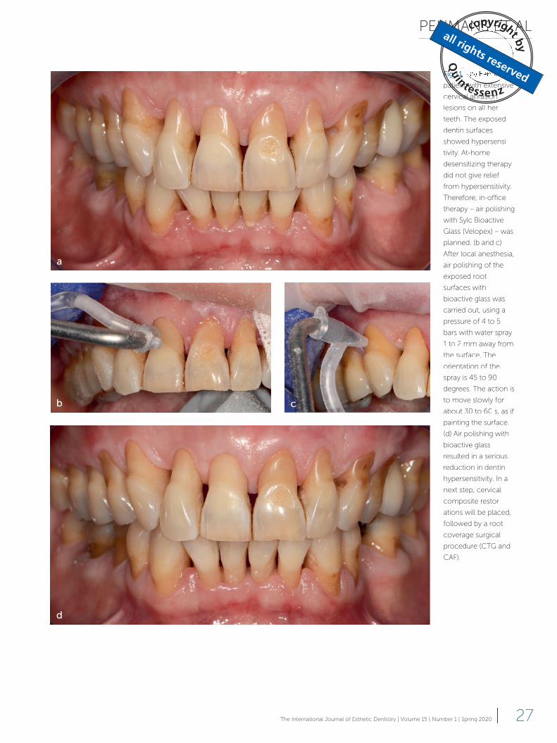
Fig 11
patient with extensive
cervical abrasion
lesions on all her
teeth. The exposed
dentin surfaces
showed hypersensi-
tivity. At-home
desensitizing therapy
did not give relief
from hypersensitivity.
Therefore, in-office
therapy – air polishing
with Sylc Bioactive
Glass (Velopex) – was
planned. (b and c)
After local anesthesia,
air polishing of the
exposed root
surfaces with
bioactive glass was
carried out, using a
pressure of 4 to 5
bars with water spray
1 to 2 mm away from
the surface. The
orientation of the
spray is 45 to 90
degrees. The action is
to move slowly for
about 30 to 60 s, as if
painting the surface.
(d) Air polishing with
bioactive glass
resulted in a serious
reduction in dentin
hypersensitivity. In a
next step, cervical
composite restor-
ations will be placed,
followed by a root
coverage surgical
procedure (CTG and
CAF).

CLINICAL RESEARCH
28 | The International Journal of Esthetic Dentistry | Volume 15 | Number 1 | Spring 2020
erties and clinical performance of compos-
ite resin (Fig 12). Although glass-ionomers,
resin-modified glass-ionomers, and the lam-
ination technique with composite resin have
been advocated for NCCL restorations, these
materials are less frequently used.42,43
Adhesively restored NCCLs can perform
quite well in the long term (Fig 13).43 Several
parameters determine the clinical behavior
of these restorations. According to a sys-
tematic review evaluating the clinical ef-
fectiveness of contemporary adhesives in
NCCLs, the selection of the adhesive sys-
tem is an important factor determining the
durability of the restoration.43 Apart from the
selection of the adhesive system, the oper-
ator plays a decisive role. The operator has
to take care that a high-quality clinical pro-
cedure is carried out: isolation, tooth prep-
aration, application of the adhesive system
and composite, finishing, polishing, and fi-
nally maintenance of the restoration. These
different parameters are successively dis-
cussed in detail below.
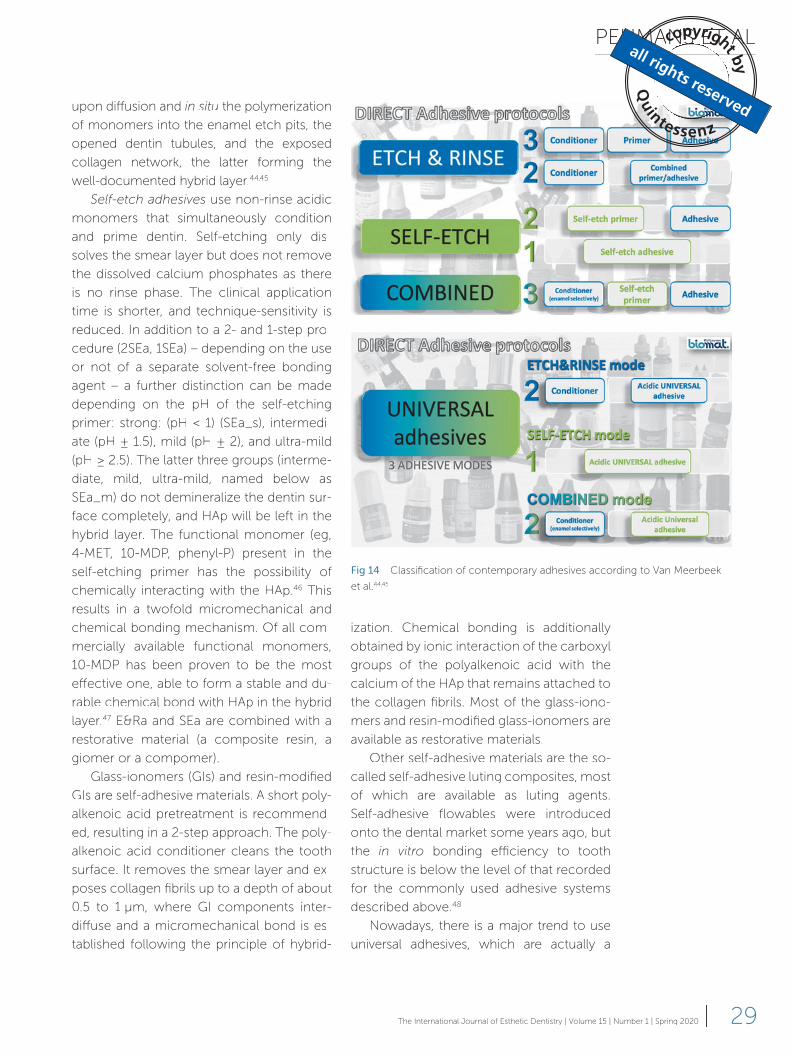
Selection of the adhesive system
According to the classification of Van Meer-
beek et al,44,45 modern adhesive approaches
can be divided into etch-and-rinse adhe-
sives (E&Ra), self-etch adhesives (SEa), and
restorative materials with a self-adhesive ap-
proach (Fig 14).
The multi-step etch-and-rinse approach
involves a phosphoric acid-etch step that at
the enamel level produces deep etch pits in
the HAp-rich substrate, and at the dentin lev-
el demineralizes up to a depth of a few mi-
crometers to expose a HAp-deprived colla-
gen mesh. The next step involves either a
separate priming step followed by the appli-
cation/curing of a combined primer/adhe-
sive resin using a 2-step procedure (2E&Ra),
or a separate primer and adhesive resin fol-
lowing a 3-step procedure (3E&Ra). The final
objective is to micromechanically interlock
Fig 12 (a) A 70-year-old female patient showing NCCLs on all her mandibular
anterior teeth. The etiology was multifactorial, with abrasion being the main
etiologic factor. The exposed dentin was not hypersensitive. The reason for the
treatment with direct composite restorations was to improve esthetics. (b) Apart
from the small lesion on tooth 43, all the NCCLs were restored with a nanohybrid
composite in combination with a mild 2SEa.
Fig 13 Two mandibular premolars with 20-year-old Class V composite restor-
ations. A 3E&R adhesive system was used in combination with a microfilled
composite (first premolar) and a hybrid composite (second premolar). After
20 years, the restorations are still clinically acceptable, presenting slight marginal
defects and superficial marginal discoloration.
a
b
PEUMANS ET AL
29The International Journal of Esthetic Dentistry | Volume 15 | Number 1 | Spring 2020 |
upon diffusion and in situ the polymerization
of monomers into the enamel etch pits, the
opened dentin tubules, and the exposed
collagen network, the latter forming the
well-documented hybrid layer.44,45
Self-etch adhesives use non-rinse acidic
monomers that simultaneously condition
and prime dentin. Self-etching only dis-
solves the smear layer but does not remove
the dissolved calcium phosphates as there
is no rinse phase. The clinical application
time is shorter, and technique-sensitivity is
reduced. In addition to a 2- and 1-step pro-
cedure (2SEa, 1SEa) – depending on the use
or not of a separate solvent-free bonding
agent – a further distinction can be made
depending on the pH of the self-etching
primer: strong: (pH < 1) (SEa_s), intermedi-
ate (pH ± 1.5), mild (pH ± 2), and ultra-mild
(pH ≥ 2.5). The latter three groups (interme-
diate, mild, ultra-mild, named below as
SEa_m) do not demineralize the dentin sur-
face completely, and HAp will be left in the
hybrid layer. The functional monomer (eg,
4-MET, 10-MDP, phenyl-P) present in the
self-etching primer has the possibility of
chemically interacting with the HAp.46 This
results in a twofold micromechanical and
chemical bonding mechanism. Of all com-
mercially available functional monomers,
10-MDP has been proven to be the most
effective one, able to form a stable and du-
rable chemical bond with HAp in the hybrid
layer.47 E&Ra and SEa are combined with a
restorative material (a composite resin, a
giomer or a compomer).
Glass-ionomers (GIs) and resin-modified
GIs are self-adhesive materials. A short poly-
alkenoic acid pretreatment is recommend-
ed, resulting in a 2-step approach. The poly-
alkenoic acid conditioner cleans the tooth
surface. It removes the smear layer and ex-
poses collagen fibrils up to a depth of about
0.5 to 1 μm, where GI components inter-
diffuse and a micromechanical bond is es-
tablished following the principle of hybrid-
ization. Chemical bonding is additionally
obtained by ionic interaction of the carboxyl
groups of the polyalkenoic acid with the
calcium of the HAp that remains attached to
the collagen fibrils. Most of the glass-iono-
mers and resin-modified glass-ionomers are
available as restorative materials.
Other self-adhesive materials are the so-
called self-adhesive luting composites, most
of which are available as luting agents.
Self-adhesive flowables were introduced
onto the dental market some years ago, but
the in vitro bonding efficiency to tooth
structure is below the level of that recorded
for the commonly used adhesive systems
described above.48
Nowadays, there is a major trend to use
universal adhesives, which are actually a
Fig 14 Classification of contemporary adhesives according to Van Meerbeek
et al.44,45

CLINICAL RESEARCH
30 | The International Journal of Esthetic Dentistry | Volume 15 | Number 1 | Spring 2020
newer version of 1SEa_m that can be applied
in different modes: an etch-and-rinse mode,
a self-etch mode, and a self-etch mode with
prior selective etching of the enamel margin
with 35% phosphoric acid.49,50
NCCLs are the most ideal lesions to test
the clinical effectiveness of adhesives in clin-
ical trials.43 These lesions involve both enam-
el and dentin; however, the largest part of
the tooth structure on which to bond con-
sists of dentin. These lesions commonly do
not provide any (or they provide minimal)
macroretention, so ineffective bonding will
result in debonding and thus restoration
loss. Loss of retention is the key objective
parameter against which the bonding per-
formance of adhesives in NCCL clinical trials
is evaluated. In a systematic review of NCCL
clinical trials, the number of lost restorations
per year (calculated as annual failure rate
[AFR]) was recorded in 87 clinical trials evalu-
ating 78 different adhesives.43 The adhesives
were classified into seven different classes:
3E&R, 2E&R, 2SEa_m; 2SEa_s, 1SEa_m,
1SEa_s, and GI. SEa_m contains the inter-
mediate, mild, and ultra-mild SEa (described
on the previous page). The first conclusion
was that the adhesive strategy is a determin-
ing factor for the clinical bonding effective-
ness in NCCLs. The best-performing classes
were GI, with the lowest AFR score, followed
closely by 2SEA_m, 3E&R, and 1SEA_m. An
inadequate bonding effectiveness was no-
ticed for 2E&Ra, 1SEa_s, and 2SEa_s. It is
obvious that the chemical bonding poten-
tial of adhesives is important for the quality
and durability of the bond in NCCLs. In ad-
dition to the adhesive strategy, a wide varia-
tion exists between the adhesives them-
selves. Practitioners should therefore select
a product that has a good proven clinical
performance. In the best-performing adhe-
sive categories described above, the lowest
AFR scores were recorded for Optibond FL
(Kerr) for 3E&Ra, Clearfil SE Bond (Kuraray
Noritake) for 2SEa_m, and G-Bond (GC) for
1SEa_m. These results accord with those of
a systematic review that evaluated the bond-
ing effectiveness of adhesives in vitro ac-
cording to microtensile bond strength
tests.51 Similarly, Optibond FL and Clearfil SE
Bond showed the best results regarding im-
mediate bond strength and bond strength
after 1 year of water storage.
Although GI-based materials perform
well with regard to retention, their rather
poor esthetic properties, inferior wear resis-
tance, and solubility particularly in acidic
oral environments may make them less ad-
equate for restoring NCCLs, especially in
the anterior and premolar regions.42,43
Isolation
Proper isolation is important for the success
of NCCL restorations. A recently published
meta-analysis stated that the use of rubber
dam positively influences the performance
of adhesive NCCL restorations, resulting in
less retention loss and better marginal adap-
tation.52 Therefore, if the clinical situation al-
lows, absolute isolation with rubber dam
should be applied. A good strategy has to be
followed, with the practitioner aware of the
tips and trips to correctly isolate under rub-
ber dam.53 For this procedure, the use of
heavy rubber dam is preferred as it results in
the best retraction of the peripherical tissues,
including the papilla. The holes must be
punched correctly so that all gingival tissues
are covered. Inverting the rubber dam around
the neck of the tooth prevents the saliva
from leaking in between the rubber dam and
the gingiva. Useful clamps for rubber dam
isolation of NCCLs are the Brinker Clamps
(Tissue Retractors) (B4, B5, B6) (Hygenic;
Coltene Whaledent) and Clamp 212 (Hu
Friedy). In addition, floss, teflon tape, and gin-
gival retraction cord can be used to obtain
additional gingival retraction (Figs 15 and 16).
Intrinsic anatomical and morphological
characteristics of the cervical region can cre-
PEUMANS ET AL
31The International Journal of Esthetic Dentistry | Volume 15 | Number 1 | Spring 2020 |
ate limitations in the placement of the rubber
dam and clamp. Difficulties can occur, for
instance, when restoring a NCCL on a molar
or on other teeth with a very large difference
in height between the buccal and lingual gin-
giva or when teeth are severely crowded.
When adequate rubber dam isolation is
not possible, another isolation method
should be employed. The insertion of a gin-
gival retraction cord in combination with
cotton rolls can help to control moisture. A
blood and hemostatic agent can be used
when needed to control blood contamina-
tion. Some studies have demonstrated that
dentin contamination with ferric sulfate or
aluminum chloride decreases the bond
strength of self-etch adhesives.54,55 Groddeck
et al56 showed that cavity contamination with
hemostatic agents, applied after blood con-
tamination and removed with water spray,
does not compromise marginal adaptation
in enamel and dentin when using an E&Ra or
an SEa,56 although element surface analysis
showed that remnants of hemostatic agents
were found on enamel and dentin surfaces
after rinsing the dark coagulum with water
spray. Apparently, remnants of hemostatic
agents on the surface of dental hard tissues
might have less influence on marginal adap-
tation than on bond strength. A last option is
a proposed association of a plastic or metal
matrix with wooden wedges and a photo-
cured gingival barrier.57
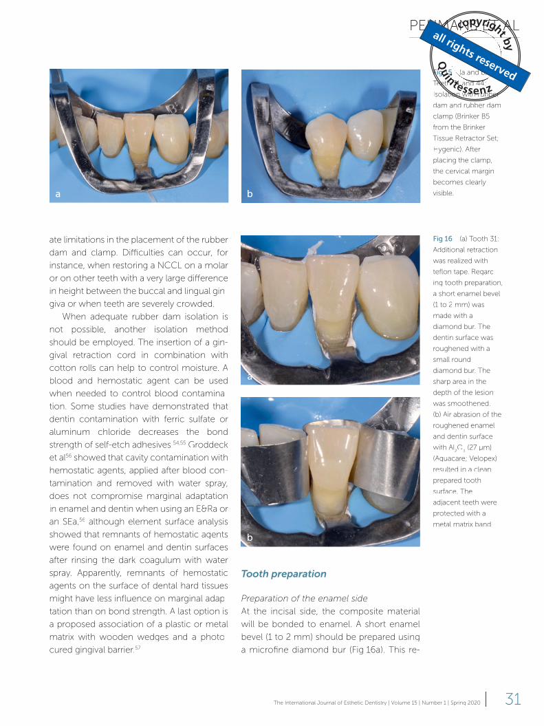
Fig 15 (a and b)
Teeth 31 and 44:
Isolation with rubber
dam and rubber dam
clamp (Brinker B5
from the Brinker
Tissue Retractor Set;
Hygenic). After
placing the clamp,
the cervical margin
becomes clearly
visible.
Fig 16 (a) Tooth 31:
Additional retraction
was realized with
teflon tape. Regard-
ing tooth preparation,
a short enamel bevel
(1 to 2 mm) was
made with a
diamond bur. The
dentin surface was
roughened with a
small round
diamond bur. The
sharp area in the
depth of the lesion
was smoothened.
(b) Air abrasion of the
roughened enamel
and dentin surface
with Al2O
3 (27 μm)
(Aquacare; Velopex)
resulted in a clean
prepared tooth
surface. The
adjacent teeth were
protected with a
metal matrix band.
a b
Tooth preparation
Preparation of the enamel side
At the incisal side, the composite material
will be bonded to enamel. A short enamel
bevel (1 to 2 mm) should be prepared using
a microfine diamond bur (Fig 16a). This re-
a
b

CLINICAL RESEARCH
32 | The International Journal of Esthetic Dentistry | Volume 15 | Number 1 | Spring 2020
moves the aprismatic enamel and increases
the bond strength to enamel.58 In addition,
placement of an enamel bevel will result in
a more gradual transition between the com-
posite restoration and tooth surface, improv-
ing the final esthetic outcome.
Preparation of the dentin surface
The dentin surface of NCCLs is often scle-
rotic and is a more difficult substrate to
bond to than sound dentin. Sclerotic dentin
is hypermineralized and has a shiny and firm
appearance. The dentin tubules are partially
or completely occluded by mineral depos-
its, resulting in a higher resistance to acid
dissolution. Morphological evaluation of the
resin–sclerotic dentin interface shows a thin
hybrid layer at the hypermineralized intertu-
bular dentin, and short and barely devel-
oped resin tags.59
To increase the bonding effectiveness to
this type of dentin, the superficial sclerotic
dentin should be roughened with a dia-
mond bur (Fig 16a).52 Another important rea-
son to roughen the dentin surface is to re-
move the contaminated layer observed on
most NCCLs during microscopic evaluation
of extracted teeth (Fig 17).11,60 This layer re-
sists adhesion and should therefore be re-
moved.
The roughness of the diamond bur is im-
portant as this can influence the thickness of
the smear layer. The creation of a thick smear
layer can interfere with the bonding efficien-
cy of mild/ultra-mild (pH ≥ 2) self-etch adhe-
sives.61 Therefore, to ensure a clean rough-
ened tooth surface with a minimal thickness
smear layer, tooth preparation should be fi-
nalized using airborne-particle abrasion with
Al2O
3 powder (27 or 50 μm) (Fig 16 b). Air
abrasion is reported to increase the surface
area available for adhesion, improve resin ad-
aptation, and enhance resin tag forma-
tion.62-64 Several in vitro studies have shown
an increased bond strength to dentin when
using an E&Ra or SEa.63-66 Other in vitro stud-
ies, however, measured no significant influ-
ence of air abrasion on the bond strength to
enamel and dentin (also for E&Ra and SEa),69
not even after aging.69
In general, the preparation must be kept
as minimally invasive as possible by only
roughening the enamel and dentin surfaces.
However, in sharp, wedge-shaped NCCLs in
teeth with heavy occlusal loading, rounding
the depth of the angular lesion can be justi-
fied to improve the biomechanical behavior
of the restorative material and consequently
promote clinical longevity (Fig 16).18,19
Application of the adhesive system
The adhesive system must be applied strict-
ly according to the manufacturer’s instruc-
tions (Figs 18 to 20). When using a non-sim-
plified adhesive (3E&Ra, 2SEa), the primer
must be applied vigorously and rigorously
to the dentin surface for the amount of
time indicated by the manufacturer. An op-
timized wetting and penetration of the ex-
posed dentin surface will result in the for-
mation of a good-quality hybrid layer. The
importance of this step is often underesti-
mated.
When using a mild SEa system, the best
clinical performance will be obtained when
the incisal enamel is selectively etched with
35% phosphoric acid (Fig 18). Small marginal
Fig 17 After isolation with gingival retraction cords and cotton rolls, the
contaminated layer on the dentin surface of the NCCLs is clearly visible. This
layer must be removed by roughening with a diamond bur or with Al2O
3 (27 μm)
to create a clean dentin surface that is perceptible for a good-quality adhesive
bond.
PEUMANS ET AL
33The International Journal of Esthetic Dentistry | Volume 15 | Number 1 | Spring 2020 |
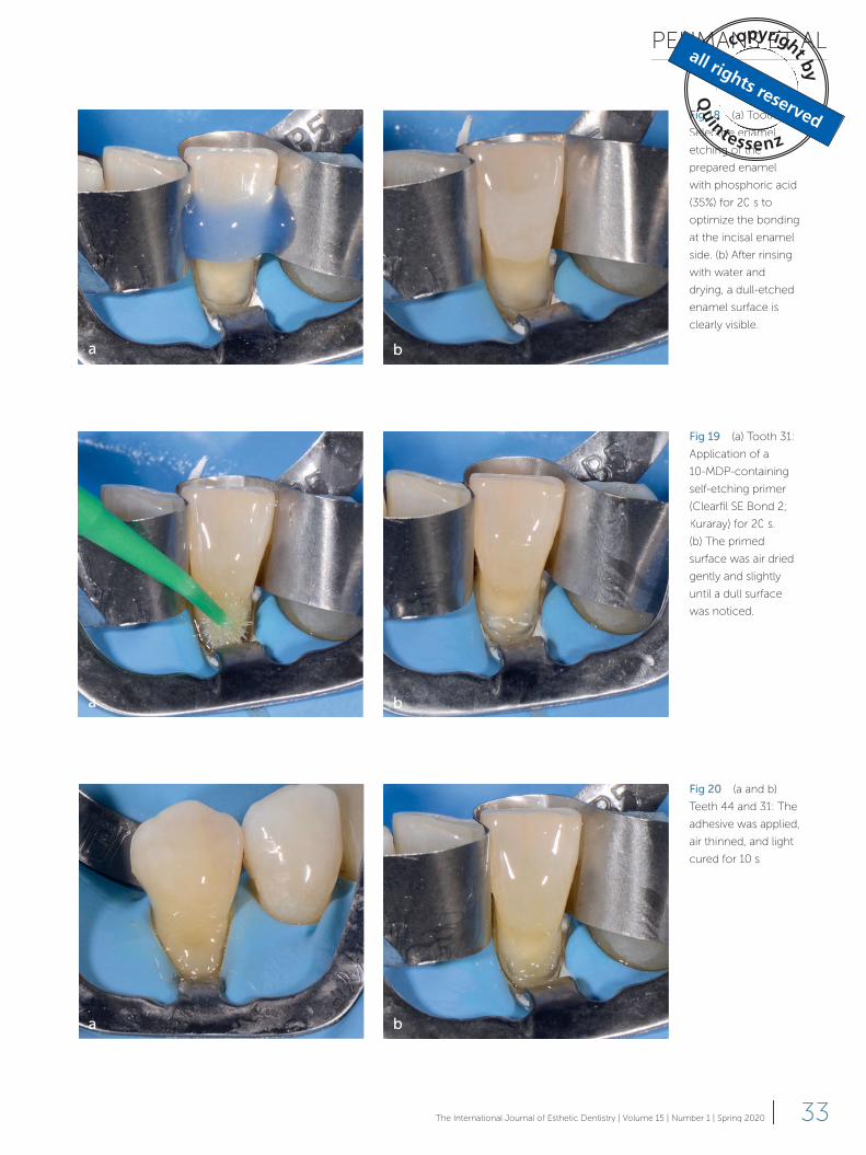
Fig 18 (a) Tooth 31:
Selective enamel
etching of the
prepared enamel
with phosphoric acid
(35%) for 20 s to
optimize the bonding
at the incisal enamel
side. (b) After rinsing
with water and
drying, a dull-etched
enamel surface is
clearly visible.
a b
Fig 19 (a) Tooth 31:
Application of a
10-MDP-containing
self-etching primer
(Clearfil SE Bond 2;
Kuraray) for 20 s.
(b) The primed
surface was air dried
gently and slightly
until a dull surface
was noticed.
a b
Fig 20 (a and b)
Teeth 44 and 31: The
adhesive was applied,
air thinned, and light
cured for 10 s.
a b

CLINICAL RESEARCH
34 | The International Journal of Esthetic Dentistry | Volume 15 | Number 1 | Spring 2020
defects and superficial marginal discolor-
ation will present less frequently at the incisal
enamel side with aging.43 Etching enamel
with phosphoric acid creates a deeper and
more pronounced etch pattern compared
with SEa and results in an increased micro-
mechanical retention and a more optimal
bond to enamel.
Selecting a universal adhesive, a 10-MDP-
based adhesive is preferred, used in a self-
etch mode with prior selective etching of
the enamel. The chemical interaction be-
tween the 10-MDP functional monomer
and calcium is only possible when HAp re-
mains in the hybrid layer. All HAp is dissolved
when one follows the E&R approach.
Finally, when using a simplified adhesive
system, it is best to cover the adhesive with a
hydrophobic resin coating. The use of this ad-
ditional coating after the application of a sim-
plified adhesive system leads to a thicker and
more uniform adhesive layer with less re-
tained water and solvent and a significant re-
duction in the fluid flow rate. In this way, a
more stable resin–dentin interface is formed.43
Selection and application
of the composite
Selection of the composite
NCCLs have a relatively small C-factor,
meaning that the mechanical properties of
the composite are less important to the out-
come of the final restoration than the actual
performance of the adhesive. Indeed, sever-
al reviews have shown that the type of com-
posite used had no influence on the bond-
ing performance of adhesives in NCCLs.42,43,70
Similarly, compomers and giomers, both
fluoride-releasing resin materials, do not
show a better performance compared with
conventional composites in NCCL clinical
trials.42,43
It has been stated that NCCLs suspected
of being caused primarily by abfraction
should be restored with a microfilled resin
composite or a flowable resin that has a low
modulus of elasticity because these will flex
with the tooth and not compromise reten-
tion. However, no definitive conclusion can
be found in the literature that addresses the
difference between failure rates of resin
composites of different stiffness used to re-
store NCCLs.71,72
Application of the composite
A multiple layering technique with a con-
ventional composite has been proposed to
restore deeper and/or larger lesions to min-
imize shrinkage due to polymerization and
also to achieve better marginal adaptation
in Class V non-carious lesions. Several in vi-
tro studies70,73,74 have shown that the best
results are obtained when using the oblique
layering technique from gingival toward in-
cisal. This technique has been applied in
most NCCL clinical trials.43 Indeed, since
enamel adhesion is stronger, more stable,
and more predictable, the insertion of ma-
terial should begin from the gingival wall,
without the surrounding enamel. Whenever
possible, the cavity should be restored with
three or two increments (Figs 21 to 25). The
last one should be placed on the enamel
margin. Small lesions can be restored with
one increment.
An alternative application method is to
cover the entire exposed dentin surface
with a thin layer of flowable composite in a
first step to optimize the adaptation of the
composite to the hybridized dentin surface.
In addition, the lower e-modulus and higher
flexibility of the flowable composite will
compensate for polymerization shrinkage.
Next, the rest of the lesion should be re-
stored with a stiffer conventional composite
(Figs 24 and 25).
As far as esthetics is concerned, the col-
or of the cervical area is easy to obtain. The
use of a medium opacity chromatic dentin
composite covered with a more translucent
slightly chromatic enamel composite – also

PEUMANS ET AL
35The International Journal of Esthetic Dentistry | Volume 15 | Number 1 | Spring 2020 |
Fig 21 (a) Tooth 44: Application of a chromatic dentin composite covering the dentin surface (Essentia Dark Dentin; GC) followed by
polymerization. (b) Next, a translucent enamel composite with low chromaticity, also called achromatic enamel75 (Essentia Dark Enamel) was
applied to restore the contour of the tooth. This layer covers the incisal enamel bevel.
a b
Fig 22 (a) Application of glycerine gel and final polymerization (20 s) to eliminate the oxygen-inhibited layer. (b) The restorations were
checked 1 week after final finishing and polishing.
a b

CLINICAL RESEARCH
36 | The International Journal of Esthetic Dentistry | Volume 15 | Number 1 | Spring 2020
Fig 23 NCCLs on teeth 44, 42, 41, and 31 (a) before and (b) after placement of direct composite restorations.
a b
Fig 24 NCCLs on teeth 34 and 35 (a) before and (b) after restorative treatment with direct composite restorations.
a b
Fig 25 (a) Tooth 35: Application of the adhesive system. (b) A thin layer of flowable composite was applied over the whole dentin surface
and polymerized. (c) A conventional microhybrid composite was placed to restore the original contour, starting from the cervical third and
covering the incisal enamel margin.
a b c

PEUMANS ET AL
37The International Journal of Esthetic Dentistry | Volume 15 | Number 1 | Spring 2020 |
called achromatic enamel75 – copies the
natural tooth structure (Figs 21 to 23). Most
practitioners prefer a simplified color build
up and use just one chromatic body com-
posite with medium opacity.
A careful placement technique is need-
ed to minimize the finishing procedure.
Finishing and polishing
Any excess or roughness should be avoided
in NCCL restorations. Plaque retention, gin-
gival inflammation, and the occurrence of
caries lesions represent not only a failure of
the restoration but also a creation of new
problems for the patient. Poorly performed
finishing and polishing procedures can lead
to soft and hard tissue damage. Techniques
that require a minimal amount of finishing
and polishing are ideal, but properly con-
toured restorations are seldom achieved
without removing excess material, especial-
ly at the margins. When needed, a good op-
tion is to use a microfine diamond finishing
point (40 μm grit size), then a coarse com-
posite finishing disc, and finally the applica-
tion of rubber polishing points with a de-
creasing grit size (Fig 26).
When a Class V restoration is performed
in a semi-direct way, as described by Fahl76
and Ritter et al,77 the composite restoration
is made and finished extraorally and is then
bonded with a luting composite. The extra-
oral finishing and polishing of these Class V
inlays simplify the finishing procedure of the
restoration after cementation. According to
these authors, this technique more easily
provides a high surface smoothness and a
good marginal adaptation compared with
the commonly used direct technique. How-
ever, clinical studies are required to prove
whether this technique is superior to the
clinically proven direct technique.
Maintenance
The condition of the restorations must be
evaluated yearly. Marginal deterioration that
presents as marginal discoloration and small
marginal defects is commonly seen in these
restorations with aging;43,78 their lifespan can
be lengthened by repolishing the restor-
Fig 26 Finishing and polishing of NCCL composite restorations. (a) Using a microfine pointed diamond bur (40 μm). (b and c) Finally,
polishing with rubber polishing points with a decreasing grit size.
a b c

CLINICAL RESEARCH
38 | The International Journal of Esthetic Dentistry | Volume 15 | Number 1 | Spring 2020
ation margins (Fig 27). If larger defects are
present, local repair is indicated.
The influence of the patient factor on
the clinical performance of the restorations
should not be underestimated. As already
mentioned, the etiology of the NCCLs must
always be kept under control to prevent re-
currence of the lesions. Importantly, special
conditions such as poor oral hygiene, high
caries risk, heavy bruxism, and erosive chal-
lenges were not included in most NCCL
clinical trials. Although one expects that
these high-risk patient factors negatively in-
fluence the lifespan of the restorations to a
large extent, the clinical longevity of resin
composites as restorative materials in such
a context has not yet been clinically investi-
gated.
Root coverage surgical procedures in combination with a restoration
NCCLs may be associated with gingival re-
cession, exposing root surfaces to the oral
cavity. Placement of a composite restor-
ation is possible, but the esthetic result can
be disappointing. If esthetics is important to
the patient, the ideal treatment for the
combination of noncarious cervical lesions
(particularly in the case of deep lesions) and
gingival recession is a combined perio-
restorative approach (Fig 28).32,79,80 In a com-
bined restorative-surgical approach, the res-
toration must be placed prior to the surgical
procedure for better visibility of the opera-
tive field and for the finished restoration. To
properly restore the dental tissue lost due to
wear, the maximum level of root coverage
needs to be predetermined.79,80
The restoration should recreate not only
the contour of the tooth crown but also that
of the lost CEJ at the root portion. Recent
systematic reviews have pointed out that
the combination of a coronally advanced
flap (CAF) with connective tissue graft (CTG)
provides the best clinical outcomes for root
coverage when appropriately performed.79,80
However, there is still a lack of long-term
clinical evidence to prove the stability of the
combined perio-restorative approach.
Conclusion
To achieve an accurate diagnosis of the eti-
ology of multifactorial NCCLs, practition-
ers should follow the following checklist:
comprehensive medical and dental history,
occlusal examination, evaluation of diet,
and oral hygiene procedures. Furthermore,
important risk factors in the formation of
NCCLs should be taken into account.
Treating NCCLs necessarily involves
problem identification, diagnosis, etiologic
factor removal, monitoring, and, if neces-
sary, treatment. Personalized treatment
Fig 27 (a) A female patient requested the replacement of cervical composite restorations due to discolored
margins. (b) Repolishing the restoration margins removed the superficial discoloration, thus increasing the longevity
of the restorations.
a b

PEUMANS ET AL
39The International Journal of Esthetic Dentistry | Volume 15 | Number 1 | Spring 2020 |
Fig 28 (a) Initial
situation: A 50-year-
old female patient
showed gingival
recession on the
maxillary anterior
teeth and deep
cervical lesions on
the maxillary
premolars and first
molars. The patient
requested an esthetic
improvement. The
deep defects on
teeth 14, 15, 16, and
26 were first restored
with direct compos-
ite restorations until
the level of the CEJ.
(b) Initial situation:
Lateral view, with
NCCLs on several
teeth. (b) After
placement of direct
composite restor-
ations on teeth 14, 15,
and 16 (restoring
deep defects). (c and
d) Further esthetic
improvement was
realized by surgical
root coverage of the
cervical lesions
(combination of CTG
and CAF) on teeth 13,
14, 15, 16, 21, 23, and
26. (e) Final result
after surgical root
coverage of the
cervical lesions
(combination of CTG
and CAF) on teeth 13,
14, 15, and 16.
a
b c
d
e

CLINICAL RESEARCH
40 | The International Journal of Esthetic Dentistry | Volume 15 | Number 1 | Spring 2020
should be applied, with the appropriate ap-
proach adopted for the specific situation.
Particular attention should be paid to the
preventive phase, as this will also determine
the longevity of the restorative treatment.
When function and esthetics are impaired,
direct composite restorations are indicated
to restore deep lesions. These restorations
show an acceptable clinical performance in
the long term provided an adhesive system
with a good proven clinical performance is
selected. In addition, a high level of oper-
ator skill combined with a careful operative
procedure is required to enhance the lon-
gevity of the restorations.
In case NCCLs are associated with gingi-
val recession, the original soft and hard tis-
sue relation and structure can be restored
esthetically and functionally by a combined
perio-restorative intervention.
References
1. Michael JA, Townsend GC, Greenwood
LF, Kaidonis JA. Abfraction: separating fact
from fiction. Aust Dent J 2009;54:2–8.
2. Bartlett DW, Shah P. A critical review of
non-carious cervical (wear) lesions and the
role of abfraction, erosion, and abrasion.
J Dent Res 2006;85:306–312.
3. Levitch LC, Bader JD, Shugars DA, Hey-
mann HO. Non-carious cervical lesions.
J Dent 1994;22:195–207.
4. Pecie R, Krejci I, Garcia-Godoy F, Bor-
tolotto T. Noncarious cervical lesions – a
clinical concept based on the literature
review. Part 1: prevention. Am J Dent
2011;24:49–56.
5. Wood I, Jawad Z, Paisley C, Brunton P.
Non-carious cervical tooth surface loss: a
literature review. J Dent 2008;36:759–766.
6. Bernhardt O, Gesch D, Schwahn C, et al.
Epidemiological evaluation of the multifac-
torial aetiology of abfractions. J Oral Rehabil
2006;33:17–25.
7. Borcic J, Anic I, Urek MM, Ferreri S. The
prevalence of non-carious cervical lesions
in permanent dentition. J Oral Rehabil
2004;31:117–123.
8. Yang J, Cai D, Wang F, et al. Non-cari-
ous cervical lesions (NCCLs) in a random
sampling community population and the
association of NCCLs with occlusive wear.
J Oral Rehabil 2016;43:960–966.
9. Zuza A, Racic M, Ivkovic N, et al. Preva-
lence of non-carious cervical lesions among
the general population of the Republic of
Srprska, Bosnia and Herzegovina. Int Dent J
2019;69:281–288.
10. Daley TJ, Harbrow DJ, Kahler B, Young
WG. The cervical wedge-shaped lesion
in teeth: a light and electron microscopic
study. Aust Dent J 2009;54:212–219.
11. Michael JA, Kaidonis JA, Townsend GC.
Non-carious cervical lesions: a scanning
electron microscopic study. Aust Dent J
2010;55:138–142.
12. Walter C, Kress E, Götz H, Taylor K,
Willershausen I, Zampelis A. The anatomy
of non-carious cervical lesions. Clin Oral
Investig 2014;18:139–146.
13. Grippo JO, Simring M, Coleman TATT .
Abfraction, abrasion, biocorrosion, and the
enigma of noncarious cervical lesions: a
20-year perspective. J Esthet Restor Dent
2012;24:10–23.
14. Hara AT, AA Ando M, Cury JA, Serra MC,
González-Cabezas C, Zero DT. Influence of
the organic matrix on root dentine erosion
by citric acid. Caries Res 2005;39:134–138.
15. Schlueter N, Hardt M, Klimek J, Ganss
C. Influence of the digestive enzymes
trypsin and pepsin in vitro on the progres-
sion of erosion in dentine. Arch Oral Biol
2010;55:294–299.
16. Young WG, Khan F. Sites of dental
erosion are saliva-dependent. J Oral Rehabil
2002;29:35–43.
17. Rees JS, Hammadeh M, Jagger DC. Ab-
fraction lesion formation in maxillary incisors,
canines and premolars: a finite element
study. Eur J Oral Sci 2003;111:149–154.
18. Soares PV, Santos-Filho PC, Soares CJ,
et al. Non-carious cervical lesions: influence
of morphology and load type on biomech-
anical behaviour of maxillary incisors. Aust
Dent J 2013;58:306–314.
19. Soares PV, Machado AC, Zeola LF, et al.
Loading and composite restoration assess-
ment of various non-carious cervical lesions
morphologies – 3D finite element analysis.
Aust Dent J 2015;60:309–316.
20. Guimarães JC, Guimarães Soella G,
Brandão Durand L, et al. Stress amplifica-
tions in dental non-carious cervical lesions.
J Biomech 2014;47:410–416.
21. Jakupovic S, Cerjakovic E, Topcic A,
Ajanovic M, Prcic A AK, Vukovic A. Analysis of
the abfraction lesions formation mechanism
by the finite element method. Acta Inform
Med 2014;22:241–245.
22. Romeed SA, Malik R, Dunne SM. Stress
analysis of occlusal forces in canine teeth
and their role in the development of
non-carious cervical lesions: abfraction.
Int J Dent 2012;2012:234845.
23. Telles D, Pegoraro LF, Pereira JC. Prev-
alence of noncarious cervical lesions and
their relation to occlusal aspects: a clinical
study. J Esthet Dent 2000;12:10–15.
24. Pegoraro LF, Scolaro JM, Conti PC, Telles
D, Pegoraro TATT . Noncarious cervical lesions
in adults: prevalence and occlusal aspects.
J Am Dent Assoc 2005;136:1694–1700.
25. Antonelli JR, Hottel TL, Brandt R, Scar-
becz M, Patel T. The role of occlusal loading
in the pathogenesis of non-carious cervical
lesions. Am J Dent 2013;26:86–92.
26. Sawlani K, Lawson NC, Burgess JO, et
al. Factors influencing the progression of
noncarious cervical lesions: a 5-year pro-

PEUMANS ET AL
41The International Journal of Esthetic Dentistry | Volume 15 | Number 1 | Spring 2020 |
spective clinical evaluation. J Prosthet Dent
2016;115:571–577.
27. Senna P, Del Bel Cury A, Rösing C.
Non-carious cervical lesions and occlusion:
a systematic review of clinical studies. J Oral
Rehabil 2012;39:450–462.
28. Silva AG, Martins CC, Zina LG, et al. The
association between occlusal factors and
noncarious cervical lesions: a systematic
review. J Dent 2013;41:9–16.
29. Azzopardi A, Bartlett DW, Watson TF,
Sherriff M. The measurement and pre-
vention of erosion and abrasion. J Dent
2001;29:395–400.
30. Eisenburger M, Shellis RP, Addy M. Com-
parative study of wear of enamel induced
by alternating and simultaneous combina-
tions of abrasion and erosion in vitro. Caries
Res 2003;37:450–455.
31. Leal NMS, Silva JL, Benigno MIM, Be-
merguy EA, Meira JBC, Ballester RY. How
mechanical stresses modulate enamel de-
mineralization in non-carious cervical lesions?
J Mech Behav Biomed Mater 2017;66:50–57.
32. Terry DA, McGuire MK, McLaren E,
Fulton R, Swift EJ Jr. Perioesthetic approach
to the diagnosis and treatment of carious
and noncarious cervical lesions: Part I.
J Esthet Restor Dent 2003;15:217–232.
33. Soares PV, Grippo JO. Noncarious Cer-
vical Lesions and Cervical Dentin Hypersen-
sitivity. Etiology, Diagnosis, and Treatment.
Quintessence Publishing, 2017.
34. Aw TC, Lepe X, Johnson GH, Mancl
L. Characteristics of noncarious cervical
lesions: a clinical investigation. J Am Dent
Assoc 2002;133:725–733.
35. Sugita I, Nakashima S, Ikeda A, et al. A
pilot study to assess the morphology and
progression of non-carious cervical lesions.
J Dent 2017;57:51–56.
36. Miglani S, Aggarwal V, Ahuja B. Dentin
hypersensitivity: recent trends in manage-
ment. J Conserv Dent 2010;13:218–224.
37. Endo H, Kawamoto R, Takahashi F, et al.
Evaluation of a calcium phosphate desensi-
tizer using an ultrasonic device. Dent Mater
J 2013;32:456–461.
38. Sauro S, Watson TF, Thompson I. Den-
tine desensitization induced by prophylactic
and air-polishing procedures: an in vitro
dentine permeability and confocal micros-
copy study. Dent Mater 2010;38:411–422.
39. Zhu M, Li J, Chen B, et al. The effect of
calcium sodium phosphosilicate on dentin
hypersensitivity: a systematic review and
meta-analysis. PLoS One 2015;10:e0140176.
40. Veitz-Keenan A, Barna JA, Strober
B, et al. Treatment for hypersensitive
noncarious cervical lesions: a Practitioners
Engaged in Applied Research and Learn-
ing (PEARL) Network randomized clinical
effectiveness study. J Am Dent Assco
2013;144:495–506.
41. Prati C, Cervellati F, Sanasi V, Monte-
bugnoli L. Treatment of cervical dentin
hypersensitivity with resin adhesives: 4-week
evaluation. Am J Dent 2001;14:378–382.
42. Pecie R, Krejci I, García-Godoy F,
Bortolotto T. Noncarious cervical lesions
(NCCL) – r clinical concept based on the
literature review. Part 2: Restoration.
Am J Dent 2011;24:183–192.
43. Peumans M, De Munck J, Mine A,
Van Meerbeek B. Clinical effectiveness of
contemporary adhesives for the restoration
of non-carious cervical lesions. A systematic
review. Dent Mater 2014;30:1089–1103.
44. Van Meerbeek B, De Munck J,
Yoshida Y, et al. Buonocore memorial
lecture. Adhesion to enamel and dentine:
current status and future challenges.
Oper Dent 2003;28:215–235.
45. Van Meerbeek B, Peumans M, Poitevin
A, et al. Relationship between bond-strength
tests and clinical outcomes. Dent Mater
2010;26:e100–e121.
46. Yoshida Y, Nagakane K, Fukuda R, et al.
Comparative study on adhesive perfor-
mance of functional monomers. J Dent Res
2004;83:454–458.
47. Yoshihara K, Yoshida Y, Hayakawa S, et
al. Nanolayering of phosphoric acid ester
monomer on enamel and dentin. Acta
Biomater 2011;7:3187–3195.
48. Poitevin A, De Munck J, Van Ende A, et
al. Bonding effectiveness of self-adhesive
composites to dentin and enamel. Dent
Mater 2013;29:221–230.
49. Rosa WL, Piva E, Silva AF. Bond strength
of universal adhesives: a systematic review
and meta-analysis. J Dent 2015;43:
765–776.
50. Chen C, Niu LN, Xie H, et al. Bonding of
universal adhesives to dentine – old wine in
new bottles? J Dent 2015;43:525–536.
51. De Munck J, Mine A, Poitevin A, et
al. Meta-analytical review of parameters
involved in dentin bonding. J Dent Res
2012;91:351–357.
52. Mahn E, Rousson V, Heintze S. Me-
ta-analysis of the influence of bonding
parameters on the clinical outcome of
tooth-colored cervical restorations.
J Adhes Dent 2015;17:391–403.
53. Browet S, Gerdolle D. Precision and
security in restorative dentistry: the synergy
of isolation and magnification. Int J Esthet
Dent 2017;12:172–185.
54. AjamiA AA, Kahnamoii MA, Kimyai S, et
al. Effect of three different contamination
removal methods on bond strength of a
self-etching adhesive to dentin contaminat-
ed with an aluminum chloride hemostatic
agent. J Contemp Dent Pract 2013;14:26–33.
55. Kuphasuk W, Harnirattisai C,
Senawongse P, Tagami J. Bond strengths of
two adhesive systems to dentin contami-
nated with a hemostatic agent. Oper Dent
2007;32:399–405.
56. Groddeck S, Attin T, Tauböck TT. Effect
of cavity contamination by blood and
hemostatic agents on marginal adaptation
of composite restorations. J Adhes Dent
2017;19:259–264.
57. Perez CR. Alternative technique for class
V resin composite restorations with mini-
mum finishing/ polishing procedures. Oper
Dent 2010;35:375–379.
58. Brännström M, Nordenvall KJ, Malmgren
O. The effect of various pretreatment meth-
ods of the enamel in bonding procedures.
Am J Orthod 1978;74:522–530.
59. Tay FR, Pashley DH. Resin bonding to
cervical sclerotic dentin: a review. J Dent
2004;32:173–196.
60. Abdalla R, Mitchell RJ, Ren YF. Non-car-
ious cervical lesions imaged by focus varia-
tion microscopy. J Dent 2017;63:14–20.
61. Ermis RB, De Munck J, Cardoso MV, et
al. Bond strength of self-etch adhesives to
dentin prepared with three different dia-
mond burs. Dent Mater 2008;24:978–985.
62. Freeman R, Varanasi S, Meyers IA,
Symons AL. Effect of air abrasion and
thermocycling on resin adaptation and
shear bond strength to dentin for an
etch-and-rinse and self-etch resin adhesive.
Dent Mater J 2012;31:180–188.

CLINICAL RESEARCH
42 | The International Journal of Esthetic Dentistry | Volume 15 | Number 1 | Spring 2020
63. Hannig M, Femerling T. Influence of
air-abrasion treatment on the interfacial
bond between composite and dentin.
Oper Dent 1998;23:258–265.
64. Manhart J, Mehl A, Schroeter R, Obster
B, Hickel R. Bond strength of composite to
dentin treated by air abrasion. Oper Dent
1999;24:223–232.
65. D‘Amario M, Piccioni C, Di Carlo S,
De Angelis F, Caruso S, Capogreco M.
Effect of airborne particle air abrasion on
microtensile bond strength of total-etch
adhesives to human dentin. Biomed Res Int
2017;2017:2432536.
66. Sutil BGDS, Susin AH. Dentin pre-
treatment and adhesive temperature as
effecting factors on bond strength of a
universal adhesive system. J Appl Oral Sci
2017;25:533–540.
67. Chaves P, Giannini M, Ambrosano GM.
Influence of smear layer pretreatments
on bond strength to dentin. J Adhes Dent
2002;4:191–196.
68. Van Meerbeek B, De Munck J, Mattar
D, Van Landuyt K, Lambrechts P. Microten-
sile bond strengths of an etch&rinse and
self-etch adhesive to enamel and dentin as
a function of surface treatment. Oper Dent
2003;28:647–660.
69. Zimmerli B, De Munck J, Lussi A,
Lambrechts P, Van Meerbeek B. Long-
term bonding to eroded dentin requires
superficial bur preparation. Clin Oral Investig
2012;16:1451–1461.
70. Kubo S, Yokota H, Yokota H, Hayashi Y.
Challenges to the clinical placement and
evaluation of adhesively-bonded, cervi-
cal composite restorations. Dent Mater
2013;29:10–27.
71. Peumans M, De Munck J, Van Landuyt
KL, Poitevin A, Lambrechts P, Van Meerbeek
B. A 13-year clinical evaluation of two three-
step etch-and-rinse adhesives in non-car-
ious class-V lesions. Clin Oral Investig
2012;16:129–137.
72. Szesz A, Parreiras S, Martini E, Reis A,
Loguercio A. Effect of flowable com-
posites on the clinical performance of
non-carious cervical lesions: a system-
atic review and meta-analysis. J Dent
2017;65:11–21.
73. Correia AMO, Tribst JPM, Matos FS, Platt
JA, Caneppele TMF, Borges ALS. Polymer-
ization shrinkage stresses in different re-
storative techniques for non-carious cervical
lesions. J Dent 2018;76:68–74.
74. Hansen EK. Effect of cavity depth and
application technique on marginal adapta-
tion of resins in dentin cavities. J Dent Res
1986;65:1319–1321.
75. Villarroel M, Fahl N, De Sousa AM,
De Oliveira OB Jr. Direct esthetic restor-
ations based on translucency and opacity
of composite resins. J Esthet Restor Dent
2011;23:73–87.
76. Fahl N Jr. Direct-Indirect class V res-
torations: a novel approach for treating
noncarious cervical lesions. J Esthet Restor
Dent 2015;27:267–284.
77. Ritter AV, Fahl N Jr, Vargas M, Maia RR. AA
The direct-indirect technique for composite
restorations revisited. Compend Contin
Educ Dent 2017;38:e9–e12.
78. Kim JH, Cho J, Lee Y, Cho BH. The
survival of Class V composite restorations
and analysis of marginal discoloration. Oper
Dent 2017;42:E93–E101.
79. Bignozzi I, Littarru C, Crea A, Vittorini
Orgeas G, Landi L. Surgical treatment op-
tions for grafting areas of gingival recession
association with cervical lesions: a review.
J Esthet Restor Dent 2013;25:371–382.
80. Zucchelli G, Gori G, Mele M, et al.
Non-carious cervical lesions associated
with gingival recessions: a decision-making
process. J Periodontol 2011;82:1713–1724.