Three new genera representing novel lineages of ...
15
Three new genera representing novel lineages of Sordariomycetidae (Sordariomycetes, Ascomycota) from tropical freshwater habitats in Costa Rica Astrid Ferrer 1 Department of Plant Biology, University of Illinois, Room 265 Morrill Hall, 505 South Goodwin Avenue, Urbana, Illinois 61801 Andrew N. Miller Illinois Natural History Survey, University of Illinois, 1816 South Oak Street, Champaign, Illinois 61820 Carolina Sarmiento Universidad de los Andes, Cra. 1 No. 18A-10, Bogota ´, Colombia Carol A. Shearer Department of Plant Biology, University of Illinois, Room 265 Morrill Hall, 505 South Goodwin Avenue, Urbana, Illinois 61801 Abstract: Three new genera are established in the Sordariomycetidae based on morphological and molecular data (SSU and LSU nrDNA) to accommo- date five ascomycete species collected from sub- merged woody debris in freshwater habitats from Costa Rica. The genus Bullimyces contains three new species, B. communis, B. costaricensis and B. auris- porus. Bullimyces is characterized by globose to subglobose, membranous, black, ostiolate ascomata; deliquescent, hyaline, globose cells that fill the center of the centrum; unitunicate asci that deliquesce early in some species; and septate, thick-walled ascospores with or without gelatinous sheaths or appendages. Bullimyces species form a well supported clade with 100% bootstrap support, but the position of the genus in the Sordariomycetidae remains unclear. The second genus, Riomyces, is represented by a single species, R. rotundus. Riomyces is characterized by globose to subglobose, membranous, black, ostiolate ascomata, unitunicate, cylindrical asci, hyaline, glo- bose cells that fill the hamathecium and septate, thick-walled ascospores with a gelatinous sheath. Although Riomyces is morphologically similar to Bullimyces, the two genera did not group together with support in any analysis. The third genus, Hydromelitis, is represented by a single species, H. pulchella. Hydromelitis is characterized by pyriform, membranous, black, ostiolate ascomata, unitunicate asci lacking an apical structure, simple, thin-walled, septate paraphyses and hyaline to golden yellow, multiseptate, thick-walled ascospores with a gelatinous sheath. Bullimyces, Riomyces and Hydromelitis were nested within an unsupported clade consisting of members of the Ophiostomatales, Magnaporthales and freshwater Annulatacaceae sensu lato and sensu stricto. Key words: aquatic, ascomycetes, fungal systemat- ics, LSU, phylogenetics, SSU INTRODUCTION Aquatic ascomycetes are microscopic, saprobic fungi that colonize and decompose submerged substrates in freshwater and marine habitats. At present 278 fresh- water and 275 marine species have been reported in the Sordariomycetes from aquatic habitats (Jones et al. 2009, http://fungi.life.illinois.edu/) and all the major lineages contain aquatic species (Zhang et al. 2006). Aquatic species share several morphological characters including the presence of elaborate gelatinous asco- spore sheaths and appendages, which presumably function in attaching spores to substrates in water (Shearer 1993; Jones 1994, 2006). Phylogenetic studies have shown that these orders have evolved indepen- dently from terrestrial ancestors (Spatafora et al. 1998, Vijaykrishna et al. 2006). The position of various genera remains incertae sedis at the ordinal level as new species from the tropics are described. During a comparative survey across four sites in Costa Rica, we found five new species of freshwater ascomycetes representing three new genera. The genus Bullimyces contains three new species, B. communis, B. costaricensis and B. aurisporus, the genus Riomyces is based on a single species, R. rotundus, and Hydromelitis is represented by a single species, H. pulchella. We provide a description of these species and use both morphological and molecular sequence data from partial 18S small subunit (SSU) and 28S large subunit (LSU) nuclear ribosomal DNA to determine their placement as novel freshwater Sordar- iomycetidae lineages. MATERIALS AND METHODS Collection, isolation and morphological examination.—Meth- ods for the collection, isolation and morphological exam- ination of specimens are presented in Ferrer and Shearer (2005). Specimens are deposited in the Fungarium of the University of Illinois at Urbana-Champaign (ILL). Molecular study.— Fungal mycelia were removed from cultures grown on potato dextrose agar (PDA, Difco) and ground in liquid nitrogen. DNA was extracted with the Submitted 6 Apr 2011; accepted for publication 16 Dec 2011. 1 Corresponding author. E-mail: [email protected] Mycologia, 104(4), 2012, pp. 865–879. DOI: 10.3852/11-111 # 2012 by The Mycological Society of America, Lawrence, KS 66044-8897 865
Transcript of Three new genera representing novel lineages of ...
Three new genera representing novel lineages of Sordariomycetidae(Sordariomycetes, Ascomycota) from tropical freshwater habitats in Costa Rica
Astrid Ferrer1
Department of Plant Biology, University of Illinois,Room 265 Morrill Hall, 505 South Goodwin Avenue,Urbana, Illinois 61801
Andrew N. MillerIllinois Natural History Survey, University of Illinois,1816 South Oak Street, Champaign, Illinois 61820
Carolina SarmientoUniversidad de los Andes, Cra. 1 No. 18A-10, Bogota,Colombia
Carol A. ShearerDepartment of Plant Biology, University of Illinois,Room 265 Morrill Hall, 505 South Goodwin Avenue,Urbana, Illinois 61801
Abstract: Three new genera are established in theSordariomycetidae based on morphological andmolecular data (SSU and LSU nrDNA) to accommo-date five ascomycete species collected from sub-merged woody debris in freshwater habitats fromCosta Rica. The genus Bullimyces contains three newspecies, B. communis, B. costaricensis and B. auris-porus. Bullimyces is characterized by globose tosubglobose, membranous, black, ostiolate ascomata;deliquescent, hyaline, globose cells that fill the centerof the centrum; unitunicate asci that deliquesce earlyin some species; and septate, thick-walled ascosporeswith or without gelatinous sheaths or appendages.Bullimyces species form a well supported clade with100% bootstrap support, but the position of thegenus in the Sordariomycetidae remains unclear. Thesecond genus, Riomyces, is represented by a singlespecies, R. rotundus. Riomyces is characterized byglobose to subglobose, membranous, black, ostiolateascomata, unitunicate, cylindrical asci, hyaline, glo-bose cells that fill the hamathecium and septate,thick-walled ascospores with a gelatinous sheath.Although Riomyces is morphologically similar toBullimyces, the two genera did not group togetherwith support in any analysis. The third genus,Hydromelitis, is represented by a single species, H.pulchella. Hydromelitis is characterized by pyriform,membranous, black, ostiolate ascomata, unitunicateasci lacking an apical structure, simple, thin-walled,septate paraphyses and hyaline to golden yellow,
multiseptate, thick-walled ascospores with a gelatinoussheath. Bullimyces, Riomyces and Hydromelitis werenested within an unsupported clade consisting ofmembers of the Ophiostomatales, Magnaporthales andfreshwater Annulatacaceae sensu lato and sensu stricto.
Key words: aquatic, ascomycetes, fungal systemat-ics, LSU, phylogenetics, SSU
INTRODUCTION
Aquatic ascomycetes are microscopic, saprobic fungithat colonize and decompose submerged substrates infreshwater and marine habitats. At present 278 fresh-water and 275 marine species have been reported in theSordariomycetes from aquatic habitats (Jones et al.2009, http://fungi.life.illinois.edu/) and all the majorlineages contain aquatic species (Zhang et al. 2006).Aquatic species share several morphological charactersincluding the presence of elaborate gelatinous asco-spore sheaths and appendages, which presumablyfunction in attaching spores to substrates in water(Shearer 1993; Jones 1994, 2006). Phylogenetic studieshave shown that these orders have evolved indepen-dently from terrestrial ancestors (Spatafora et al. 1998,Vijaykrishna et al. 2006). The position of various generaremains incertae sedis at the ordinal level as new speciesfrom the tropics are described. During a comparativesurvey across four sites in Costa Rica, we found five newspecies of freshwater ascomycetes representing threenew genera. The genus Bullimyces contains three newspecies, B. communis, B. costaricensis and B. aurisporus,the genus Riomyces is based on a single species, R.rotundus, and Hydromelitis is represented by a singlespecies, H. pulchella. We provide a description of thesespecies and use both morphological and molecularsequence data from partial 18S small subunit (SSU) and28S large subunit (LSU) nuclear ribosomal DNA todetermine their placement as novel freshwater Sordar-iomycetidae lineages.
MATERIALS AND METHODS
Collection, isolation and morphological examination.—Meth-ods for the collection, isolation and morphological exam-ination of specimens are presented in Ferrer and Shearer(2005). Specimens are deposited in the Fungarium of theUniversity of Illinois at Urbana-Champaign (ILL).
Molecular study.—Fungal mycelia were removed fromcultures grown on potato dextrose agar (PDA, Difco) andground in liquid nitrogen. DNA was extracted with the
Submitted 6 Apr 2011; accepted for publication 16 Dec 2011.1 Corresponding author. E-mail: [email protected]
Mycologia, 104(4), 2012, pp. 865–879. DOI: 10.3852/11-111# 2012 by The Mycological Society of America, Lawrence, KS 66044-8897
865
DNeasy Plant Mini Kit (QIAGEN Inc., Valencia, California)according to the kit’s instructions. Fragments of SSU andLSU were PCR amplified with PureTaqTM Ready-To-Go PCRbeads (Amersham Bioscience Corp., Piscataway, New York)according to Promputtha and Miller (2010). Primers NS1and NS4 (White et al. 1990) for SSU and LROR and LR6 forLSU (Rehner and Samuels 1994, Vilgalys and Hester 1990)were used for PCR amplification. PCR products werepurified with the QIAquick PCR purification kit (QIAGENInc., Valencia, California) and subsequently used in 11 mLsequencing reactions with BigDyeH Terminators 3.1 (Ap-plied Biosystems, Foster City, California) and primers NS1,NS2, NS3 and NS4 for SSU and LROR, LR3, LR3R and LR6for LSU. Sequences were generated at the University ofIllinois Biotechnology Center using an ABI 3730XL high-throughput automated sequencer. Sequences were assem-bled and edited manually in Sequencher 4.7 (Gene CodesCorp. 1991).
Phylogenetic analyses.—DNA sequences for Bullimyces,Hydromelitis and Riomyces are provided (TABLE I). Todetermine the familial placement of the new genera, taxasequenced in this study along with sequences obtained fromGenBank from various subclasses currently circumscribedwithin the Sordariomycetes based on Zhang et al. (2006)and Reblova (2006, 2009) were aligned with MUSCLEH(Edgar 2004) as implemented in the program SeaView 4.1(Galtier et al. 1996, Guoy et al. 2010). The SSU and LSUdatasets were initially analyzed separately and then concat-enated and analyzed as a single dataset after no conflictamong the bootstrap values of well supported clades wasobserved (Wiens 1998). A second analysis was performed forLSU data with a more extensive taxon sampling andincluding members in the Annulatascaceae sensu lato andstricto clades (Reblova 2006, 2009). Models of evolutionarychange were determined with Modeltest 3.7 (Posada andCrandall 1998). Maximum likelihood analyses (ML) wereconducted with an online version of RAxML (http://phylobench.vital-it.ch/raxml-bb/index.phpwith) (Stamata-kis 2008) under the GAMMA model with 100 bootstrapreplicates. Bayesian analyses employing Markov chainMonte Carlo (MCMC) were performed with MrBayes 3.1.2(Huelsenbeck et al. 2001, Huelsenbeck and Ronquist 2001)as an additional means of assessing branch support.Bayesian analyses using a uniform model (GTR + I + G)
were conducted using 10 000 000 generations with treessampled every 100th generation resulting in 100 000 totaltrees. Two independent analyses were performed with fourchains using default settings to ensure that trees were beingsampled from the same tree space. The first 10 000 trees,which extended beyond the burn-in phase in each analysis,were discarded, and the remaining 90 000 trees were used tocalculate posterior probabilities. The consensus of 90 000trees was generated in PAUP 4.0b10 (Swofford 2002).Phylogenetic trees were drawn with Figtree 1.2.2.
RESULTS
Phylogenetic analyses.—The combined SSU and LSUincluded 2407 characters from 46 taxa. A total of 364ambiguously aligned characters were excluded, yield-ing a total of 2043 characters. The ML analysisresulted in a single most likely tree (FIG. 1), whichwas similar in topology and placement of the majororders of Sordariomycetes to the phylogeny presentedby Zhang et al. (2006). Bullimyces, Riomyces andHydromelitis grouped within an unsupported cladeconsisting of members of the Ophiostomatales andMagnaporthales along with species of Sordariomyce-tidae inc. sed. The Bullimyces clade was well supportedwith 100% maximum likelihood bootstrap (MLB)support and 100% Bayesian posterior probability(BPP); the three sequences of B. communis formeda well supported clade and were sister to B.costaricensis and B. aurisporus. Riomyces was basal toBullimyces with no support. Ceratostomella pyreneicawas basal to Bullimyces but was not supported by MLB.Hydromelitis formed a clade with Lentomitella cirrhosawith no support.
Final alignment of the partial LSU sequences ofnrDNA included 1179 characters from 67 taxa. A totalof 154 ambiguously aligned characters were excluded,yielding a total of 1025 characters. The LSU data wasexpanded to include additional related species fromGenBank, most of which lacked a corresponding SSUsequence. The tree obtained with the ML analysisshowed a topology similar to the SSU and LSU tree
TABLE I. Strains used in this study, their origin and GenBank accession numbers
Taxa Isolate number Origin SSU LSU
Bullimyces aurisporus AF316-1a Costa Rica: Alajuela JF758615 ———Bullimyces aurisporus AF316-1b Costa Rica: Alajuela JF758614 JF775590Bullimyces communis AF281-3 Costa Rica: Heredia JF758617 JF775585Bullimyces communis AF281-4 Costa Rica: Alajuela JF758618 JF775586Bullimyces communis AF281-5 Costa Rica: Alajuela JF758619 JF775587Bullimyces costaricensis AF317-1a Costa Rica: Limon ——— JF775591Bullimyces costaricensis AF317-1b Costa Rica: Limon JF758616 JF775592Hydromelitis pulchella AF284-2 Costa Rica: Alajuela JF758613 JF775588Riomyces rotundus AF303-1 Costa Rica: Heredia JF758612 JF775589
866 MYCOLOGIA

FIG. 1. Most likely tree based on a combined SSU and LSU nrDNA sequence data obtained with RAxML. Numbers abovethe branches indicate ML bootstrap support $ 75% and numbers below indicate Bayesian posterior probabilities $ 95%.GenBank accession numbers are given after taxon names. Species sequenced for this study are highlighted.
FERRER ET AL.: FRESHWATER ASCOMYCETES 867

(FIG. 2), with Bullimyces, Riomyces and Hydromelitiswithin an unsupported clade consisting of membersof the Ophiostomatales, Magnaporthales and fresh-water Annulatascaceae sensu lato and sensu stricto.The three sequences of B. communis formed amonophyletic group with 99% MLB and 100% BPPvalues and formed a well supported clade to B.costaricensis and B. aurisporus with 98% MLB and100% BPP. Because ordinal or familial placement forthe three genera was not supported, they shouldremain in the Sordariomycetidae inc. sed.
TAXONOMY
Bullimyces A. Ferrer, A.N. Mill., C. Sarmiento etShearer gen. nov.
MycoBank MB561094Ascomata in ligno submerso, partim immersa vel im-
mersa, globosa vel subglobosa, membranacea, nigra, ostio-lati. Collum cylindricum, atrobrunneae. Centris ascocar-piorum cellulis psedoparenchymaticis, globosis, hyalinis,deliquescentibus. Asci unitunicati, cylindrici, tenuitunicati,sine poro apicali, octospori, uniseriati, persistentes veldeliquescentes. Ascosporae ellipsoideae-fusiformes, hyali-nae vel lutea, septatae, ad septum non constrictae,pachydermaticae, cum vel sine tunica gelatinosa praeditaeet appendages.
Typus generic: Bullimyces communis A. Ferrer, A.N.Mill., C. Sarmiento et Shearer
Etymology: From Latin Bulla 5 bubble; + Greek Myces5 fungus, in reference to the round cells filling thehamathecium.
Ascomata on submerged wood immersed to erum-pent in the substrate, globose to subglobose, mem-branous, black, ostiolate. Neck cylindrical, darkbrown. Pseudoparenchyma of thin-walled globose,hyaline cells, filling the centrum, deliquescent. Asciunitunicate, cylindrical, thin-walled, lacking an apicalpore and apical structures, eight-spored, uniseriate,deliquescing early in some species. Ascospores broad-ly ellipsoidal-fusiform to ellipsoidal, hyaline becomingdark yellow with age, septate, not constricted at septa,thick-walled, with or without a gelatinous sheath andappendages.
Bullimyces communis A. Ferrer, A.N. Mill., C.Sarmiento et Shearer sp. nov. FIGS. 3–16
MycoBank MB561095Ascomata in ligno submerso, 350–650 3 340–660 mm,
immersa, globosa, membranacea, nigra, ostiolati. Collum100–170 3 60–80 mm, cylindricum, atrobrunneae. Centrisascocarpiorum cellulis psedoparenchymaticis, 38–90 mmdiam, globosis, hyalinis, deliquescentibus. Asci 310–430 3
18–22 mm, unitunicati, cylindrici, tenuitunicati, sine poroapicali, octospori, uniseriati, deliquescentes. Ascosporae 40–48 3 19–22 mm, ellipsoideae-fusiformes, hyalinae, 3-septatae,
ad septum non constrictae, pachydermaticae, hyaline calyp-tra bipolare.
Holotype: COSTA RICA. ALAJUELA, Cano NegroReserve, Rio Frio, 10u539N, 84u459W, water 27.5 C, pH 5,on submerged wood, 15 Dec 2005, Astrid Ferrer & MarlonSalazar, AF281-1 (ILL).
Etymology: From Latin Communis 5 common, inreference to being the most collected species in Costa Rica.
Ascomata on submerged wood, 350–650 3 340–660 mm, immersed in the substrate, globose, membra-nous, black, ostiolate, brown. Neck 100–170 3 60–80 mm, cylindrical, dark brown. Pseudoparenchyma ofthin-walled cells 38–90 mm diam, globose, hyaline,filling the centrum, deliquescent. Asci 310–430 3 18–22 mm, unitunicate, cylindrical, thin-walled, lacking anapical pore and apical structures, floating free withinthe centrum, eight-spored, uniseriate. Ascospores 40–48 3 19–22 mm (mean 5 43.70 3 20.20 mm, n 5 30),broadly ellipsoidal-fusiform to ellipsoidal, hyaline,three-septate, not constricted at septa, thick-walled(2–3 mm), with hyaline, gelatinous caps at both apices,2–3 mm long, staining in aqueous nigrosin, ascosporesaccumulate as a mass at tip of the neck after discharge.
Known distribution: COSTA RICA: Alajuela, Heredia.Habitat: On submerged, dead woody debris.Additional specimens examined: COSTA RICA. ALA-
JUELA: Cano Negro Reserve, Rio Frio, 10u539N, 84u459W,water 27.5 C, pH 5, on submerged wood, 15 Dec 2005,Astrid Ferrer & Marlon Salazar, AF281-4, AF281-5, AF281-6,AF281-7. HEREDIA: La Selva Biological Station, La SelvaStream, 10u259N, 84u019W, water 25 C, pH 5, on submergedwood, 10 Jan 2006, Marlon Salazar, AF281-3.
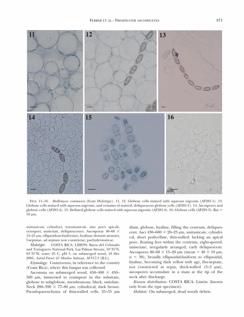
Commentary: Bullimyces communis was the mostcommon freshwater ascomycete reported in this studyfrom Costa Rica with six collections. The presence ofglobose cells in the hamathecium was observedconsistently in all collections, although in water theglobose, chain-like cells deliquesce promptly (FIGS. 11–16). Another interesting aspect of this fungus is that theasci were never observed to be arranged in a hymeniallayer but instead floating among the globose cells.Bullimyces communis is characterized by three-septateascospores with gelatinous caps at both apices. Onecollection, AF281-6, had hyaline ascospores that becamedark yellow with age but was identical to the typespecimen in every other feature.
Bullimyces costaricensis A. Ferrer, A.N. Mill., C.Sarmiento et Shearer sp. nov. FIGS. 17–24
MycoBank MB561097Ascomata in ligno submerso, 450–490 3 450–500 mm,
immersa, globosa vel subglobosa, membranacea, nigra,ostiolati. Collum 380–390 3 77–80 mm, cylindricum, atro-brunneae. Centris ascocarpiorum cellulis psedoparenchyma-ticis, 35–55 mm diam, latae, globosis, hyalinis, deliquescenti-bus. Collum 280–390 3 77–80 mm. Asci 430–600 3 20–23 mm,
868 MYCOLOGIA

FIG. 2. Most likely tree from LSU nrDNA sequence data obtained with RAxML. Support values and highlighting asillustrated (FIG. 1).
FERRER ET AL.: FRESHWATER ASCOMYCETES 869
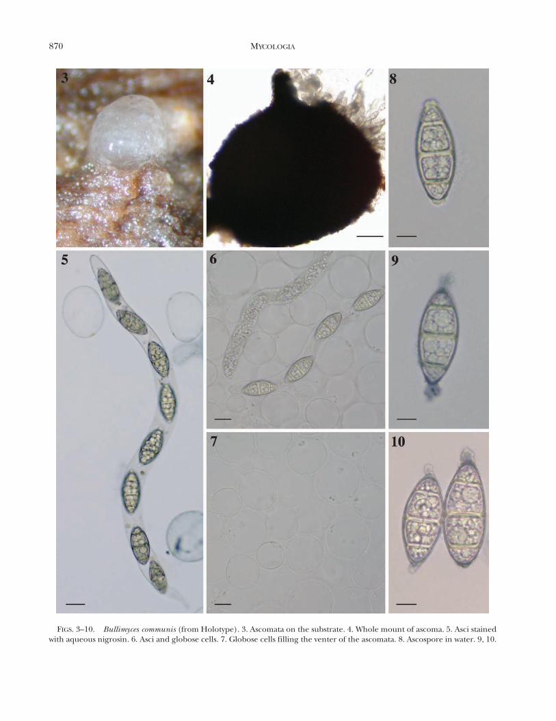
FIGS. 3–10. Bullimyces communis (from Holotype). 3. Ascomata on the substrate. 4. Whole mount of ascoma. 5. Asci stainedwith aqueous nigrosin. 6. Asci and globose cells. 7. Globose cells filling the venter of the ascomata. 8. Ascospore in water. 9, 10.
870 MYCOLOGIA
unitunicati, cylindrici, tenuitunicati, sine poro apicali,octospori, uniseriati, deliquescentes. Ascosporae 40–68 3
15–21 mm, ellipsoideae-fusiformes, hyalinae demum atrantes,5-septatae, ad septum non constrictae, pachydermaticae.
Holotype: COSTA RICA. LIMON: Barra del Coloradoand Tortuguero National Park, Las Palmas Stream, 10u359N,83u319W, water 25 C, pH 5, on submerged wood, 18 Dec2005, Astrid Ferrer & Marlon Salazar, AF317-1 (ILL).
Etymology: Costaricensis, in reference to the country(Costa Rica), where this fungus was collected.
Ascomata on submerged wood, 450–490 3 450–500 mm, immersed to erumpent in the substrate,globose to subglobose, membranous, black, ostiolate.Neck 280–390 3 77–80 mm, cylindrical, dark brown.Pseudoparenchyma of thin-walled cells, 35–55 mm
diam, globose, hyaline, filling the centrum, deliques-cent. Asci 430–600 3 20–23 mm, unitunicate, cylindri-cal, short pedicellate, thin-walled, lacking an apicalpore, floating free within the centrum, eight-spored,uniseriate, irregularly arranged, early deliquescent.Ascospores 40–68 3 15–20 mm (mean 5 48 3 19 mm,n 5 30), broadly ellipsoidal-fusiform to ellipsoidal,hyaline, becoming dark yellow with age, five-septate,not constricted at septa, thick-walled (3–5 mm),ascospores accumulate in a mass at the tip of theneck after discharge.
Known distribution: COSTA RICA: Limon (knownonly from the type specimen).
Habitat: On submerged, dead woody debris.
FIGS. 11–16. Bullimyces communis (from Holotype). 11, 12. Globose cells stained with aqueous nigrosin (AF281-1). 13.Globose cells stained with aqueous nigrosin, and remains of stained, deliquescent globose cells (AF281-1). 14. Ascospores andglobose cells (AF281-4). 15. Deflated globose cells stained with aqueous nigrosin (AF281-4). 16. Globose cells (AF281-5). Bar 5
10 mm.
FERRER ET AL.: FRESHWATER ASCOMYCETES 871

FIGS. 17–24. Bullimyces costaricensis. (AF317-1). 17. Ascomata on the substrate (note mass of hyaline ascospores. 18. Wholemount of ascoma. 19. Ascospore in water. 20. Elongated asci with eight ascospores. 21. Ascospore becoming pale brown withage. 22, 23, 24. Globose cells. Bar 5 10 mm.
872 MYCOLOGIA

Commentary: Bullimyces costaricensis is morphological-ly similar to the type species B. communis, with darkascomata, globose hamathecial cells, thin-walled ascilacking an apical pore, and thick-walled, hyaline, multi-septate ascospores. However B. costaricensis differs fromB. communis in ascus and ascospore morphology. Theasci of B. costaricensis are particularly long (430–600 mmcompared to 310–430 mm in B. communis), and theascospores of B. costaricensis are larger (40–68 mm long inB. costaricensis compared to 40–48 mm in B. communis),have more septa and lack appendages.
Bullimyces aurisporus A. Ferrer, A.N. Mill., C.Sarmiento et Shearer sp. nov. FIGS. 25–29
MycoBank MB561098Ascomata in ligno submerso, 260–310 3 290 mm, partim
immersa vel immersa, subglobosa vel obpyriforma, mem-
branacea, nigra, ostiolati. Collum 224 3 62 mm, cylindricum,
atrobrunneae. Asci deliquescentes. Ascosporae 34–38 3 17–
20 mm, ellipsoideae, lutea, three-septatae, ad septum non
constrictae, pachydermaticae, tunica gelatinosa praeditae.
Holotype: COSTA RICA. ALAJUELA: Cano NegroReserve, Cano Blanco stream 10u509N, 84u79W, water 26 C,pH 5, on submerged wood, 16 Dec 2005, Astrid Ferrer &Marlon Salazar, AF316-1 (ILL).
Etymology: From Latin Aurum 5 gold; Spora 5
spore, in reference to the color of the ascospores.Ascomata on submerged wood, 260–310 3 290 mm,
immersed to erumpent in the substrate, subglobose toobpyriform, membranous, black, ostiolate. Neckcylindrical, dark brown, 224 3 62 mm. Asci notobserved, early deliquescent. Ascospores 34–38 3
17–20 mm, ellipsoidal, dark yellow, three-septate, notconstricted at septa, thick-walled (2.0 mm), surround-
FIGS. 25–29. Bullimyces aurisporus (AF316-1). 25. Whole mount of ascoma. 26 & 27. Ascospores in water showing gelatinoussheath. 28. Germinating ascospores. 29. Top view of ascospores. Bar 5 10 mm.
FERRER ET AL.: FRESHWATER ASCOMYCETES 873

FIGS. 30–38. Riomyces rotundus from the holotype (AF303-1). 30. Whole mount of ascoma. 31. Neck. 32. Asci surroundedby globose cells. 33. Unitunicate ascus. 34. Empty Asci. 35, 36. Globose cells. 37, 38. Ascospores showing gelatinous sheathexpanding in water. Bar 5 10 mm.
874 MYCOLOGIA

FIGS. 39–46. Hydromelitis pulchella from the holotype (AF284-1). 39. Ascomata on the substrate. 40. Whole mount ofascoma. 41. Paraphyses. 42, 45. Young ascus. 43. Ascospores in water. 44. Ascospore becoming pale yellow with age. 46.Ascospore stained with aqueous nigrosin. Bar 5 10 mm.
FERRER ET AL.: FRESHWATER ASCOMYCETES 875

ed by an irregular gelatinous sheath about 3–5 mmwide, ascospores accumulate in a mass at the tip of theneck after discharge.
Known distribution: COSTA RICA: Alajuela (knownonly from the type specimen).
Habitat: On submerged, dead woody debris.Commentary: Bullimyces aurisporus is morphologi-
cally similar to the type species of the genus, B.communis, with dark ascomata and thick-walled, three-septate ascospores. However the ascospores of B.aurisporus are shorter (34–38 mm in B. aurisporuscompared to 40–48 mm in B. communis) and have athick, irregular mucilaginous sheath surrounding theascospore. Although we do not have informationabout ascus and hamathecium morphogy, moleculardata support its placement in this genus. We were notable to induce sexual reproduction in culture for B.aurisporus.
Riomyces A. Ferrer, A.N. Mill., C. Sarmiento etShearer gen. nov.
MycoBank MB561099Ascomata in ligno submerso, partim immersa vel im-
mersa, globosa ad subglobosa, membranacea, nigra, ostio-lati. Collum breve, atrobrunneae. Hamathecium ascocar-piorum cellulis psedoparenchymaticis, globosis, hyalinis,deliquescentibus. Asci unitunicati, cylindrici, tenuitunicati,breve deicellati, sine poro apicali, octospori, uniseriati.Ascosporae, ellipsoideo-fusiformes, hyalinae, septatae, adseptum non constrictae, pachydermaticae, tunica gelatinosapraeditae.
Typus generic: Riomyces rotundus A. Ferrer, A.N. Mill.,C. Sarmiento et Shearer
Etymology: From the Spanish word Rıo 5 river +Greek Myces 5 fungus, in reference to the freshwaterhabitat of the fungus.
Ascomata on submerged wood, immersed toerumpent in the substrate, globose to subglobose,membranous, black, ostiolate. Neck short, darkbrown. Hamathecium composed of globose, hyaline,deliquescent cells. Asci unitunicate, broadly cylindri-cal, thin-walled, short-pedicellate, lacking an apicalpore and other apical structures, eight-spored, uni-seriate. Ascospores ellipsoidal-fusiform, hyaline, sep-tate, not constricted at septa, thick-walled, surround-ed by a gelatinous sheath.
Riomyces rotundus A. Ferrer, A.N. Mill., C. Sarmientoet Shearer sp. nov. FIGS. 30–38
MycoBank MB561100Ascomata in ligno submerso, 440–720 3 440–600 mm,
partim immersa vel immersa, globosa vel subglobosa,membranacea, nigra, ostiolati. Collum 76 3 84 mm, breve,atrobrunneae. Hamathecium ascocarpiorum cellulis psedo-parenchymaticis, 35–100 mm diam, globosis, hyalinis, deli-
quescentibus. Asci 250–340 3 25–29 mm, unitunicati,cylindrici, tenuitunicati, breve deicellati, sine poro apicali,octospori, uniseriati, deliquescentes. Ascosporae 30–40 3
20–25 mm, ellipsoideo-fusiformes, hyalinae, 3-septatae, adseptum non constrictae, pachydermaticae, tunica gelatinosapraeditae.
Holotype: COSTA RICA. HEREDIA: La Selva Biologi-cal Station, Arboleda Stream 10u259N, 84u009W, water 25 C,pH 7, on submerged wood, 9 Jan 2006, Marlon Salazar,AF303-1 (ILL).
Etymology: From Latin rotunda 5 round, globular,in reference to the cells of the hamathecium.
Ascomata on submerged wood, 440–720 3 440–600 mm, immersed to erumpent in the substrate,globose to subglobose, membranous, black, ostiolate.Neck short, dark brown, 76 3 84 mm. Hamatheciumcomposed of globose, wide, hyaline, deliquescentcells, 35–100 mm diam. Asci 250–340 3 25–30 mm,unitunicate, broadly cylindrical, thin-walled, with ashort pedicel, lacking an apical pore and other apicalstructures, eight-spored, uniseriate. Ascospores 30–403 20–25 mm (mean 5 35 3 22 mm, n 5 30), broadlyellipsoidal-fusiform, hyaline, three-septate, not con-stricted at septa, thick-walled (4–5 mm), surroundedby an irregular gelatinous sheath extending about ca.10–20 mm from the ascospore wall; sheath marginsstain in aqueous nigrosin, ascospores accumulate in amass at the tip of the neck after discharge.
Known distribution: COSTA RICA: Alajuela andHeredia.
Habitat: On submerged, dead woody debris.Additional specimens examined: COSTA RICA. HEREDIA:
La Selva Biological Station, Esquina Stream, 10u249N,84u009W, water 25 C, pH 5, on submerged wood, 9 Jan2006, Marlon Salazar, AF303-2. ALAJUELA: Cano NegroReserve, Rıo Frıo, 10u539N, 84u459W, water 27 C, pH 5, onsubmerged wood, 15 Dec 2005, Astrid Ferrer & MarlonSalazar, AF303-3.
Commentary: Riomyces rotundus is morphologicallysimilar to species in the genus Bullimyces in havingdark ascomata, the unique hyaline, globose cells fillingthe centrum, thin-walled asci lacking an apical pore,and thick-walled, hyaline, multiseptate ascospores. Itwas observed in this fungus that the asci were attachedto a hymenial layer but with time the asci becomeunattached and surrounded by the globose cells fillingthe centrum, some empty asci were observed stillattached to the hymenium (FIG. 34).
Hydromelitis A. Ferrer, A.N. Mill., C. Sarmiento etShearer gen. nov.
MycoBank MB561101Ascomata in ligno submerso, partim immersa vel immersa,
pyriformia, membranacea, nigra, ostiolati. Paraphysibussimplicis, sepatatis. Asci unitunicati, clavati, tenuitunicati,breve deicellati, sine poro apicali, octospori, biseriati vel
876 MYCOLOGIA

interdum triseriati. Ascosporae, ellipsoideae, hyalinae velflavus, septatae, ad septum non constrictae, pachydermati-cae, tunica gelatinosa praeditae.
Ascomata on submerged wood, immersed toerumpent, pyriform, membranous, black, ostiolate.Paraphyses simple, thin-walled, septate. Asci unituni-cate, clavate, thin-walled, short-pedicellate, lacking anapical pore and other apical structures, eight-spored,biseriate. Ascospores ellipsoidal, hyaline to goldenyellow, multiseptate, septa with or without a centralchannel, not constricted at septa, thick-walled, sur-rounded by a gelatinous sheath.
Typus generic: Hydromelitis pulchella A. Ferrer, A.N.Mill., C. Sarmiento et Shearer.
Etymology: From Latin Hydromelitis 5 honey-water,in reference to the color of the ascospores and thespore mass on the wood.
Hydromelitis pulchella A. Ferrer, A.N. Mill., C.Sarmiento et Shearer sp. nov. FIGS. 39–46
MycoBank MB561102Ascomata in ligno submerso, 220–280 3 190–250 mm,
partim immersa vel immersa, pyriformia, membranacea,nigra, ostiolati. Paraphysibus 3–5 mm diam, simplicis,sepatatis, hyalinis. Asci 150–170 3 30–33 mm, unitunicati,clavati, tenuitunicati, breve deicellati, sine poro apicali,octospori, biseriati vel interdum triseriati. Ascosporae 35–513 13–18 mm, ellipsoideae, hyalinae vel flavus, 6-septatae, adseptum non constrictae, pachydermaticae, tunica gelatinosapraeditae.
Holotype: COSTA RICA. ALAJUELA: Cano NegroReserve, Rıo Frıo 10u539N, 84u459W, water 27.5 C, pH 5, onsubmerged wood, 15 Dec 2005, Astrid Ferrer & MarlonSalazar, AF284-2 (ILL).
Etymology: From Latin Pulchella 5 beautiful, inreference to the ascospores.
Ascomata on submerged wood, 220–280 3 190–250 mm, immersed to erumpent in the substrate,pyriform, membranous, black, ostiolate. Paraphysescylindrical, 3–5 mm diam, simple, thin-walled, septate,hyaline. Asci 150–210 3 30–33 mm, unitunicate,clavate, thin-walled, with a short pedicel, lacking anapical pore and other apical structures, eight-spored,biseriate, occasionally triseriate. Ascospores 35–51 3
13–18 mm, ellipsoidal, hyaline to golden yellow, mostlysix-septate (3–7), with or without a central channel inthe septal walls, not constricted at septa, thick-walled(up to 5 mm); ascospores surrounded by a gelatinoussheath that stains in aqueous nigrosin, 3–7 mm wide atlateral walls of ascospores, tapering toward the apices,ascospores accumulate in a yellow mass at the tip ofthe neck after discharge.
Known distribution: COSTA RICA: Alajuela andHeredia.
Habitat: On submerged, dead woody debris.
Additional specimens examined: HEREDIA: La SelvaBiological Station, El Sura Stream 10u259N, 84u009W, water25 C, pH 7, on submerged wood, 10 Jan 2006, MarlonSalazar, AF284-1; La Selva Biological Station, Panteno,10u25980N, 84u09220W, water 25 C, pH 5.5, on submergeddecorticated wood, 19 May 2000, Jennifer L. Anderson,Rebecca Wulffen, A468-1; La Selva Biological Station, RioPuerto Viejo, 10u259480N, 84u09170W, water 23 C, pH 5.5, onsubmerged, soft, decorticated wood, 20 May 2000, JenniferL. Anderson, Rebecca Wulffen, A468-2; La Selva BiologicalStation, Piper, 10u259570N, 84u19440W, on submergeddecorticated wood, 6 Feb 2001, Cathy Pringle, A468-3; LaSelva Biological Station, Arboleda 30, 10u259470N,84u09390W, submerged, soft, decorticated wood, 17 May2000, Jennifer L. Anderson, Rebecca Wulffen, A468-4.
Commentary: Hydromelitis pulchella is morphologi-cally similar to the type species of Bullimyces, B.communis, with dark ascomata, thin-walled asci lackingan apical pore, and thick-walled, multiseptate asco-spores with a sheath. However H. pulchella differs fromB. communis in ascomal and hamathecial morphology.The ascomata of H. pulchella are pyriform comparedto globose in B. communis, and the hamathecium ofH. pulchella is formed by simple, thin-walled, septate,hyaline paraphyses, while B. communis has hyaline,globose cells filling the centrum.
DISCUSSION
Bullimyces.—This genus with three new speciesrepresents a distinct taxonomic entity based onmorphological and molecular data. The three newspecies placed within Bullimyces share several mor-phological characteristics including globose to sub-globose, membranous, black, ostiolate ascomata,hyaline, globose cells filling the centrum, unitunicateasci; and multiseptate, thick-walled ascospores, whichdarken with age into a golden color, with or without agelatinous sheath or appendages. Nonetheless Bulli-myces species can be identified based on ascosporemorphology and septation. B. communis is character-ized by three-septate ascospores with gelatinous capsat both apices; ascospores of B. costaricensis are five-septate and lack appendages, and B. aurisporus hasthree-septate ascospores and a mucilaginous sheathsurrounding the ascospore.
Bullimyces is very similar to Riomyces describedherein in ascomata and ascus morphology, but thecells filling the centrum in Riomyces are irregular andhave thicker walls. Bullimyces and Riomyces bothgroup with members of the Sordariomycetidae butwere not sister taxa. Bullimyces shows affinities toCeratostomella pyrenaica based on SSU and LSU dataand to Ceratostomella pyrenaica, Rhamphoria deliculataand Xylomelasma sordida based on LSU data. Thesegenera have similar ascomatal morphology to Bulli-
FERRER ET AL.: FRESHWATER ASCOMYCETES 877
myces with dark, nonstromatic ascomata with acylindrical neck, but otherwise they differ greatly inthat these genera have asci with prominent apicalrings, septate paraphyses that are wide at the base andtapering at the apex and mostly thin-walled asco-spores (Reblova 2006). Furthermore Bullimyces andRiomyces differ in habitat from these other generabecause Bullimyces and Riomyces occur only infreshwater while Ceratostomella pyrenaica, Rhamphoriadeliculata and Xylomelasma sordida have been col-lected from decayed wood in terrestrial habitats.
The species of Bullimyces have a unique combina-tion of morphological characters, which makes itdifficult to place them taxonomically within theSordariomycetes without supporting molecular data.For instance Bullimyces shares some striking morpho-logical similarities to the freshwater genus LuttrelliaShearer, a member of the Halosphaeriaceae (Hypo-creomycetideae) (Shearer 1978). The genus Luttrelliacurrently accommodates four aquatic species (Shear-er 1978, Ferrer and Shearer 2007), with similarascomatal and ascospore morphology, however thehamathecium of Luttrellia is composed of septate,hyaline catenophyses, with the exception of Luttrelliahalonata, where catenophyses are hyaline, wide andglobose (Ferrer and Shearer 2007). Unfortunately nomolecular data are available at present for Luttrellia.Future molecular phylogenetic analysis may clarifywhether Luttrellia halonata shares affinities with thegenus Bullimyces.
Globose cells that fill the centrum of Bullimyces andRiomyces have been reported by Nakagiri and Tokura(1987) in the marine genus Corollospora (Halosphaer-iaceae). Corollospora has pseudoparenchyma of hya-line, thin-walled, rounded cells filling the center ofyoung ascocarps. Nonetheless they are distinct fromthose of Bullimyces and Riomyces because the pseudo-parenchyma cells of Corollospora are joined one cell toanother by pit connections (Nakagiri and Tokura1987), a feature not seen in Bullimyces.
Riomyces.—The position of Riomyces remains incertaesedis because Riomyces was basal to Bullimyces andgrouped with other members of the Ophiostomatalesand Magnaporthaceae without support (FIGS. 1, 2).Riomyces and Bullimyces may be closely related based ontheir morphological similarities. However additionaltaxon sampling in the LSU analyses revealed thatRiomyces and Hydromelitis also could be related tofreshwater ascomycetes in the Annulatascaceae sensulato and stricto. Unfortunately no SSU data are availablefor the Annulatascaceae, perhaps due to the difficulty insequencing this group (Ferrer pers comm).
Hydromelitis.—Among the Sordariomycetidae Hydro-melitis shows some similarities to the genus Bullimyces in
having multiseptate, thick-walled ascospores surround-ed by a gelatinous sheath that darkens into a goldencolor with age. However the ascomata of Hydromelitisare distinctly pyriform rather than globose as in mostBullimyces species. In addition the hamathecium ofHydromelitis are formed by simple, thin-walled, septate,hyaline paraphyses while Bullimyces species have hya-line, globose cells filling the centrum.
The placement of the new genera at the family level isuncertain. The combination of morphological charac-teristics found in Hydromelitis including the membra-nous, pyriform ascomata and phragmoseptate, thick-walled ascospores with a sheath are not present in anyfamily currently included in the Sordariomycetidae.
Freshwater ascomycetes possess unique morpholog-ical characteristics that might be adaptive to freshwaterhabitats, such as ascospores with elaborate appendagesand sheaths (Shearer 1993, Jones 2006). Also in theaquatic Dothideomycetes order Jahnulales the vegeta-tive hyphae are almost 10 times wider than otherhyphae reported in the Ascomycota (Inderbitzin et al.2001, Raja and Shearer 2006). We are of the opinionthat these wide hyphae aid the attachment of theascomata to the soft, decorticated, woody substrateswhile in water, analogous to an anchor. Bullimyces andRiomyces possess wide, globose cells in the hamathe-cium that might be adaptive in providing buoyancy tothe asci and the ascospores when they are dischargedinto water. The functional significance of these traitsremains unclear, but we hypothesize that these globosecells may be important for dispersion and establish-ment of fungi on wood in flowing water.
ACKNOWLEDGMENTS
We thank Marlon Salazar, Jennifer Anderson, RebeccaWulffen and Catherine Pringle for great help withcollecting. We appreciate the Organization for TropicalStudies (OTS) for logistic support to collect in Costa Rica.We thank Huzefa Raja for reviewing the manuscript beforesubmission. Financial support by the National ScienceFoundation (NSF grants Nos. DEB 0316496, DEB0515558) is gratefully acknowledged. Any opinions, findingsand conclusions or recommendations expressed in thispublication are those of the author(s) and do notnecessarily reflect the views of the National ScienceFoundation.
LITERATURE CITED
Edgar R. 2004. MUSCLE: multiple sequence alignment withhigh accuracy and high throughput. Nucleic Acids Res32:1792–1797, doi:10.1093/nar/gkh340
Ferrer A, Shearer CA. 2005. New records and a new speciesof Canalisporium from aquatic habitats in Panama.Mycotaxon 93:179–188.
878 MYCOLOGIA

———. 2007. Three new species of Luttrellia fromtemperate and tropical freshwater habitats. Mycologia99:144–151, doi:10.3852/mycologia.99.1.144
Galtier N, Gouy M, Gautier C. 1996. SeaView and Phylo_win:two graphic tools for sequence alignment and molec-ular phylogeny. Comput Appl Biosci 12:543–548.
Gouy M, Guindon S, Gascuel O. 2010. SeaView 4: amultiplatform graphical user interface for sequencealignment and phylogenetic tree building. Mol BiolEvol 27:221–224, doi:10.1093/molbev/msp259
Huelsenbeck JP, Mark PVD, Ronquist F. 2001. MrBayes:Bayesian inference of phylogenetic trees.3.1.2. http:mrbayes.csit.fsu.edu/download.php [Accessed Jan2009].
———, Ronquist FR. 2001. MrBayes: Bayesian inference ofphylogenetic trees. Biometrics 17:754–755.
Inderbitzin P, Landvik S, Abdel-Wahab A, Berbee ML. 2001.Aliquandostipitaceae, a new family for two new tropicalascomycetes with unusually wide hyphae and dimor-phic ascomata. Am J Bot 88:52–61, doi:10.2307/2657126
Jones EBG. 1994. Ultrastructure and taxonomy of theaquatic ascomycetous order Halosphaeriales. Can J Bot73(Suppl. 1):S790–S801.
———. 2006. Form and function of fungal spore append-ages. Mycoscience 47:167–183, doi:10.1007/s10267-006-0295-7
———, Sakayaroj J, Suetrong S, Somrithipol S, Pang KL.2009. Classification of marine Ascomycota, anamorphicand Basidiomycota. Fungal Divers 35:1–187.
Nakagiri A, Tokura R. 1987. Taxonomic studies of the genusCorollospora (Halosphaeriales, Ascomycotina) with de-scriptions of seven new species. Trans Mycol Soc Japan28:413–436.
Posada D, Crandall KA. 1998. Modeltest: testing the modelof DNA substitution. Bioinformatics 49:817–818,doi:10.1093/bioinformatics/14.9.817
Promputtha I, Miller AN. 2010. Three new species ofAcanthostigma (Tubeufiaceae, Pleosporales) from theGreat Smoky Mountains National Park. Mycologia 102:574–587, doi:10.3852/09-051
Raja HA, Shearer CA. 2006. Jahnula species from North andCentral America, including three new species. Mycolo-gia 98:312–332, doi:10.3852/mycologia.98.2.319
Rehner SA, Samuels GJ. 1994. Taxonomy and phylogeny ofGliocladium analysed from nuclear large subunit
ribosomal DNA sequences. Mycol Res 98:625–634,doi:10.1016/S0953-7562(09)80409-7
Reblova M. 2006. Molecular systematics of Ceratostomellasensu lato and morphologically similar fungi. Mycolo-gia 98:68–93, doi:10.3852/mycologia.98.1.68
———. 2009. Teleomorph of Rhodoveronae (Sordariomyce-tidae) discovered and re-evaluation of Pleurophrag-mium. Fungal Divers 36:129–139.
Shearer CA. 1978. Fungi of the Chesapeake bay and itstributaries VII. Luttrellia estuarina gen. et sp. nov.(Ascomycetes) Mycologia 70:692–697.
———. 1993. The freshwater ascomycetes. Nova Hedwig 56:1–33.
Spatafora JW, Volkmann-Kohlmeyer B, Kohlmeyer J. 1998.Independent terrestrial origins of the Halosphaeriales(Marine Ascomycota). Am J Bot 85:1569–1580,doi:10.2307/2446483
Stamatakis A. 2006. RAxML-VI-HPC: maximum likelihood-based phylogenetic analyses with thousands of taxa andmixed models. Bioinformatics 22:2688–260, doi:10.1093/bioinformatics/btl446
Swofford DL. 2002. PAUP*4: phylogenetic analysis usingparsimony (*and other methods). Sunderland, Massa-chusetts: Sinauer Associates.
Vijaykrishna D, Jeewon R, Hyde KD. 2006. Moleculartaxonomy, origins and evolution of freshwater ascomy-cetes. Fungal Divers 23:367–406.
Vilgalys R, Hester M. 1990. Rapid identification andmapping of enzymatically amplified ribosomal DNAfrom several Cryptococcus species. J Bacteriol 172:4238–4246.
White TJ, Bruns T, Lee S, Taylor J. 1990. Amplification anddirect sequencing of fungal ribosomal RNA genes forphylogenetics. In: Innis MA, Gelfand DH, Sninsky JS,White TJ, eds. PCR protocol: a guide to methods andapplications. San Diego: Academic Press. p 315–322.
Wiens JJ. 1998. Combining datasets with different phyloge-netic histories. Syst Biol 57:568–581, doi:10.1080/106351598260581
Zhang N, Castlebury LA, Miller AN, Huhndorf S, SchochCL, Seifert KA, Rossman AY, Rogers JD, Kohlmeyer J,Volkmann-Kohlmeyer B, Sung G–H. 2006. An overviewof the systematics of the Sordariomycetes based on afour-gene phylogeny. Mycologia 98:1076–1087,doi:10.3852/mycologia.98.6.1076
FERRER ET AL.: FRESHWATER ASCOMYCETES 879