The Ultimate Healing Beam: The Future is Now John Han-Chih Chang, M D Radiation Oncologist CDH...
50
The Ultimate Healing The Ultimate Healing Beam: Beam: The Future is Now The Future is Now John Han-Chih Chang John Han-Chih Chang , , M M D D Radiation Oncologist CDH Proton Center, a ProCure Center Primary Investigator for the Radiation Therapy Oncology Group Primary Investigator for the Children’s Oncology Group Children’s Memorial Hospital Vice Chair of the Midwest Children’s Brain Tumor Clinic
-
Upload
alexia-kerry-jackson -
Category
Documents
-
view
216 -
download
0
Transcript of The Ultimate Healing Beam: The Future is Now John Han-Chih Chang, M D Radiation Oncologist CDH...

The Ultimate Healing Beam:The Ultimate Healing Beam:The Future is NowThe Future is Now
The Ultimate Healing Beam:The Ultimate Healing Beam:The Future is NowThe Future is Now
John Han-Chih ChangJohn Han-Chih Chang, , MMDDRadiation Oncologist
CDH Proton Center, a ProCure CenterPrimary Investigator for the Radiation Therapy Oncology Group
Primary Investigator for the Children’s Oncology GroupChildren’s Memorial Hospital
Vice Chair of the Midwest Children’s Brain Tumor Clinic

Road MapRoad Map
Background History – When and Where Proton Mechanics – How to Applications/Prostate Cancer – What for Conclusion

High End Image Guided High End Image Guided Glorified Tanning BoothsGlorified Tanning Booths

Modality and Delivery Must Work Modality and Delivery Must Work TogetherTogether
Techniques to Improve Radiation Delivery
Radiation Modality
OPTIMAL RADIATION THERAPY

Protons through the AgesProtons through the Ages
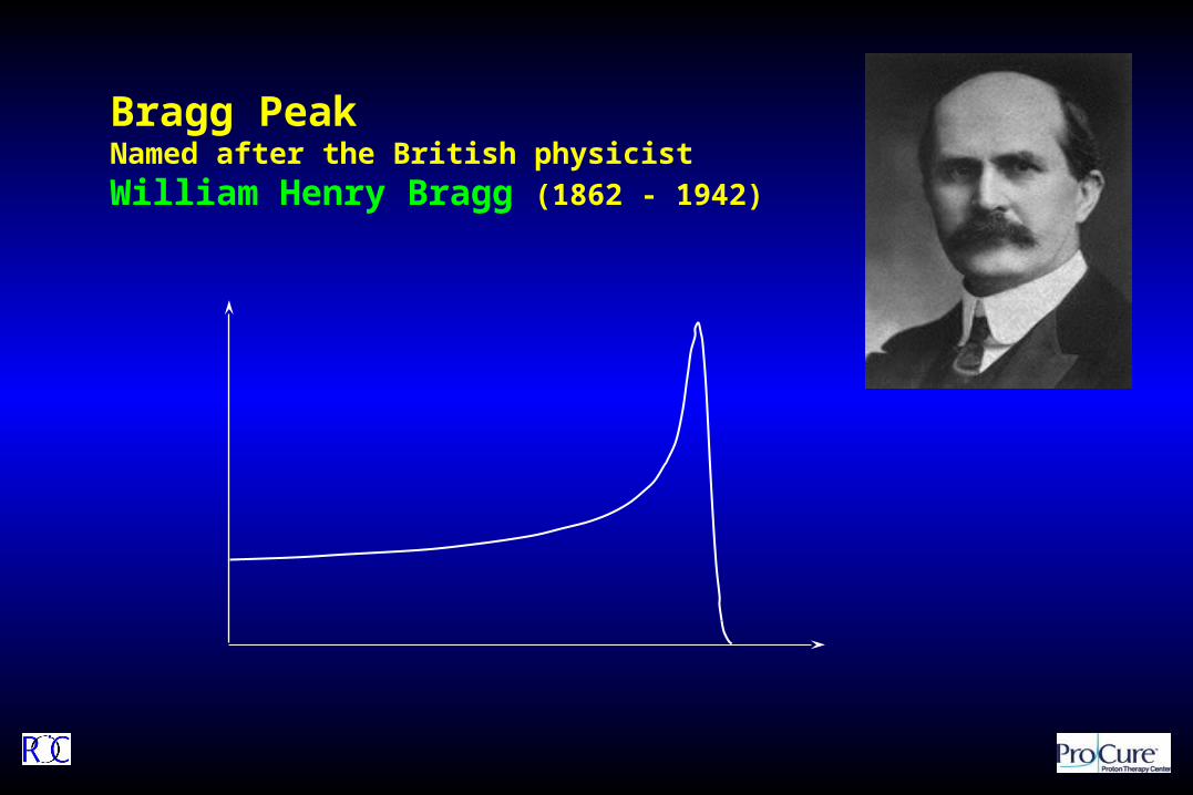
Bragg PeakNamed after the British physicist
William Henry Bragg (1862 - 1942)

R.R. Wilson, Radiology 1946; 47:487-491
Hydrogen Atom
Protons: “Ancient” History 101
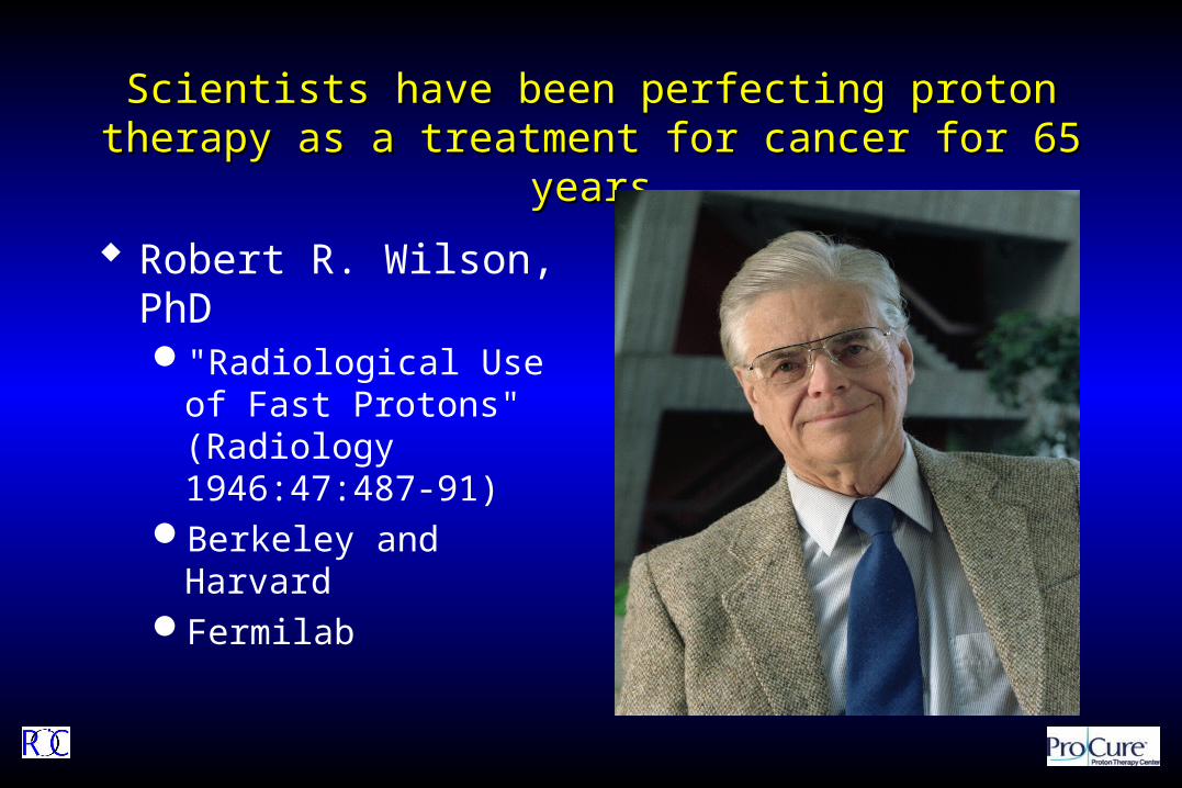
Scientists have been perfecting proton therapy as a Scientists have been perfecting proton therapy as a treatment for cancer for 65 yearstreatment for cancer for 65 years
Robert R. Wilson, PhD"Radiological Use of Fast
Protons" (Radiology 1946:47:487-91)
Berkeley and HarvardFermilab

1946 – Robert Wilson proposes using protons clinically
1955 – The first patient is treated at Berkley
1961 – The Harvard Cyclotron Lab (HCL) begins therapy
1991 – Loma Linda (LL) operates the first proton gantry
2001 – HCL closes NPTC opens
Protons: History 101
10
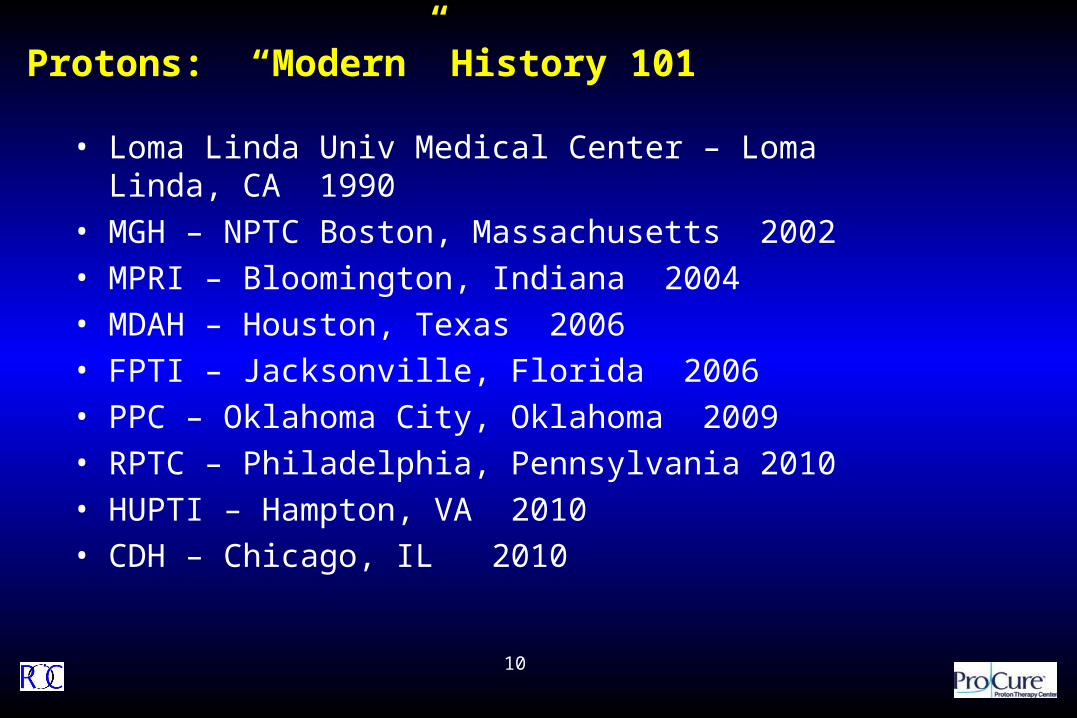
Protons: “Modern” History 101
• Loma Linda Univ Medical Center – Loma Linda, CA 1990
• MGH – NPTC Boston, Massachusetts 2002
• MPRI – Bloomington, Indiana 2004
• MDAH – Houston, Texas 2006
• FPTI – Jacksonville, Florida 2006
• PPC – Oklahoma City, Oklahoma 2009
• RPTC – Philadelphia, Pennsylvania 2010
• HUPTI – Hampton, VA 2010
• CDH – Chicago, IL 2010

Proton therapy found its first clinical home in CaliforniaProton therapy found its first clinical home in California Loma Linda
First patient 1990
First facility designed as patient treatment center
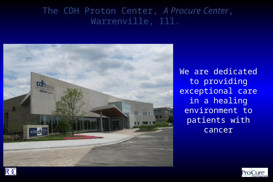
We are dedicated to providing exceptional
care in a healing environment to patients
with cancer
The CDH Proton Center, A Procure Center, Warrenville, Ill.

Mechanism of ActionMechanism of Action

Protons have Fewer Side Effects than PhotonsProtons have Fewer Side Effects than Photons
In order for photons to reach a prescribed dose at the tumor depth, healthy tissue gets four times the radiation as the tumor
Protons put 80% of their energy into the tumor and only 20% into healthy tissue
Protons deposit more than 80% of their energy in the tumor
There is no reason to irradiate healthy tissue
Lower
Higher
Rad
iati
on
Do
se
20%70% 10%
Photons deposit only 20% of their energy in the tumor
Prescribed Doseto Kill Tumor
Depth in Tissue
Photons deposit only 20% of their energy in the tumor
Lower
Higher
Ra
dia
tio
n D
os
e
80%20% 0%Prescribed Dose
to Kill Tumor
Depth in Tissue

0 5 10
15
20
25
30
0
20
40
60
80
100
Depth in Tissue (cm)
Rela
tive D
ose
High Energy X-Rays
200 MeV Protons
Spread Out Bragg Peak (SOBP)
Tumor
The Physics of ProtonsThe Physics of Protons
Depth Dose Curves for Different Treatment Types
Healthy Tissue Healthy Tissue

Protons are physically superior to X-rays
Protons behave differently than x-rays:
Protons
X-Rays do not
Protons improve the “therapeutic ratio”
maximizing tumor control while minimizing side effects
At a given radiation dose to a tumor protons deliver, on average, less than half the radiation dose to normal tissues than do x-rays 1
The Value of ProtonsThe Value of Protons
16
(1) Jay Loeffler, Massachusetts General Hospital, “Proton Therapy 2009”
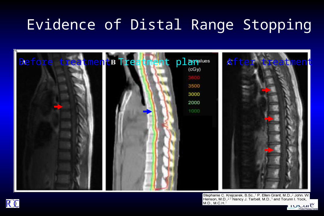
Evidence of Distal Range Stopping
Before treatment Treatment plan After treatment

Why would we chose Protons?Why would we chose Protons?
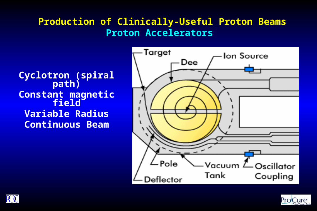
Cyclotron (spiral path)Constant magnetic field
Variable RadiusContinuous Beam
Production of Clinically-Useful Proton BeamsProton Accelerators

Treatment DeliveryTreatment Delivery
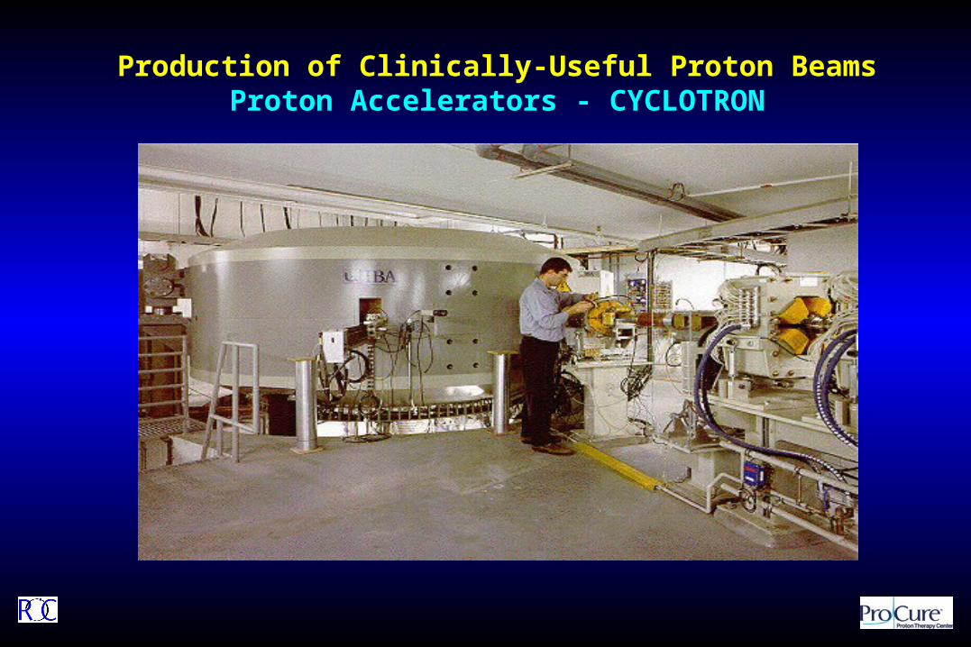
Production of Clinically-Useful Proton Beams Proton Accelerators - CYCLOTRON

Production of Clinically-Useful Proton Beams Beam Line
Energy Selection System (230 MeV 70 MeV)Beam Transport and Switching System

Nozzle Snout (with aperture & compensator)
6-axis patient positioner
Gantry 1 NPTC- Harvard

Robotic TableRobotic Table
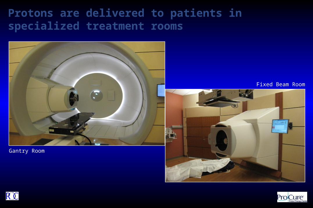
Protons are delivered to patients in specialized treatment rooms
Gantry Room
Fixed Beam Room

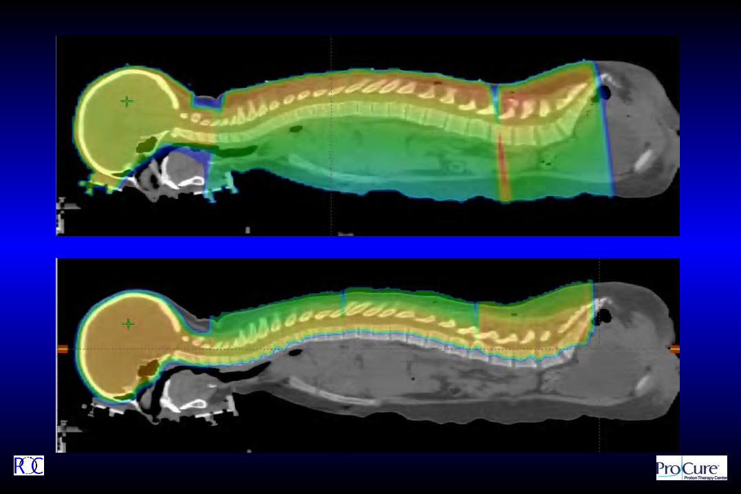
Clinical IndicationsClinical Indications
CurrentPediatrics
Paraspinal Ewing’s Optic pathway glioma Optic nerve meningioma Prostate/Pelvic RMS Exophytic BSG Craniospinal irradiation Suprasellar NGGCT
ProstateHead and Neck/Base of Skull Intracranial
MeningiomaParaspinal/Sacrum
Chordoma
PlannedLung
Organ motion Density changes
Tumor response Inspiration: Expiration
GI Organ motion Density changes
Breast - APILymphomaOcular

Prostate CancerProstate Cancer

Radiation Treatment OptionsRadiation Treatment Options
Radiation therapy options includeBrachytherapy (BT) is vastly underutilized
Effective Safe (in the appropriately selected patients) Convenient (1 to 2 day procedure)
External Beam Radiation Therapy (EBRT): Proton beam is superior to IMRT Higher cure rates Lower complication rates
Stereotactic Body Radiotherapy (SBRT) Effective Convenient (3 to 5 day non-invasive procedure) Safety seems to be similar to IMRT (it is still X-rays)

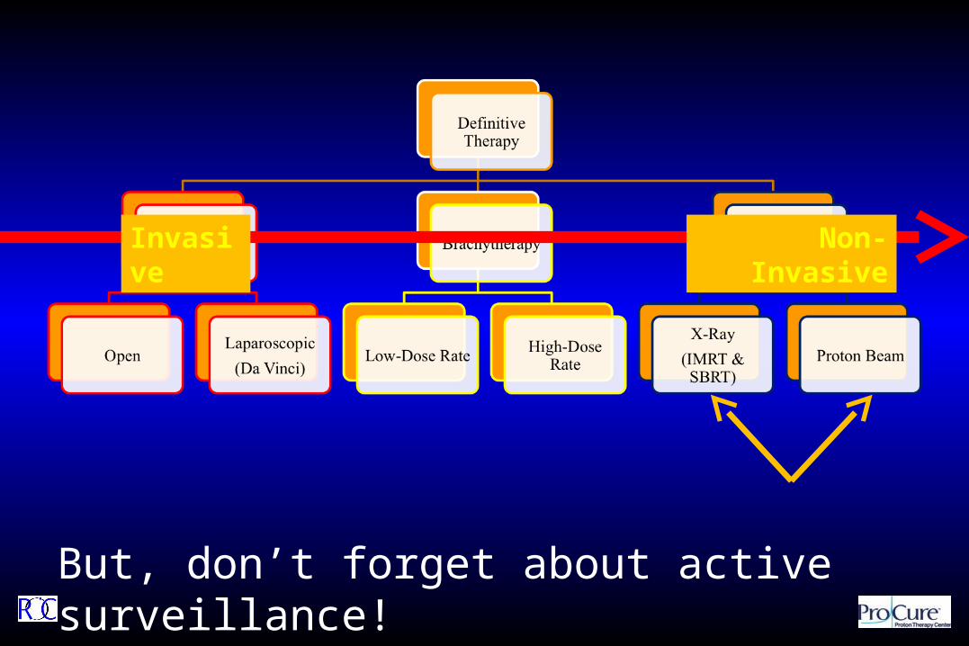
But, don’t forget about active surveillance!
Invasive Non-Invasive

Misconceptions About Misconceptions About Proton TherapyProton Therapy
“Just because someone keeps saying it doesn’t make it true.”
The typical quote: “The DVH of IMRT is better than the DVH of protons in the high dose region, and that’s what really counts”FACT: Protons, regardless of delivery method, and with
equivalent PTVs, should yield superior DVH curves without overlap
The typical quote: “There is no data showing protons have better control rates”FACT: Protons do have better controlBy definition, protons will never have worse control rates
than x-rays

Misconceptions About Misconceptions About Proton TherapyProton Therapy
“Just because someone keeps saying it doesn’t make it true.”
Patient access must not be based on misconceptions. We must rely on science and data to drive these decisions.
The typical quote: “There is no data showing that side effects and complications (“toxicity”) are lower with protons”FACT: At a similar treatment dose and volumes, the toxicity
is lower with protons The typical quote: “Protons are 2x to 5x times more expensive
than IMRT”FACT: Protons are at most 40% – 60% more than IMRT,
based on Medicare, and offer a much better valueThe lifetime costs of protons are much less than IMRT

Protons have Fewer Side Effects than PhotonsProtons have Fewer Side Effects than Photons
In order for photons to reach a prescribed dose at the tumor depth, healthy tissue gets four times the radiation as the tumor
Protons put 80% of their energy into the tumor and only 20% into healthy tissue
Protons deposit more than 80% of their energy in the tumor
There is no reason to irradiate healthy tissue
Lower
Higher
Rad
iati
on
Do
se
20%70% 10%
Photons deposit only 20% of their energy in the tumor
Prescribed Doseto Kill Tumor
Depth in Tissue
Photons deposit only 20% of their energy in the tumor
Lower
Higher
Ra
dia
tio
n D
os
e
80%20% 0%Prescribed Dose
to Kill Tumor
Depth in Tissue
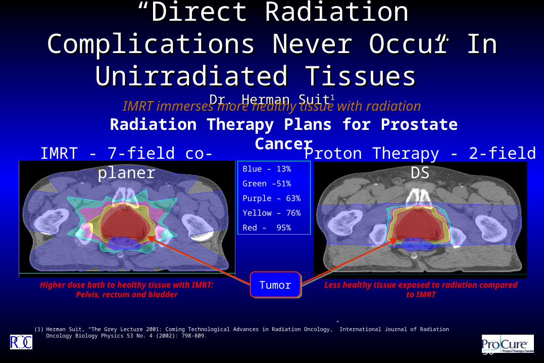
““Direct Radiation Complications Never Direct Radiation Complications Never Occur In Unirradiated Tissues” Occur In Unirradiated Tissues”
Dr. Herman SuitDr. Herman Suit11
IMRT - 7-field co-planer Proton Therapy - 2-field DS
Radiation Therapy Plans for Prostate Cancer
Less healthy tissue exposed to radiation compared to IMRT
Higher dose bath to healthy tissue with IMRT:Pelvis, rectum and bladder
Blue – 13%
Green – 51%
Purple – 63%
Yellow – 76%
Red – 95%
(1) Herman Suit, “The Grey Lecture 2001: Coming Technological Advances in Radiation Oncology,” International Journal of Radiation Oncology Biology Physics 53 No. 4 (2002): 798-809.
IMRT immerses more healthy tissue with radiation
TumorTumor
36
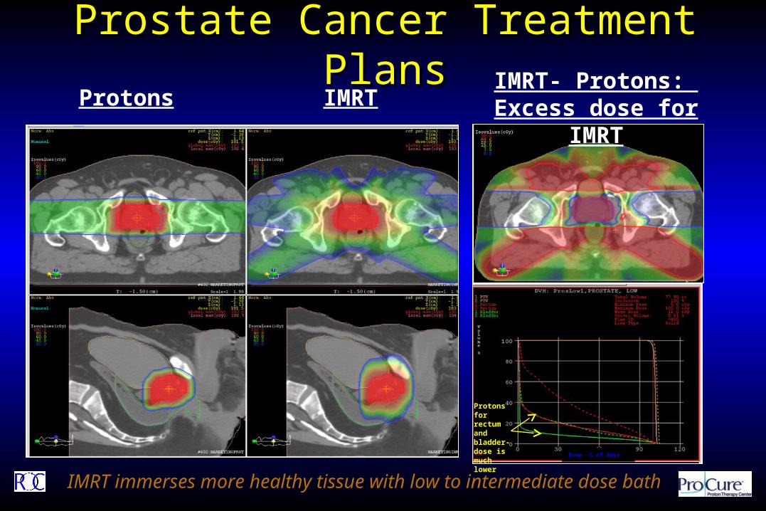
Prostate Cancer Treatment PlansProstate Cancer Treatment PlansProtons IMRT
Dose -% of dose
Protons for rectum and bladder-dose is much lower
IMRT- Protons: Excess dose for IMRT
IMRT immerses more healthy tissue with low to intermediate dose bath

The Data: Photons vs ProtonsThe Data: Photons vs Protons

Prostate CancerProstate Cancer
Modality Dose Recurrence Complication
Conventional Radiation <60 GY 38% 22%
Conventional Radiation 60 – 65 GY 36% 35%
Conventional Radiation 70 GY 28% 45%
Conventional Radiation >75 GY 20% 60%
Protons 75 GY 15% 12%
Source: Presentation by Dr. N. Mendenhall, University of Florida, IBA
Proton Therapy vs. Conventional Radiation (by dose) in Locally Advanced Prostate Cancer

RectumRectum
IMRT - MSK
3D CRT - MSK
IMRT - UFPTI
Proton - UFPTI
The limit of the photon modality
IMRT - MGH
Proton - MGH
Adapted from Zelefsky 2000, Trofimov 2007 and Vargas 2008

Rectal dose comparisonRectal dose comparisonIMRT plans
Rectum V70
MSKCC 14%
MGH 14.5%
MDACC 15.5%
UF 14%
Protons UF 8%
Zelefsky et al Radiotherapy and Oncology 2000; 55:241-249
Trofimov et al IJROBP 2007; 69:pp. 444–453,
Zhang et al IJROBP 2007; 67: 620–629
Vargas et al IJROBP 2008; 70: pp. 744–751

University of Florida Dosimetry Data Show Protons University of Florida Dosimetry Data Show Protons Reduce Dose To The Rectum By 59%Reduce Dose To The Rectum By 59%
IJROBP 2008Radiation dose to the rectum – proton therapy and IMRT1
Radiation Dose (CGE/Gy)0 10 20 30 40 50 60 70 80 90
0%
10%
20%
30%
40%
50%
60%
70%
80%
90%
Rec
tal V
olum
e R
ecei
ving
Rad
iati
on (
%)
Proton
IMRT
Dose to rectum is more than 2x with
IMRT vs. protons at 32 Gy
Background on study
First prostate patients seen at University of Florida Proton Therapy Institute (“UFPTI”)
Both proton and IMRT plans were planned prospectively for each patient
The results
Relative and absolute mean rectal dose savings of 59.2% and 20.1%, respectively, with proton therapy
Why this is important
Entire Dose Volume Histogram (“DVH”) does matter, not just high the dose region
– Rectal wall volume irradiated at 32.4 Gy is biggest predictor of rectal toxicity2
Extremely high correlation between rectal volume irradiation to 70 Gy and 5-year toxicity rates3
(1) Carlos Vargas et al., “Dose-Volume Comparison of Proton Therapy and Intensity-Modulated Radiotherapy for Prostate Cancer,” International Journal of Radiation Oncology Biology Physics 70 No.3 (2008): 744-751.(2) Susan Tucker, Lei Dong, Rex Cheung, et al., “Comparison of Rectal Dose-Wall Histogram Versus Dose-Volume Histogram for Modeling the Incidence of Late Rectal Bleeding After Radiotherapy,” International Journal of Radiation Oncology Biology Physics 60 (2004):
1589-1601.(3) Mark Storey, Alan Pollack, Gunar Zagars et al., “Complications from Radiotherapy Dose Escalation in Prostate Cancer: Preliminary Results of a Randomized Trial,” International Journal of Radiation Oncology Biology Physics 48 (2000): 635-642.
Dose to rectum is almost 2x with IMRT vs. protons at 70 Gy

GI (Rectal) Side Effects and ComplicationsGI (Rectal) Side Effects and Complications
43
Inflammation causedby radiation
Chronic Radiation Proctitis in the GI tract
Necrosis and ulcer
The probability of damage to the GI tract is much higherwith x-rays than protons
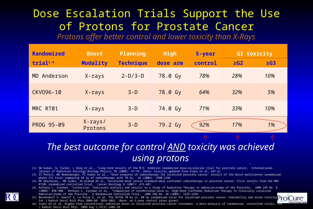
Dose Escalation Trials Support the Use of Protons for Dose Escalation Trials Support the Use of Protons for Prostate CancerProstate Cancer
Randomized Boost Planning High 5-year GI toxicity
trial1-4 Modality Technique dose arm control ≥G2 ≥G3
MD Anderson X-rays 2-D/3-D 78.0 Gy 78% 28% 10%
CKVO96-10 X-rays 3-D 78.0 Gy 64% 32% 5%
MRC RT01 X-rays 3-D 74.0 Gy 71% 33% 10%
PROG 95-09 X-rays/Protons 3-D 79.2 Gy 92% 17% 1%
(1) DA Kuban, SL Tucker, L Dong et al., “Long-term results of the M.D. Anderson randomized dose-escalation trial for prostate cancer,” International Journal of Radiation Oncology Biology Physics 70 (2008): 67-74. (Note: toxicity updated from Viani et al, ref 6)
(2) ST Peters, WD Heemsbergen, PC Koper et al., “Dose-response in radiotherapy for localized prostate cancer: results of the Dutch multicenter randomized phase III trial comparing 68 Gy of radiotherapy with 78 Gy,” 24 (2006): 1990-1196.
(3) DP Dearnaley, MR Sydes, JD Graham et al, “Escalated-dose versus standard-dose conformal radiotherapy in prostate cancer: first results from the MRC RT101 randomized controlled trial,” Lancet Oncology 8 (2007): 475-487.
(4) Anthony L. Zietman, “Correction: Inaccurate analysis and results in a Study of Radiation Therapy in Adenocarcinoma of the Prostate,” JAMA 299 No. 8 (2008): 898-900. Anthony L. Zietman et al., “Comparison of Conventional-Dose vs. High-Dose Conformal Radiation Therapy in Clinically Localized Adenocarcinoma of the Prostate. A Randomized Controlled Trial,” JAMA 294 No. 10 (2005): 1233-1239.
(5) Beckendorf V, Guerif S, Le Prise E, et al. The GETUG 70 Gy vs. 80 Gy randomized trial for localized prostate cancer: Feasibility and acute toxicity. Int J Radiat Oncol Biol Phys 2004;60: 1056–1065. (Note: no 5-year control rates given)
(6) Viani GA et al. Higher-than-conventional radiation doses in localized prostate cancer treatment: a meta-analysis of randomized, controlled trials. Int J Radiat Oncol Biol Phys. 2009 Aug 1;74(5):1405-18.
Protons offer better control and lower toxicity than X-Rays
The best outcome for control AND toxicity was achieved using protons

Reviewing the DataReviewing the DataParameter PROG 9509 MSK
Collection Prospective Retrospective
Institutions Multi-instituion Single-instituion
Follow-up >10 year 8 years
Photon RT 3D CRT IMRT
Image Guidance? No Yes
The Only Difference – Proton Boosti.e., this wasn’t even all protons – this was protons tacked onto what would be
considered, by today’s standards, inferior radiation therapy
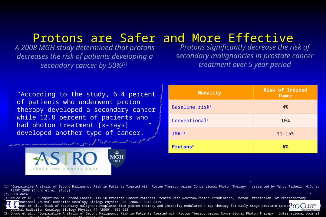
Protons are Safer and More EffectiveProtons are Safer and More Effective
“According to the study, 6.4 percent of patients who underwent proton therapy developed a secondary cancer while 12.8 percent of patients who had photon treatment [x-rays] developed another type of cancer.”
A 2008 MGH study determined that protons decreases the risk of patients developing a
secondary cancer by 50%(1)
(1) “Comparative Analysis of Second Malignancy Risk in Patients Treated with Proton Therapy versus Conventional Photon Therapy,” presented by Nancy Tarbell, M.D. at ASTRO 2008 (Chung et al. study)(2) SEER data(3) McGee et al., “Comparison of Second Cancer Risk in Prostate Cancer Patients Treated with Neutron/Photon Irradiation, Photon Irradiation, or Prostatectomy,” International Journal Radiation Oncology Biology Physics 66 (2006): S318-
S319(4) Fontenot et al., “Risk of secondary malignant neoplasms from proton therapy and intensity-modulated x-ray therapy for early-stage prostate cancer,” International Journal Radiation Oncology Biology Physics 74 (2009): 616-622(5) Chung et al., “Comparative Analysis of Second Malignancy Risk in Patients Treated with Proton Therapy versus Conventional Photon Therapy,” International Journal Radiation Oncology Biology Physics 72 (2008) :S8
Protons significantly decrease the risk of secondary malignancies in prostate cancer treatment over 5
year period
Modality Risk of Induced Tumor
Baseline risk2 4%
Conventional3 10%
IMRT4 11-15%
Protons5 6%

Prostate Cancer SummaryProstate Cancer Summary
Protons are AN option for prostate cancer treatment
Protons are superior to IMRT Protons are different from surgery and
brachytherapy Active surveillance is perfectly acceptable for
many men with prostate cancer Discussions should be had with patients about
ALL the options

Parting ShotsParting Shots Take home points:
All cancers should be approached in a multi-specialty or multi-disciplinary fashion
Patient care should be performed in team approach: Concierge/Receptionists, Nurses, Therapists,
Physicists/Dosimetrists, Physicians
State of the Art Radiation Therapy @ CDH/Procure FULL Spectrum of Radiation Treatment options
HDR Brachytherapy SBRT/SRS IMRT/3D CRT/IGRT Proton Beam Therapy

Photons/Electrons will still be needed Brachytherapy will still be utilized Image guidance will remain critical for all
modalities of radiation therapy Proton beam therapy can improve the side effects
profile in many of the disease we currently treat with photon radiation.
We are seeing just the tip of the iceberg
Parting ShotsParting Shots

Tumors we are and will be able to treat:
Neurologic
• Brain
• Spinal Cord
Other Solid Tumors
• Breast Cancer (2011)
• Lung Cancer (2011)
• Colorectal Cancer
• Prostate
Head / Neck
• Eye
• Sinus/nasal
• Throat
• Ear
Pediatric
• Brain
• Spinal Cord
• Bone

QuestionsQuestions