The Synthesis of Novel Kinase Inhibitors using Click Chemistry
160
The Synthesis of Novel Kinase Inhibitors using Click Chemistry Thesis presented in fulfilment of the requirements for the degree of Master of Science in the Faculty of Science at Stellenbosch University Supervisor: Prof Willem A. L. van Otterlo Co-supervisor: Dr Stephen C. Pelly Faculty of Natural Science Department of Chemistry and Polymer Science by Luke Hodson
Transcript of The Synthesis of Novel Kinase Inhibitors using Click Chemistry

The Synthesis of Novel Kinase Inhibitors using Click
Chemistry
Thesis presented in fulfilment of the requirements for the degree of
Master of Science in the Faculty of Science at Stellenbosch University
Supervisor: Prof Willem A. L. van Otterlo
Co-supervisor: Dr Stephen C. Pelly Faculty of Natural Science
Department of Chemistry and Polymer Science
April 2014
by
Luke Hodson

2
DECLARATION
By submitting this thesis electronically, I declare that the entirety of the work contained
therein is my own, original work, that I am the sole author thereof (save to the extent
explicitly otherwise stated), that reproduction and publication thereof by Stellenbosch
University will not infringe any third party rights and that I have not previously in its entirety
or in part submitted it for obtaining any qualification.
February 2014
Copyright © 2014 Stellenbosch University
All rights reserved
Stellenbosch University http://scholar.sun.ac.za

3
ABSTRACT
Cancer is the leading cause of death on the planet, killing an estimated 8.2 million people in
the year of 2012.The disease is associated with two families of genes, namely oncogenes and
tumour suppressor genes. The hallmarks of cancer pathogenesis include gene amplification,
point mutations or chromosomal rearrangements within these genes. Kinases are responsible
for the reversible phosphorylation of proteins, which plays a significant and extensive role in
cellular signal transduction. Aberrant kinase activity provokes overexpression, mutations and
chromosomal translocation and results in the onset of onco- and tumorogenesis, ultimately
leading to cancer.
Inactivation of this class of enzyme is thus critical as it would result in the suppression of
these unwanted activities. For this, researchers have developed kinase inhibitors, specifically
targeting these proteins and thus inhibiting signal transduction pathways and tumour growth.
This has resulted in great successes, particularly in the case of the commercial inhibitor,
imatinib. However, resistance to approved therapeutic agents through mutations has resulted
in the search for more potent and selective inhibitors to overcome these obstacles.
This project involved the synthesis of bioactive heterocycles linked to 1,2,3-triazoles using
either a C-C or C-N bond forming strategy. The synthetic methodology followed included the
use of Sonogashira coupling reactions between3-bromoquinoline, 7-chloro-4-iodoquinoline,
4-bromoisoquinolineand5-bromoisoquinolineand trimethylsilylacetylene (TMSA), followed
by deprotection of the TMS group to yield heterocycles bearing terminal alkynes. The
synthesis of both benzyl azide and 2-(azidomethyl)pyridine as azide fragments, allowed for
subsequent coupling of the synthesized azide and alkyne fragments through copper-mediated
click chemistry, affording a library of 1,4-substituted 1,2,3-triazole based reversible kinase
inhibitors. Synthesis of a second library of o-, m- and p-substituted nitro benzyl azides,
allowed for both copper- and ruthenium-mediated click reactions, between the alkynes and
nitro benzyl azides synthesized, to yield 1,4- and 1,5-substituted 1,2,3-triazoles, respectively.
Finally, reduction of the incorporated o-, m- and p- substituted nitro group, and acylation of
the resultant amine with acryloyl chloride, resulted in the incorporation of the important
Michael acceptor moiety required for irreversible inhibition. This afforded a library of both
reversible and potential irreversible triazole-based kinase inhibitors through efficient copper-
and ruthenium-mediated click chemistry.
Stellenbosch University http://scholar.sun.ac.za

4
Biological screening and activity assays against the wildtype, and two mutated forms of the
EGFR kinase, were undertaken with these synthesized compounds.A number of synthesized
inhibitors showed good selectivity for the mutated forms of the EGFR kinase only.The most
potent inhibitor N-{2-{[4-(isoquinolin-4-yl)-1H-1,2,3-triazol-1-
yl]methyl}phenyl}acrylamide,displayed efficacy in the low μM range - comparable to that of
the FDA approved drug, gefitinib.
The synthetic methodology derived in this project could be applied to the use of biological
space probes with further investigatory research. Furthermore, from the biological screening
results obtained, and the selectivity profile shown by these inhibitors, the synthesis of a
second generation library of compounds is an additional research possibility.
Stellenbosch University http://scholar.sun.ac.za

5
UITTREKSEL
Kanker is die hoof oorsaak van sterftes ter wêreld, wat verantwoordelik is vir die dood van
ongeveer 8.2 miljoen mense in die jaar 2012. Die siekte word geassosieer met twee
geenfamilies, naamlik onkogene en gewasonderdrukkingsgene. Die kenmerke van kanker
pathiogene behels geenversterking, puntmutasies of chromosomale herrangskikking binne in
die gene. Kinase is verantwoordelik vir die omkeerbare fosforilering van proteine wat n uiters
belangrike rol in sellulere sein transduksie speel. Abnormale kinase aktiwiteit lei tot
ooruitdrukking, mutasies en chromosomale translokasie wat tot die ontwikkeling van onko-
en gewasgroei en wat eindelik tot kanker lei.
Deaktivering van die klas van ensieme is dus krities want dit sal die ongewenste abnormale
aktiwiteite onderdruk. As gevolg van die bogenoemde, het navorsers kinase inhibeerders
ontwikkel wat die spesifieke protein teiken en hiermee die sein transduksie roete asook gewas
groei inhibeer. Hiermee het die sukses van inhibeerders veral die kommersiele inhibeerder,
imatinib, grootliks toegeneem. Oor die afgelope jare het die belangstelling in die
ontwikkeling van meer selektiewe en kragtige inhibeerders toegeneem as gevolg van die
weerstand wat goedgekeurde terapeutiese middels opbou.
In hierdie projek is daar gebruik gemaak van „n C-C of C-N bindingsvorming strategie om
bioaktiewe heterosikliese molekules te sintetiseer wat gekoppel is aan 1,2,3-triasool
funksionele groepe. Die sintetiese metode maak gebruik van Sonogashira reaksies vir die 3-
bromo-kwinolien, 7-chloro-4-iodokwinolien, 4-bromoisokwinolien en 5-bromoisokwinolien
met trimetielsilielasetileen (TMSA), gevolg met die ontskerming van die TMS-groep om die
terminale alkyn op die heterosiklusse te ontbloot. Die asied fragmente, bensiel asied en 2-
(asidometiel)piridien, was toe gesintetiseer om met die gevormde heterosiklus alkyne „n
koper ondersteunende kliek chemie te ondergaan. „n Reeks van 1,4-digesubstitueerde 1,2,3-
triasool gebaseerde omkeerbare kinase inhibitore is toe gevorm. „n Tweede reeks met o-, m-,
en p- gesubtitueerde nitro bensiel asiede was gesintetiseer om 1,4- en 1,5- digesubtitueerde
1,2,3-triasole te sintetiseer met behulp van koper- en ruthenium ondersteunende kliek chemie.
Laastens was die o-, m-, en p- nitro groepe gereduseer om „n primêre amien te vorm. Die
gevormende amien het „n asileringsreaksie met akriloïel chloried ondergaan om die kern, die
Michael akseptor, te inkorporeer. Die Michael akseptor word benodig om „n onomkeerbare
inhibitoriese aktiwiteit te kan uitvoer. Die projek het dus met behulp van kliek chemie, twee
Stellenbosch University http://scholar.sun.ac.za

6
1,2,3-triasool reekse gelewer wat omkeerbare en onomkeerbare inhibitoriese aktiwiteit kan
uitvoer.
Die verbindings gesintetiseerd in hierdie projek het keuringstoetse ondergaan teen die wilde
tipe en teen twee gemuteerde forme van die EGFR kinase ensiem. Van hierdie verbindings
het goeie selektiwiteit vertoon teenoor die gemuteerde EGFR kinase ensiem. Die mees
aktiewe inhibeerder, N-{2-{[4-isokwinolin-4-iel)-1H-1,2,3-triasool-1-iel]feniel}akrielamied,
het aktiwiteit in die lae µM reeks vertoon. Dié inhibisie waarde is vergelykbaar met die FDA
goedgekeurde medikasie, gefitinib.
In hierdie projek is sintetiese metodes ontwikkel wat toegepas kan word op meer intensiewe
biologiese ondersoeke en asook meer navorsing. Die resultate vekry van die biologiese
aktiwiteit, asook die verbindings se selektiwiteit, gee die moontlikheid vir die ontwikkeling
en sintese van „n tweede generasie verbindings.
Stellenbosch University http://scholar.sun.ac.za

7
ACKNOWLEDGEMENTS
Personal Acknowledgements
It is with a fond and gracious heart that I look back on the years of 2012 and 2013. I can‟t
think of a time that I‟ve learnt more about life than in the span of these last two years.
Throughout this time I‟ve acquired a great deal of memories, experiences, triumphs and
losses. Ive learnt that in chemistry, a passion and love for what you do is not enough. A
stalwart determination and patience is required in this field, something I am grateful to have
learnt. I like to think I am an adult now, or at least approaching that landmark. The very
toughness and unforgiving nature of research in organic chemistry - which has caused an
abundance of frustration and vexation in my life to put it mildly – has consequencely taught
me that nothing in life worth having, is not achieved through hard work. Smooth sailing
never made a skilled sailor, and it is through these hardships and trials and tribulations that
we learn and grow as human beings. I have my sweetest mistress, organic chemistry, to thank
for this.
At the top of my list I would like to thank my supervisor Professor Willem van Otterlo. You
have been a father figure to me in the time I have known you since 2011. You are a role
model not only in academia, but in the diplomatic and fair way you treat everyone, no matter
what their title or role. I have so much respect for the love you have for your family, and how
you put them first.It really does shine through. I am grateful for all the opportunities and
guidance you have given me and I look forward to working with you for the next few years.
This luckily equates to more biltong for you.
To my co-supervisor Dr Steven Pelly, there are two things in this world you have a colossal
knowledge of - beer and molecular modelling. I would thus like to thank you for the fine beer
that you brew and the complete education you have given me in molecular modelling. It is a
fine skill to possess and I believe I have learnt from the best teacher on the continent.
I would like to especially thank Dr Gareth Arnott - you reached this kid! Thank you for all
the advice and input in my project and always having a free minute to talk to me any time
during the day. You are a metaphorical well of synthetic information and you inspire me to
want to always inspect and question the finer details, which really is the beauty of science.
For all the NMR analytical data I would like to thank Dr Jaco Brand and Ms Elsa Malherbe.
For the MS data I would like to acknowledge CAF and Dr Marietjie Stander. Elsa cheered me
Stellenbosch University http://scholar.sun.ac.za

8
up on numerous occasions upon days of experiencing that dreaded feeling of not acquiringthe
correct spectra. I would also like to thank Dr Daniel Rauh and Julian Engel at the Techniese
Universiteit Dortmund for their work on the biological screening assays. I look forward to
further colloboration and perhaps sampling some of your world renowned beer in the future.
To the group of medicinal and organic chemistry, I find myself extremely privileged and
grateful to work with such an amazing, if not slightly weird, assembly of people. The best
people are the crazy ones and for this I must single out Leon Jacobs. Thank you for teaching
me how to do my first column, I like to think I have come a long way from my humbe
beginnings. Derik Wilbers, the epitomy of a true gentleman, thank you for being such a loyal
friend. To Jacques Vogeli, my oldest friend, we‟ve seen quite a lot since we met in 1999.
Here‟s to the next 15 years.
I am a person that believes family comes first. So I feel extremely blessed to be able to call
the most extraordinary people I know, my family members. Thank you for always being so
strong mom. Words would never be able to express the gratitude and respect I have for you.
For all the sacrifices you have made and putting your children in front of yourself in all
things. You are the best person I know. I call myself lucky to such a good relationship with
my sister. Thank you for always looking after me Loreen. You know me better than anyone
else, and only we can laugh the way we do. Thank you dad for all the words of wisdom and
fatherly advice you‟ve given me sitting around a fire at many hunting trips.
Lastly, but most certainly not least, I would like to thank my best friend, who just so happens
to be my girlfriend and soulmate. Thank you Alet, for being the beautiful person you are.
You‟ve taught me what life is all about. Thank you for always wanting whats best for me,
and more importantly showing me to want it for myself as well.
Im sure the piano would have started playing by now if I were at the Emmys or Oscars, but I
feel very blessed and privileged to be able to say that when I wake up in the morning the first
thing I think and look forward to, is getting into the lab to start working. Even on Mondays.
Every day brings challenges but most importantly to me, it brings something new to learn. I
count myself very lucky and amongst and rich in the world to have this.
Acknowledgements for Funding
Money makes the world go round and this is no more evident than in the field of science.
Without proper funding, this MSc project would not have been possible.
Stellenbosch University http://scholar.sun.ac.za

9
Therefore I would like to extend my thanks to The National Research Foundation (NRF) for
their generous contribution in the form of a bursary.
Stellenbosch University http://scholar.sun.ac.za

10
Declaration ............................................................................................................................................................. 2
Abstract................................................................................................................................................................... 3
Uittreksel ................................................................................................................................................................ 5
Acknowledgements ................................................................................................................................................ 7
Personal Acknowledgements ......................................................................................................................... 7
Acknowledgements for Funding .................................................................................................................... 8
List of Abbreviations ............................................................................................................................................ 13
CHAPTER 1 – CANCER AND TARGETED TREATMENT ....................................................................... 14
1.1 Cancer ............................................................................................................................................................. 14
1.1.1 Statistics, Definition and History ........................................................................................................ 14
1.1.2 Causation of Cancer and Prevention ................................................................................................... 20
1.1.3 The Evolution of Cancer Treatment .................................................................................................... 27
1.2 Targeted Therapy ............................................................................................................................................ 35
1.2.1 The Revolution of Targeted Therapy .................................................................................................. 35
1.2.2 Molecular-Targeted Treatments – Successes and Challenges. ............................................................ 36
1.2.3 The Scope of Targeted Therapy and the 21st Century. ........................................................................ 41
CHAPTER 2 – KINASES AND KINASE INHIBITORS ............................................................................... 47
2.1 Kinases ........................................................................................................................................................... 47
2.1.1 The Origins and Mechanism of Protein Phosphorylation ................................................................... 47
2.1.2 The Eukaryotic Protein Kinase Superfamily and the Protein Kinase Complement of the Human
Genome ........................................................................................................................................................ 50
2.1.3 Oncogenic Kinase Signalling .............................................................................................................. 54
2.2 Kinase Inhibitors ............................................................................................................................................. 57
2.2.1 Protein Kinase Inhibitors – an Overview. ........................................................................................... 57
2.2.2 Design, Synthesis and Modes of Binding. .......................................................................................... 60
CHAPTER 3 – 1ST
GENERATION REVERSIBLE INHIBITOR LIBRARY .............................................. 64
3.1 Synthetic Methodology, Target Strategy and Driving Group ......................................................................... 64
3.1.1 The Paper that started it all. ................................................................................................................ 64
3.1.2 Rationale of Target Kinase: EGFR ..................................................................................................... 66
3.1.3 Choice of Heterocyclic Driving Groups and Side Chains. .................................................................. 68
Stellenbosch University http://scholar.sun.ac.za

11
3.2 Synthesis of Alkyne Fragments ...................................................................................................................... 70
3.2.1 Sonogashira Coupling and Silyl Deprotection. ................................................................................... 70
3.2.2 Optimization of Reaction Conditions. ................................................................................................. 72
3.3 Synthesis of Azide Side Chain Fragments ...................................................................................................... 76
3.3.1 Synthesis of benzyl azide and 2-(azidomethyl)pyridine. .................................................................... 76
3.4 Generation of Combinatorial Library from Azide and Alkyne Fragments ..................................................... 78
3.4.1 Defining “Click” Chemistry ................................................................................................................ 78
3.4.2 The 1,4-Copper Catalysed Azide Alkyne Cycloaddition (CuAAC). .................................................. 79
3.4.3 Catalytic Cycle and Mechanism. ........................................................................................................ 80
3.4.4 Synthetic Optimization and Generation of Library of Novel Reversible Inhibitors ........................... 81
CHAPTER 4 – 1ST
GENERATION IRREVERSIBLE INHIBITOR LIBRARY ......................................... 86
4.1 Motivation, Rationale and Strategy for Synthesis of Irreversible Inhibitor Library ....................................... 86
4.1.1 Irreversible Inhibitors – Successes and Advantages. .......................................................................... 86
4.1.2 Rationale and Proposed Synthetic Strategy for Irreversible “Space Probing” Inhibitor Library. ....... 89
4.2 Synthesis of Library of 1,5- Substituted Potential Irreversible Inhibitors. ..................................................... 91
4.2.1 Synthesis of Nitrobenzyl Azide Fragments. ........................................................................................ 91
4.2.2 The Ruthenium Catalysed Azide Alkyne Cycloaddition Reaction. .................................................... 91
4.2.2 Catalytic Cycle and Mechanism. ........................................................................................................ 93
4.2.3 Synthetic Optimization and Generation of Library of Novel Irreversible Inhibitors .......................... 94
4.2.4 Reduction of Aromatic Nitro and Addition of Acryl Amide Moiety to Clicked Compounds. ........... 98
CHAPTER 5 – BIOLOGICAL EVALUATION OF SYNTHESIZED POTENTIAL KINASE
INHIBITORS AND CONCLUSIONS ............................................................................................................ 102
5.1 Biological Screening Methodology .............................................................................................................. 102
5.1.1 Professor Daniel Rauh‟s Research Focus – Fluorescent Labelling in Kinases. ................................ 102
5.1.2 Methodology Used in Screening of Reversible and Irreversible Inhibitors against EGFR. .............. 104
5.2 Results and Discussion ................................................................................................................................. 105
5.3 Conclusions .................................................................................................................................................. 108
CHAPTER 6 – FUTURE WORK ................................................................................................................... 110
6.1 Future Work .................................................................................................................................................. 110
6.1.1 2nd
Generation Library Based on Compound AT5. ........................................................................... 110
6.1.2 Biological Space Filling and Cysteine Targeting Probes. ................................................................. 112
Stellenbosch University http://scholar.sun.ac.za

12
CHAPTER 7 – EXPERIMENTAL ................................................................................................................. 114
7.1 General procedures ....................................................................................................................................... 114
7.1.1 Purification of solvents and reagents. ............................................................................................... 114
7.1.2 Chromatography ............................................................................................................................... 114
7.1.3 Spectroscopic and physical data ....................................................................................................... 114
7.1.4 Other general procedures .................................................................................................................. 115
7.2 Synthesis Pertaining to Chapter 3. ................................................................................................................ 115
7.2.1 Compounds 2a-b. .............................................................................................................................. 115
7.2.2 Compounds 2c-d. .............................................................................................................................. 117
7.2.2 Compounds 3a-b. .............................................................................................................................. 118
7.2.2 Compounds 4a-d. .............................................................................................................................. 119
7.2.3 Compounds 5a-d. .............................................................................................................................. 122
7.3 Synthesis Pertaining to Chapter 4. ................................................................................................................ 124
7.3.1 Compounds 6a-c. .............................................................................................................................. 124
7.3.1 Compounds 7a-l. ............................................................................................................................... 126
7.3.2 Compounds 8a-l. ............................................................................................................................... 133
CHAPTER 8 – REFERENCES ....................................................................................................................... 142
Stellenbosch University http://scholar.sun.ac.za

13
LIST OF ABBREVIATIONS
AAC – Azide alkyne cycloaddition mAbs – Monoclonal antibodies
Abl – Abelson murine leukemia viral oncogene homolog 1 MAP – Mitogen-activated protein kinases
ADP – Adenosine diphosphate NCI – National Cancer Institute
ALL – Acute lymphoblastic leukemia NGF – Nerve growth factor
ALK – Anaplastic lymphoma kinase NSCLC – Non-small-cell lung carcinoma
aPK – Atypical lymphoma kinase PARP – Poly ADP ribose polymerase
ATP – Adenosine triphosphate PDB – Protein data bank
BCR – Breakpoint cluster region PDGF – Platelt derived growth factor
BRM – Biological response modifier PDK1 – Phosphoinositide-dependent kinase-1
cAMP –Cyclic adenosine monophosphate PKA – Protein kinase A
CMCK – Calcium -dependent protein kinase PKC – Protein kinase C
CML – Chronic myelogenous (or myeloid) leukemia PTK – Protein tyrosine kinase
COD –Cyclooctadiene RPTK – Receptor protein tyrosine kinase
Cp –Cyclopentadiene Ser – Serine
CuAAC – Copper catalaysed azide alkyne cycloaddition SERM – Selective estrogen-receptor modulator
Cys – Cysteine Src – Sarcoma
EGF – Epidermal growth factor SVCP – Special virus cancer program
EGFR –Epidermal growth factor receptor T790M – Threonine790 mutation
ePK –Eukaryotic protein kinases TAF – Tumour angiogenesis factor
EPR – Electron paramagnetic resonance TBAF – Tetrabutyl ammonium fluoride
FLiK – Florescent labelling in phosphatases TBAI – Tetrabutyl ammonium iodide
FLiP – Florescent labelling in phosphatases TGF – Transforming growth factor
FRET – Fluorescence resonance energy transfer Thr – Threonine
GIST – Gastrointestinal stroma tumors TK – Tyrosine kinase
Glu– Glutamic acid TMSA – Trimethylsilyl acetylene
HBV –Hepatitis B Tyr – Tyrosine
HER2 – Human epidermal growth factor VEGF – Vascular endothelial growth factor
HGP – Human Genome Project
HPV – Human papillomavirus
IARC - International Agency for Research on Cancer
JAK – Just another kinase
KSP – Kinesin spindle protein
L858R – Leucine 858 mutation
LK – Lipid kinase
Stellenbosch University http://scholar.sun.ac.za

Chapter 1 – Cancer and Targeted Treatment
_______________________________
14
CHAPTER 1 – CANCER AND TARGETED TREATMENT
1.1CANCER
1.1.1 Statistics, Definition and History
Cancer is the chief cause of death worldwide, leading mortality causation in economically
developed countries andsecondin developing countries.1 In 2002, the inaugural GLOBOCAN
series of the International Agency for Research on Cancer (IARC) estimated that there were
10.9 million new cases and 6.7 million deaths in the year due to cancer.2 In 2004, deaths due
to cancer amounted to 12.6% of total deaths worldwide, coming second only to heart disease
related deaths with 15.1%.3 In 2008, the number increased to an estimated 12.7 million new
cases and 7.6 million deaths for the year. This translates to approximately 21000 deaths per
day and 13% of all deaths worldwideper annum. Globally, cancer is the culprit for one in
eight deaths and is the cause of more deaths than AIDS, tuberculosis and malaria combined.4
Figure 1
Illustration of worldwide statistics gathered from GLOBOCAN 2008.5
Most recently, in 2012, 14.1 million new cases and 8.2 million deaths were estimated in the
year – an 11% and 7.8% increase, respectively, since 2008. Prevalence estimates exhibited
that there were 32.6 million people alive, above the age of 15, who had been diagnosed with
cancer in the last 5 years. 57% (8 million) of new cancer cases, 65% (5.3 million) of the
cancer deaths and 48% (15.6 million) of the 5-year prevalent cancer cases occurred in the less
Stellenbosch University http://scholar.sun.ac.za

Chapter 1 – Cancer and Targeted Treatment
_______________________________
15
developed regions.The most commonly diagnosed cancers worldwide illustrated in Figure 1,
were those of the lung (1.8 million, 13.0% of the total), breast (1.7 million, 11.9%), and
colorectum (1.4 million, 9.7%). The most common causes of cancer death were cancers of the
lung (1.6 million, 19.4% of the total), liver (0.8 million, 9.1%), and stomach (0.7 million,
8.8%).6Projections based on these estimationsdisplay a substantial increase to 19.3 million
cases by the year 2025, and 21.4 million cases and 13.2 million deaths by 2030.7 These
increases can largely be attributed tothe growth and aging of the human population, a
consequence of reductions in childhood mortality and deaths due to infectious diseases in
developing nations. As developing countries advance through accelerated societal and
economic transitions, they are ultimately more likely to become “Westernized”. With
Westernization and the adoption of Western lifestyles - which includes the use of tobacco,
poor diet choice and lack of physical activity – it can be deduced that the pattern of cancer
incidences, especially that of lung, breast and colorectal cancer,so evident in developed
countries, will follow.8
Closer to home, these predictions and evaluations are becoming a reality. With over 715 000
newly recorded cancer cases and 542 000 deaths occurring in the year 2008 on the African
continent due to cancer, a significant number which is estimated to double in the next 20
years, the burden of cancer is becoming heavier every day.
Figure 2
Illustration of most common cancers by gender and country in Africa gathered from GLOBOCAN 2008.9
Stellenbosch University http://scholar.sun.ac.za

Chapter 1 – Cancer and Targeted Treatment
_______________________________
16
Furthermore, an increasing number of lung, female breast and prostate cancer cases are being
documented as shown in Figure 2.9 In the face of these daunting statistics and harsh realities,
opportunities for cancer prevention and control on our native soil, as well as worldwide, must
be undertaken.
Cancer, which is medically termed malignant neoplasm, is the collective name given to an
expansive group of diseases which are mainly characterised by unregulated cellular division
and growth. In cancer, the rapid division of abnormal cells leads to the formation of
malignant tumours which grow beyond their usual capacity and boundaries. This results in
the invasion of adjoining body parts and possibly vital organs.10
Cancer may also spread via
the lymphatic system and bloodstream to other remote areas of the body. This process is
referred to as metastasis, and if the spread is not controlled or regulated it can result in death.
Metastases are the major cause of death from cancer. Cancer can evolve from almost any type
of cell and you can develop cancer in any organ of the body. There are over 200 different
types of cancer known and over 60 different organs in the human body where cancer can
grow.11
Figure 3
a) (left) The Edwin Smith Papyrus and b) (right) The printed collected writing of Hippocrates circa 1588
in Venice.12
It has been said that cancer is as old as the human race;however discoveries in the field of
paleopathology indicate the existence of tumours in prehistoric animals, predating the advent
of man. The oldest medical description of what we call cancer, dates back to approximately
Stellenbosch University http://scholar.sun.ac.za

Chapter 1 – Cancer and Targeted Treatment
_______________________________
17
3000 BC and can be found written in the Edwin Smith Papyrus, an ancient Egyptian manual
for surgery illustrated in Figure 3a. It recounts 8 incidents of tumours or ulcers in the breast
area, with the writer concluding that there is no treatment or cure for the disease. The
attempted treatment entailed cauterisation by making use of a fire drill. It is interesting to
note that the Egyptian manner of remedy for tumours and cancerous growths using arsenic
paste, known as “Egyptian ointment,” remained in use up until the 19th
century.Fast
forwarding approximately 2600 years brings us to the time of the Greek physician
Hippocrates (460-375 BC), considered the “Father of Medicine.” Hippocrates is credited with
the coining of the term “cancer” from the Greek words carcinos and carcinoma, names
Hippocrates gave to describe non-ulcer- and ulcer- forming tumours respectively. The Greek
words translate to crab - what the bulging, finger like protrusions of tumours reminded him
of.Treatment at the time included salves and ointments and cautery for surface lesions, whilst
deep tumours were removed by knife or regarded as untreatable.12
A printed collection of his
writings, still effective at the end of the 16th
century, is pictured in Figure 3b.
With the incorporation of Greece into the Roman Empire in 146 BC, Greek physicians now
became Roman citizens. Among these was Aulus Celsus (25 BC-AD 50), an authoritative
physician who modelled Latin as the language of medicine and transcribed the Greek terms
Hippocrates used, to the Latin name for crab – cancer. Another native of Greece, Claudius
Galen (130-200), used the Greek term for swelling, oncos, to describe tumours. Both the
terms that Hippocrates, Celsus and Galen brought into existence are still used today – to
describe the study malignant growths and cancer specialists respectively, namely oncology
andoncologists.12,13
The Renaissance was a time of great development in scientific thinking and the application
thereof. Thinkers such as Galileo Galilei (1564-1642) and Isaac Newton (1643-1727)
introduced the use of scientific method which was later applied to study diseases. William
Harvey (1578-1657), who shed light on human circulation by performing autopsies.12
The
pioneering work of Giovanni Batista Morgagni (1682-1771) also propelled cancer research
forth. He is remembered as a giant of the time because of the relations he drew from patient‟s
conditions and post-mortem conclusions derived from autopsies, and the importance of
persuading his colleagues - and the medicinal field in general – that the further advancement
of medicine lay in clinicopathologic correlation. His published work in 1761, De Sedibus, et
Stellenbosch University http://scholar.sun.ac.za

Chapter 1 – Cancer and Targeted Treatment
_______________________________
18
Causis Morborum, connected and linked analytic and post-mortem data from over 700
autopsies.14
Significant advances were made in anatomy, pathology and surgical techniques which served
as a foundation for the modern advancements in the 18th
and 19th
centuries. Despite the
innovation and improvement achieved at the time, surgery was used as a last ditch remedy to
save the patient‟s life. This was predominately due to the pain subjects had to endure sans
anaesthesia. It is noted that during these centuries most patients would rather die than go
through the agony of surgery. William T.G. Morton (1819-1868) is considered the founder of
modern anaesthesia, and administered the anaesthetic (sulphuric ether), in the first surgical
operation under general anaesthetic. The surgery - removal of a large vascular tumour on the
neck - was carried out by John Collins Warren on 16 October 1846 in the Massachusetts
Hospital in Boston.15
It is important to note that were it not for the introduction of “germ
theory” by Louis Pasteur (1822-1895), and its ensuing utilization in the operating theatre by
Joseph Lister (1827-1912), patients that survived operations would almost certainly have
become stricken with infection, and death by sepsis would ensue. Lister was one of the first
to see the link between Pasteur‟s findings on the fermentation and decomposition of meat by
microscopic living organisms and the infection suffered by patients. Surgeons were now able
to operate with a considerably diminished chance of infection.16
The combination of these two
milestones in medicine allowed surgery to flourish, leading to the development of long-
established cancer operations such as the radical mastectomy which was devised by William
Stewart Halsted (1852-1922), the first to perform such an operation in the United States in
1882. This revolutionary operation remained the standard for breast cancer treatment surgery,
until the 1970s which ushered in the use of a modified radical mastectomy.17
The19th
century saw the employment of the modern microscope in the field of oncology.
Pathologists, or sometimes surgeons who assumed the role of pathologists, would perform
preoperative and postoperative microscopic investigation on the tumours of their patients.
From this was born the field of surgical pathology, of which Rudolf Virchow (1821-1902)
ofGermany was a colossus. Rudolf coined a number of terms including chromatin and
leukaemia, deduced that cancer cells are abnormal in comparison to benign cells and
proposed that all cells arise from the division of pre-existing cells. He presented the first
microscopic description of benign and malignant tumours and very importantly speculated
that cancer in fluids of the body is caused by a growth-stimulating substance.18
In short, he
Stellenbosch University http://scholar.sun.ac.za

Chapter 1 – Cancer and Targeted Treatment
_______________________________
19
deciphered the cellular origin of cancer19
and like Morgagni before him, conceived a
correlation between the microscopic pathology of the patient with cancer.This paradigm shift
aided in the progression of cancer surgery and allowed for precise prognosis for patients and
a way of knowing whether operations were successful in the removal of tumours.
In the early decades of the 20th
century, two major breakthroughs provided impetus for the
use of animals in cancer research.20
These were the discovery of the Rous sarcoma virus,21
a
retrovirus that cause sarcoma in chickens and the first oncovirus to be described, by Francis
Peyton Rous (1879-1970). There was also an important proposal by Theodor Boveri (1862-
1915) that cancerous tumours arise from chromosomal mutation, the causation of cells to
grow and divide uncontrollably, which was later proved correct by Thomas Hunt Morgan in
1915.22
Figure 4
Table illustrating the discoveries and events in the field of cancer and the effects on patient survival rates
in the United States.18
Stellenbosch University http://scholar.sun.ac.za

Chapter 1 – Cancer and Targeted Treatment
_______________________________
20
Following the timeline illustrated in Figure 4, a number of colossal discoveries throughout
the 20th
century, including the deciphering of structure of DNA, the breaking of the genetic
code, and an array of advances in the understanding of molecular biology of cancer itself –
which will be divulged in full - have led to an ever increasing survival rate and the first
decrease in the total number of deaths from cancer in human history. Building on the path
that brought us to this milestone, further scientific research and experimental evaluation will
be paramount in bringing an end to the war on cancer.
1.1.2 Causation of Cancer and Prevention
Since the dawn of medicine and medical science, physicians were perplexed and pondered
the causes of cancer. Humorism, or humoralism, was a theory postulated by the ancient
Egyptians, but only established and adopted by Hippocrates, and the ancient Greek and
Roman physicians and philosophers, circa 400 BC. This theory stated that the human body
held within it four humours or bodily fluids, namely: black bile, yellow bile, phlegm and
blood. For a healthy person, these humours would be in balance. Diseases and illness were
believed to originate from an excess or deficiency of any one or more humours. Galen
promulgated this theory and it is largely due to his influence and writings - and the religious
prohibition of autopsies and studying of the body – that humorism remained undisputed for
many years.13
In 1858, Rudolf Virchow conclusively discredited this theory with his
published work on cellular pathology.18
Opposition to the humoral theory was founded by the discovery of lymph and the absence of
black bile by numerous dissections of animals, conducted by Andreas Vesalius (1514-1564).
His published work provoked rivalry antagonism from the devotees of Galen. Lymph
eventually replaced black bile in the world of medicine and it was believed that the flow of
blood and lymph throughout the body was essential for life. With this knowledge, the French
philosopher and mathematician René Descartes (1596-1650) put forward his “lymphatic
theory of cancer.” This theory hypothesized that leakage of lymph through the lymph ducts
resulted in benign tumours, whilst local fermentation and deterioration of lymph resulted in a
malignant tumour. Lymph theory was endorsed by the German duo of Georg Ernst Stahl
(1659-1734) and Friedrich Hoffman (1660-1742), who further expounded that the etiology of
cancer was as result of the degeneration of lymph, with variable density, acidity and
alkalinity.23
The renowned Scottish surgeon, John Hunter (1728-1793), a pioneer in work of
Stellenbosch University http://scholar.sun.ac.za

Chapter 1 – Cancer and Targeted Treatment
_______________________________
21
the lymphatic system,agreed that tumours were formed as a result of lymph being thrown out
by the blood.13
In 1838, the German pathologist Johannes Müller (1801-1858) determined the cellular nature
of tumours. According to his research,cancerous growths did not consist of lymph but rather
of cells and that the makeup of these growths was virtually identical to that of normal tissue.
However, Müller speculated that the origin of cancer cells were not from normal tissue, but
grew from blastema – analogues cells between normal tissue – giving rise to “Blastema
Theory” as the cause of cancer. Müller also propounded that the basisfor cancer was the
accumulation of cells in diseased organs that could be potentially harmful in growing and
spreading to other organs. His student, Rudolf Virchow, later showed that all cells, including
cancerous cells, are copied from other cells.14, 23
In the 18th
century, the birth of the field of cancer epidemiology – the study of the patterns,
causes, distribution and control of the disease - can be attributed to three major findings. The
first was in 1713 by the Italian physician Bernardino Ramazzini (1633-1714), who noticed
the almost entire absence of cervical cancer and higher frequency of breast cancer in nuns, in
comparison to that of married woman at the time. He speculated whether this was due to their
celibacy, an important observation in advancing the understanding the role of hormones,
especially during pregnancy, as well as STDs and their contribution towards cancer risk.24
His
work also divulged the hazards of metals in certain occupations and dangers of occupational
disease.25
Secondly, in 1761 the London surgeon John Hill (1716-1775) confirmed in his
book,Cautions against the Immoderate Use of Snuff,the correlation between the use of
tobacco snuff and the formation of swellings and polyps in the nose. This confirmed the
concerns of Thomas Venneron the threat of excessive use of tobacco 150 years
earlier.25
These conclusions eventually led to further research years later, which found that
smoking causes lung cancer and the United States Surgeon General‟s warning of the hazards
of tobacco usein 1964.26
Lastly, Percival Pott (1714-1788) noticed a considerable number of
cases of scrotal cancer in chimney sweeps, caused by the chronic exposure to soot collecting
in the area of the scrotum. This is not surprising as chimney sweeps of the time would “buff
it” or proceed to work in the nude. Pott identified the association between the soot being the
causation of the cancer and in so doing, became the first scientist to demonstrate a work-
related link to cancer and that cancer may be caused by an environmentalcarcinogen.14
This
Stellenbosch University http://scholar.sun.ac.za

Chapter 1 – Cancer and Targeted Treatment
_______________________________
22
led to the induction of the Chimney Sweepers Act of 1788, which attempted to stop child
labour.27
In 1915, Katsusaburo Yamagiwa (1863-1930), a Tokyo pathologist, and his associates were
the first to invoke cancer in laboratory animals. By applying a solution of crude tar to the
rabbit‟s ears daily for two years, Yamagiwa and co-workers were able to induce invasive skin
cancer in seven of the 137 rabbits – demonstrating for the first time experimentally that
chemicals can be carcinogenic.20
In the decades of, and following 1940, a nationwide fervour in researchers in the United
States searching for chemical carcinogens was witnessed. Amongst others, oils, tar,
petroleum, rubbers and other chemicals were irrefutably shown to lead to cancer. By the
1960s, a lengthy list of organ-specific cancer-causing agents had been identified by
experimental evaluation.28
Preceding the 1950s, only a handful of viruses were known to
cause malignant neoplasm in laboratory animals. In less than 20 years, the field of viral
oncology exploded with the identification of a myriad of viruses being the causative agent of
cancer. This included the discovery of the origin of Rous chicken sarcoma, the first cancer
seen in animals, to be a virus through electron microscopy. Rous chicken sarcoma is shown
on a hen in Figure 5, induced through the virus. In addition, an unusually high number of
leukaemia cases in radiologists were attributed to continua exposure to X-rays.28
Figure 5
Rous sarcoma that was induced by a filterable virus is shown on a hen.18
Stellenbosch University http://scholar.sun.ac.za

Chapter 1 – Cancer and Targeted Treatment
_______________________________
23
Diet and nutrition was determined to be the responsible factor in a large proportion of molar
pregnancies in Asia.28
The discovery that external amounts of oestrogen were able to induce
mammary cancer across a range of species of laboratory animals, and that oestrogen was
responsible for breast cancer in both female and male humans, was also achieved in this
period.28
With the onset of the 1950s, scientists were far more capable of tackling the intricate enigmas
of chemistry and biology, owing largely to a number of experiments carried out in the period
of two decades. The misconception that genetic information was contained and transmitted
by cell proteins was experimentally abolished by Oswald Avery (1877-1955) in 1944. His
work with pneumococcal bacillus, indicated that DNA was the carrier of cellular information
and contributed principally to the colossal revelation, that was the unravelling of the structure
of DNA, by Francis Crick (1916-2004) and James Watson (1928- ) in 1953.19
In the same
year, the Finnish researcher CarlNordling (1919-2007), first postulated the multi-mutation
theory of cancer in his publication in the British Journal of Cancer.29
Nordling observed that
the occurrence of cancer in industrialised nations increased with age, and theorized that
cancer cells contain mutations in a number of various genes which amass with age. This work
was the first to associate cancer with ageing and would later become known as the“Knudson
Hypothesis”, after Alfred G. Knudson‟s (1922- ) work confirming that the accumulation of
mutationsin cellular DNA is a causation of cancer. His statistical analysis on instances of
retinoblastoma in children, found that inherent retinoblastoma occurs at an earlier age and in
both eyes of patients, in comparison to the sporadic disease. This was linked to a genetic
propensity for cancer and Knudson further suggested that multiple mutations to DNA were
necessary to induce cancer.30
The discovery of two significant families of genes associated with cancer, namely oncogenes
and tumour suppressor genes, occurred in the 1970s. With the proposal of the oncogene
hypothesis of cancer and the coining of the term oncogene by Robert Huebner (1914-1998)
and George Todaro (1937- ) in 1969,31
it was postulated that oncogenes were genes that could
cause uncontrolled cellular growth in other cells, resulting in cancer. The discovery of the
first oncogene, Src (short for sarcoma), by J. Michael Bishop (1936- ) and Harold E. Varmus
(1939- ), who found that DNA in normal chicken cells contained the gene responsible for
avian sarcoma, a source for the causation of cancer in chickens, followed shortly thereafter in
1976. This suggested that the gene that regulated cell growth and differentiation could also be
Stellenbosch University http://scholar.sun.ac.za

Chapter 1 – Cancer and Targeted Treatment
_______________________________
24
responsible for the formation of malignant tumours.32
The origin of oncogenes was identified
as the alteration or over expression of normal cellular genes called proto-oncogenes, a
process known as activation. Proto-oncogenes provide the blueprint for their corresponding
onco-proteins, which oversee cell growth and differentiation and play a role in cellular signal
transduction pathways.33
Activation of proto-oncogenes to become oncogenes can occur
through mutations, an increase in protein concentration or over expression and chromosomal
translocations. These processes are illustrated in Figure 6. Bishop and Varmus‟ work on
oncogenes led directly to the discovery of the first human proto-oncogene, c-Src, for which
they were awarded the Nobel Prize in Physiology or Medicine in 1989.32
Figure 6
Representation of the various mechanisms of proto-oncogene activation.34
Examples of proto-oncogenes include the protein subfamily rat sarcoma or RAS – found in
~20-25% of all carcinomas and in up to 90% of pancreatic cancer tumours, myelocytomatosis
viral oncogene homolog or MYC – a regulator gene that codes for transcription factors - and
the WNT signal transduction pathways which are responsible for cell proliferation and
embryonic development.35
Genes that could inhibittumourigenicity were first reported to exist in experiments conducted
by Stanbridge in 1976, and later confirmed by Klinger and co-workers.36
Molecular genetic
studies identified and subsequently characterized the role of tumour suppressor genes and
their involvement in cancer. It is now known that these genes regulate an array of cellular
activities including the retardation of cellular growth, repair of damaged DNA, differentiation
and apoptosis.37
Defective tumour suppressor genes can lead to uncontrolled cellular growth
and have been associated with the cause of multiple cancers.33
With the aid of research, a
Stellenbosch University http://scholar.sun.ac.za
Chapter 1 – Cancer and Targeted Treatment
_______________________________
25
number of tumour suppressor genes have been identified which include p53, RB, INK4a and
ARF – all of which have been linked to the causation of many tumour types.37
Scientists had now discovered that the causation of cancer by carcinogens or viruses was in
fact damage by chemicals and radiation to DNA, or the addition of new DNA sequences by
viruses respectively. It was understood that defective genes or mutated DNA in cells could be
inherited, and that these hereditary anomalies were similar to mutations caused by
carcinogens and could result in further instances of cancer with additional mutations.
Regardless of the cause of mutation, inherent, sporadic or environmental, cells that arose
through activation of proto-oncogenes to form oncogenes led to the growth of clusters of
irregular cells named clones of the abnormal cell.Through cell differentiation and division,
mutant clones would evolve and become more damaged with more mutations over time,
causing the cancer to grow and spread. The most notable difference between normal cells and
cancerous cells, is that regular cells with mutations undergo apoptosis whilst cancerous cells
with damaged DNA do not, a discovery that shed light on the mystery that was the etiology
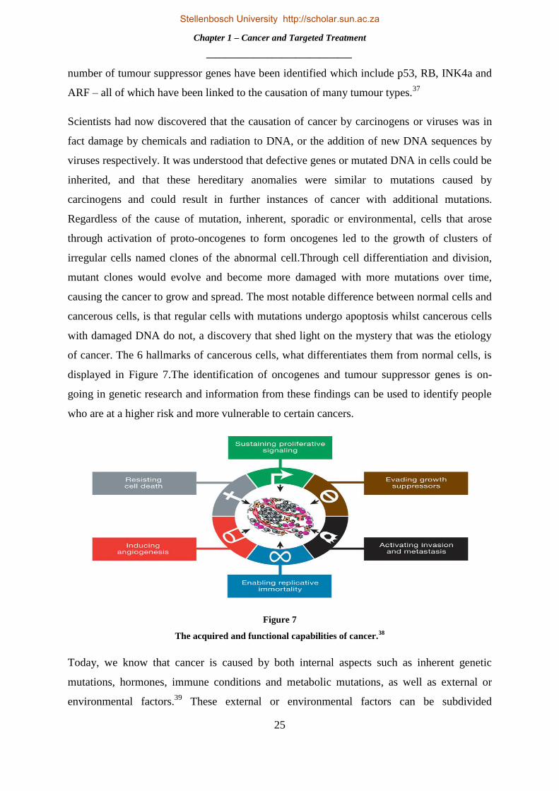
of cancer. The 6 hallmarks of cancerous cells, what differentiates them from normal cells, is
displayed in Figure 7.The identification of oncogenes and tumour suppressor genes is on-
going in genetic research and information from these findings can be used to identify people
who are at a higher risk and more vulnerable to certain cancers.
Figure 7
The acquired and functional capabilities of cancer.38
Today, we know that cancer is caused by both internal aspects such as inherent genetic
mutations, hormones, immune conditions and metabolic mutations, as well as external or
environmental factors.39
These external or environmental factors can be subdivided
Stellenbosch University http://scholar.sun.ac.za

Chapter 1 – Cancer and Targeted Treatment
_______________________________
26
intothreecategories, namely physical carcinogens such as UV radiation, chemical carcinogens
such as tobacco, asbestos and arsenic and biological carcinogens, such as infectious
organisms and viruses.10
As is clearly evident in the graphic representations of Figure 8, tobacco is one of the main
cancer risk factor in the world, increasing risk of developing at least 14 different types of
cancer, containing at least 50 carcinogens and being the culprit for 22% of global cancer
mortalities and 72% of lung cancer deathsworldwide. It has been estimated that poor diet can
be linkedto 30-35% of all cancer deaths in United States, up to 70% of these being colorectal
cancers. Chronic alcohol consumption has been connected to a multitude of cancers,
including those of the upper aerodigestive tract, liver, pancreas and even breast. Persistent
infections of amongst others, the Human Papilloma virus (HPV), hepatitis B (HBV), hepatitis
C and HIV, have been related to cervical, anogential and skin cancer, as well as Hodgkin‟s
Lymphoma and T-cell leukaemia to name a few. Infections are associated with 17.8% of all
cancer cases internationally and in up to 20% in the United States as shown in Figure 8.10, 39
Figure 8
The role of genes and the environment in the development of cancer. A is the percentage contribution of
each factor. B is familial risk ratios for certain cancers. C is the percentage contribution of each
environmental factor.37
Stellenbosch University http://scholar.sun.ac.za

Chapter 1 – Cancer and Targeted Treatment
_______________________________
27
It is estimated that more than 30% of all cancer incidents can be prevented. This can be
achieved by avoiding known carcinogens such as tobacco, alcohol and other chemical
carcinogens, a healthy diet and lifestyle, vaccinations against HPV and HBV and reduced
exposure to UV radiation. It is a case of cancer prevention is better than cure and if detected
early and treated effectively a large number of cancers have a high probability of going into
remission. Awareness of early signs and symptoms and undergoing screening at regular
intervals when older, can both decrease the cancer mortality rate.10
As an important example,
the invention of the “pap smear” or “pap test” by George Papanicolaou (1883-1962) and its
implementation as test in the 1960s by the American Cancer Society (ACS) has led to a
decline of about 70% in cervical cancer.13
A greater understanding of the pathogenesis and
causation of malignant neoplasm has enabled researchers to develop effective therapeutic
treatments for the disease. From humble beginnings, these treatments now make use of state
of the art technology and cutting edge research techniques to halt the advance of cancer.
1.1.3 The Evolution of Cancer Treatment
Similar to ascertaining and understanding the cause and diagnosis of cancer, the progression
and development of cancer treatment has been a slow endeavour. In antiquity, putting under
the knife and cautery remained the best method for removal of tumours and remedy of
cancer. Since 1500 BC, and for 300 years afterwards, various concoctions of herbal remedies
containing boiled cabbage, tea or fruit amongst other things, as well as elixirs and pastes of
assorted metals like mercury, lead, iron and copper, were made use of internally or externally
in different concentrations. Galen, as Hippocrates did, held firm beliefs that cancer was
incurable and considered the patient untreatable. With his prolific writings and influence at
the time, progress in the comprehension and treatment of cancer was brought to a standstill
for an age, as others throughout history adopted his approach.12
Surgery, the only real tool of
cancer treatment at the time, was a primeval and ghastly affair with many complications, the
main obstacle being blood loss. As discussed previously, it wasn‟t until the 19th
and early 20th
century with the pioneering introduction of antiseptic practice, use of anaesthesia and surgical
pathology that cancer surgery, and surgery in general, blossomed. So much so, that the
subsequent 100 years were named, “the century of the surgeon.”13
A surge of surgical prototypes followed in this century, with a number of them being
accomplished by Theodor Billroth (1829-1894). His repertoire included the first
esophagectomy,laryngectomy and the first successful gastrectomy or treatment of gastric
Stellenbosch University http://scholar.sun.ac.za

Chapter 1 – Cancer and Targeted Treatment
_______________________________
28
cancer.18
However, the most monumental moment in cancer surgery transpired in 1882, with
the introduction of the radical mastectomy technique by William Halsted. Halsted believed
that cancer radiated from the primarytumourto adjacent points.Figure 9 shows the en bloc
abscission of all neighbouring tissue technique he endorsed, to ensure removal of all
cancerous cells.19
The surgery entailed removal of the full breast, axillary lymph nodes
andthe pectoralis major and minor in one piece under his recommendation.
Figure 9
A diagram of Halsted’s showing his technique for the radical mastectomy.18
Halsted‟s radical mastectomy remained the cream of the crop of cancer operations for a
century with only slight alterations being made by others.18
Stephen Paget (1855-1926) made
an important contribution to the knowledge of how cancer spreads by the bloodstream to all
organs, but could only develop in particular organs, with his “seed and soil” theory of
metastasis.40
This appreciation of metastasis was truthful andaided physicians in ascertaining
the limits of cancer surgery and the development of systemic therapy – the future of cancer
treatment. Cancer surgery eventually evolved into a combinational therapy with
chemotherapy and radiation. With the vast escalation in superior technology of today, the use
of ultrasound, computed tomography (CT scans), magnetic resonance imaging and positron
emission tomography (PET scans) has become standard procedure and replaced the need for
investigative surgery.In addition, through the use of fibre optic technology, the endoscope,
Stellenbosch University http://scholar.sun.ac.za

Chapter 1 – Cancer and Targeted Treatment
_______________________________
29
cryosurgery, radiofrequency ablation and laser technology, surgeons have been devising
lesser invasive ways of eliminating tumours.13
The foundation of hormone therapy, among the most prominent of modern approaches of
cancer treatment, was laid by George Thomas Beatson (1848-1933) and his inquisitive
nature. Beatson perceived, and concluded experimentally, that removal of the ovaries in
rabbits led to fatty degeneration in normal cell division. In work he published in 1896, a
paper entitled, “On Treatment of Inoperable Cases of Carcinoma of the Mamma: Suggestions
for a New Method of Treatment, with Illustrative Cases,” Beatson showed improvement of
advanced breast cancer after bilateral oopherectomy (removal of the ovaries) in 3 patients.
Beatson surmised that the ovaries were indeed the cause of breast cancer and had
unknowinglycorrelated the hormone oestrogen and its complications in cancer causation even
before it had been discovered. He has been the named the “father of endocrine ablation” in
cancer treatment.41
45 years later Charles Huggins (1901-1997) established the validity of this
relationship with hormones and cancer, by experimental removal of the testicles
(orchiectomy) in male patients with prostate cancer. Striking reversion of metastatic prostate
cancer through this surgical procedure, as well as with dispensing of oestrogen, was
witnessed by Huggins.42
The combined work of these two giants in their field led to the use of
modern hormonal therapy used to treat breast cancer today, such as tamoxifen and aromatase
inhibitors. The development of new hormone therapeutic agents is on-going and this,
combined with research in understanding how hormones manipulate cancer growth, has
revolutionized cancer risk and treatment, especially in prostate and breast cancer.13
The German physics professor Wilhelm Röntgen (1845-1923) became the first man to
produce and detect X-rays in 1895, whilst experimenting with gaseous discharge tubes. In
fact, the first x-ray image is that of his wife‟s hand. He later held a lecture disclosing his
discovery, generating excitement worldwide. His work earned him the first Nobel Prize in
physics in 1901,43
and coupled with the discovery of the radioactive elements radium and
polonium in 1898 by Pierre and Marie Curie, the era radiation therapy for cancer had
emerged. The first physician to use X-rays to treat cancer was Emil Grubbe (1875-1960),
only months after Röntgen‟s discovery, on Gordon Isaacs for retinoblastoma as pictured in
Figure 10.44
Radiotherapy showed marked success in skin cancer, as well breast cancer and
cervical cancer.20
Fractionated radiation treatment, where the doses of radiation are spread
out over time, became a landmark in the field, and in 1928 it was shown to treat head and
Stellenbosch University http://scholar.sun.ac.za

Chapter 1 – Cancer and Targeted Treatment
_______________________________
30
neck cancers.19
Modern radiation oncology can be traced back to the introduction of cobalt
therapy and medical linear accelerator technology as treatment. The first patient that received
radiotherapy from a clinical linear accelerator was in 1953.45
Henry Kaplan (1918-1984) was
hugely influential in the design and development of these machines, which has become a
cornerstone in cancer radiotherapy.46
As with surgery, breakthroughs in technology,
especially in the field of computation, have in recent years allowed radiologists to deliver
accurate and precise energy to tumours, limiting damage to adjacent regular tissue.19
Figure 10
The first patient treated with a linear accelerator for retinoblastoma, Gordan Isaacs, who lived to
adulthood with normal vision after radiotherapy.47
It became clear to surgeons, radiologists and all individuals involved in the field of oncology
that regardless of the quality of surgery or radiotherapy treatment, or the combination of the
two, cure rates had levelled and progression had thus halted. In the 1950s only a third of
cancers could be treated successfully with the methods and techniques available.19
The closing
of the 19th
century saw Paul Ehrlich (1854-1915) postulate his “side chain theory” of 1897 -
the origin of targeted therapy and the introduction of the notion that chemicals could be used
to fight cancer.48
Ehrlich coined the term “chemotherapy”19
and later won the Nobel Prize in
Stellenbosch University http://scholar.sun.ac.za

Chapter 1 – Cancer and Targeted Treatment
_______________________________
31
Physiology or Medicine in 1908, for his work on immunology, a field he brought into
being.48
His proposition that the human immune system stems tumour growth, gave impetus
for research directed at exploiting the immune system in cancer treatment, as well as the
widespread search for vaccines andchemicals to extinguish cancer. Animal models of
transplantable tumourswere developed at the dawn of the 20th
century and a basis of being
able to consistently predict the antitumour effect of these agents for humans from these
models was thoroughly investigated. This research remained largely fruitless, mostly due to a
restricted clinical trial for humans.19
This changed however, after examination of soldiers
exposed to mustard gas in World War I and II showed acute reduction and harmful changes
in bone marrow that developed in blood cells. During this period, experimental evaluation
showed that nitrogen mustard, an analogue of mustard gas, had beneficial effects in treating
lymphoma, as well as leukaemia.Nitrogen mustard was the first of its kind, an alkylating
agent that damaged the DNA of abnormal, rapidly growing cells, effectively killing them, and
the blueprint for the design of a series of similar but more efficacious compounds.49
In
addition, folic acid, a vitamin present in leafy vegetable, was shown to be associated with
bone marrow function andits associated deficiency in megaloblastic anaemia.49
Sidney Farber
(1903-1973) and colleagues, probed the effects it had on leukaemia patients, during which it
surprisingly showed to accelerate proliferation of the cancerous cells. Joining forces with
medicinal chemists at Lederle Laboratories, a series of similar compounds were generated
which included aminopterin and amethopterin, now known as methotrexate, which acted as
folate antagonists.50
Farber dispensed these drugs to children with acute lymphoblastic
leukaemia (ALL) and remarkably, by blocking the folate-needing enzymes activity, induced
remission of ALL. Methotrexate proved to show inhibition across a wide range of cancers,
including breast, bladder and choriocarcinoma, a malignancy inherent in trophoblastic cells
of the placenta, and formed the basis for the first cure for metastatic cancer.49
With the
knowledge that chemotherapeutic agents could act by inhibiting a vital chemical reaction for
DNA duplication, researchers discovered inhibitors of other critical cellular functions. The
early activity and success of nitrogen mustard and methotrexate, resulted in the founding of
the National Cancer Chemotherapy Service Center (NCCSC) in 1955, of which Farber
became a director.28
This heralded the age of chemotherapy.
The migration to the pioneering use of combination chemotherapy, where multiple drugs with
different modes of action are used collectively, can be traced back to the long term remissions
and even curing of ALL developed by Emil Frei (1924-2013) and colleagues, and the
Stellenbosch University http://scholar.sun.ac.za

Chapter 1 – Cancer and Targeted Treatment
_______________________________
32
extension of this to Hodgkin‟s and non-Hodgkin‟s lymphoma by Vincent DeVita (1935- )
and co-workers.49
In the case of Hodgkin‟s lymphoma, a combination of nitrogen mustard,
oncovin, procarbazine and prednisone administered as treatment, lead to remission rates of
effectively zero to 80%, with 60% of these patients never relapsing. Today, this cancer is
curable 90% of the time, with treatment consisting of integration of radiotherapy and
chemotherapy.50
The success of combination therapyhad a positive effect use of chemotherapeutic agents as an
adjuvant to surgery and radiotherapy.19
Physicians became more lenient and tolerable to
using drugs after surgical resection, with no greater success than in that of breast and
colorectal cancer. As a result of the long but ever improving methodology and techniques
used in the treatment of cancer, shown in Figure 11, the breast cancer mortality rate began to
fall in 1991 - a trend that has been maintained – and a drop of 40% in the mortality rate of
colorectal cancer patients has been witnessed in the last 4 decades.19
Figure 11
Timeline of pivotal events in cancer treatment.19
Stellenbosch University http://scholar.sun.ac.za

Chapter 1 – Cancer and Targeted Treatment
_______________________________
33
Research today in chemotherapy, mainly involves improving the activity and reducing the
side effects these drugs have. This includes the search and use for new drugs and drug
combinations, the development of liposomal therapy51
which increases selectivity towards
cancer cells and decreases side effects, and chemo protective agents such as amifostine which
reduces renal injury in chemo patients.52
The great Paul Ehrlich did put forward the idea that the human immune system could halt and
prevent tumour growth.53
With scientists growing understanding of the biology of cancer
cells, biological agents that could imitate natural cellular signals that are responsible for cell
growth, were developed and unveiled. The isolation, synthesis and application of these agents
in treatment, is known biological response modifier (BRM) therapy or immunotherapy.These
agents are administered to patients in order to either emulate or manipulate a natural immune
response. This can be achieved by directly changing abnormal cellular growth or indirectly
by aiding normal cells bring the cancer under control.13
The first real embodiment of Ehrlich‟s
vision was the production of monoclonal antibodies (mAbs) by Georges Köhler (1946-1995)
and César Milstein (1927-2002), for which they won the Nobel Prize in Physiology or
Medicine in 1984.53
Scientists recognized and identified certain tumour targets, name
antigens, which bind specifically to certain antibodies. With technological advancements in
the 1970s, mass production of mAbs could be undertaken and used to target specific
antigens.13
With the accessibility to substantial amounts of mAbs with singular specificity,
developments and enhancement of this method by making use of recombinant DNA
technology improved efficacy and diminished side effects and resulted in the first Food and
Drug Administration (FDA) approved monoclonal antibody, rituximab, in 1997, for the
treatment of B-cell lymphoma.19
Since then eleven other antibodies have obtained approval
for cancer immunotherapy for a wide variety of cancers, which include trastuzumab in 1998
for the treatment of breast cancer.53
It became clear that cellular, rather than hormonal,
immunity played a much larger role in the immunotherapy of cancer.A name synonymous
with immunotherapy is that of Steven Rosenberg(1940- ) who first introduced immuno cell
therapy in 1985, after the description of T-cell growth factor or interleukin-2 in 1976. By
administering interleukin-2 to patients with metastatic melanoma and renal cancers,
regression was witnessed in an invasive metastatic disease for the first time by immune
therapy.19
This became known as adoptive cell transfer treatment.54
Immunotherapy has also
been shown to increase survival probability, regress malignant tumour growth55
and result in
regression and disease free periods.56
It has also been shown to be more efficacious as a
Stellenbosch University http://scholar.sun.ac.za

Chapter 1 – Cancer and Targeted Treatment
_______________________________
34
combination or adjuvant therapy.57
Therapeutic vaccines that treat cancer by aiding the human
body‟s immune system in attacking cancer cells - not unlike preventative vaccines which
prevent infectious diseases - have been researched and developed.Sipuleucel-Tor Provenge, is
an example of this, and received FDA approval in 2010 for treatment of metastatic hormone
efractory prostate or prostate cancer that no longer responds to immunotherapy and is
castration resistant.58
The signing of the National Cancer Act of 1971 by Richard Nixon and the subsequent
declaration of the “war on cancer” was explosive for the development of chemotherapy. The
act effectively quadrupled the budget of the National Cancer Institute (NCI) by the end of the
decade.19
Clinical evaluation of novel drugs and innovative programs of chemotherapy
improvement were broadened.50
This led to the development of a new series of screening
techniques, as throughout the 1960s and 70s, screening was done strictly in vivo in mouse
models. However, these screening evaluations proved inadequate and in 1976 human
xenografts were introduced either by implantation or inoculation of mice. Today, over 300
xenografts have been established, including all the key human tumour types. Regrettably,
these newly implemented screening systems are yet to accurately predict the outcome of
clinical trials correctly.49
In the 1980s, chemotherapy research and development decelerated, due to extended periods
of time required for trials which afforded only minor increases in efficacy against malignant
tumours, coupled with the dismal performances of screening methods. Whilst endeavours to
recover the development and progression of cytotoxic drugs continued, new insights in
cellular biology revealed brand new cellular signal transduction pathways that regulate cell
growth and differentiation. These new signal networks were also found to be fundamentally
modified in cancerous cells. Researchers set out on amending these inherent flaws by
targeting growth factors, signalling molecules, cell-cycle proteins, apoptosis regulators and
angiogenesis catalysts.49
Until the 1990s, virtually all drugs employed in cancer therapeutics -
excluding that of hormonal therapy - operated by destroying proliferating abnormal cells. At
this very time, an outbreak of novel drug targets revolutionized the cancer industry, from a
government funded research program to a multi-billion dollar enterprise.49
The last two
decades has seen a transition from the use of cytotoxic agents and immunotherapy, to the
rational design of anticancer drugs, which are mechanism based.59
This shift can best be
described as the dawn of the era of targeted therapy.
Stellenbosch University http://scholar.sun.ac.za

Chapter 1 – Cancer and Targeted Treatment
_______________________________
35
1.2 TARGETED THERAPY
1.2.1 The Revolution of Targeted Therapy
The past two decades has witnessed a metamorphosis in our understanding of cancer
epidemiology and our approach to the treatment thereof, a statement no more evidently
substantiated with a recent study showing a 20% decline in cancer mortality in the United
States since its peak in 1991. In the period from 1991-2010 this accounts for 1.3 million lives
being saved, a decline of more than 30% in the death rate of colorectal cancer, breast cancer
in woman and lung cancer in men, and more than 40% in prostate cancer. In the five year
period of 2006-2010, an annual decrease of 1.8% in men and 1.4% in woman has been
observed.60
These statistics can largely be attributed to tremendous advancements in the
fields of molecular and cellular biology which has led to improved insight into the neoplastic
cellular mechanisms of proliferation, differentiation, apoptosis, tumour immunology and
causation of cancer in viral and chemical carcinogens.61
The origin of identification of these novel signalling pathways, or molecular targets, can be
traced back to the 1960s Special Virus Cancer Program (SVCP), a program dedicated to
identifying viruses linked to cancer. When this project failed, it altered its research program
to study genes that were co-opted by tumour viruses, which led to the description of
numerous oncogenes, suppressor oncogenes and signal transduction pathways critical in
formative biology.50
This scientific knowledge led to the identification of the majority of
contemporary drug targets that are currently the focal point of targeted therapy.50
It also
expedited the Human Genome Project (HGP) and the complete sequencing of the human
genome in 2001,62
a milestone in advancing our understanding of cancer.63
A flash flood of
small biotechnology firms were founded during this time, filled with researchers intent on
rectifying the molecular faults in cancer cells. Today there are over 1300 of these enterprises,
with more than half concerned with cancer research and treatment.49
In 2013, the worldwide
market for oncology-related products was estimated to be approximately $77.3 billion and it
is projected to reach an estimated $143.7 billion by 2023.64
The targeted cancer therapy
market value is expected to double from $25 billion in 2008 to $51 billion by the year 2015.65
Biochemist Stanley Cohen (1922- ) and neurologist Rita Levi-Montalcini (1909-2012)
discovered and isolated nerve growth factor (NGF),for which they received the Nobel prize in
1986.66
Cohen went on to discover epidermal growth factor (EGF),67
a fundamental discovery
in our understanding of cancer for the design of modern cancer therapeutics. Research was
Stellenbosch University http://scholar.sun.ac.za

Chapter 1 – Cancer and Targeted Treatment
_______________________________
36
also beginning to reveal the importance of signal transduction in cancer translation.Signal
transduction is the conversion of extracellular signalling such as hormones, growth factors,
neurotransmitters and cytokines which elicit a specific internal cellular response such as gene
expression, proliferation or even apoptosis.68
The transformation of cancer treatment from
sweeping, non-tailored, large dose chemotherapy with undesirable side effects, to the
exploration and hunt for inhibitors that maintained negligible adverse effects with superlative
anticancer activity was thus desired.69
In addition the required features of auspicious drugs
became apparent - a high metabolic stability which meant a long half-life and slow
decomposition rate by enzymes such as cytochrome-P450, high specificity, good
bioavailability and low toxicity.49
1.2.2 Molecular-Targeted Treatments – Successes and Challenges.
Tamoxifen (Nolvadex; AstraZeneca) has been hailed as the foundation of targeted therapy
and was the first of its kind - a selective oestrogen receptor modulator (SERM).70
Tamoxifen
was originally developed as a prospective contraceptive some 40 years ago in the era of
discovery and application of chemotherapy.71
Prompting its failure as the post coital
prophylactic ICI 46474, it was reinvented as tamoxifen – the first targeted therapeutic for
breast cancer.70
Its first clinical trials in 1971 showed encouraging effects in advance breast
cancer72
and subsequent oestrogen-assay evaluations and further experimentation shed light
on its mechanism of action. It was found that tamoxifen itself was a prodrug, having little
affinity for its target, the oestrogen receptor. Metabolic breakdown of tamoxifen in the liver
yielded its active compound 4-hydroxytamoxifen, an anti-oestrogenic metabolite with a 100
fold higher affinity for the oestrogen receptor.71
This metabolite acts as an antagonist of the
oestrogen receptor in breast tissue, blocking gene transcription and preventing proliferation of
cancerous cells.73
This breakthrough in the understanding of anti-oestrogen activity further
advanced structure-activity relationship studies and led to the synthesis of nearly all other
SERMs including raloxifene. It has been estimated that over 400 000 woman have their lives
today because of tamoxifen therapy which has remained the benchmark in treatment for
endocrine treatment of all stages of oestrogen-receptor positive breast cancer, and is one of
the most widely used anticancer agents today. Although tamoxifen has been shown to have
an excellent risk benefit ratio in midlife and older woman, it has also been shown to augment
the risk of some positive events, such as prevention of osteoporosis, and others negative, such
as an increased risk of endometrial cancer in woman.71
Stellenbosch University http://scholar.sun.ac.za
Chapter 1 – Cancer and Targeted Treatment
_______________________________
37
The first monoclonal antibody to match the criteria of a targeted therapeutic agent was
trastuzumab(Herceptin; Genentech). Human epidermal growth factor receptor type 2 (HER2),
also known as HER2/neu and ErbB-2, is over expressed in up to 30% of early stage invasive
breast tumours and a noteworthy association between this aberrant gene activity and a
diminished rate of survival in patients has been found.74
The receptor consists of an
extracellular binding domain, a transmembrane lipophilic segment and a functional
intracellular tyrosine kinase domain.75
HER2 signalling is responsible for certain cellular
responses and processes which include cell proliferation, differentiation, apoptosis and
migration. Activation of the tyrosine kinase domain occurs by homo- or heterodimerisation,
usually brought about by ligand binding; however, in the case of HER2, dimerisation may
occur without a ligand. Upon activation, the signal transduction pathways of HER2 stimulate
cell growth and survival. In some types of cancer this mechanism is utilized, as mutations or
over expression of this class of receptor has been known to result in uncontrolled cellular
growth, ergo leading to tumour formation.75
Trastuzumab prevents the activation and thus
dimerisation of the HER2 receptor, thus inhibiting tumour growth and inducing cell death. It
has also been suggested that trastuzumab may enlist immune effector cells accountable for
antibody-dependant cytotoxicity, which results in increased tumour assault by lymphoid
cells.75
Trastuzumab received FDA approval in 1998, shows mild adverse effects and when
coupled with chemotherapy, has shown considerable improvements in response, duration and
survival time of therapy in contrast to chemotherapy only. Furthermore, studies have shown
that trastuzumab might enhance the effects of radiotherapy, as well as the action of
tamoxifen.74
If tamoxifen and trastuzumab shed light on the capability and efficacy of these molecular
targeted therapeutic agents in cancer treatment, imatinib mesylate (Glivec, Novartis) – used
in the treatment of chronic myeloid leukaemia (CML) - was the monumental paradigm shift
in exhibiting the potential and power of rationally designed inhibitors in a number of facets. It
graced the cover of Time magazine as shown in Figure 12, hailed as the new cure for cancer.
With tremendous advancements in technology, the first step to identifying the pathogenesis
of CML was taken by Peter Nowell (1928- ) and David Hungerford (1927-1993) whilst
studying human cellular mitosis. They detected a persistent chromosomal abnormality,
namely a shortening in chromosome 22, which would later become known as the
Philadelphia chromosome (Ph).
Stellenbosch University http://scholar.sun.ac.za
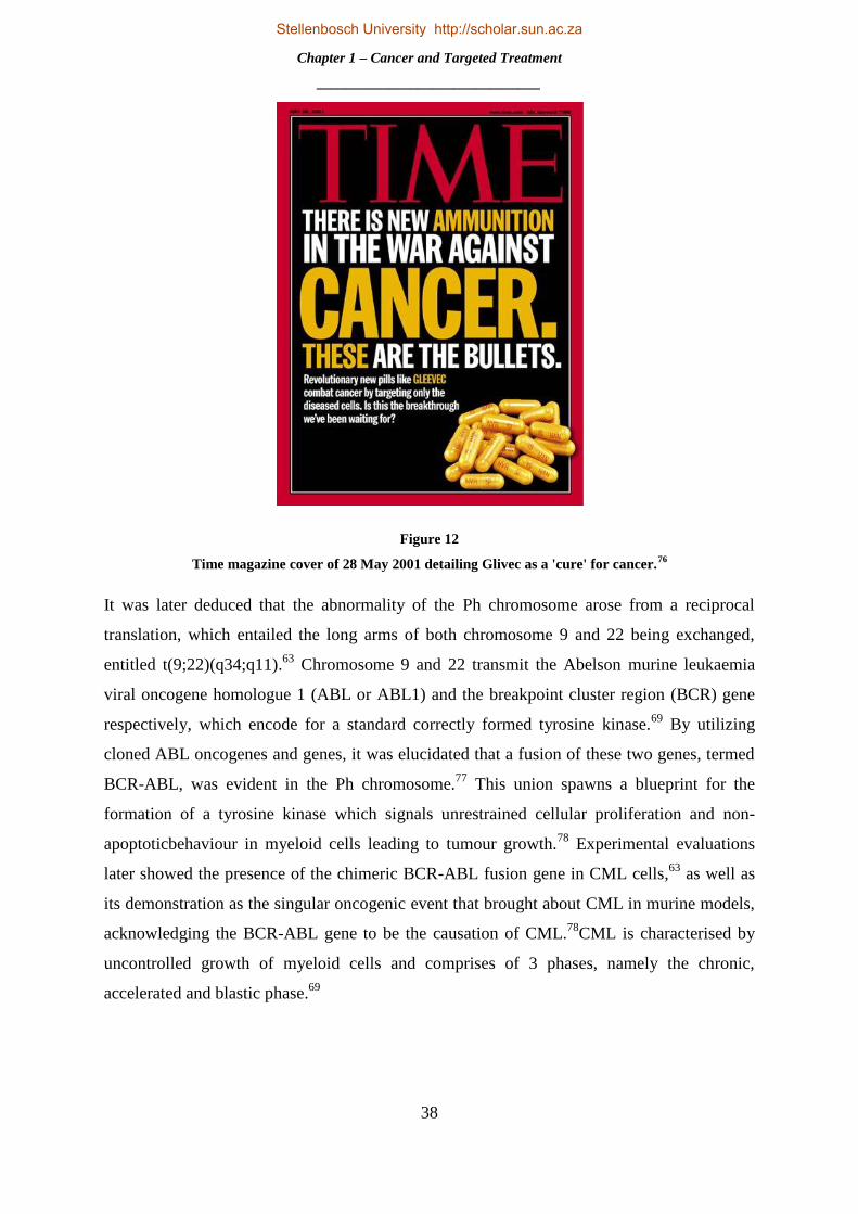
Chapter 1 – Cancer and Targeted Treatment
_______________________________
38
Figure 12
Time magazine cover of 28 May 2001 detailing Glivec as a 'cure' for cancer.76
It was later deduced that the abnormality of the Ph chromosome arose from a reciprocal
translation, which entailed the long arms of both chromosome 9 and 22 being exchanged,
entitled t(9;22)(q34;q11).63
Chromosome 9 and 22 transmit the Abelson murine leukaemia
viral oncogene homologue 1 (ABL or ABL1) and the breakpoint cluster region (BCR) gene
respectively, which encode for a standard correctly formed tyrosine kinase.69
By utilizing
cloned ABL oncogenes and genes, it was elucidated that a fusion of these two genes, termed
BCR-ABL, was evident in the Ph chromosome.77
This union spawns a blueprint for the
formation of a tyrosine kinase which signals unrestrained cellular proliferation and non-
apoptoticbehaviour in myeloid cells leading to tumour growth.78
Experimental evaluations
later showed the presence of the chimeric BCR-ABL fusion gene in CML cells,63
as well as
its demonstration as the singular oncogenic event that brought about CML in murine models,
acknowledging the BCR-ABL gene to be the causation of CML.78
CML is characterised by
uncontrolled growth of myeloid cells and comprises of 3 phases, namely the chronic,
accelerated and blastic phase.69
Stellenbosch University http://scholar.sun.ac.za

Chapter 1 – Cancer and Targeted Treatment
_______________________________
39
Figure 13
Illustrative summary of the chemical optimization of imatinib.78
With the identification of the molecular pathogenic event for CML, an attractive drug target
had been exposed in the BCR-ABL tyrosine kinase and its enzymatic activity.78
This
resultedin CIBA–Geigy, now Novartis, establishing a kinase inhibitor development program
in 1984.63
Following screening for inhibitors of protein kinase C (PKC), a lead compound
was identified with a phenylaminopyrimidine backbone which can be seen in Figure 13a
PKC inhibitions were markedly increased with the addition of a 3-pyridyl group at the 3
position of the pyrimidine (Fig. 13a, blue). Furthermore, tyrosine kinase inhibitory activity
benefitted largely from the inclusion of an amide group on the phenyl ring of the lead
structure (Fig. 13b, red). Interestingly, substitution at the 6 position of the diaminophenyl ring
completely nullified any PKC inhibitory activity, but the addition of a methyl, increased
activity against tyrosine kinases whilst sacrificing loss of PKC inhibition(Fig. 13c, pink).
Lastly, to combat poor solubility and oral bioavailability, an extremely polar, hydrophilic side
chain was attached (Fig. 13c, green). STI571, or what is now imatinib, was selected as the
most encouraging candidate for further development. With the aid of molecular modelling
and crystallographic data, the binding mode in the ATP active site was further understood. A
crystal structure of imatinib in complex with the c-ABL kinase domain can be seen in Figure
14. In vitroscreening of the candidate compound against a number of kinases showed
Stellenbosch University http://scholar.sun.ac.za

Chapter 1 – Cancer and Targeted Treatment
_______________________________
40
inhibitory activity of phosphorylation for three target kinases, namely BCR–ABL, c-KITand
the platelet-derived growth factor (PDGF) receptor.78
Figure 14
Crystal structures of the kinase domain of c-Abl in complex with imatinib (PDB_ID:1IEP)
The candidate compound was found to not only inhibit the growth of CML and BCR-ABL
cells in cultures, but also in vitro.63
Following this, in vivo experiments with mouse models
showed continuous inhibition of the fundamentally active p210BCR–ABL
tyrosine kinase
phosphorylation, and resulted in tumour free existence of the mice. The first clinical trials
carried out by Brian Druker (1955- ) showed a remarkable response of 98% of the patient‟s
blood counts returning to normal.78
This propelled imatinib‟s FDA approval application
forward and in 2001 it received FDA approval for treatment of CML. A disease that was
regarded as untreatable, and thus inevitably fatal before –with an estimated survival of 15
months for patients - is now treatable with a survival expectation of 15 years.63
In a 5 year
evaluation of patients treated with imatinib, 89% of these patients survived with complete
remission after 60 months.79
Imatinib is not as selective as previously thought, but fortunately
this promiscuity extends to treatment and inhibition of other kinases as previously stipulated.
Imatinib has undergone clinical trials for the inhibition of c-kit in which overexpression can
lead to the formation of gastrointestinal stromal tumours (GIST), as well as being effective in
treatment of proliferative premalignant hematopoietic diseases such as hypereosinophilia
syndrome and chronic eosinophilia leukaemia, associated with the expression of an activated
form of PDGFR.63
Imatinib is a champion for targeted therapy and through its development
Stellenbosch University http://scholar.sun.ac.za

Chapter 1 – Cancer and Targeted Treatment
_______________________________
41
by identification of its molecular pathogen and drug target, medicinal chemistry endeavours,
as well as biological assays and application to human clinical trials, has shown that is
possible to produce a rationally designed, molecularly-targeted agent for the treatment of a
specific cancer. Importantly, success was attained in the predictive quality of in vitro and
animal model trials, as the results obtained in these trials agreed well and substantiated the
results obtained in the clinical trials.63
In its success; it presented a proof of principle and
concept, following a more traditional route of drug discovery which had never before been
successful for this target – cancer. This was a definitive advancement in the campaign of
targeted therapy.
1.2.3 The Scope of Targeted Therapy and the 21st Century.
Although examples of growth factor receptor inhibitors - which target signal transduction
pathways to bring a halt to aberrant and uncontrolled cellular growth - have been discussed,
there are various other mechanisms that are focussed and exploited and different ways in
which targeted therapeutic agents are implemented in the treatment of cancer. Angiogenesis
is derived from the two Greek words angio, meaning “blood vessel,” and genesis, meaning
“origin” or “beginning.” From this it is clearly deduced that the term describes the
physiological process by which new blood vessels are created. The creator and titan of the
field of angiogenesis, Judah Folkman (1933-2008), first postulated in 1971 that tumour
growth is angiogenesis dependant and correctly predicted that the inhibition of angiogenesis
or the concept of “antiangiogenesis” could be used as a possible future cancer therapeutic.80
Whilst conducting research on different solutions of haemoglobin as possible blood
transfusion substitutes, Folkman determined the viability of this hypothesis by perfusing
canine thyroid glands in these solutions. The glands subsisted for up to two weeks. To
ascertain whether or not these glands could support growth, they were implanted with murine
melanomas. Tumour development was witnessed, but only to about 1-2mm in size
whereupon growth was halted, even though both the tumour and thyroid gland remained
alive. Upon transplantation back to the mice, the tumour underwent
acceleratedvascularisation and swelled to sizes of up to 1cm3. It was on these experiments
that Folkman based his deductions divulged in his landmark paper.81
The paper he wrote
provided a schematic and the foundation for the rest of his research based on a number of
observations. Folkman recognized that solid tumours were much more reliant and possibly
dependant on the enlistment of new blood vessels for growth. Another crucial perception
made during experimentation with resting endothelial cells of blood vessels was that tumours
Stellenbosch University http://scholar.sun.ac.za

Chapter 1 – Cancer and Targeted Treatment
_______________________________
42
were capable and readily made use of dispersing factors, termed tumour angiogenesis factor
(TAF) by Folkman, which promoted and stimulated endothelial cell proliferation, a
mechanism implemented by tumours for tumour angiogenesis.
Figure 15
The principle of anti-angiogenesis therapy.82
Logically, Folkman rationalized that antiangiogenesis therapy by inhibition of TAF, could
provide control on the growth of malignant neoplasms, effectively cutting it off from
resources for further proliferation – the principal of angiogenesis inhibitors as illustrated in
Figure 15. This would not lead to complete tumour removal, but rather withdrawal to a
dormant state and a diminutive size of the tumour. Folkman recognized this fragile
instrument necessary for tumour growth and saw a target for future cancer therapeutics,
foreseeing the evolution of anti-angiogenic antibodies in use today, decades prior to their
arrival onto the scene.82
Today, it is universally acknowledged that oncogene activation leads to carcinogenesis.83
The
use of in vitro bioassays, which were later extended to murine models, displayed evidence
that the conditional expression of activated oncogenes in cells resulted in a heightened rate of
proliferation and a marked reduction in apoptosis, resulting in rapid tumour growth leading to
the death of the host. Oncogene deactivation resulted in the tumours receding, leaving either
shrunken malignancies or no trace of them at all. This verified the reliance of tumour
proliferation and survival on oncogene activation, what is today known as oncogene
addiction.83, 84
Further studies and experiments have been undertaken indicative that this is
not the only requisite for the conceiving of a malignant tumour. The omission of angiogenesis
in tumour growth results in inert, miniscule malignancies of ~ 1mm3
or less, in a dormant like
state, shown to be innocuous to patients. In addition, transformation of oncogenes in tumour
Stellenbosch University http://scholar.sun.ac.za

Chapter 1 – Cancer and Targeted Treatment
_______________________________
43
cells have shown to considerably escalate angiogenic activity by means of abnormal
expression of vascular endothelial growth factor (VEGF) – one of many signal proteins
produced by cells that act as a mitogen for endothelial cells, promoting angiogenesis and
vasculogenesis. A decrease in the expression of antiangiogenic proteins was also witnessed.
Combined with experiments conducted by Fernandezet al.85
, which showed tumour growth
restriction by an angiogenesis inhibitor, led to the premise that formation of a malignant mass
is unachievable without both tumour and endothelial cell proliferation, a requirement and
component of angiogenesis.83
The discovery of the existence of factors promoting endothelial cell proliferation, coupled
with the notion of tumour growth dependency on angiogenesis, led to various scientific
endeavours to identify and isolate these angiogenic mitogen factors. It was not until 1989 that
Ferraraet al. became the first to isolate VEGF, an endothelial cell specific factor, from the
follicular cells of bovine thyroids.86
Arrays of in vivo and in vitro models have displayed
VEGF to prompt a potent angiogenic reaction, as well as the dependency of cells of newly
formed blood vessel on VEGF in malignancies. Overexpression of this factor has been
documented in a myriad of cancers including lung, breast, gastrointestinal tract, renal and
ovarian carcinomas.87
It is estimated that in the order of 60% of the roughly 200 different
types of human cancers express VEGF.83
This led to the development of bevacizumab
(avastin, Genentech/Roche), a humanized monoclonal antibody which is known as a „direct‟
angiogenesis inhibitor. Its mode of action entails blocking signalling from the pro-angiogenic
protein VEGF and thus thwarting vascular endothelial cell proliferation, migration and
evasion of apoptosis.88
Clinical trials of avastin ensued, where it passed with flying colours,
showing great efficacy, especially in cases of colorectal cancer.87
In February 2004, FDA
commissioner Mark McClellan declared that, in conjunction with surgery, radiotherapy and
chemotherapy, „„antiangiogenic therapy can now be considered the fourth modality of cancer
treatment.‟‟ Avastin subsequently received FDA approval for colorectal cancer.With the
closing stages of 2007, 23 therapeutic agents with antiangiogenic activity were in phase III
and 30 were in phase II clinical trials.89
The challenges faced with antiangiogenic therapy is
mainly concerned with the recognition that tumours may generate a range of angiogenic
molecules, and depending on the stage of development may be dependent on different factors
for angiogenesis. Thus the inhibition of a singular angiogenic factor may have little to no
effect of halting tumour growth. Antiangiogenic agents are now used in combination with
other angiogenesis inhibitors, as well as in adjuvant therapy to greater effect.81
Stellenbosch University http://scholar.sun.ac.za

Chapter 1 – Cancer and Targeted Treatment
_______________________________
44
Apoptotic and non-apoptotic mechanisms of cellular death have been acknowledged to be
conducive to the growth of cancer and the development of resistance to anticancer
therapeutics.Irregularities in DNA maintenance and preservation facilitate genetic mutations
and ultimately causation of cancer. Regular cells possess vital mechanisms to disrupt and
ensure that these inherent anomalies do not perpetuate, leading to carcinogenesis. The tumour
suppressor gene p53, aptly labelled the “guardian of the human genome”, plays an essential
role in this defence of normal tissue and prevention of cancer. P53 stands sentry to cellular
strain, hypoxia and DNA deterioration and following detection of any of the aforementioned
stresses, the suppressor gene becomes stabilized, inducing cell cycle arrest or apoptosis.90
The nature of this response is to destroy deviant cells undergoing uncontrolled cellular
growth, discontinuing the progression to malignancies. It is now understood that cells can
also be eradicated following DNA impairment by necrosis, mitotic catastrophe and
autophagy.90
It is not surprising that researchers have found ways to utilize these processes in
bringing about the programmed cellular death of tumours. An example would bebortezomib
(Velcade, Millennium Pharmaceuticals), a boronic acid dipeptide and novel reversible
proteasome inhibitor, which was shown to be effective amongst patients with multiple
myeloma which was obstinate to conventional chemotherapy.91
It has been determined that
the mechanism of action of this new class of anticancer therapeutic involves induction of
apoptosis, the reversal of resistance to therapy of multiple myeloma cells, and blocking
angiogenesis in vivo.92
The ubiquitin-proteasome signal transduction pathway is extensively
involved in the removal of damaged or redundant proteins in the cells of eukaryotic
organisms. It has been shown that proteasome inhibition is associated with sensitization to
apoptosis and more importantly, that cancerous cells are significantly more receptive to
proteasomal obstruction than are normal cells.93
Following success in clinical trials,
bortezomib received FDA approval and it has been shown to be superior to other available
treatments in cases of multiple myeloma.92
In addition to the expansion of monoclonal antibodies and small molecule signal transduction
pathway inhibitors available, researchers have identified new classes of molecules and
therapies such as small interfering RNA (siRNA) and poly (ADP-ribose) polymerase enzyme
(PARP) inhibitors. The involvement of siRNA in the RNA interference (RNAi) pathway,
where the expression of specific genes can be tampered with or even dismantled, has led to
sparks of interest in targeted therapy and more recently clinical trials of patients with
advanced liver metastasized cancer through lipid nanoparticles targeting VEGF and kinesin
Stellenbosch University http://scholar.sun.ac.za

Chapter 1 – Cancer and Targeted Treatment
_______________________________
45
spindle protein (KSP). The trials were a success, halting the growth of the metastases or even
promoting remission in certain patients.94
PARP activation plays a critical role in DNA harm
response processes or the ability of cells to maintain damaged DNA, and has been shown to
enhance the cytotoxicity of anticancer therapeutics by tumour chemo and radio
sensitization.95
The exploitation of defects in these DNA repair pathways has generated
considerable interest in the targeted inhibition thereof, spawning a number of inhibitors in
clinical phase III and phase II trials.96
With the steady and continual innovation of state-of-the-art, superior technology and a
greater understanding of the biological process of cancer at the molecular level, researchers
will gain, and keep gaining, additional novel targets for promising therapeutic agents. The
precise targeting of specific inhibitors to a distinct breed of tumour, increasing the
effectiveness and lowering adverse events, has always been problematic. The use of gene
expression profiling by DNA microarrays has facilitated in the comprehensive identification
of new cancer classes (class discovery) and the classification and assignment of known
tumours to classes (class prediction), a powerful new tool to be sharpened for use in target
therapy.97
Stellenbosch University http://scholar.sun.ac.za

Chapter 1 – Cancer and Targeted Treatment
_______________________________
46
Figure 16
Targeted therapeutic agents, their targets and mechanisms.99
Proteomics, or the extensive study of proteins structure and functions, has found its footing in
cancer research as biomarkers for certain cancers and early cancer prognosis.98
Specificity,
fewer and milder adverse effects, greater efficacy and lower toxicity is the trademark of
targeted therapy. The future of research in this area will see the pursuit of improvement of
these qualities as anticancer agents, as well as the identification and understanding of new
therapeutic targets.
The field of targeted therapy has grown enormously over the last twenty years, targeting a
host of different cancer through an assortment of disease mechanisms. A few of these
therapeutic agents with their corresponding targets are shown in Figure 16. Of all the targeted
treatments, none have shown as much promise and delivered as many results, both clinically
and in gaining an understanding of oncogenesis as that of the targeted inactivation of kinases
through kinase inhibitors. With a collection of FDA approved drugs being used to treat
cancer and a variety of other disorders, an impetus has been provided for further research in
this area of cancer treatment and the development of new and improved kinase inhibitors.
Stellenbosch University http://scholar.sun.ac.za

Chapter 2 – Kinases and Kinase Inhibitors
_______________________________
47
CHAPTER 2 – KINASES AND KINASE INHIBITORS
2.1 KINASES
2.1.1 The Origins and Mechanism of Protein Phosphorylation
The collaboration of spouses Carl (1896-1984) and Gerti Cori (1896-1957) and their work on
carbohydrate metabolism, the discovery of the “Cori Cycle,” “Cori Ester” and glycogen
synthesis earned them the Nobel Prize in physiology or medicine in 1947, as well as
illuminating the presence of phosphorylation as a regulatory process. Experimentation with
diced skeletal muscle tissue of frogs revealed an intermediate from glycogenolysis – the
breakdown of glycogen – designated glucose-1-phosphate, which would later be termed the
“Cori Ester.” Further studies elucidated its structure and the enzyme glycogen phosphorylase,
which catalyses the rate-determining step of this process.100,101
Phosphorylase was seen to
have two forms, namely a and b, and the inter-conversion of these two enzymes from a to b,
by another enzyme discovered in 1943 by Gerty Cori, was the first example of allosteric
activation.101
However, it was not until many years later that the real significance of this
reaction was grasped.
It was in the middle of the 1950s thatEdwin Krebs (1918-2009) and Edmond Fischer (1920- )
determined that the processes of phosphorylation and dephosphorylation were implicated in
the mutual conversion of phosphorylase a into phosphorylase b, work for which they received
the Nobel Prize for physiology or medicine in 1992. They exposed that this interconversion,
or more specifically the conversion of form b into form a, occurred readily in the presence of
magnesium adenosine triphosphate (Mg-ATP or ATP) and phosphorylase kinase, an enzyme
they discovered and coined.101,102,103
This enzyme was later shown to facilitate the conversion
from b into a by assignmentof a γ- phosphoryl group of ATP to an explicit serine residue of
phosphorylase b, an example of phosphorylation.104
Dephosphorylation, or the reversion from
a back to b, was activated by removal of thisγ- phosphorylgroup by a phosphate releasing
(PR) enzyme known today as phosphatase 1.101
The first example of a protein kinase cascade
-the activation of one protein kinase by another – was identified with the revealing of 3′, 5′-
cyclic adenosine monophosphate (cAMP) dependant protein kinase A (PKA) and its
activation of phosphorylase kinase. PKA was subsequently shown to suppress another
enzyme, namely glycogen synthase, the first example to display the inhibitory activity of
phosphorylation.101
Phosphorylation however, remained dormant in research – owing to the
Stellenbosch University http://scholar.sun.ac.za

Chapter 2 – Kinases and Kinase Inhibitors
_______________________________
48
belief that it was exclusive to the regulation of only the glycogen metabolic pathway – until
the 1970s and 80s.
A number of the most notable serine/threonine specific protein phosphatases and their
regulatory mechanisms were identified during decades of the 1970s and 80s.105
The
unravelling of the amino-acid sequence for the first protein kinase PKA was accomplished in
1981.106
The amalgamation of these and other discoveries, led to insight into the target of
cyclosporin, the immunosuppressant drug that made organ transplants possible. Multisite
phosphorylation, the phosphorylation of two or more residues by two or more protein
kinases, was observed in the 1970s.101
Perhaps the greatest milestone in these 20 years was the
discovery by Ray Erikson (1936- )that v-Src, a gene found in Rous sarcoma virus, encoded
aprotein kinase.107
This led Tony Hunter (1943- ), or the “kinase king,” to uncover that the v-
Src gene was responsible for phosphorylation of tyrosine kinase residues in 1980.108
Within
a year, the realization that tyrosine phosphorylation played a role in regular cellular
preservation was grasped, and in the following years the magnitude of phosphorylation as an
extensive, complex and crucial cellular activity regulatory mechanism was revealed.109
The
discovery of epidermal growth factor receptor (EGFR) as a protein kinase,110
shed light on its
activation by epidermal growth factor (EGF) interacting with the receptor itself. The
exposure that this was the case with a number of other growth factor receptors provided an
impetus for researchers to find the natural substances these enzymes acted upon.101
This led
to the peculiar finding that the receptors themselves were becoming repeatedly
phosphorylated at multiple tyrosine residues - being the most prominent suitable cellular
substrates themselves - and the discovery of receptor autophosphorylation, which today is
recognized as pivotal in downstream signal transduction and mediation of these signals.101
It
was not long thereafter that the first protein tyrosine phosphatase(PTP1B) was purified, as
well as the cloning of the family of “just another kinase” (JAK) which were shown to play a
role in signal transduction and activation of transcription.101
The 1990s have been conferred
the title of “decade of protein kinase cascades”101
and the unearthing of an insulin activated
protein kinase that phosphorylates microtubule-associated protein 2 (MAP2), later changed to
mitogen-activated kinase (MAP) kinase - a family of protein kinases that are activated in
response to diverse mitogens, stresses and developmental signals.111
Both tyrosine and
threonine residues are phosphorylated – activated by a dual specific MAP kinase. Further
experimental evaluation on these MAP kinases showed their importance in cellular protection
against stress, damaging agents and infection.101
Stellenbosch University http://scholar.sun.ac.za
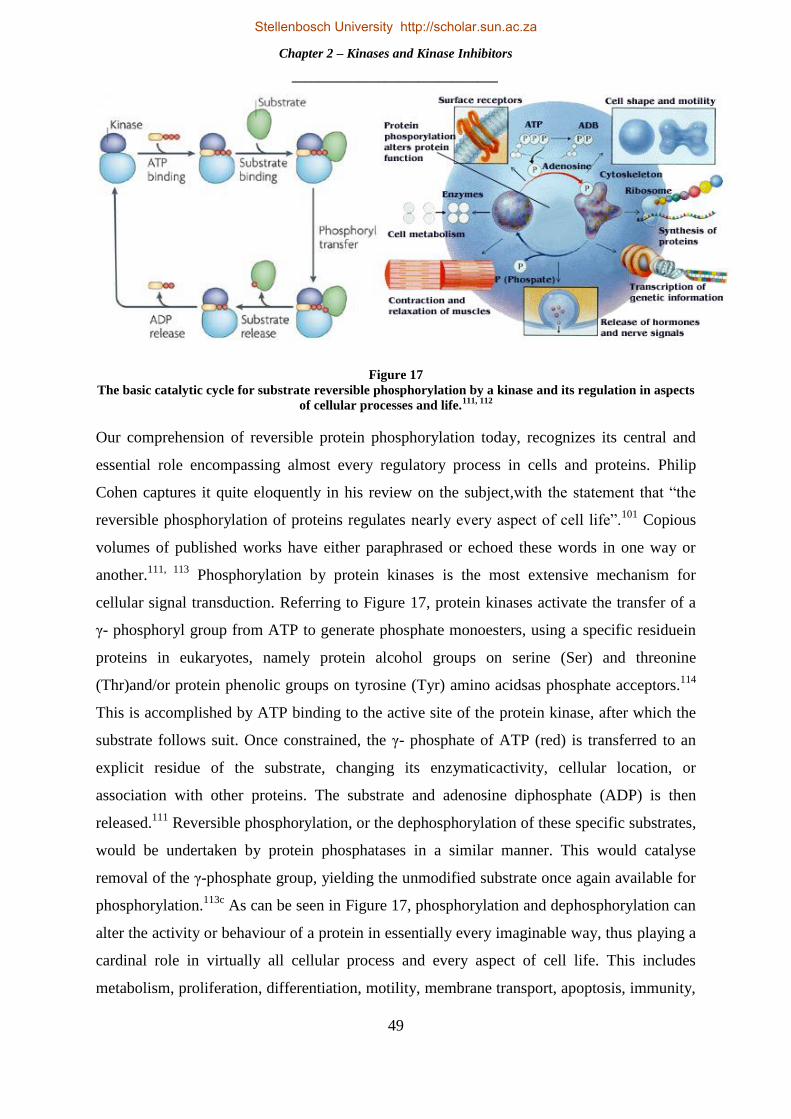
Chapter 2 – Kinases and Kinase Inhibitors
_______________________________
49
Figure 17
The basic catalytic cycle for substrate reversible phosphorylation by a kinase and its regulation in aspects
of cellular processes and life.111, 112
Our comprehension of reversible protein phosphorylation today, recognizes its central and
essential role encompassing almost every regulatory process in cells and proteins. Philip
Cohen captures it quite eloquently in his review on the subject,with the statement that “the
reversible phosphorylation of proteins regulates nearly every aspect of cell life”.101
Copious
volumes of published works have either paraphrased or echoed these words in one way or
another.111, 113
Phosphorylation by protein kinases is the most extensive mechanism for
cellular signal transduction. Referring to Figure 17, protein kinases activate the transfer of a
γ- phosphoryl group from ATP to generate phosphate monoesters, using a specific residuein
proteins in eukaryotes, namely protein alcohol groups on serine (Ser) and threonine
(Thr)and/or protein phenolic groups on tyrosine (Tyr) amino acidsas phosphate acceptors.114
This is accomplished by ATP binding to the active site of the protein kinase, after which the
substrate follows suit. Once constrained, the γ- phosphate of ATP (red) is transferred to an
explicit residue of the substrate, changing its enzymaticactivity, cellular location, or
association with other proteins. The substrate and adenosine diphosphate (ADP) is then
released.111
Reversible phosphorylation, or the dephosphorylation of these specific substrates,
would be undertaken by protein phosphatases in a similar manner. This would catalyse
removal of the γ-phosphate group, yielding the unmodified substrate once again available for
phosphorylation.113c
As can be seen in Figure 17, phosphorylation and dephosphorylation can
alter the activity or behaviour of a protein in essentially every imaginable way, thus playing a
cardinal role in virtually all cellular process and every aspect of cell life. This includes
metabolism, proliferation, differentiation, motility, membrane transport, apoptosis, immunity,
Stellenbosch University http://scholar.sun.ac.za

Chapter 2 – Kinases and Kinase Inhibitors
_______________________________
50
learning and memory.101,111
The selection of reversible phosphorylation as the universal
cellular regulative mechanism utilized by eukaryotic cells lies in its simplicity, adaptability
and the large concentration of ATP as a γ-phosphate donor.101
It has been predicted that
phosphorylation takes place on at least one suitable residue of up to 30% of all proteins. If
there are approximately 10000 different proteins in a normal cell, with an average length of
roughly 400 amino acids, of which 17% are Ser (8.5%), Thr (5.7%) and Tyr (3.0%) residues,
then there are a staggering 700 000 potential locations for phosphorylation to take place for
any given kinase.111, 115,116
The knowledge of phosphorylation, and its role, propelled
extensive research into protein kinases to answer questions pertaining to the specificity of
phosphorylation and its regulatory roles and its involvement in disease.
2.1.2 The Eukaryotic Protein Kinase Superfamily and the Protein Kinase Complement
of the Human Genome
With the last piece of the human genome sequence puzzle fitted in 2001, an opportunity to
determine the completeaccompaniment of human protein kinases presented itself.
Bioinformaticians at SUGEN, Inc. -a drug discovery company focused on development of
protein kinase inhibitors - led by Gerard Manning and Sucha Sudarsanam and including Tony
Hunter and Greg Plowman, embarked on a comprehensive effort to classify, characterise and
describe the full eukaryotic protein kinase (ePK) complement from genomic sequence
data.113a
The group exhausted all available human genome sequence sources - including all
those publicly available, Celera genomic databases, Genbank and many others not publicly
available - with a hidden Markov model profile to detect fragments bearing any similarity to
any known kinase. Following this, the use of homology modelling, predictive software and
verification of the accuracy of these techniques by DNA cloning, resulted in a complete
catalogue of 518 putative protein kinase genes. This eukaryotic superfamily, or kinome,
consisted of 478 ePK and 40 atypical protein kinases (aPK) – that is a variety of protein
kinases that do not share a substantial sequence overlap to the ePK domain, but have proven
kinase activity – approximating to 1.7% of all predicted human genes. Their efforts also
identified 106 kinase pseudogenes.113d
Classification of an extensive and intricate group such
as the human kinome, and the addition of model organism‟s kinases, meant division into a
hierarchy of groups, families and sub-families as can be seen in Fig 16.Thereare two main
subdivisions within the superfamily:the protein-serine/threonine kinases and theprotein-
tyrosine kinases.114
This superfamily contains 9 broad groups including tyrosine kinase (TK),
tyrosine kinase like (TKL), calcium/calmodulin-dependent protein kinase (CMCK), and
Stellenbosch University http://scholar.sun.ac.za

Chapter 2 – Kinases and Kinase Inhibitors
_______________________________
51
others shown in large on Figure 18. The 9th
group is not displayed and contains the aPK
family.Each of these broad groups is further sub-divided into a total of 134 families and 196
subfamilies. The classification of these kinases was carried out according to a number of
fundamental criteria, by sequence correlation of the active catalytic domains, supported by
sequence similarity outside the catalytic domain and physiological function.113d
The
knowledge acquired with the full complement of the ePK superfamily provided a
colossalimpact on further research on kinases and kinase inhibitors.113a
Figure 18
An overview of the human kinome.117
Stellenbosch University http://scholar.sun.ac.za

Chapter 2 – Kinases and Kinase Inhibitors
_______________________________
52
Figure 19
3D and 2D representation of the kinase ATP binding site, illustratingstructure and nomenclature.118
Most ePKs are structurally similar and are analogous by their canonical, homologous
catalytic domains consisting of approximately 250 – 300 amino acid residues.114
The majority
of kinasespossess a common fold composed of two lobes, a smaller N-terminus lobe
consisting of 5 antiparallel β-sheets and 1 helix and the C-terminus lobe consisting of α-
helices as can be seen in Figure 19a.111,118
A cleft or narrow hydrophobic pocket situated
between these two lobes houses the ATP binding site, where ATP resides with its phosphate
backbone protruding into the solvent tail – the area of the protein exposed to the solvent. The
ATP binding site is linked by a flexible hinge region which commonly consists of one
hydrogen bond donor flanked by two hydrogen bond acceptors arising from the protein spine
as can be seen in a 2D schematic inFigure 19c.118
The activation loop of the kinase represents
Ser, Thr and Tyr residues which may be phosphorylated and occupy a sector of the binding
site when not phosphorylated. After activation, this hydrophilic charged loop moves into the
solvent to expose the ATP catalytic domain allowing the protein or substrate to bind along
the cleft of the active site whereby transfer of the terminal γ-phosphate of ATPto the
hydroxyl oxygen of the Ser, Thr or Tyr residue of the substrate occurs.111
These substrates
can be seen in Figure 20. A large role in this phosphate transfer is played by the aspartate
residue which typically engages in a salt bridge interaction with a conserved lysine residue
shown in Figure 19c. The gatekeeper (gk) residue plays a significant role in defining kinase
inhibitor selectivity with the specificity pocket shown in Figure 19b. The size/volume of this
side chain governs entrance to the hydrophobic pocket located behind it as seen in Figure 19b
and c.118
a b c
Stellenbosch University http://scholar.sun.ac.za

Chapter 2 – Kinases and Kinase Inhibitors
_______________________________
53
Figure 20
Chemical structures of amino acids and ATP involved in protein phosphorylation.
Although protein kinases share a common fold and many structural affinities, differences in
relation to charge and hydrophobicity in the surface area of the active site result in distinct
kinase specificity.111
A number of structural relationships kinases have in common with one
another reflect in related cell regulatory functions.114
Neighbouring determinants in and
around the ATP binding site play a large role in determining substrate specificity and
particular phosphorylation sequences which take place in the protein
kinase.114
Exemplification of this is the difference in the depth of the catalytic cleft between
the two major classes of kinases namely the Ser/Thr - specific kinases, which constitute about
80% of protein kinases, and the Tyr-specific kinases.114
The difference in depth allows a Tyr
residue to traverse the length amid the protein backbone and the γ-phosphate of ATP, whilst
Ser/Thr residues cannot. This attribute is instrumental not only for the specificity of Tyr and
Ser/Thr kinases, but also their phosphatase counterparts.114
Distal binding motifs and docking
sites or additional binding sites, which are often spatially isolated from the catalytic domain,
provide supplementary binding interactions and sometimes allosteric regulation and
localization – leading to an increase in the affinity of kinases for particular substrates.Various
other mechanisms, such as competition and multisite phosphorylation, ensure proper
phosphorylation of corresponding substrates selectively to maintain the specificity that is
observed in signalling pathways.114
An understanding of these differences in selectivity and
specificity of phosphorylation and the regulatory effects thereof, as well as the catalytic
domain of the vast number of kinases, has increased the understanding in designing
increasingly efficacious and selective kinase inhibitors.
Stellenbosch University http://scholar.sun.ac.za
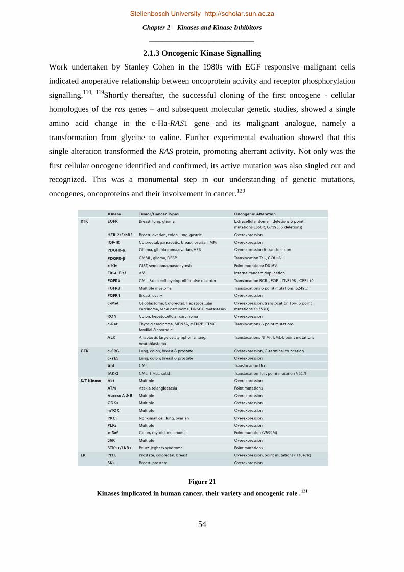
Chapter 2 – Kinases and Kinase Inhibitors
_______________________________
54
2.1.3 Oncogenic Kinase Signalling
Work undertaken by Stanley Cohen in the 1980s with EGF responsive malignant cells
indicated anoperative relationship between oncoprotein activity and receptor phosphorylation
signalling.110, 119
Shortly thereafter, the successful cloning of the first oncogene - cellular
homologues of the ras genes – and subsequent molecular genetic studies, showed a single
amino acid change in the c-Ha-RAS1 gene and its malignant analogue, namely a
transformation from glycine to valine. Further experimental evaluation showed that this
single alteration transformed the RAS protein, promoting aberrant activity. Not only was the
first cellular oncogene identified and confirmed, its active mutation was also singled out and
recognized. This was a monumental step in our understanding of genetic mutations,
oncogenes, oncoproteins and their involvement in cancer.120
Figure 21
Kinases implicated in human cancer, their variety and oncogenic role .121
Stellenbosch University http://scholar.sun.ac.za

Chapter 2 – Kinases and Kinase Inhibitors
_______________________________
55
Fast forward 20 years – with the unravelling of the full human kinome complement and
chromosomal mapping, 244 protein kinases were revealed to map to disease loci or cancer
amplicons.113d
164 of these were seen to be implicated with tumour amplicons - or replicative
genes, whilst 80 kinases mapped to loci associated with other diseases.113d
Figure 21
illustrates known kinase targets that has been intensely investigated and shown to be involved
in carcinogenesis, where RTK is receptor tyrosine kinase, CTK is cytoplasmic tyrosine
kinase, S/T Kinase is serine/threonine kinase and LK is lipid kinase.121
With the incredible
amount of regulatory activity and responsibility protein kinases have with regards to all
cellular processes, it is not surprising that perturbations of these signal transduction pathways
by mutations and other genetic alterations results in deregulation of kinase activity and
malignant neoplasm transformation.122
Certain classes of signalling proteins,i.e. those that
oversee cellular proliferation, differentiation and development, are targeted much more
sporadically by oncogenic mutations.122
Receptor protein tyrosine kinases (RPTK) represent
a significant allocation of all oncoproteins that are responsible for causation of a variety of
cancers as can be seen in Figure 21. Protein tyrosine kinases (PTK) phosphorylation activity
in resting, untransformed cells is usually tightly controlled by the antagonizing effect of
tyrosine kinase andphosphatases.123
However, with even slight genetic or structural mutations,
these PTK change into powerful oncoproteins which can wreak cellular havoc.122
Oncogenic
deregulation or transforming functions acquired by PTK can principally be attributed to
disruption or constitutive activation of one or more of the signalling pathways which are
regulated by PTKs to ensure conservation of catalytic domains and cellular function. This
results in activation of further signalling pathways, proteins and messengers impeding regular
cellular processes.122, 123
Aberrant activity may be brought about by a variety of mechanisms;
however, there are four main principals to oncogenesis or oncogene addiction in PTKs.
Firstly, abnormal or over-expression of PTKs, or an over expression of its associated ligand,
is concomitant with autocrine-paracrine stimulation which has been linked to array of human
cancers. Overexpression of EGFR is reported in 40-80% of non-small cell lung
cancer(NSCL) and up to 50% of primary lung cancer. EGFR and ligand over-expression is
depicted in panel A of Figure 22, with PTKs known to be activated through that mechanism
listed on the right. Secondly, gain of function (GOF) mutations or mutations within the
extracellular domain of RPTKs and CTKs has been associated with several neoplasmic
malignancies, e.g mutations in EGFR amino acids have led to uncontrolled cellular growth of
ovarian tumours and non-small cell lung carcinoma.
Stellenbosch University http://scholar.sun.ac.za
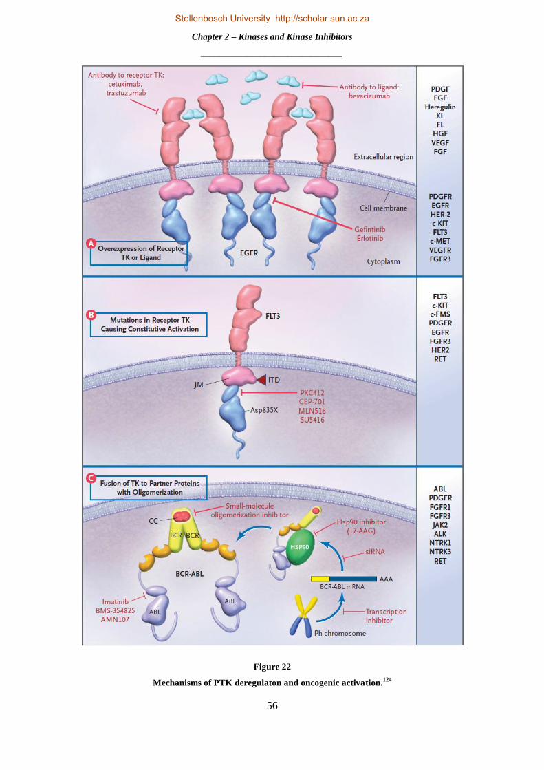
Chapter 2 – Kinases and Kinase Inhibitors
_______________________________
56
Figure 22
Mechanisms of PTK deregulaton and oncogenic activation.124
Stellenbosch University http://scholar.sun.ac.za

Chapter 2 – Kinases and Kinase Inhibitors
_______________________________
57
Panel B of Figure 22 shows constitutional activation of Fms-like tyrosine kinase 3 (FLT3)
through a point mutation.Thirdly, abnormal chromosomal translocation and genomic
rearrangements may result in the cancerous fusion of proteins and resulting in irregular
functionality. A representation of this is CML, which arises from a reciprocal translocation
between chromosomes 9- and 22, as highlighted in Section 2.1.2. This fusion is illustrated in
panel C of Figure22.Lastly, in rodents and chickens, retroviral transduction of proto-
oncogenes related to a PTK leads to constitutional transitions – resulting in malignant
activity.122, 123,124
With deregulation of signalling, mutation and over expression of PTKs
playing a colossal role in the molecular pathogenesis of cancer, as well asthe wealth of
knowledge on the enzymes themselves and their congruency as drug targets, the design and
synthesis of kinase inhibitors as targeted therapeutic agents of the PTks has been a very
successful and attractive endeavour for the last 20 years.
2.2 KINASE INHIBITORS
2.2.1Protein Kinase Inhibitors – an Overview.
A search on Scifinder for the term “kinase inhibitor” yields ~90 000 references containing the
word as entered and ~310 000 containing the concept. To date 26 protein kinase inhibitors
have received FDA approval of which 17 are small molecules, with the remainder consisting
of both anti-angiogenic and monoclonal antibody inhibitors of Ser/Thr and Tyr kinases.125
Approximately 142 inhibitors, ranging over 42 different kinase targets have progressed to
some phase of clinical assessment.126
Although the vast majority of these are related to the
treatment of cancer, kinase inhibition has been implicated in the remedy of an array of
ailments inclusive of immunological, neurological, metabolic and infectious disease.121
With
protein kinases comprising a large share of all oncoproteins and aberrant cellular activity
playing such a pivotal role in the pathogenesis of a myriad of diseases, it is not surprising that
widespread interest and impetus into the targeted inactivation of this class of enzyme has
been witnessed. This has resulted in a tremendous investment of resources, especially from
the pharmaceutical sector, towards the development of numerous efficient strategies for
designing potent and selective kinase inhibitorsfor suppression of these unwanted cellular
processes and treatment of these disorders.121, 123
The prominence of kinases as one of the most intensively pursued classes of drug targets may
be attributed to a number of factors. Standing true to the adage, “kinases are responsible for
the regulation of nearly every aspect of cellular life,” and with 518 kinases in the human
Stellenbosch University http://scholar.sun.ac.za

Chapter 2 – Kinases and Kinase Inhibitors
_______________________________
58
kinome, almost every signal transduction pathway goes via a phosphotransfer cascade,
meaning the inactivation of this target is viable and will yield a real biological response.124
As
was discussed before, the emergence of imatinib and its success displayed this aspect of
kinase inhibition and was the herald for a proof of principle of this targeted therapy.78
Despite
a high degree of similarity and preservation in the active site of kinases, highly selective
inhibitors with positive therapeutic characteristics can and have been developed.127
Moreover,
and quite surprisingly, inhibition of regular cell kinase activity is well tolerated in the human
body, allowing for the selective felling of malignant cells. This is illustrative in dasatinib, an
inhibitor of BCR/Abl and Src tyrosine kinases and used in the treatment of imatinib resistant
CML, which potentially inhibits all 9 members of the Src family, and possibly others, whilst
showing minimal adverse events and a favourable side effect profile.124
Targeting and
inhibition of genetically mutated or activated kinases has been fortuitous in the relative ease
of designing mechanistically relevant cellular assays, as these kinases are promptly identified
by DNA sequencing due to their inherent anomalies. This gives a distinct advantage in
inhibitor discovery and optmization.124
Kinase inhibitors have shown marked success in
clinical trials and as approved therapeutic agents. In an evaluation of 974 anticancer drugs,
kinase inhibitors shone with a clinical attrition rate of 53% from phase I to approval,
considerably lower in contrast to the 82% achieved for other antitumour remedies.126
Besides
imatinib, exhibiting up to 80% reduction in tumour growth in chronic phase CML patients,79
other FDA approved inhibitors have shownbeneficial effects in patients. Crizotinib, an
inhibitor of an anaplastic lymphoma kinase (ALK) fusion protein linked with carcinogenesis,
showed tumour shrinkage and growth halt in 90% of 82 patients carrying this ALKfusion
gene.128
Lapatinib is associated with the inhibition of two oncogenes namely, EGFR and
HER2. Overexpression of HER2 has been shown to be conducive to breast cancer and in
combination therapy with capecitabine, showed 51% reduction in the risk of disease
progression.129
Erlotinib is an inhibitor of EGFR, which is over expressed in certain malignant
cells such as those of breast and lung carcinoma. In combination with the use of cytotoxic
drugs, erlotinib has shown improved overall survival in patients by 19%, whilst progression
free survival (cancer is not exacerbated) was increased by 29%, in comparison to the use of
cytotoxic drugs alone.130
However, despite the inspiring successes and advantages of this branch of targeted therapy,
the field of kinase inhibition faces a number of challenges. These include drug resistance due
to further mutations, lack of inhibitor selectivity and efficacy and challenges in drug target
Stellenbosch University http://scholar.sun.ac.za

Chapter 2 – Kinases and Kinase Inhibitors
_______________________________
59
validation associated with its particular disease.124
Kinase inhibitors are very efficacious for
patients with single genetic alteration type cancers, in contrast to those carcinomas that carry
a large number of different mutations where treatment by kinase inhibition is limited. Thus,
proper subject selection has become a main concern in kinase drug discovery.126
Resistance to
small molecule kinase inhibitors is best exemplified with gefitinib, which targets EGFR
which is over expressed in 40-80% of NSCLC and other epithelial malignancies.131
Tumours
of patients that exhibited strong responses to therapy usually contained abnormalities in the
EGFR gene. These genetic mutations were found to most frequently be either deletions in the
amino acid chains between 747 – 750 (delL747-S752), or a point mutation - a replacement of
one gene with another. These consist virtually always of substitution of arginine with leucine
at codon 858 (L858R) and methionine which is substituted for threonine at position 790
(T790M).132
These mutations lead directly to an increase in alteration of signalling pathways
and carcinogenesis and thus facilitate oncogenic activity. Patients with the delL747-S752
mutation wereseen to show increased tumour susceptibilty to gefitinib, which resulted in
interim regression of tumour growth and succesful treatment. However, following a
remission period a second mutation was seen to develop, in this case the T790 mutation,
which shows high level resistance to the anilinoquinazoline inhibitor. This showed that
relapses in patients with NSCLC usually contained mutations in EGFR which conferred
resistance to gefitinib.132
In another example, mutations in BCR-ABL associated with imatinib
resistance usually result in replacement of threonine with isoleucine at position 315, which
corresponds structurally to a very similar change to that of the T790 mutation.132
A greater
understanding of the mechanisms of resistance in CML has resulted in the development of a
2nd
generation library of inhibitors.133
With the advancement of large selectivity screening
platforms, it has been shown that the majority of approved kinase inhibitors show inadequate
selectivity and are promiscuous in targeting mulitple kinases. With the exception of lapatinib,
this places restrictions on the applicability of late stage cancer treatment and its use as
therapeutic agents in general.126
Accessibility of high-resolution crystal structures
hasmarkedly expedited the design of increasingly selective inhibitors and coupled with the
development of allosteric and irreversible inhibitors, has done much to combat these
issues.134
Target validation remains problematic in kinases and has resulted in a constricted
selection of kinase targets. More than 50% of all inhibitors in clinical evaluation targets
kinases with already approved drugs on the market. This suggests that a proof of concept is
necessary in kinase drug discovery. Almost the same percentage of kinases are
Stellenbosch University http://scholar.sun.ac.za

Chapter 2 – Kinases and Kinase Inhibitors
_______________________________
60
uncharacterized with nearly 25% having completely unknown function, with little to no
explanation as to their implication in signalling pathways.126
However, the introduction of
specificity screening platforms and chemical probes that target a number of these unknown
kinases have shown that current clinical and preclinical candidates display inhibitory activity
of these kinases.126
The use of modern cytogenetics to identify chromosomal transformations
and array comparative genomic hybridization techniques may also be utlized for the
identification of new tyrosine kinase drug targets.124
2.2.2Design, Synthesis and Modes of Binding.
In modern drug discovery, advances in technology, protein ligand interactions and cellular
based assays and screening methods, has resulted in the design, development, synthesis and
optimization of hugely succesful anti cancer agents.59,123
A search for the term “kinase” in the
protein data bank (PDB), yields 6540 crystal structure with nearly 2700 ligand hits. The
importance of structural biology in the campaign of kinase inhibitor drug discovery is no
more evident than in these numbers. Technological leaps in x-ray crystallography and related
disciplines has facilitated the rapid and low cost solution of de novo compounds and
structures, yielding important protein:drug interactions in a typically cheaper and faster
method than that of compound synthesis and in vitro evaluation.113b
The world wide protein
data bank (wwPDB) acts as a deposition for all protein related data, and thus a plethora of
crystal structures are available for scrutiny and research. This provides structural insight into
kinase inhibitors and their specific binding modes in the active site and assists in the
structure-guided design of specific kinase inhibitors.135
Thorough profiling of kinase
inhibitors through biochemical, cell base assays such as fluorescent polarization assay and
nonradioactive high throughput assays, have provided insight into target validation and
selectivity.123
Pharmacaphore and molecular modelling, docking and structure-aided ligand
design tools of increasing refinement have also found a place in drug design and
optimization.118
The combined and cumulative usage of 2D NMR spectroscopy,in silico
computer-assisted methods, structural biology, enzymatic and cellular based assays,
medicinal chemistry and synthetic experimental evaluation efforts together, can defined as
structure assisted design and optimization in modern drug discovery.59
Kinase inhibitors consist of different classes which are distinguished by their mode of
binding in the ATP active site, as well as their location in the enzyme itself. This class of
anticancer agents can thus be divided into three types, namely: type I or ATP
Stellenbosch University http://scholar.sun.ac.za

Chapter 2 – Kinases and Kinase Inhibitors
_______________________________
61
competitive,type II or allosteric and type III or covalent inhibitors. In spite of the large
amount of conservation in the sequencing of the ATP binding site of kinases, the majority of
reported and approved inhibitors are ATP competitive or reversible inhibitors.113b
Figure 23
Kinase inhibitor type I and II binding modes.121
These type I inhibitors exploit the active conformation of the kinase. These inhibitors usually
consist of a heterocyclic core, which serves to form weak non-covalent hydrogen
bondacceptor/donor interactionswith the hinge binding region in the active site, as can be
seen in Figure 23a. Furthermore, this heterocyclic system acts a scaffold for side chains
which are used to fill the hydrophobic pockets located in the active site seen shaded in grey
and yellow in Figure 23a. This provides further hydrophobic interactions and increases
selectivity in the inhibitors. Examples of these inhibitors include imatinib and nilotinib.
Type II or allosteric inhibitors in contrast, target the inactive form of the kinase, also known
as the DFG out conformation due to the configuration of this chain.121
This configuration of
the DFG out motif, from movement of the activation loop, bares a peripheral hydrophobic
binding site flanking the ATP active site, where type II inhibitors reversibly inhibit the
Stellenbosch University http://scholar.sun.ac.za

Chapter 2 – Kinases and Kinase Inhibitors
_______________________________
62
protein kinase as illustrated in Figure 23b (the blue shaded area defines this additional
hydrophobic binding site.)121
Allosteric inhibitors bind outside the ATP binding site, at an
allosteric site separate from the enzyme active site, where ligand binding takes place to effect
kinase activity. They are non ATP competitive as they block ATP phosphorylation remotely
from the active site.113b
Allosteric inhibitors are thus highly selective as they target explicit
binding sites that are unique to certain kinases.121
The last class of kinase inhibitors, type III, are irreversible or covalent inhibitors. This class
of drug inhibits irreversibly, by forming a covalent bond to a specific cysteine residue located
in the ATP active site.121
Irreversible inhibitors, like their reversible counterparts, consist of a
heterocyclic core or “driving group” which fulfils the same role as in reversible inhibitors,
forming weak hydrogen bond interactions with the hinge region. However, covalent
inhibitors carry at a designated position a suitable electrophilic functionality known as a
“warhead.” It is this “warhead” that forms a covalent bond with a nucleophilic cysteine
residue located inside the binding site, usually through a Michael addition reaction as an
example. The mechanism of this covalent bond formation and structure of the irreversible
inhibitor canertinib, which has shown activity against EGFR and HER2, can be seen on
Figure 24 below.In this reaction the electron rich sulphur group of the cysteine residue reacts
at the electrophilic β-carbon of the acrylamide warhead. The intermediate enolate then forms
the 1,4- addition product.136
Figure 24
Notable structural moieties and mechanism of covalentinhibition with Canertinib as example.136
Stellenbosch University http://scholar.sun.ac.za

Chapter 2 – Kinases and Kinase Inhibitors
_______________________________
63
Irreversible inhibition holds a number of advantages over ATP competitive inhibition. Firstly,
irreversible inhibitors contain the structural motifs necessary for reversible inhibition, thus
irreversible inhibitors benefit from weak hydrogen bond interactions in the active site, as well
as stronger covalent bond interactions. Irreversible inhibitors are not in competition with high
endogenous concentrations of ATP in cells. This also means irreversible inhibition is
unaffected by changes in the ATP binding affinity of the kinase through point mutations, a
mechanism to overcome some forms of drug resistance. Irreversible inhibition has a longer
duration of effect and has been to shown to persist in deactivation of the enzyme even after
the free inhibitor has left circulation. Lastly, a high selectivity profile is associated with these
inhibitors as only kinases with the appropriate cysteine residue in the correct place should
undergo covalent bond formation and irreversible inhibition.134b, 136
Of the 518 kinases in the
human kinome only 43% (or 211) have at least one cysteine residue in the ATP binding site
spread over 27 different positions. This means that a finite number of these candidates are
druggable, which brings about an increased chance of developing potent and selective
inhibitors.137
However, promiscuity and toxicity remain the biggest challenges in the
development of irreversible inhibitors. Selectivity, amongst not only the kinome, but also
other cell constituents becomes an issue. However, with a vast library of inhibitor kinase co-
crystal structures, refinement of driving portions, as well as proper placement of warheads,
has resulted in overcoming these complications.136
Protein kinases and kinase inhibitor design, development and synthesis has become one of the
most significant drug discovery categories in oncology.126
Technological advancements,
developments in high throughput sequence screening for target validation and innovation in
structural biology all aid the rational design and synthesis of novel kinase inhibitors, thereby
increasing the viability and opportunities for drug discovery development in this field. This is
therefore an attractive area for medicinal chemists to investigate and has become the main
focus of many research groups.
Stellenbosch University http://scholar.sun.ac.za

Chapter 3 – 1st Generation Reversible Inhibitor Library
_______________________________
64
CHAPTER 3 – 1ST
GENERATION REVERSIBLE INHIBITOR LIBRARY
3.1 SYNTHETIC METHODOLOGY, TARGET STRATEGY AND DRIVING GROUP
3.1.1 The Paper that started it all.
Recently, Müller and co-workers described an innovative way to prepare mimics of naturally-
occurring kinase inhibitors, such as meriolin, by following synthetic methodology they
developed.138
This synthetic procedure, which can be seen in Scheme 1b, involved a one pot
Sonogashira coupling, followed by a TMS deprotection and a copper-mediated click reaction
to afford 1,2,3-substituted triazoles, in good yields, from the corresponding halogenated
precursors shown as in Scheme 1a.138
Scheme 1
Synthetic concept and methodology used by Müller for N-Boc protected heterocycles.138
b)
Stellenbosch University http://scholar.sun.ac.za

Chapter 3 – 1st Generation Reversible Inhibitor Library
_______________________________
65
A library of these triazolyl NH-heterocycles was synthesized and not surprisingly, as these
mimics contain a heterocyclic core with a hinge binding motif corresponding with reversible
kinase inhibition, the group found that a number of the compounds were excellent inhibitors
of pyruvate dehydrogenase lipoamide kinase isozyme 1 (PDK1). To determine whether the
triazole moiety simply acted as a linker or retained additional functionality important for
inhibition an analogue of the most efficacious compound was prepared whilstsubstituting the
triazole moiety with a pyrazole ring. Interestingly, the triazole-bearing compound8f was
shown to act as more than just a simple linker, displaying a pharmacophore character
essential for biological activity. It was shown that the pyrazole compound11 was
significantly less active with an IC50 value of 2.6 µM for PDK1 compared with 0.8 µM for
the triazole as can be seen in Figure 25.Furthermore, an isomeric compound 10 was
synthesized, varying only in the positioning of the triazole moiety‟s carbon and nitrogen,
which showed no activity against PDK1. A crystal structure of compound 8f with kinase
PDK1 indicated the presence of hydrogen bond interactions between the triazole nitrogenN3
with Thr222 and a bridging interaction between N2 and H2O and Lys111.138
Figure 25
Comparison of IC50values of PDK1 inhibition between triazole, isomeric and pyrazole compounds and
crystal structure of compound 8f in PDK1.138
Stellenbosch University http://scholar.sun.ac.za

Chapter 3 – 1st Generation Reversible Inhibitor Library
_______________________________
66
Heterocycles containing 1,2,3-triazole systems have been reported to possess various
biological activities including anti-HIV, anti-allergic, anti-fungal, anti-viral and anti-
microbial activity.139
1,2,3-Triazole moieties have also been shown to be useful building
blocks in chemistry and have been modified to play important roles in pharmacological
applications due to their stability towards light, moisture, oxygen and metabolism in the
body.140
Moreover, these moieties have found a range of applications in industry as dyes,
corrosion inhibitors, photographic materials and agrochemicals.141
In 1,4-disubstituted 1,2,3-
triazoles, N2 and N3 have been shown to act as H-bond acceptors.142, 143
Their resistance to
hydrolytic cleavage and oxidative transformations has also earned them their ability to act as
bioisosteres for moieties sensitive to these conditions, such as esters, thioesters, and
thioamides.143
Triazoles used as linkers have been shown to form planar structures, which
would promote eligibility for the narrow ATP binding site.144
This has generated considerable
interest in the developmentof new and more efficient synthetic pathways for the design of a
diverse array of 1,2,3-triazole-containing heterocycles as possible drug candidates, as well as
investigation of their ability as pharmacophores. Thus, on this basis, it was our endeavour to
adopt the synthetic methodology formulated by Müller and his team, to be applied to various
halogenated heterocyclic analogues of our choosing, which also contained the important
hinge binding motif for kinases. This would be an excellent platform to determine the
triazole‟s ability to act as a hydrogen bond acceptor with the hinge region or within the active
site of the target kinase and to generate a 1st generation library of novel potential reversible
inhibitors and determine their efficacy.
3.1.2Rationale of Target Kinase: EGFR
Lung cancer accounts for over a third of all cancer deaths and is the leading cause of death in
men worldwide, coming second in woman only to breast cancer.145,146
It can be seen as a
miscellany of diseases largely divided into two groups namely: small cell lung cancer
(SCLC) accounting for ~20% of lung cancers and non-small cell lung cancer (NSCLC)
comprising of the remaining ~80% of lung cancers.146
NSCLC is postulated to originate in
lung epithelial cells, and is made up of a large variety and number of cellular malignancies
such as adenocarcinoma, bronchioloalveolar, squamous, anaplastic and large-cell
carcinomas.146
The EGFR family or ErbB tyrosine kinase receptors, constitutes four
homologous receptors namely: the epidermal growth factor receptor (ErbB1/EGFr/HER1),
ErbB2 (HER2/neu), ErbB3 (HER3), and ErbB4 (HER4), whose functions has been
investigated and studied extensively.147
The role of EGFR driven signalling in the aetiology
Stellenbosch University http://scholar.sun.ac.za

Chapter 3 – 1st Generation Reversible Inhibitor Library
_______________________________
67
of human canceris well known148
and as has been elaborated and clarified before. Over
expression of these receptors is abundant in tumours, as well as autocrine or paracrine-
stimulated growthin malignancies.147a
EGFR has been correlated to a multitude of cancers,
including that of the head and neck, brain, colorectal, bladder, breast and lung.146
Recent
analyses have shown that EGFR is over expressed in 62% of NSCLC patients.Also, the
chromosomal region of EGFR, 7p12, was found to be augmented in genomic examinations.
Additional to over expression and chromosomal transformations, the receptor ligands
epidermal growth factor (EGF) and transforming growth factor (TGF) have also been found
to be commonly over expressed in NSCLC. This leads to aberrant autocrine functionality and
uncontrolled activity and growth of the receptor.146
Approximately 40% of patients diagnosed
with NSCLC today are at an advanced stage. First line therapy for this disease consists of
platinum-based combination chemotherapy; however prognosis is poor with median survival
rates of 8-11 months, 1 year survival rates of ~35% and 2 year survival rates of 10-20% being
reported.145
The treatment of NSCLC is thus an on-going struggle and new therapeutic targets
of the molecular mechanisms that govern the growth of these malignancies are desperately
needed.
A range of drug discovery strategies has been developed to address this need, including the
use of monoclonal antibodies such as cetuximab and bevacizumab and small molecule kinase
inhibitors such as gefitinib, erlotinib and lapatinib whose molecular structures can be seen in
Figure 26 below. Gefitinib, a reversible inhibitor, has increased our understanding of NSCLC
and mutations of EGFR through specific resistance mechanisms to this treatment.Erlotinib, as
discussed before in Section 2.2.1, has shown a benefit to patients with advanced lung cancer,
some showing complete response, whilst others only partial to the treatment.145
Lapatinib is
seen as a dual tyrosine kinase inhibitor as it interrupts both the HER2 and EGFR signalling
pathways and is used primarily for the treatment of breast cancer where it has shown marked
success.129
Figure 26
Molecular structures of gefitinib, erlotinib and lapatinib.
Stellenbosch University http://scholar.sun.ac.za

Chapter 3 – 1st Generation Reversible Inhibitor Library
_______________________________
68
Following the lead of these FDA approved drug candidates, the EGFR kinase domain would
seem a logical and attractive target for drug discovery. For this project we would be
collaborating with a team of German biochemists from the Technische Universiteit
Dortmund, led by Prof. Daniel Rauh. A student in Prof. Rauh‟s group, Julian Engel, would be
responsible for carrying out the biological assays and determining the potency and efficacy of
our library of potential inhibitors. Julian‟s main focus of research is that of EGFR, which was
accordingly selected as our target kinase for this project as screening could be facilitated.
3.1.3 Choice of Heterocyclic Driving Groups and Side Chains.
Quinoline, a heterocyclic aromatic compound, was first extracted from coal tar in 1834,
which still remains the primary source for its industrial usage. Although the heterocycle itself
has limited application, derivatives thereof have been shown to possess an enormous variety
of uses in extensive fields, and italso occurs in a number of natural products and alkaloids.149
A natural product derivative, quinine, and further synthetic analogues thereof such as
chloroquine,150
primaquine151
and mefloquine,152
have been extensively researched and found
great success in the treatment of malaria. Biological activity of compounds containing a
quinoline backbone has been found in anti-tuberculosis,153
anti-microbial,154
anti-
convulsant,155
anti-inflammatory,156
cardiovascular157
and most importantly, anticancer
applications. Quinoline as a privileged anticancer scaffold shows activity in a number of
different target sites such as topoisomerase I, telomerase, farnasyl transferase, Src tyrosine
kinase, protein kinase CK-II and others.149
As protein tyrosine kinase inhibitors, quinoline-
based driving groups have shown potent IC50 values for a range of kinases and different types
of cancers.61, 158
Isoquinoline, a benzopyridine derivative like quinoline, was also first isolated from coal tar
51 years after its structural isomer. This heterocycle is the structural spine of a large number
of natural alkaloids. Isoquinoline analogues have found a variety of uses as anaesthetics,
antihypertension agents and vasodilators.159
A number of derivatives have been applied in
anti-malarial, anti-HIV, anti-tumour, anti-microbial and anti-bacterial usage.160
Most
importantly, isoquinoline-derived drugs have shown protein tyrosine kinase inhibitory
activity against a number of kinases including EGFR.161
Stellenbosch University http://scholar.sun.ac.za

Chapter 3 – 1st Generation Reversible Inhibitor Library
_______________________________
69
Scheme 2
Synthetic scheme illustrating choice of heterocyclic driving group, side chains and reactions.
On the premise of these findings and research already carried out using these heterocyclic
cores, we decided to make use of two halogenated structures of both quinoline and
isoquinoline which can be seen in Scheme 2 above (1a-d). Following the synthetic approach
devised by Müller, Sonogashira coupling and deprotection would afford heterocyclic alkynes
which could subsequently undergo a copper catalysed azide alkyne cycloaddition (CuAAC)
with appropriate azides, affording a library of reversible, 1,4 1,2,3- triazole-based potential
kinase inhibitors.A number of molecular modelling and docking calculations were
undertaken making use of Acclerys Discovery Studio software. The investigation visually
showed promising results and binding modes adopted by these compounds. A number of
hydrogen bond acceptor/donor interactions were seen between the quinoline and isoquinoline
moieties, and the methionine and threonine hinge binding region. The choice of benzyl azide
and 2-(azidomethyl)pyridine (Scheme 2) as suitable side azide side chain fragments to
undergo this CuAAC was made on the grounds of simplicity, as well as potential
hydrophobic interactions, as seen in the molecular modelling and docking calculations. As
both moieties were predicted to fit in the hydrophobic pocket of the active site, an aromatic
core with a flexible methyl linker for rotation was logically chosen.The addition of nitrogen
in the pyridine moiety, and a lack thereof in the benzyl variation, would give an indication as
to whether the heterocyclic counterpart took part in any favourable hydrogen bond
interactions, possibly increasing efficacy and selectivity. This provided further
encouragement for the use of these driving groups and side chain linkers for the synthesis of
our potential reversible inhibitors.
Stellenbosch University http://scholar.sun.ac.za

Chapter 3 – 1st Generation Reversible Inhibitor Library
_______________________________
70
3.2 SYNTHESIS OF ALKYNE FRAGMENTS
3.2.1 Sonogashira Coupling and Silyl Deprotection.
The palladium-catalysed coupling of aryl or vinyl halides (or triflates) with terminal
acetylenes is generally referred to as a Sonogashira coupling reaction.162
Garnering its name
from Kenkichi Sonogashira, Yasuo Tohda, and Nobue Hagihara, who first reported usage of
the reaction in 1975,163
it was an extension of the work done by Heck but allowed for much
milder conditions to be used.162
Nearly 30 years on, the robust Sonogashira coupling has
transitioned to one of the most central and well established methods for sp2–sp carbon–
carbon bond formation in organic synthesis,162
finding widespread application in the
preparation of arylalkynes and conjugated enynes,a lynchpin in natural product synthesis and
in heterocylic chemistry.164
This has also extended to use in the synthesis of molecular
electronics, dendrimers and conjugated polymers or nanostructures.162
Conventionally, a
copper co-catalyst is added, which provides increased reactivity, but copper-free Sonogashira
synthetic methodology has been explored and developed.165
Standard Sonogashira reaction
conditions employs some source of palladium such as PdCl2(PPh3)2 as catalyst, in
conjunction with a co-catalytic amount of Cu(I) such as CuI, with an amine as solvent or an
inorganic base as shown in scheme 3.162
The precise mechanism for the Sonogashira coupling is not yet accurately understood, due to
complications in analysis as two metal catalysts are present, which sheds doubt on certain
intermediates that are formed. However, it is generally accepted that the reaction progresses
through two separate catalytic cycles, namely the palladium and copper cycle as illustrated in
Scheme 3. Firstly, the palladium cycle is well agreed upon as it is derived from classical
carbon-carbon cross-coupling reactions.166
The cycle begins with the inactive Pd(II) catalyst,
such as PdCl2(PPh3)2, being reduced to the active 14 electron palladium species
[Pd(0)L2],shown as complex A in scheme 3. This complex then undergoes oxidative addition
of the aryl or vinyl halide, which is reasoned to be the rate-limiting step of the reaction,
forming the adduct [Pd(II)R1L2X], or complex B in Scheme 3. This adduct then undergoes
transmetallation with the copper acetylide complex F, formed during the copper cycle, which
in turn results in complex C after expulsion of the copper halide complex G which is
regenerated. The trans organic ligands of complex C undergo cis/trans-isomerisation,
yielding complex D, following subsequent reductive elimination affording the alkyne product
and regenerating the [Pd(0)L2] catalyst.162
Stellenbosch University http://scholar.sun.ac.za

Chapter 3 – 1st Generation Reversible Inhibitor Library
_______________________________
71
Scheme 3
Standard Sonogashira coupling reaction conditions and catalytic cycle adapted from Chinchilla et al.162
In comparison to the palladium cycle, the copper cycle is unexplored and esoteric, with most
of the mechanistic details hypothesized. It is believed that the presence of base promotes
copper acetylide formation through the formation of an intermediateπ-alkyne copper complex
E, which in turns makes the terminal alkyne‟s proton more acidic, resulting in the copper
acetylide complex F which undergoes transmetallation in the palladium cycle, regenerating
the copper halide catalyst.162
The use of silyl protecting groups in organic synthesis has become a routine procedure.167
The utilization of these groups in organic synthesis has extended across a wide variety of
Complex A
Complex B
Complex D
Complex C
Complex F
Complex E
Complex G
Transmetallation
Stellenbosch University http://scholar.sun.ac.za

Chapter 3 – 1st Generation Reversible Inhibitor Library
_______________________________
72
functional groups including formation of silyl ethers in protection of hydroxyl groups, as well
acetylene hydrogen protection. In the case of acetylene, the use of a protecting group for an
acetylinic hydrogen is often necessary due to its acidic nature.168
In addition the steric bulk of
these silane moieties prohibits catalytic hydrogenation.169
The use of trialkylsilylacetylenes in
organic synthesisis convenient as they tend to be liquids or solids, as opposed to the
unprotected gaseous counterparts.168
These protecting groups are frequently cleaved or
removed through a fluoride source, the driving force of this reaction being the formation of
the stronger Si-F bond (564.8 kJ/mol) in comparison to that of the Si-C bond (451 kJ/mol).
Trimethylsilylacetylene (TMSA) is frequently used in Sonogashira coupling reactions with
aryl or vinyl halides.162
3.2.2 Optimization of Reaction Conditions.
For the synthesis of our alkyne fragments, the halogenated heterocyclic scaffold of 3-
bromoquinoline (1a) was selected for optimization of this synthetic procedure. As this
reaction sequence had not been previously applied to quinoline or isoquinoline scaffolds, the
one pot methodology was provisionally abandoned and products were isolated and purified at
various steps throughout the synthesis of this alkyne fragment. The reaction sequence that
followed would entail a Sonogashira coupling of the heterocyclic halide with TMSA
followed by in situ deprotection of the silyl protected alkyne intermediate. This was done via
an appropriate source of fluoride, namely tetrabutyl ammonium fluoride (TBAF) as the tri-
hydrate, as can be seen in Table 1. Our first attempt entailed recreating conditions as used by
Müller and co-workers,138
using appropriate inert Schlenk and degassing techniques.
However, the yields initially obtained were poor and the results obtained by Müller could not
be reproduced under these conditions. A number of optimization modifications were
undertaken, including increasing the amount of catalyst and co-catalyst, the amount of base
added, as well as the source and amount of fluoride for deprotection which can be seen
summarized in Table 1. THF was kept as solvent and reaction times were kept constant at 12
h for the optimization process. As can be deduced from this experimental evaluation,
changing of the parameters afforded little profit in the yield obtained, with exception to the
use of liquid TBAF as fluoride source and longer degassing periods. It was only after
employing the freeze-pump-thaw cycling technique for degassing of liquids that the alkyne
heterocyclic compound was afforded in very good yields.
Stellenbosch University http://scholar.sun.ac.za

Chapter 3 – 1st Generation Reversible Inhibitor Library
_______________________________
73
Table 1
Optimization of Sonogashira coupling for 3-bromoquinoline.
Attempt
PdCl2(PPh3)2
(mol. %)
CuI
(mol. %)
Et3N
(equiv.)
Flouride source
(equiv.)
Degassing
(min)a
Yield
(%)
1 2 4 2
TBAF.3H2O
(1.5)
- 37
2 4 4 2
TBAF.3H2O
(1.5)
15 50
3 4 6 3
TBAF.3H2O
(2)
30 52
4 4 4 2
Liquid TBAF
(1.5)
30 64
5b 4 4 2
Liquid TBAF
(1.5)
30 85
a = Performed by passing N2 through reaction.
b = Freeze-pump-thaw technique was also applied for degassing.
This laboratory-scale technique, involves the flash freezing of the fluid to be degassed in a
Schlenk tube usually with liquid nitrogen and the application of a vacuum thereafter. Upon
subsequent thawing, bubbles of gas are seen to form and escape to the top head space of the
flask. This process is typically repeated between 3-6 times until all air is removed from the
reaction vessel and fluid.170
This afforded our first alkyne fragment2a in good yield.
Subsequent 1H NMR spectroscopic analysis confirmed formation of
the unprotected alkyne product. The acetylenic proton 1 was
identified as a singlet at 3.28 ppm integrating for 1H. The aromatic
proton peaks of 4 and 5, were seen as a triplet of doublets
Stellenbosch University http://scholar.sun.ac.za

Chapter 3 – 1st Generation Reversible Inhibitor Library
_______________________________
74
integrating for 1H each at 7.56 and 7.63 ppm. Peaks corresponding to proton6 and3,were
witnessed as doublets at 7.77 and 8.09 ppm, both integrating for 1. Proton 2 was seen as a
singlet at 8.27 ppm integrating for 1H and proton 7 could also be seen as a singlet at 8.94
ppm, integrating for 1H.
Application of these synthetic conditions and parameters for 7-chloro-4-iodoquinoline (1b)
did not afford the yields obtained for the 3-bromoquinoline (1a) counterpart. After
experimentation with different sources of fluorine, including potassium fluoride (KF) and
caesium fluoride (CsF), it was determined that the use of CsF facilitated the maximum yield
obtained for this specific heterocycle.Similar application of the optimized synthetic
conditions to both 4- and 5-bromoisoquinoline scaffolds (1c-d) resulted in no reaction taking
place.
After scouring the literature for halogenated isoquinolines and metal-mediated coupling
reactions and further experimental evaluation, it was found that Et3N was required as solvent
and elevated temperatures were necessary for the reaction to proceed.171
This afforded both
heterocyclic alkynes 2c and 2d in very good yields. The necessity of these conditions could
possibly be explained by the isoquinoline moieties higher pKa value of 5.46, and thus
increased basicity, in comparison to that of the quinoline moieties pKa value of 4.85.172
Studies on the basicity of these heterocycles, in both the liquid and gaseous phase, have been
indicative of a “peri” steric effect - hindering protonation in aqueous media of both quinoline
and acridine that is absent in isoquinoline. This is displayed in Figure 27, where the presence
of two peri hydrogens results in a more pronounced steric hindrance effect, which in turn is
reflected in a lower pKa value in comparison to quinoline, with one peri hydrogen, and
pyridine with none.172
Basicity measurements undertaken in the gas phase showed the trend of
increasing basicity in the order pyridine < isoquinoline ~ quinoline < acridine, a complete
reversal in the trend seen in solution.172
Figure 27
Peri steric effects on various heterocylic scaffolds and their corresponding pKa values in solution.
Stellenbosch University http://scholar.sun.ac.za

Chapter 3 – 1st Generation Reversible Inhibitor Library
_______________________________
75
The solvent free values support the hypothesis of this peri steric effect. With the lack of this
peri steric effect in the isoquinoline moiety, coordination to the copper catalyst could be
occurring more freely, hindering formation of the π-alkyne copper complex and in turn the
reaction itself. Furthermore, with a higher pKa value and increased basicity, the isoquinoline
moiety could be acting as a base. The use of excesstriethylamine and increased temperatures
in the reaction could be counteracting these factors. A summary of the final optimized
reaction conditions for each halogenated heterocycle, as well as the best yield obtained, can
be seen in Scheme 4. All heterocylic alkyne compounds 2a-d were fully characterized and
product formation was confirmed through the analysis thereof. This furnished our fully
characterized alkyne heterocycle fragment library, compounds2a-d, and allowed us to
continue in the synthesis of the azide fragments.
Scheme 4
Optimized conditions for Sonogashira coupling and in situ deprotection for all heterocycles.
Stellenbosch University http://scholar.sun.ac.za

Chapter 3 – 1st Generation Reversible Inhibitor Library
_______________________________
76
3.3 SYNTHESIS OF AZIDE SIDE CHAIN FRAGMENTS
3.3.1Synthesis of benzyl azide and 2-(azidomethyl)pyridine.
The azide side chain fragments chosen to undergo the CuAAC reaction with our already
synthesized alkyne fragment library, consisted of benzyl azide and 2-(azidomethyl)pyridine.
As the halogenated precursors, benzyl bromide and 2-(bromomethyl)pyridine hydrobromide,
were commercially available and a number of literature procedures for the synthesis of these
fragments were accessible, few complications arose.
The conversion of benzyl bromide into benzyl azide is a well-documented, simple reaction.
Sodium azide undergoes a simple nucleophilic substitution reaction with benzyl bromide. The
azide group acts as the nucleophile and salt formation of NaBr is the driving force behind the
reaction. Our reaction conditions entailed the use of acetone and water as solvents in a 4:1
ratio at ambient temperature, as can be seen in scheme 5.173
This afforded a quantitative yield
of benzyl azide as a brown oil (3a).
The synthesis of 2-(azidomethyl)pyridine was not as straight forward. A first attempt at the
synthesis of this fragment using the procedures for compound 3a, afforded the product in a
very poor yield. Further research on the literature of these compounds unearthed a procedure
which made use of a catalytic amount of both 18-crown-6 and tetrabutylammonium iodide
with DMF as solvent, as can be seen in Scheme 5.174
Scheme 5
Reaction conditions for azide fragment synthesis.
The reaction proceeded smoothly at ambient temperature and afforded the product 3b in a
good yield of 78%. The role of 18-crown-6 and tetrabutylammonium iodide (TBAI) in this
Stellenbosch University http://scholar.sun.ac.za
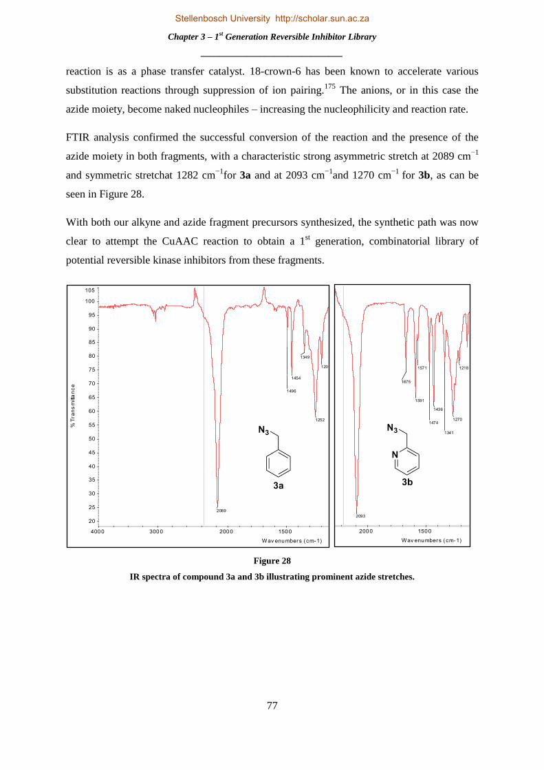
Chapter 3 – 1st Generation Reversible Inhibitor Library
_______________________________
77
696
735
8751201
1252
1349
1454
1496
2089
20
25
30
35
40
45
50
55
60
65
70
75
80
85
90
95
100
105
%Tr
ans
mitta
nce
600 800 1000 1000 1500 2000 3000 4000
Wav enumbers (cm-1)
reaction is as a phase transfer catalyst. 18-crown-6 has been known to accelerate various
substitution reactions through suppression of ion pairing.175
The anions, or in this case the
azide moiety, become naked nucleophiles – increasing the nucleophilicity and reaction rate.
FTIR analysis confirmed the successful conversion of the reaction and the presence of the
azide moiety in both fragments, with a characteristic strong asymmetric stretch at 2089 cm−1
and symmetric stretchat 1282 cm−1
for 3a and at 2093 cm−1
and 1270 cm−1
for 3b, as can be
seen in Figure 28.
With both our alkyne and azide fragment precursors synthesized, the synthetic path was now
clear to attempt the CuAAC reaction to obtain a 1st generation, combinatorial library of
potential reversible kinase inhibitors from these fragments.
Figure 28
IR spectra of compound 3a and 3b illustrating prominent azide stretches.
626
660
751
1218
1270
1341
1436
1474
1571
1591
1675
2093 25
30
35
40
45
50
55
60
65
70
75
80
85
90
95
100
%Tr
ans
mitta
nce
600 800 1000 1000 1500 2000 3000 4000
Wav enumbers (cm-1)
Stellenbosch University http://scholar.sun.ac.za

Chapter 3 – 1st Generation Reversible Inhibitor Library
_______________________________
78
3.4 GENERATION OF COMBINATORIAL LIBRARY FROM AZIDE AND ALKYNE
FRAGMENTS
3.4.1Defining “Click” Chemistry
The term “Click” chemistry was coined by K. Barry Sharpless and his group in a landmark
paper in 2001.176
With particular emphasis on following the strategy adopted by nature in
generating large molecules built from smaller components via heteroatom to carbon linkers, a
rigorous and stringent set of criteria was defined which must be observed by a process, to fall
within this category. Thus a click chemistry reaction is delineated and exemplified best as
one that is modular, broad in application, affording high yields with minimal and innocuous
by-products, stereospecific and have a high atom economy. Furthermore, the reaction should
proceed under ambient reaction conditions, using commercially and readily available
reagents and starting materials, in a solvent that is benign to the environment and easily
removed with simple separation, isolation and purification.176
A number of reactions have been found to satisfy these criteria exceptionally and have
become benchmark click reactions. These include: thiol-ene click reactions,177
Diels-Alder
and inverse electron demand Diels-Alder reactions,178
nucleophilic substitutions tostrained
epoxides,179
and cycloaddition reactions such as the [4+1] cycloaddition between isonitriles
and tetrazines180
and the [3+2] Huisgen 1,3-dipolar cycloadditions, frequently called “click”
reactions, including the copper-181
and ruthenium-catalysed variants.182
Click chemistry has found a plethora of applications in chemistry, biochemistry,
supramolecular chemistry, polymer and materials science.183
This has extended to its use in
2D gel electrophoresis, macromolecular or nanostructures,184
peptidomimetics or its peptide
chains,185
macrocyclizations,186
dendrimer design,187
nanotechnology and drug
discovery.183,188
Click chemistry has also been used to label biomolecules within systems,
through biorthogonal click reactions. This means that thesereactions possess good selectivity
and biocompatibility,189
overcoming toxicity such that their participating reagents can form
covalent bonds within abundantly functionalized biological systems - in some cases, living
organisms.195, 196
This has won click chemistry world recognition as a household term for
chemists.
Stellenbosch University http://scholar.sun.ac.za

Chapter 3 – 1st Generation Reversible Inhibitor Library
_______________________________
79
3.4.2 The 1,4-Copper Catalysed Azide Alkyne Cycloaddition (CuAAC).
The azide alkyne Huisgen cycloaddition has been referred to as the “cream of the crop” of
click chemistry by Sharpless himself.176
In particular, the copper-catalysed cycloaddition has
become one of the most popular and recurrently used reactions in click chemistry today.183,188
It was discovered simultaneously and separately by the groups of Sharpless and Valery V.
Fokin,190
as well as the Meldal group in Denmark.191
Whilst the Meldal group were the first
to document and report the reaction in the synthesis of peptidotriazoles on solid support, they
did not fully appreciate the potential of the reaction and its applicability as a click reaction.
Sharpless on the other hand, lauded it as a reliable and robust catalytic process, bringing
unparalleled selectivity, consistency and broad spectrum utilization in the organic synthesis
for the formation of covalent linkers between assorted components.190
Scheme 6
General reaction conditions and product formed for CuAAC.
The copper-catalysed azide alkyne cycloaddition reaction occurs between organic azides and
terminal alkynes which are united to exclusively form 1,4-regioisomers of 1,2,3-triazoles as
product,shown in scheme 6.192
Although commercial sources of copper(I) such as cuprous
bromide or iodide may be used for the cycloaddition, the reaction has been found to proceed
more efficiently with the use of a copper(II) source such as copper sulphate and an applicable
reducing agent such as sodium ascorbate or ascorbic acid, as seen in scheme 6. This is due to
Cu(I) species being unstable in aqueous solvents, which can be overcome with the addition of
stabilising ligands. An advantage of generating the Cu(I) species in situ, whereby the Cu(II)
complex is reduced from the +2 oxidation state to the +1, is the non-requirement of a base. If
copper(I) sources are used directly, triethylamine (Et3N) orN,N-diisopropylethylamine
(DIPEA) are frequently used as bases.
This economical reaction excels in a variety of solvents.Whilst water without organic co-
solvent or a mixture of water/alcohol such as methanol, iso-proponal or tert-butanol is most
Stellenbosch University http://scholar.sun.ac.za

Chapter 3 – 1st Generation Reversible Inhibitor Library
_______________________________
80
commonly used, organic solvents such as dimethyl sulfoxide, tetrahydrofuran, acetone,
dimethylformamide and acetonitrile are also effective.192
The reaction functions ina wide pH
range, from 4-12, with pH 7-9 yielding best results. Reactions have been shown to proceed at
0 °C and as high as 160 °C. The isolation and purification is also simple, usually requiring
filtration of product or extraction, avoiding laborious and time consuming chromatographical
methods or recrystallization.192
A huge boon of this reaction is its inertness to many of the
most common functional groups without protection.188
For these reasons, it is clear to see
why this reaction has gained such relevance and esteem in organic synthesis.
3.4.3 Catalytic Cycle and Mechanism.
The current proposed mechanism for the CuAAC is based on density functional theory (DFT)
calculations undertaken by Sharpless and his group.192
Although there was a deficiency in
certain aspects of chief transitions and intermediates, the study elucidated much in
understanding, through qualitative and quantitative analysis, of the mechanistic path followed
in this reaction as shown in Figure 29. Nevertheless, the exact DFT calculations agree and
overlap with a number of experimental observations and evidence. These include a lower
activation barrier calculated in comparison to related cycloadditions, which accounts for a
dramatic increase in reaction rate observed in the CuAAC. Secondly, the regiosomer obtained
in the experimental reaction, the 1,4-substituted variant, is predicted by DFT. Lastly, the DFT
calculations successfully predicted the reaction to proceed with other dipoles such as nitrile
oxides, which is observed experimentally adhering strictly to the regioselectivity. This
enforces the mechanism put forward by Sharpless and his group.192
The reaction mechanism begins with either the Cu(I) catalyst or the Cu(I) species generated
in situ through reduction of a suitable Cu(II) pre-catalyst. This alkyne coordinates to the
Cu(I) species, which results in the expulsion of a ligand from the copper complex and
formation of the copper acetylide as shown in step AFigure 29. This conversion proceeds
through the formation of a π-alkyne copper complex as has been seen in various other copper
mediated coupling reactions.162
Thereafter, step B involves the replacement of the previously
expelled ligand with the coordination of the azide to the copper through the nitrogen proximal
to the carbon forming the copper-azide-acetylide complex. Subsequent attack, in step C, from
the distalnitrogen of the azide of the alkyne carbon bonded to R1 affords the six-membered
copper(III) metallacycle. From this point the barrier for ring formation is very low, which
furnishes the triazolyl-copper derivative through step D.
Stellenbosch University http://scholar.sun.ac.za

Chapter 3 – 1st Generation Reversible Inhibitor Library
_______________________________
81
Figure 29
Proposed reaction mechanism and catalytic cycle for CuAAC adapted from Sharpless et al.192
Subsequent release of the triazole through step E affords the 1,4-substituted 1,2,3,-triazole
and regeneration of the copper catalyst.192
3.4.4Synthetic Optimization and Generation of Library of Novel Reversible Inhibitors
With our alkyne and azide side chain moietiesin hand, it was our intention to generate a small
“combinatorial” library derived from these previously synthesized fragments, through the use
of the CuAAC. As can be imagined, a large amount of literature procedures and reaction
conditions have been reported for this reaction, which resulted in a number of experimental
evaluation and optimization reactions being performed, in order to determine what conditions
afforded the highest yield in the most efficient manner as can be seen in Table 2. For this,
compound 2a and benzyl azide(3a) were selected and examined against an assortment of
different sources of copper, including Cu(I) and Cu(II) species with reducing agents, and a
variety of different solvents in different ratios. The reaction time was kept constant at 24h and
all reactions were carried out at rt.
Stellenbosch University http://scholar.sun.ac.za

Chapter 3 – 1st Generation Reversible Inhibitor Library
_______________________________
82
Table 2
Optimization of CuAAC for compound 4a.
Attempt
Source of
copper
Amount
(mol. %)
Reducing
agent
Amount
(mol. %)
Solvents
used
Solvent
ratio
Yield
(%)
1193
CuSO4.5H2O 2 Sodium
ascorbate 5 H2O:t-BuOH 1:1 18
2193
CuSO4.5H2O 5 Sodium
ascorbate 5 H2O:t-BuOH 1:3 20
3193
CuSO4.5H2O 5 Sodium
ascorbate 5 H2O:MeOH 1:1 28
4194
CuI 5 DIPEA 1 equiv. ACN - 12
5174
Cu(OAc)2 10 - - H2O:t-BuOH 1:1 62
6174
Cu(OAc)2 10 - - H2O:MeOH 1:4 73
Our initial reactions condition were based on the one of the most popular and frequently used
copper sources, CuSO4, with the equally as prominent sodium ascorbate as reducing agent in
aqueous medium – a procedure which often affords quantitative yields as outlined by one of
the co-discoverers and founder of the principle of click chemistry, Valery V. Fokin.193
Sodium
ascorbate, a mild reductant, is a suitable and advantageous alternative to oxygen free
conditions as any dissolved dioxygen is reduced, eliminating the appearance of by-products
from copper catalysed oxidative side reactions.193
Water is a conducive solvent for the
formation of reactive copper acetylides, especially when formed in situ.193
However, as
shown above in attempt 1-3 in Table 2, use of these reagents and conditions with a variety of
solvents and ratios and amount of catalyst loadings afforded very poor yields. This led us to
evaluate the use of a Cu(I) halide species in an organic solvent, such as CuI in acetonitrile
with the use of DIPEA as base (attempt 4). However this afforded an even lower yield which
Stellenbosch University http://scholar.sun.ac.za

Chapter 3 – 1st Generation Reversible Inhibitor Library
_______________________________
83
can be explained by the formation of oxidative by-products, which seemingly halted
progression of the reaction and destroyed the catalyst. With this methodology, elimination of
oxygen in the reaction vessel is a priority, but with the proclaimed robust nature of this
reaction we sought out other synthetic conditions which would afford a reasonable yield.
Scheme 7
General reaction conditions and product formed for CuAAC.174
After combing through the literature, a procedure was found which makes use of copper (II)
acetate without inclusion of a reducing agent as devised by Zhu and co-workers.174
The group
found that the CuAAC proceedswith a number of Cu(II) salts, without the need for a reducing
agent, in readily oxidizable alcohols such as MeOH, EtOH and i-PrOH, but not t-BuOH. This
led to the hypothesis that a Cu(I) species was generated via the reduction of the Cu(II)
catalyst through oxidation of these alcohol solvents as shown in Scheme 7a below. However,
the reaction was seen to proceed with a very high yield with Cu(OAc)2 in t-BuOH which is
not inclined to oxidation. This led to the group putting forward a second postulation, that a
Glaser-type reaction - or in the case of copper (II) acetate salts, a related Eglinton-type
reaction, shown in Scheme 7b - was taking place. This furnished the catalytically active Cu(I)
species, which has been shown to be generated in these reactions,195, 196
which in turn
facilitated reaction of the azide alkyne cycloaddition (AAC). According to the authors,
electron paramagnetic resonance (EPR) and absorption spectroscopy both supported these
hypotheses.174
Use of Cu(OAc)2in a mixed solvent system H2O:t-BuOH afforded a satisfactory yield and
with replacement of MeOH a yield of 73% was found to be acceptable for the optimization of
this reaction, as seen in Table 2. With a suitable synthetic procedure optimized, we applied
these parameters to the rest of our heterocyclic alkyne and azide fragments in a
“combinatorial” fashion, generating a library of clicked compounds in good yield as shown in
Table 3.
a)
b)
Stellenbosch University http://scholar.sun.ac.za

Chapter 3 – 1st Generation Reversible Inhibitor Library
_______________________________
84
Table 3
Combinatorial library generated through CuAAC with yields.
These novel compounds were fully characterized using 1H
NMR, 13
C NMR, FTIR spectroscopy and HRMS-TOF MS.
This confirmed the formation of the clicked products. Showing
compound 4aas example, both the benzylic protons and the
Stellenbosch University http://scholar.sun.ac.za

Chapter 3 – 1st Generation Reversible Inhibitor Library
_______________________________
85
CH2 proton in the triazole ring were clearly accounted for in the 1H NMR spectra, with
proton 1 showing up as a singlet at 5.61 ppm integrating for 2H and proton 2 being observed
as a singlet at 7.86 integrating for 1H as was expected. All other protons were accounted for
in the spectra. For the 13
C NMR spectra, carbon1 was at 54.5 ppm and the triazole formation
was confirmed by both carbons 2 and 3 showing on the spectra at and 129.36 and 145.49
ppm. All other corresponding carbon peaks were accounted for. The [M+H]+
ion found in the
MS spectra was 287.1297, which matched identically with the calculated 287.1297 for
C18H15N4.
Through use of the CuAAC we were able to generate 8 novel, 1,4-substitued 1,2,3,-triazole
based potential reversible kinase inhibitors. These compounds would later be sent for
biological assays to determine efficacy and potency. With the synthetic methodology adopted
and developed in the synthesis of our potential reversible inhibitor library, we set our sights
on utilizing this click chemistry approach in the synthesis of potential irreversible inhibitors.
Irreversible kinase inhibitors have been shown to possess a number of distinct advantages
over their ATP competitive counterparts, and thus it was our endeavour to move one step
forward in the design of potent and selective inhibitors.
Stellenbosch University http://scholar.sun.ac.za
Chapter 4 – 1st Generation Irreversible Inhibitor Library
_______________________________
86
CHAPTER 4 – 1ST
GENERATION IRREVERSIBLE INHIBITOR LIBRARY
4.1 MOTIVATION, RATIONALE AND STRATEGY FOR SYNTHESIS OF IRREVERSIBLE
INHIBITOR LIBRARY
4.1.1 Irreversible Inhibitors – Successes and Advantages.
The successes attained by reversible, ATP competitive kinase inhibitors as anticancer agents,
brought drug discovery in kinase inhibition to the forefrontof the targeted therapeutic
treatment of cancer. However, challenges met in the clinical and approved phases of these
drugs, which include promiscuity across the widely conserved human kinome, high
intracellular concentrations of endogenous ATP, the competitive substrate in reversible
inhibition and resistance to prescribed drugs due to mutations in the enzyme, has sparked a
search for alternative therapeutic mechanisms to combat these challenges.197
A resurgence in
the field of covalent drugs or irreversible inhibitors,198
has been paramount to addressing and
overcoming these challenges. This revival has been brought on by a greater understanding of
the advantages and disadvantages of irreversible inhibition and the approval of a number of
potent and non-toxic covalent drugs.199
Covalent drugs, in general, have been an extremely
lucrative and successful class of treatment for the pharmaceutical sector, with 3 of the top ten
selling drugs in the United States in 2009 being covalent drugs, $33 billion dollars in
worldwide sales from the 26 covalent drugs for which there is data and the most used
irreversible drug globally, aspirin, having 80 million tablets consumed per year in the United
States alone.198
A breakdown of the 39 drugs that had received FDA approval in mid-2011
shows that ~33% are used as anti-infectives, 20% as anti-cancer agents, 15% for
gastrointestinal disorders and 10% and 5% for cardiovascular and central nervous system
disorders respectively as can be seen in Figure 30. Inflammation targets make up 3% of these
FDA approved drugs with 13% targeting other disorders.198
This has shown irreversible
inhibition to be a successful, proven approach in many classes of drug targets.197
In the last
decade, the distinct advantages of targeted, irreversible kinase inhibitors include benefits such
as extraordinary efficacy and IC50 profiles in biological assays, success in overcoming ATP
competition, an inherent selectivity that is exclusive to its mechanism of action and very
importantly, an ability to retain efficacy against resistance mutations in target kinases.197
This
has resulted in a number of irreversible kinase inhibitors undergoing clinical investigation
and this field of research can be considered the vanguard of protein kinase inhibition.
Stellenbosch University http://scholar.sun.ac.za

Chapter 4 – 1st Generation Irreversible Inhibitor Library
_______________________________
87
Figure 30
Pie chart illustrating the spread of covalent inhibitors in treatment.198
With no greater PTK are these benefits greater exemplified than with EGFR. Figure 31 shows
the chemical structure of 6 irreversible inhibitors in particular, all targeting the EGFR/HER2
pathway and in clinical trials, that have shown the promise and potential of this mechanism of
treatment. IC50 values in nM, based on preclinical in vitro EGFR and HER2 kinase assays,
are indicated below each compounds name.197, 200
Not only do these inhibitors show
remarkable antitumour activity and efficacy in in vitro and in vivo assays, they also exhibit
potent selectivity and the ability to overcome mutations that have sparked resistance to
approved ATP competitive inhibitors on the market.134b, 200-201
The compound WZ4002, as
example, has shown remarkable selectivity in targeting the T790M specific mutation within
the EGFR kinase domain.201c, 201d
This secondary mutation at the gatekeeper residue, which
occurs by substitution of the threonine 790 amino acid with methionine, is one of the most
common causes of resistance to known therapeutic agents.134b
The result of this substitution is
a bulkier side chain at the gatekeeper residue which restricts and hinders access to the active
site by the inhibitor through sterics, thus lowering efficacy. It has also been shown that this
mutation increases affinity for the natural substrate ATP, thus meaning an increased dosage
of inhibitors are needed to remain competitive.134b
Drug discovery stratagems targeting this
mutation through irreversible inhibition have resulted in failure due to a unacceptable toxicity
profile which is linked to simultaneous inhibition of the wildtype EGFR.201c
Stellenbosch University http://scholar.sun.ac.za

Chapter 4 – 1st Generation Irreversible Inhibitor Library
_______________________________
88
Figure 31
A number of irreversible inhibitors currently in clinical trials with IC50 values shown in nM.
The wild type can be thought of as the normal or phenotypic enzyme, a product of standard
alleles, present in the body. However, WZ4002 has shown an incredible selectivity for this
mutated form of the EGFR kinase, being 30-100 fold more potent against EGFR T790M, and
up to 100- fold less potent against the wildtype EGFR, in comparison to other quinazoline-
based EGFR inhibitors in vitro.201c
With these results it is clear to see why the field of
irreversible inhibition has generated so much interest and has opened up a dominant approach
to identifying new classes of mutant-selective kinase inhibitors.
Stellenbosch University http://scholar.sun.ac.za
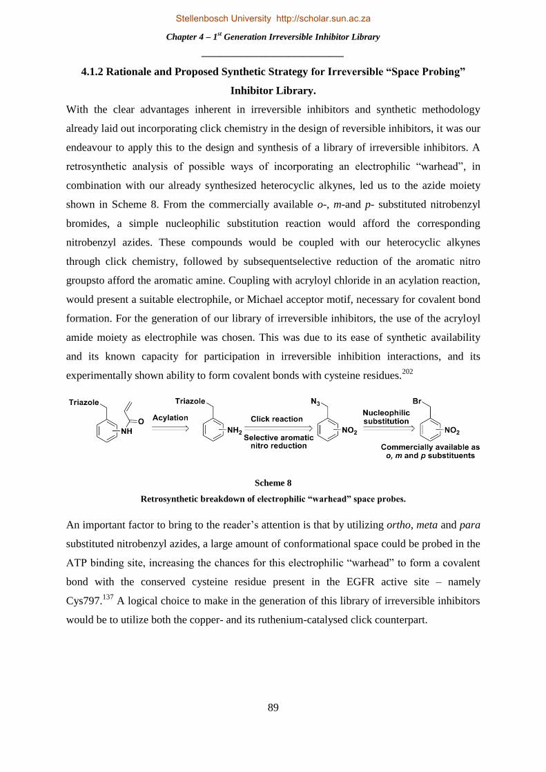
Chapter 4 – 1st Generation Irreversible Inhibitor Library
_______________________________
89
4.1.2 Rationale and Proposed Synthetic Strategy for Irreversible “Space Probing”
Inhibitor Library.
With the clear advantages inherent in irreversible inhibitors and synthetic methodology
already laid out incorporating click chemistry in the design of reversible inhibitors, it was our
endeavour to apply this to the design and synthesis of a library of irreversible inhibitors. A
retrosynthetic analysis of possible ways of incorporating an electrophilic “warhead”, in
combination with our already synthesized heterocyclic alkynes, led us to the azide moiety
shown in Scheme 8. From the commercially available o-, m-and p- substituted nitrobenzyl
bromides, a simple nucleophilic substitution reaction would afford the corresponding
nitrobenzyl azides. These compounds would be coupled with our heterocyclic alkynes
through click chemistry, followed by subsequentselective reduction of the aromatic nitro
groupsto afford the aromatic amine. Coupling with acryloyl chloride in an acylation reaction,
would present a suitable electrophile, or Michael acceptor motif, necessary for covalent bond
formation. For the generation of our library of irreversible inhibitors, the use of the acryloyl
amide moiety as electrophile was chosen. This was due to its ease of synthetic availability
and its known capacity for participation in irreversible inhibition interactions, and its
experimentally shown ability to form covalent bonds with cysteine residues.202
Scheme 8
Retrosynthetic breakdown of electrophilic “warhead” space probes.
An important factor to bring to the reader‟s attention is that by utilizing ortho, meta and para
substituted nitrobenzyl azides, a large amount of conformational space could be probed in the
ATP binding site, increasing the chances for this electrophilic “warhead” to form a covalent
bond with the conserved cysteine residue present in the EGFR active site – namely
Cys797.137
A logical choice to make in the generation of this library of irreversible inhibitors
would be to utilize both the copper- and its ruthenium-catalysed click counterpart.
Stellenbosch University http://scholar.sun.ac.za

Chapter 4 – 1st Generation Irreversible Inhibitor Library
_______________________________
90
Scheme 9
Proposed synthetic route to potential irreversible inhibitors and illustration of space probing effect of
these compounds.
This would allow the generation of 1,4- and 1,5- substituted CuAAC and ruthenium catalysed
azide alkyne cycloaddition (RuAAC) products respectively (see Section 4.2.2 for more
information), affording two different regioisomers – thus expanding the conformational space
filled and effectively augmenting the probing affect brought on by the use of o-, m- and p-
substituted electrophiles. An illustration of the proposed synthesis for this library of space
probing potential irreversible inhibitors can be seen in Scheme 9 above. A copper- or
ruthenium-mediated click reaction between the quinoline and isoquinoline heterocyclic
alkynes and synthesized azides would afford the corresponding triazole carrying an aromatic
nitro group. Reduction of this moiety and subsequent coupling with acryloyl chloride would
provide our fully furnished 1,4- and 1,5-, o-, m- and p-substituted library of potential
irreversible inhibitors. To fully show the space probing effect of these compounds, and
illustrated example is shown at the bottom of Scheme 9. This shows that for each heterocyclic
alkyne undergoing this reaction sequence, six potential irreversible inhibitors, adopting
unique spatial conformation and thus occupying a different volume of space in the active site,
will be generated. This can be seen as a powerful tool for generating a large library of
compounds targeting the cysteine residue and irreversible inhibition. The generation of the
1,4-substituted library through CuAAC was carried out and completed by a postdoc
researcher in our labs, Dr. Abu Taher and will not be discussed here.
Stellenbosch University http://scholar.sun.ac.za

Chapter 4 – 1st Generation Irreversible Inhibitor Library
_______________________________
91
4.2 SYNTHESIS OF LIBRARY OF 1,5- SUBSTITUTED POTENTIAL IRREVERSIBLE
INHIBITORS.
4.2.1 Synthesis of Nitrobenzyl Azide Fragments.
With the heterocyclic alkynes synthesized already in hand the first step in the generation of
the irreversible inhibitor library was to synthesize theo-, m- and p-substituted nitrobenzyl
azide fragments. This was accomplished with relative ease as conditions used for the
synthesis of compound 3a were utilized, affording quantitative or near quantitative yields.
The reaction conditions and yields are summarized in Scheme 10 below.
Scheme 10
Synthesis of nitrobenzyl azide fragments 6a-c.
Subsequent FTIR analysis confirmed the presence of the azide species in compounds 6a-c.
As an example, compound 6ashowed acharacteristic strong asymmetric stretch at 2091 cm−1
and symmetric stretchat 1254 cm−1
. This afforded the nitrobenzyl azide fragments and
allowed us to move on to synthesizing a combinatorial library of these fragments and the
previously synthesized alkyne fragments, through RuAAC reactions.
4.2.2The Ruthenium Catalysed Azide Alkyne Cycloaddition Reaction.
Thesuccess, popularity and applicability attained by the regioselective 1,4-substituted triazole
CuAAC reaction demanded a viable synthetic route and methodology to be crafted for its
corresponding regioisomer, the 1,5-substituted triazole.203
Although synthetic procedures to
these 1,5-substituted regioisomers are available, which include the use of stabilized
phosphonium ylides,204
the addition of bromomagnesium acetylides to organic azides205
and
the use of enamines with aryl azides,206
they lack the simplicity, scope and directness of the
CuAAC.182, 203
This led to the development of a ruthenium-mediated azide alkyne
cycloaddition reaction by the groups of Fokin and Jia.203
By making use of ruthenium-based
catalysts such as such as CpRuCl(PPh3)2 and Cp*RuCl(PPh3)2, the groups found a
regioselective catalytic route to these 1,5-substituted triazoles.207
A notable dampener on the
Stellenbosch University http://scholar.sun.ac.za
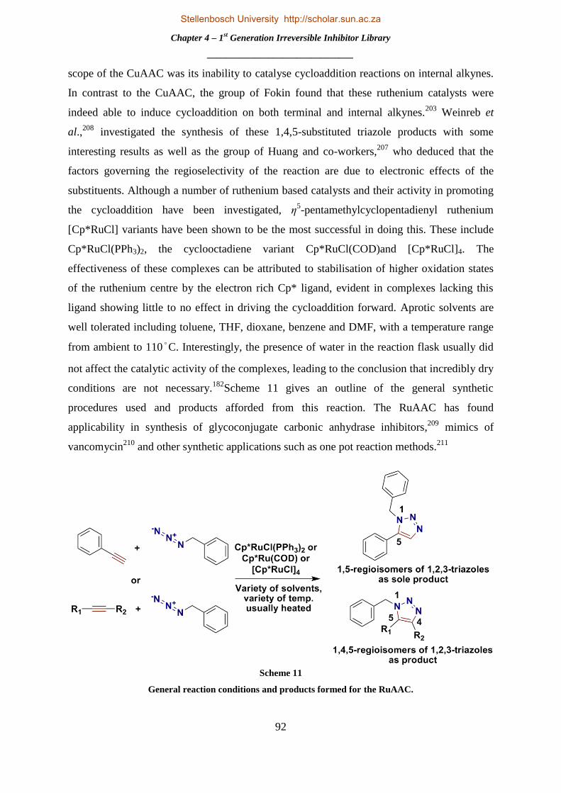
Chapter 4 – 1st Generation Irreversible Inhibitor Library
_______________________________
92
scope of the CuAAC was its inability to catalyse cycloaddition reactions on internal alkynes.
In contrast to the CuAAC, the group of Fokin found that these ruthenium catalysts were
indeed able to induce cycloaddition on both terminal and internal alkynes.203
Weinreb et
al.,208
investigated the synthesis of these 1,4,5-substituted triazole products with some
interesting results as well as the group of Huang and co-workers,207
who deduced that the
factors governing the regioselectivity of the reaction are due to electronic effects of the
substituents. Although a number of ruthenium based catalysts and their activity in promoting
the cycloaddition have been investigated, η5-pentamethylcyclopentadienyl ruthenium
[Cp*RuCl] variants have been shown to be the most successful in doing this. These include
Cp*RuCl(PPh3)2, the cyclooctadiene variant Cp*RuCl(COD)and [Cp*RuCl]4. The
effectiveness of these complexes can be attributed to stabilisation of higher oxidation states
of the ruthenium centre by the electron rich Cp* ligand, evident in complexes lacking this
ligand showing little to no effect in driving the cycloaddition forward. Aprotic solvents are
well tolerated including toluene, THF, dioxane, benzene and DMF, with a temperature range
from ambient to 110°C. Interestingly, the presence of water in the reaction flask usually did
not affect the catalytic activity of the complexes, leading to the conclusion that incredibly dry
conditions are not necessary.182
Scheme 11 gives an outline of the general synthetic
procedures used and products afforded from this reaction. The RuAAC has found
applicability in synthesis of glycoconjugate carbonic anhydrase inhibitors,209
mimics of
vancomycin210
and other synthetic applications such as one pot reaction methods.211
Scheme 11
General reaction conditions and products formed for the RuAAC.
Stellenbosch University http://scholar.sun.ac.za
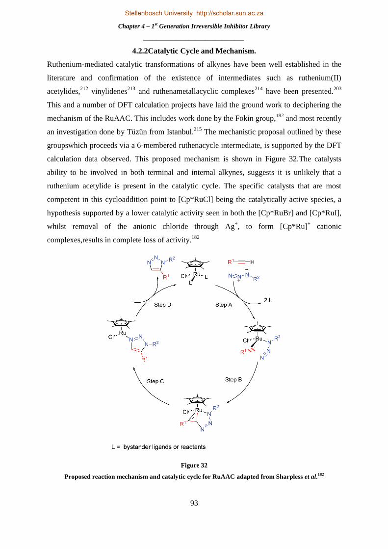
Chapter 4 – 1st Generation Irreversible Inhibitor Library
_______________________________
93
4.2.2Catalytic Cycle and Mechanism.
Ruthenium-mediated catalytic transformations of alkynes have been well established in the
literature and confirmation of the existence of intermediates such as ruthenium(II)
acetylides,212
vinylidenes213
and ruthenametallacyclic complexes214
have been presented.203
This and a number of DFT calculation projects have laid the ground work to deciphering the
mechanism of the RuAAC. This includes work done by the Fokin group,182
and most recently
an investigation done by Tüzün from Istanbul.215
The mechanistic proposal outlined by these
groupswhich proceeds via a 6-membered ruthenacycle intermediate, is supported by the DFT
calculation data observed. This proposed mechanism is shown in Figure 32.The catalysts
ability to be involved in both terminal and internal alkynes, suggests it is unlikely that a
ruthenium acetylide is present in the catalytic cycle. The specific catalysts that are most
competent in this cycloaddition point to [Cp*RuCl] being the catalytically active species, a
hypothesis supported by a lower catalytic activity seen in both the [Cp*RuBr] and [Cp*RuI],
whilst removal of the anionic chloride through Ag+, to form [Cp*Ru]
+ cationic
complexes,results in complete loss of activity.182
Figure 32
Proposed reaction mechanism and catalytic cycle for RuAAC adapted from Sharpless et al.182
Stellenbosch University http://scholar.sun.ac.za

Chapter 4 – 1st Generation Irreversible Inhibitor Library
_______________________________
94
The group hypothesized that the reaction mechanism begins with displacement of the
spectator ligands to form an activated complex as shown in step A of Figure 32. This
complex is converted, via oxidative coupling of the alkyne and azide, to the ruthenacycle
illustrated by step B in Figure 32. The regioselectivity is determined by this step or the first
new carbon-nitrogen bond formed, which can be seen as a nucleophilic attack of the activated
alkyne at the electrophilic terminal nitrogen of the azide. Computational studies support the
irreversible configuration of this ruthenacycle. Subsequent reductive elimination (step C)
releases the 1,5 substituted triazole product and the catalyst is regenerated in as shown in step
D. DFT studies also indicate that the reductive elimination is the rate determining step in the
catalytic cycle.182
4.2.3 Synthetic Optimization and Generation of Library of Novel Irreversible Inhibitors
With both the heterocyclic alkyne fragments (2a-d)and the newly synthesized nitrobenzyl
azide fragments (6a-c) in hand, the synthesis of the exclusively 1,5-substituted, RuAAC-
mediated library of potential irreversible kinase inhibitors could be undertaken. For this
purpose, the catalyst Cp*RuCl(COD) was chosen and purchased for all reactions on the
grounds that it is the most labile catalyst, with the cyclooctadiene group being more easily
displaced than say that of the phosphine ligands, and proceeds in the least demanding
reaction conditions.182
Following suit as was previously done in the synthetic endeavours of
our reversible library, the coupling between the heterocyclic alkyne 2a and ortho substituted
nitrobenzyl azide 6a was chosen to be optimized, as can be seen in Table 4. The
Cp*RuCl(COD) has been shown to be active at catalytic amounts of 2 mol. % at ambient
temperatures producing high yields and 100% regioselectivity.182
Thus our first reaction
conditions implemented a minimum amount of catalyst at rt with anhydrous tertrahydrofuran
as solvent. Careful consideration was taken to keep conditions inert and a period of 45min of
degassing with nitrogen was implemented. This however, resulted in very poor yields which
were solely attributed to the incomplete consumption of both starting reagents as deteremined
by TLC. The reaction was seen not to proceed to completion, and after 24h the reaction was
stopped and both the starting materials were salvaged and the product isolated. This meant
that whilst we were not losing both our heterocyclic alkyne and azide fragment reagents, the
amount of product afforded in this reaction was unacceptable (<20%). Thus it was decided to
make use of more catalyst in the second attempt2, but againto no avail, it was seen to have
little effect on the yield and more importantly not successful in driving the reaction to
completion.
Stellenbosch University http://scholar.sun.ac.za

Chapter 4 – 1st Generation Irreversible Inhibitor Library
_______________________________
95
Table 4
Optimization of RuAAC for compound 7a.
Attempt182
Cp*RuCl(COD) (mol. %) Solvent Temp (°C) Yield(%)
1 2 THFa
rt 18
2 4 THFa rt 19
3 4 THFb rt 23
4 4 Dioxanea rt 18
5 4 Toluenea Gradually to 80 71
6 4 Toluenea 80 85
a = Nitrogen was passed through reaction for 45min for degassing
b = Freeze-pump-thaw technique and nitrogen was passed through for 45min for degassing of
reaction.
At this point we investigated whether or not the catalyst was being deactivated due to
contaminants such as molecular oxygen or water in the reaction flask and so a freeze-pump-
thaw technique, in combination with passing nitrogen through the reaction, was applied for
attempt 3.
This had little effect on the yield and the next variable to investigate was that of solvents.
Attempt 4 saw the utilization of anhydrous dioxane, which resulted in a lower yield than that
obtained for attempt 3 – the main culprit still being incompletion of the reaction. Finally it
was decided to make use of a high boiling point solvent and by increasing the temperature of
the reaction, give external energy to drive the reaction to completion. For this purpose,
toluene was chosen as solvent and the reaction was gradually heated to 80°C, starting at rt in
20°C increments every hour for attempt 5. Satisfyingly, this resulted in the reaction going to
completion and a good yield of 71% being obtained for the reaction. The reason we delayed
with increasing the temperature to allow it to go to completion, was that the Cp*RuCl(COD)
Stellenbosch University http://scholar.sun.ac.za

Chapter 4 – 1st Generation Irreversible Inhibitor Library
_______________________________
96
catalyst has specificallybeen shown to proceed better at ambient conditions and that elevated
temperatures are actually detrimental to the reaction, resulting in deactivation, decomposition
and low yields.182
Interestingly, this was not the case for our reactions and the final attempt 6
kept the temperature of the reaction constant at 80°C from start to finish, resulting in our
optimized yield of 85% for compound 7a. These reaction conditions were then applied to the
rest of the heterocyclic alkynes 2a-d and our o-, m- and p- substituted nitrobenzyl azides 6a-
c, the results of which are summarized below in Table 5.
Table 5
Summary of synthesis of 1,5- substituted triazole compounds 7a-l.
It is interesting to note that the products formed from the isoquinoline alkynes resulted in far
lower yields than those products built from the quinoline alkynes. A number of control
reactions, changing the solvent, temperature and the use of freeze-pump-thaw techniques,
similar to those done in the optimization reactions, were undertaken to determine whether
these had any effect on the yield. As was the case with our optimization results, the reactions
Stellenbosch University http://scholar.sun.ac.za

Chapter 4 – 1st Generation Irreversible Inhibitor Library
_______________________________
97
did not proceed to completion with a lowering of the temperature and other variables altered
only worsened the afforded yield. The difference in yields between these two scaffolds could
be attributed to coordinative and basic effects of the isoquinoline driving group as discussed
in Section 3.2.2. In the end the yields obtained were very good for the quinoline backbone
products and acceptable for those containing isoquinoline as the driving group.
The compounds synthesized were fully characterized using 1H
NMR, 13
C NMR, FTIR spectroscopy and HRMS-TOF MS. This
confirmed the synthesis thereof. Using compound 7a, as
example,1H NMR spectroscopic analysis showed the benzylic
peak of protons 1 at 6.07 ppm integrating for 2H and proton2 in
the triazole ring being witnessed as a singlet at 8 ppm integrating for 1H, as was expected.
All other protons were accounted for in the spectra. 13
C NMR spectroscopy also confirmed
product formation as the benzylic carbon 1was witnessed at 49.6 ppmand both triazole
carbons2 and 3 were seen at 129.6 and 136.1 ppm respectively. The remaining carbon peaks
were all accounted for in the spectrum.MS analysis and spectra showed the [M+H]+ ion as
332.1156,which correlates well with the calculated 332.1147 for C18H14N5O2.
Confirmation of the synthesis of both the 1,4- and 1,5-
regioisomers was gained through comparison of the peaks
obtained in both the 1H NMR and
13C NMR spectra of the
appropriate CuAAC analogues synthesized by Dr. Abu
Taher, and the library synthesized through the RuAAC.
For example, the triazole proton 4 of the analogue of compound 7a, shown above on the left,
was observed at 8.17 ppm, a notable shift from that seen for proton 2 of compound 7a (8.00
ppm). A similar trend was observed for all compounds synthesized and on the basis of these
findings, it was deduced that both the 1,4- and 1,5-regioisomer series had been synthesized. It
should be noted that these correlations do not confirm which regioisomer is which; however,
there is significant literature precedent concerning the regiochemical outcomes of CuAAC
and RuAAC reactions.190, 203
It is thus our assumption that the 1,4-regiosomer was furnished
through the CuAAC reaction, whilst the 1,5-regiosomer was provided under the RuAAC
conditions. It should also be noted that 2D NMR spectroscopy, such as Nuclear Overhauser
effect spectroscopy (NOESY), or the obtaining of a crystal structure, could be used to
confirm the absolute structure of each click reaction.
Stellenbosch University http://scholar.sun.ac.za
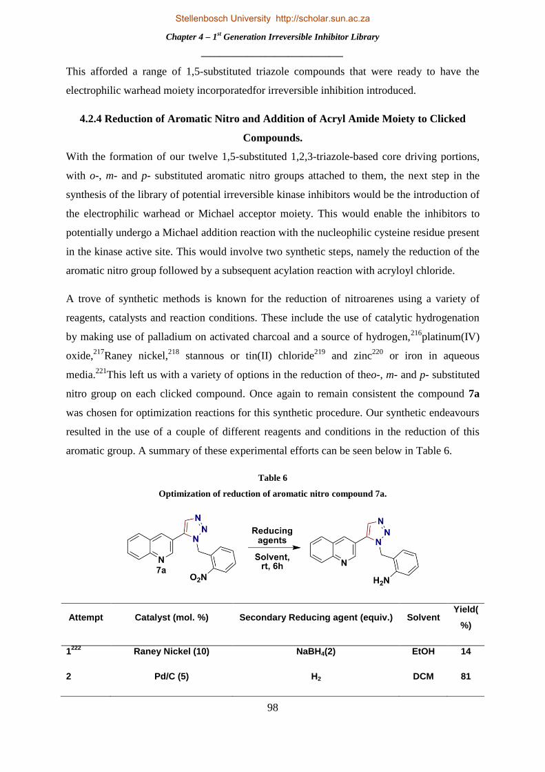
Chapter 4 – 1st Generation Irreversible Inhibitor Library
_______________________________
98
This afforded a range of 1,5-substituted triazole compounds that were ready to have the
electrophilic warhead moiety incorporatedfor irreversible inhibition introduced.
4.2.4 Reduction of Aromatic Nitro and Addition of Acryl Amide Moiety to Clicked
Compounds.
With the formation of our twelve 1,5-substituted 1,2,3-triazole-based core driving portions,
with o-, m- and p- substituted aromatic nitro groups attached to them, the next step in the
synthesis of the library of potential irreversible kinase inhibitors would be the introduction of
the electrophilic warhead or Michael acceptor moiety. This would enable the inhibitors to
potentially undergo a Michael addition reaction with the nucleophilic cysteine residue present
in the kinase active site. This would involve two synthetic steps, namely the reduction of the
aromatic nitro group followed by a subsequent acylation reaction with acryloyl chloride.
A trove of synthetic methods is known for the reduction of nitroarenes using a variety of
reagents, catalysts and reaction conditions. These include the use of catalytic hydrogenation
by making use of palladium on activated charcoal and a source of hydrogen,216
platinum(IV)
oxide,217
Raney nickel,218
stannous or tin(II) chloride219
and zinc220
or iron in aqueous
media.221
This left us with a variety of options in the reduction of theo-, m- and p- substituted
nitro group on each clicked compound. Once again to remain consistent the compound 7a
was chosen for optimization reactions for this synthetic procedure. Our synthetic endeavours
resulted in the use of a couple of different reagents and conditions in the reduction of this
aromatic group. A summary of these experimental efforts can be seen below in Table 6.
Table 6
Optimization of reduction of aromatic nitro compound 7a.
Attempt Catalyst (mol. %) Secondary Reducing agent (equiv.) Solvent Yield(
%)
1222
Raney Nickel (10) NaBH4(2) EtOH
14
2 Pd/C (5) H2 DCM 81
Stellenbosch University http://scholar.sun.ac.za

Chapter 4 – 1st Generation Irreversible Inhibitor Library
_______________________________
99
For attempt 1, we synthesized our own Raney nickel following a procedure set out by
Mozingo.223
The use of this reducing agent in tandem with NaBH4 as outlined by Litvi´c and
co-workers,222
resulted in a very poor yield and discouraged us from further use of these
reducing agents. Our next choice of reductant system, in attempt 2, made use of palladium on
activated charcoal with hydrogen gas. DCM was chosen as solvent purely for solubility issues
and the reaction was seen to proceed smoothly and effectively with a simple filtration through
Celite required for product isolation. This afforded the aniline in a good yield and became our
choice for the further reduction of all aromatic nitro groups.
It was deemed unnecessary to purify the product obtained after reduction, due to the lack of
formation of by-products and simple isolation procedure, and thus the acylation reaction was
performed directly on the aromatic amine afforded after reduction. The reaction of acryloyl
chloride with triethylamine as base in DCM, proceeded smoothly with acceptable yields
between 56 and 66%. It is important to note that the yields obtained were calculated over two
steps. As this reaction afforded our final product, it was seen as redundant to labour further
on optimization of the reaction conditions. The formal reaction conditions used for both the
aromatic nitro reduction and acylation reaction can be seen below in Table 7, as well as a full
summary of yields obtained over the two steps for products 8a-l.
A full characterisation of all synthesized compounds was
completed. The spectral data obtained from this confirmed the
formation of our desired products. With compound 8a as
example, analysis of the 1H NMR spectra obtained clearly showed
the ethylene protons 1 and 2both as doublets of doublets at 6.12
and 5.64 ppm respectively, each integrating for 1H. The 3rd
ethylene proton 3was also witnessed as a doublet of doublets at
6.28 ppm integrating for 1H. The amide proton 4could be seen far downfield at 9.84 ppm
integrating for the required 1H. Lastly, the benzyl protons of 5were observed as a singlet at
5.77 ppm, integrating for 2H and indicative of the triazole and driving portions still being
attached to the molecule. All other protons were accounted for in the spectra. The 13
C NMR
spectrum for compound 8aclearly showed the benzyl carbon of 5 at 48.8 ppm and that of
carbon 6 further downfield at 163.4 ppm. All other carbons were observed as expected in the
spectra. Finally, the HRMS spectra for compound 8a showed agreement between the [M+H]+
ion 356.1510 and that calculated for C21H18N5O as 356.1511.
Stellenbosch University http://scholar.sun.ac.za

Chapter 4 – 1st Generation Irreversible Inhibitor Library
_______________________________
100
Table 7
Reaction conditions and yields obtained for products 8a-l.
This afforded our twelve potential irreversible kinase inhibitors and furnished our 1st
generation library of potential irreversible inhibitors. With both a 1st generation library of
eight reversible and twelve 1,5-substituted potential irreversible inhibitors synthesized and
another twelve 1,4-substituted irreversible inhibitors synthesized by Dr. Abu Taher, we were
ready to send these compounds to our collaborators at the Technische Universiteit Dortmund
Stellenbosch University http://scholar.sun.ac.za

Chapter 4 – 1st Generation Irreversible Inhibitor Library
_______________________________
101
in Germany for biological screening. The assays results could determine efficacy, selectivity
and other important factors and data that could be analysed to provide further knowledge on
these potential kinase inhibitors. Further use of molecular modelling could be used to
rationalize the results obtained and determine where the most feasible and profitable synthetic
modifications could be made in the possible synthesis of a 2nd
generation library based on the
most efficacious potential inhibitors. The methods and results of these biological assays will
be discussed in full in the following chapter.
Stellenbosch University http://scholar.sun.ac.za

Chapter 5 – Biological Evaluation of Synthesized Potential Kinase Inhibitors and Conclusions
_______________________________
102
CHAPTER 5 – BIOLOGICAL EVALUATION OF SYNTHESIZED
POTENTIAL KINASE INHIBITORS AND CONCLUSIONS
5.1 BIOLOGICAL SCREENING METHODOLOGY
5.1.1 Professor Daniel Rauh’s Research Focus – Fluorescent Labelling in Kinases.
Professor Daniel Rauh is the head of chemical biology at the Technische Universiteit
Dortmund and has filled this post since the end of 2010. With nearly 70 publications in
journals such as Nature Chemistry, Nature Chemical Biology, Nature Genetics, Journal of the
American Chemistry Society and the American Chemistry Society Chemical Biology, he has
distinguished himself in his field. His research focus is mainly on the development of
biological assays for protein kinases and phosphatases, which is why we have been privileged
to collaborate with him and his group. Prof. Rauh‟s group were the first to develop detection
methods in biological assays called fluorescent labels in kinases (FLiK) or the FLiK
approach,224
and for their phosphatase counterparts florescent labels in phosphatases
(FLiP).225
The active reversible phosphorylation of a protein kinase may change the enzyme
conformation and thus its state of activity towards further phosphorylation or biological
processes.226
Important residues in this reversible process are the conserved triplet of the
activation loop in the kinase domain, consisting of aspartate (D), phenylalanine (F), and
glycine (G) residues – making up the DFG motif. This can be seen on the right of Figure 33.
The aspartate residue (D) in particular, plays a catalytic role in phosphate transfer.118
Two
important conformations protein kinases may adopt is the DFG-out, or inactive state, and the
DFG-in or active state. The reversible transition between these two conformations is
controlled by the rotational movement of the aspartate residue (D), accompanied by
corresponding rotation of both the adjacent phenylalanine (F) and glycine (G) residues. In the
DFG-out conformation the aspartate residue (D) is rotated out of the ATP binding pocket,
exposing a large hydrophobic pocket for ligands or inhibitors to utilize.118
For the DFG-in
conformation the opposite is true, with the aspartate residue (D) moving back into the ATP
active site. This reversible process is illustrated on the left side of Figure 33.
Stellenbosch University http://scholar.sun.ac.za

Chapter 5 – Biological Evaluation of Synthesized Potential Kinase Inhibitors and Conclusions
_______________________________
103
Figure 33
Ilustration of reversible conformational transition of kinases and the DFG motif.224
Certain inhibitors make use of, and in doing so stabilize, a specific conformation of a protein
kinase for ligand binding. Type II, or allosteric inhibitors and type III, non ATP competitive
or irreversible inhibitors – shown in blue - make use of and stabilize the inactive or DFG-out
conformational state,whilst classicATP competitive, reversible or Type I inhibitors – shown
in yellow - make use of and stabilize the DFG-in or active state.226
It is this precise
mechanism and conformational preference and stabilization of the protein kinase by ligands
and inhibitors that the FLiK biological assay exploits.
The FLiK assay makes use of the environmentally sensitive, thiol-reactive fluorophore
acrylodan, which is attached to a specific site of the target kinase – shown as a green and red
sphere in the DFG-out and DFG-in conformation respectively in Figure 33 above. During
ligand or inhibitor binding, the protein kinase undergoes a conformational change. This
conformational change and stabilization brought on by the inhibitor binding, produces an
alteration in the microenvironment, which may change the solvent shell of the fluorophore
acrylodan, seen as a change between the red and green sphere illustrated in Figure 33. This
alteration is witnessed as a significant change in itsemission spectra, which is characterised
by two emission maxima (λem,max) at ∼470 and ∼510 nm. Conformational transitions bring
about changes in the total intensity of these two emission maxima relative to each other,
resulting in an attractive ratiometric fluorescence readout, which has been shown to be
superior to its monochromic counterpart.224,226
The FLiK assay is thus a conformation-specific
binding assay and has been shown to be viable for a number of kinases including p38α
kinase,227
abl228
and cSrc,229
to name a few. This methodology has been used in high
Stellenbosch University http://scholar.sun.ac.za

Chapter 5 – Biological Evaluation of Synthesized Potential Kinase Inhibitors and Conclusions
_______________________________
104
throughput screening and in cooperation with the pharmaceutical industry,226
and is
especially significant when deducing the relative binding modes and specificity of potent
inhibitors. The ratiometric fluorescence readout explained above is also made use of in
activity-based assays for kinases.
5.1.2 Methodology Used in Screening of Reversible and Irreversible Inhibitors against
EGFR.
For the biological assays and IC50 values determined for our compounds, a student in Prof.
Rauh‟s group, Julian Engel, made use of the HTRF KinEASE-TK assay from Cisbio. For our
specific biological screening, this technique involved use of the wildtype form of the kinase
and the two medicinally relevant mutant deviants, more specifically the inhibitor-sensitizing
mutation EGFR Leu858Arg (L858R) and the double mutant drug resistant variant, EGFR
Leu858Arg-Thr790Met (L858R_T790M). As a control reaction these kinases were each
incubated in the presence or absence of our potential reversible and irreversible inhibitors,
followed by the addition of a biotinylated poly Glu-Tyr substrate peptidewhich is
phosphorylated by the EGFR kinase. Completion of the reaction allows for subsequent
introduction of an antiphosphotyrosine antibody labelled with europium cryptate, and
streptavidin which is labelled with a fluorophoreXL665. The fluorescence resonance energy
transfer or FRET is then measured between europium cryptate and XL665 to quantify the
phosphorylation of the substrate peptide. This allows for a control experiment and value for
kinase activity and following this, ATP concentrations were set at their respective Km values
(30 μM for the EGFR-WT, 60 μM for EGFR-L858R, and 30 μM for EGFR-L858R_T790M).
Our synthesized inhibitors were then preincubated with the specific kinase and peptide for 2h,
where after ATP was added to initiate the reaction and the fluorescence of these samples
were documented at 620 nm (Eu-labelled antibody) and 665 nm (XL665 labelled
streptavidin) 60 μs after excitation at 317 nm by a Tecan Safire plate reader. The quotient of
these two readings, done in an eight fold dilution series and carried out in triplicate, were then
plotted against inhibitor concentrations and fitted to a Hill 4-parameter equation to determine
relevant IC50 values.230
The principle of the FRET analysis, using the attachedeuropium
cryptate andstreptavidin groups, as well as the data acquired and an example of IC50 value
determination in these biological assays is shown in Figure 34.
Stellenbosch University http://scholar.sun.ac.za

Chapter 5 – Biological Evaluation of Synthesized Potential Kinase Inhibitors and Conclusions
_______________________________
105
Figure 34
Principle of FRET analysis as well as IC50 value through use of this technique and plotting of 8-fold
dilution series.
Gefitinib and WZ4002 were used as control groups for the kinase inhibitory activity tests and
in determination of the IC50 values. A total of 33 inhibitors were sent to our collaborators and
the kinase inhibitory activity results against the EGFR wildtype, L858R and L858R_T790M
mutation can be seen in Figure 35.
5.2 RESULTS AND DISCUSSION
The biological screening yielded a few important and interesting results.The first point is
evident in the screen against the wildtype. Whilst the control inhibitors exhibit very potent
inhibition of the wildtype EGFR kinase at low concentrations, it is encouraging to see that
our synthesized inhibitors showed little to no activity against the phenotypic kinase, even
athigh concentrations of 50μM. This can be interpreted as a very good result as high
inhibitory activity against the wildtype kinase is usually linked to a high toxicity profile, as it
targets the unmutated form of the kinase.
EGFR-wt_IC50
RL1147 [nM]0.1 10 1000 100000
665/
620
nm [a
u]
0.2
0.3
0.4
0.5
0.6
0.7
0.8
0.9
1
1.1
Stellenbosch University http://scholar.sun.ac.za

Chapter 5 – Biological Evaluation of Synthesized Potential Kinase Inhibitors and Conclusions
_______________________________
106
Figure 35
Kinase inhibitory activity results against wildtype, L858R and L858R_T790M mutation EGFR kinases.
-20
0
20
40
60
80
100
120
140
160
DM
SOG
efi
tin
ibW
Z40
02
LH9
A
T9
LH5
LH
3
HL8
9
HL4
5
RL4
33
H
L59
LH
7
HL4
8
HL4
9
HL6
0
HL9
6
HL7
1
AT7
AT1
2
HL7
2A
T3
HL1
04
H
L58
LH
8
HL9
0
LH1
A
T2
AT6
LH
2LH
6
SP1
LH
4
AT8
SP
2
AT1
A
T11
A
T10
A
T4
AT5
kin
ase
act
ivit
y/
%
2.5µM
25µM
50µM
Screen Wildtype
-20
0
20
40
60
80
100
120
DM
SO
Ge
fiti
nib
WZ4
002
AT9
AT3
HL9
6
AT7
HL7
2
HL4
8
LH9
HL7
1
AT8
AT1
2
HL9
0
HL4
9
AT2
AT6
SP1
HL5
9
LH5
LH7
LH1
LH6
LH8
HL8
9
LH2
AT4
AT1
0
AT1
1
SP2
HL6
0
HL1
04
LH3
HL4
5
LH4
AT1
HL5
8
AT5
Kin
ase
act
ivit
y/
%
2.5µM
25µM
50µM
Screen L858R
-40
-20
0
20
40
60
80
100
120
140
160
DM
SO
WZ4
002
Ge
fiti
nib
AT9
HL9
6
LH9
HL8
9
AT3
LH5
HL4
9
HL7
1
HL7
2
HL4
8
HL9
0
LH1
HL4
5
AT1
2
HL1
04
LH7
AT2
LH3
AT7
AT1
1
LH6
AT8
HL5
9
AT1
0
SP1
LH2
AT6
HL5
8
LH8
AT4
SP2
LH4
AT1
AT5
HL6
0
kin
ase
act
ivit
y/
%
2.5µM
25µM
50µM
Screen L858R_T790M
Stellenbosch University http://scholar.sun.ac.za

Chapter 5 – Biological Evaluation of Synthesized Potential Kinase Inhibitors and Conclusions
_______________________________
107
Looking at both mutated forms of the EGFR kinase and the biological screening data, it is
encouraging to see that not only are the inhibitors active, but in the case of the L858R screen,
a number compounds including AT5, HL58 (8c), AT1 and LH4 (4d) are seen to be similarly
active to the FDA approved drug gefitinib, used specifically for treatment of breast and lung
cancer as an EGFR inhibitor. This is also the case for the double mutant variant
L858R_T790M, where compounds HL60 (8b), AT5 and AT1 show comparable activity to
the control group. An important conclusion to draw from this data is that whilst our
synthesized inhibitors are not effective in inhibiting the wildtype variant, they show promise
and efficacy against the mutant strains. This indicates a positive selectivity profile for our
inhibitors, in contrast to that found for gefitinib and WZ4002, which show activity across the
board for all the EGFR screens. This shows promise for our synthesized inhibitors as a high
selectivity and moderate to good inhibition trend and profile was witnessed.
Table 8
IC50 values in μM of most efficacious and selective synthesised inhibitors.
From these results corresponding IC50 values were derived for each of our inhibitors. A
summary of the most efficacious and selective inhibitors is show in Table 8.Whilst the IC50
values of these compounds are not inside of the nanomolar range and cannot compete with
WZ4002, a number of interesting conclusions and future work can be derived from these
results. Compound AT5,highlighted in red in Table 8, was shown to be the most efficacious
inhibitor of those synthesized, in this case by Dr Taher. This was based on the isoquinoline
scaffold with the 1,4-substituted regioisomer of the triazoleand the Michael acceptor in the
Stellenbosch University http://scholar.sun.ac.za

Chapter 5 – Biological Evaluation of Synthesized Potential Kinase Inhibitors and Conclusions
_______________________________
108
ortho positionand thus an irreversible inhibitor. AT5 showed good inhibitoryactivity and an
excellent selectivity profile for the mutated kinases. Its IC50 value was comparable to gefitinib
and lower in the case of the double mutant variant. The very impressive selectivity could be
investigated further with molecular modeling, as well as the determination of whether
synthetic tweaking to the molecule could result in a lower IC50 value whilst still retaining the
selectivity. Compound LH4, highlighted in green in Table 8, was a reversible inhibitor
synthesized in this project and showed slightly higher IC50 values, yet retained the selectivity
seen for compound AT5. With further investigation into this compound through molecular
modeling, a suitable position for an electrophilic warhead to be placed on the molecule could
be found, perhaps increasing its efficacy. Lastly, compounds HL58 (7c) and HL60 (7b)
showed remarkable selectivity for the L858R and L858R_T790M mutations alone
respectively. This is highlighted in blue in Table 8. The use of molecular modelling to further
determine the differences in selectivity and their binding modes is definitely a venture worth
pursuing.
5.3 CONCLUSIONS
The aim of this project was to synthesize a library of reversible and potential irreversible
kinase inhibitors. This project has seen the synthesis of 20 of the 32 potential inhibitors
includingeight reversible inhibitors, compounds 4a-d and 5a-d, as well as twelve irreversible
inhibitors, compounds 8a-l. The synthesis of these compounds made use of palladium, copper
and ruthenium metal-mediated chemistry including Sonogashira coupling reactions, CuAAC
and RuAAC.All novel compounds were fully characterized using a range of analytical
techniques.
Biological screening assays against the wildtype, and two mutant forms of the EGFR kinase –
L858R and L858R_T790m – were undertaken by our collaborators at the Techniese
Universiteit Dortmund in Germany. The potential kinase inhibitors synthesized showed good
inhibitory activity and a favourable selectivity profile for both the mutated strains of the
EGFR kinase. Compound AT5 showed the greatest inhibitory activity and was the most
efficacious inhibitor with an IC50 value of 4.53 and 1.85 μM L858R and L858R_T790M
mutants respectively. This inhibitor shows greater activity against the double mutant variant
L858R_T790M than the FDA approved drug, gefitinib. Compound AT5is exceedingly
selective towards the mutant forms of the EGFR kinase as it is essentially non-active against
the EGFR wild type. Compound LH4(4d), shows similar efficacy, being slightly less potent,
Stellenbosch University http://scholar.sun.ac.za

Chapter 5 – Biological Evaluation of Synthesized Potential Kinase Inhibitors and Conclusions
_______________________________
109
and a similar selectivity profile. Compounds HL58 (7c) and HL60 (7b) showed selectivity
for the L858R and L858R_T790M mutation respectively.
With the encouraging results obtained from the biological screening assays, molecular
modelling and docking studies could be undertaken to determine the synthetic alterations that
are required to generate a 2nd
generation library of compounds. Based on compound AT5,
LH4(4d),HL58 (7c) and HL60 (7b),this library would build on the inherent selectivity of
these molecules for the mutant EGFR kinase variations and explore the origin of this
selectivity. This could possibly lead to more potent inhibitors with an even higher selectivity
profile.
The synthetic methodology developed in this project could also be utilized in the synthesis of
possible biological space and cysteine probes. With the synthesis of these probes, large
libraries could potentially be synthesized quickly and efficiently, allowing for a large scale,
broad range kinase screening.
Stellenbosch University http://scholar.sun.ac.za

Chapter 6 – Future Work
_______________________________
110
CHAPTER 6 – FUTURE WORK
6.1 FUTURE WORK
6.1.1 2nd
Generation Library Based on Compound AT5.
Molecular modelling calculations and docking studies were undertaken to postulate the
specific binding mode of AT5,andinspection of the conformation adopted by the compound
provided insight into possible synthetic modifications. Scrutiny of the inhibitor in the active
site and its adopted spatial arrangement revealed two possible sites for further investigation
shown in Figure 36 below. The first of these promising sites is the introduction of another,
secondary hydrogen bond interaction with the hinge region at Glu791.
Figure 36
Illustration of possible sites for further synthetic investigation and modification.
GLN791
Stellenbosch University http://scholar.sun.ac.za

Chapter 6 – Future Work
_______________________________
111
As can be seen in Figure 36, there is a considerable amount of space between the carbon
alpha to the nitrogen of the isoquinoline ring and the carbonyl bond of GLN791. This space,
whilst large enough to warrant the introduction of a functional group, is also small enough to
maintain a hydrogen bond effect. Thus we hypothesize the introduction of a hydrogen bond
donor group, such as a hydroxyl or amine moiety at this position of the isoquinoline driving
group, could result in higher efficacy and selectivity for member of a 2nd
generation library.
Secondly, on the right side of the scaffold there exists a large open hydrophobic pocket,
shown by a yellow caged sphere in Figure 36. It is our belief that functionalizing the
isoquinoline core at relevant positions to extened into this hydrophobic pocket would increase
the inhibitor selectivity profile and possibly the potency.
To test this hypothesis docking calculations were undertaken with a large group of potential
second generation inhibitors. These included the addition of the amine and hydroxyl hinge
binding motif ortho to the isoquinoline nitrogen at R1, as well as various functional groups on
position R2 and R3 of the isoquinoline ring in different combinations. This included a methyl,
ethyl, methoxy and cyano functional group for possible space filling. The results were
promising and showed the highest score and visual impression with the groups illustrated in
Figure 37 below. The synthesis of a 2nd
generation library could thus yield compounds with
increased efficacy and selectivity profiles for these irreversible inhibitors.
Figure 37
Possible structures for 2nd
generation library of AT5.
Stellenbosch University http://scholar.sun.ac.za

Chapter 6 – Future Work
_______________________________
112
6.1.2 Biological Space Filling and Cysteine Targeting Probes.
As was shown in the proposed synthesis of our irreversible compounds in Section 4.1.2, the
benefits of using a benzyl azide with an o-, m- and p- substituted functional group, which
would later become an electrophilic warhead capable of forming covalent bond interactions
with the cysteine residue in the active site, is a largely untapped potential biological probe. If
the synthetic methodology and route developed and employed in our synthesis of irreversible
inhibitors were to be shortened and simplified to have the o-, m- and p- substituted
electrophilic warhead already in place on the benzyl azide moiety, as shown in Scheme 12
below, this would open the door to a vast number of possibilities that these moieties could be
used as biological probes of the kinase active site. Through the use of both CuAAC and
RuAAC click chemistry as we have shown in our synthesis, a potentially wide ranging library
could probe a significant amount of space in the active site; in addition, this library could be
synthesized simply and effectively.
Scheme 12
Proposed synthetic plan for use of biological space probes to generate a large library of compounds.
Stellenbosch University http://scholar.sun.ac.za

Chapter 6 – Future Work
_______________________________
113
This could be done by propargylation, or the addition of an alkyne group to heterocycles or
driving groups that are known to form hydrogen bond interactions effectively with the hinge
region of kinases, followed by subsequent coupling through both the 1,4-substituted CuAAC
and the 1,5-substituted RuAAC with the o-, m- and p- substituted electrophilic warhead
moieties as shown in Scheme 12. This would generate a large number of different spatial and
space probing conformations from one heterocycle or driving group and thus a large library
of potential irreversible inhibitors targeting the cysteine residue across a broad panel of
kinases. From this library biological assays will hopefully lead to a few compounds that show
high efficacy and selectivity for a certain kinase, and further synthetic modifications could
result in an extremely potent and selective inhibitor.
Furthermore, if a certain compound generated through the use of this synthetic methodology
shows promise by displaying high efficacy and potent selectivity, a simple and effective way
to give an indication of whether or not this is through covalent bond formation with the
cysteine residue would be to synthesize an analogue of the compound with the saturated
propionamide probe derivative as shown in Scheme 13. This will afford critical information
on whether or not the compounds inhibit irreversibly or not, as a large drop in the efficacy of
the compound would be expected without the neccasary Michael acceptor moiety. With the
use of these both of these biological probes a large amount of information regarding the
targeting of the cysteine residue, chemical space probed in the active site of the kinase as well
as the type of inhibition that is undertaken by these synthesized compound will be afforded.
Scheme 13
Proposed synthetic method for insight into compounds mechanism of inhibition.
Stellenbosch University http://scholar.sun.ac.za

Chapter 7 – Experimental
_______________________________
114
CHAPTER 7 – EXPERIMENTAL
7.1 GENERAL PROCEDURES
7.1.1 Purification of solvents and reagents.
Chemicals used in these experiments were purchased from Merck or Sigma Aldrich. Solvents
used for chromatographic purposes were distilled by means of conventional distillation
procedures. Solvents used for reaction purposes were dried over the appropriate drying agents
and then distilled under N2gas. Tetrahydrofuran was distilled from sodium metal, using
benzophenone as indicator. Dichloromethane, dichloroethane, dimethylformamide and
acetonitrile were distilled from calcium hydride. Ethanol was distilled from magnesium
turnings and iodine. Diethyl ether was bought with a ≥98% purity grade from Sigma Aldrich
and then dried on activated 3Å molecular sieves.Other reagents requiring purification were
purified according to standard procedures.
7.1.2 Chromatography
Thin layer chromatography was performed using Merck silica gel 60 F254 coated on
aluminium sheets. Visualization was performed with a UV lamp, using iodine on silica, or by
spraying with a Cerium Ammonium Molybdate (CAM) or ninhydrin (NIN) or potassium
permanganate (KMnO4) solution followed by heating.
All column chromatography was performed on Merck silica gel 60 (particle size 0.040-0.063
mm) using one of or combinations of petroleum ether,EtOAc, toluene, dichloromethane,
ethanol and/orMeOH as a solvent.
7.1.3 Spectroscopic and physical data
NMR spectra (1H,
13C) were recorded on a 300 MHz Varian VNMRS
(75 MHz for 13
C), a 400 MHz Varian Unity Inova (101 MHz for 13
C), or a 600 MHz Varian
Unity Inova (150 MHz for 13
C). Chemical shifts (δ) are reported in ppm and J-values are
given in Hz. Chemical shifts were recorded using the residual solvent peak or external
reference. All spectra were obtained at 25 °C unless otherwise reported. Data was processed
using MestReNova.
Mass spectrometry was performed on a Waters SYNAPT G2. Infrared spectra were recorded
on a Thermo Nicolet Nexus 470 by means of Attenuated Total Reflectance (ATR) mode.
Stellenbosch University http://scholar.sun.ac.za

Chapter 7 – Experimental
_______________________________
115
Melting points were obtained using a Gallenkamp Melting Point Apparatus and are
uncorrected.
7.1.4 Other general procedures
All reactions were performed under a positive pressure of 5.0 gradeN2 or Ar unless water was
used as a solvent. The glassware was flame-dried while under vacuum before being purged
with N2 gas. Condensers were pre-dried at 120 °C for a minimum of two hours. Standard
Schlenk techniques were employed when necessary.
Solvents were removed in vacuo, where we arereferring to removal of the solvent using a
rotary evaporator followed by removal of trace amounts of solvent using a high vacuum
pump at ca. 0.08 mm Hg.
7.2 SYNTHESIS PERTAINING TO CHAPTER 3.
7.2.1 Compounds 2a-b.
General Procedure for Sonogashira Coupling of Quinoline Alkynes
A dry Schlenk tube was evacuated under high vacuum and filled with Ar (3×). To this
Schlenk tube was added by syringe, under Ar, in anhydrous THF (15 mL). The solvent was
then degassed by 3 - 5 cycles of freeze-pump-thaw technique. The solvent was subsequently
degassed for 15 min with Ar purge. This was followed by the addition of the catalysts,
PdCl2(PPh3)2(4 mol. %) and CuI (4 mol. %), under positive pressure Ar. Et3N (2 equiv) was
then added by syringe and the mixture was allowed to stir for 5 min. Following this,
trimethylsilylacetylene (TMSA) (1.5 equiv) was added by syringe. A colour change from
yellow to black was witnessed, indicating the consumption of the catalyst and that the
reaction was working. The reaction mixture was allowed to stir at rt, under N2, for up to 6 h
or until complete consumption of the starting materials (monitored by TLC). This resulted in
the intermediate TMS-protected quinoline alkyne, requiring no characterisation or
purification, which allowed for in situ deprotection. To the reaction mixture was added
liquidTBAF - or CsF - (1.5 equiv.), which was then left to stir for 0.5 h until the deprotection
was complete (monitored by TLC). The reaction mixture was then filtered through Celite,
followed by removal of the Et3Nin vacuo. The reaction mixture was then diluted with EtOAc
(30 mL) and washed with a saturated NH4Cl solution (60 mL). The layers were then
separated and the organic layer was rinsed with a saturated solution of brine (50 mL), dried
over MgSO4 and filtered. After removal of the solvents in vacuo, the residue was purified by
Stellenbosch University http://scholar.sun.ac.za

Chapter 7 – Experimental
_______________________________
116
flash column chromatography over silica gel eluted first with EtOAc and hexane
(EtOAc:Hexane, 5:95) and then (EtOAc:Hexane, 60:40). This afforded the pure product as a
white solid. The details for the compounds synthesized are described below.
3-ethynylquinoline - 2a
Scale: 3-bromoquinoline (800 mg, 3.84 mmol)
This compound was synthesized following the general procedure for
Sonogashira coupling of quinoline alkynes. The compound was purified by silica gel column
chromatography, eluted first with EtOAc and hexane (EtOAc:Hexane, 5:95) and then
(EtOAc:Hexane, 60:40). This afforded the pure product as a white solid (500 mg, 3.26 mmol,
85%).
Rf = 0.3 (EtOAc:Hexane, 5:95) ; Mp 73-75 °C; IR (ATR, cm-1
):3159(s, stretch, –C≡C–H),
2092(w, stretch, –C≡C–), 1120 (s, stretch, C–N); 1H NMR (400 MHz, CDCl3) δ 8.94 (d, J =
2.1 Hz, 1H, ArH), 8.28 (d, J = 1.9 Hz, 1H, ArH), 8.12 – 8.05 (m, 1H, ArH), 7.79 – 7.75 (m,
1H, ArH), 7.73 (ddd, J = 8.4, 6.9, 1.5 Hz, 1H, ArH), 7.56 (ddd, J = 8.1, 7.0, 1.1 Hz, 1H,
ArH), 3.28 (s, 1H, CCH); 13
C NMR (101 MHz,CDCl3) δ 152.4 (ArC), 147.2 (ArC), 139.4
(ArC), 130.5 (ArC), 129.6 (ArC), 127.7 (ArC), 127.5 (ArC), 127.1 (ArC), 116.4 (ArC),
81.1(CCH), 80.6 (CCC); HRMS-TOF MS ES+: m/z [M+H]+ calcd for C11H8N: 154.0657;
found: 154.0657
7-chloro-4-ethynylquinoline - 2b
Scale: 7-chloro-4-iodoquinoline (1500 mg, 5.18 mmol)
This compound was synthesized following the general procedure for
Sonogashira coupling of quinoline alkynes. However,CsF (1.2 equiv) was
used as the deprotecting agent for the synthesis of this compound. The compound was
purified by silica gel column chromatography, eluted first with EtOAc and hexane
(EtOAc:Hexane, 5:95) and then (EtOAc:Hexane, 60:40). This afforded the pure product as a
white solid (603 mg, 3.21 mmol, 62%).
Rf = 0.33 (EtOAc:Hexane, 5:95) ; Mp 109-111 °C; IR (ATR, cm-1
):3170 (s, stretch, –C≡C–
H), 2097(w, stretch, –C≡C–), 1151 (s, stretch, C–N); 1H NMR (400 MHz, CDCl3) δ 8.87 (d,
J = 4.4 Hz, 1H,ArH), 8.19 (t, J = 8.6 Hz, 1H,ArH), 8.11 (d, J = 2.0 Hz, 1H,ArH), 7.56 (dd, J
Stellenbosch University http://scholar.sun.ac.za

Chapter 7 – Experimental
_______________________________
117
= 8.9, 2.1 Hz, 1H,ArH), 7.52 (d, J = 4.4 Hz, 1H,ArH), 3.68 (s, 1H,CCH); 13
C NMR (101
MHz,CDCl3) δ 150.8 (ArC), 148.4 (ArC), 136.1 (ArC), 128.8 (ArC), 128.8 (ArC), 128.5
(ArC), 127.3 (ArC), 126.4 (ArC), 124.6 (ArC), 87.1 (CCH), 78.9 (CCC); HRMS-TOF MS
ES+: m/z [M+H]+ calcd for C11H7ClN: 188.0267; found: 188.0265
7.2.2 Compounds 2c-d.
General procedure for Sonogashira coupling of isoquinoline alkynes
A dry Schlenk tube was evacuated under high vacuum and filled with Ar (3×). To this
Schlenk tube was added by syringe, under Ar, anhydrous Et3N (15 mL). The solvent was then
degassed by 3 to 5 cycles of freeze-pump-thaw technique. The solvent was subsequently
degassed for 15 min with Ar purge. This was followed by the addition of the catalysts,
PdCl2(PPh3)2(4 mol. %) and CuI (4 mol. %), under positive pressure Ar. The reaction mixture
was then allowed to stir for 5 min. Following this, TMSA (1.5 equiv) was added by syringe.
The reaction mixture was placed in an oil bath heated to 65 °C and a colour change from
yellow to black was witnessed, indicating the consumption of the catalyst and that the
reaction was working. The reaction mixture was allowed to stir at 65 °C, under N2, for up to 8
h or until complete consumption of the starting materials (monitored by TLC). This resulted
in the intermediate TMS-protected quinoline alkyne, requiring no characterisation or
purification, which allowed for in situ deprotection. To the reaction mixture was added liquid
TBAF (1.5 equiv.), which was then left to stir for 0.5 h until the deprotection was complete
(monitored by TLC). The reaction mixture was then filtered through celite, followed by
removal of the Et3Nin vacuo. The reaction mixture was then diluted with EtOAc (30 mL) and
washed with a saturated ammonium chloride solution (60 mL). The layers were then
separated and the organic layer was rinsed with a saturated solution of brine (50 mL), dried
over MgSO4 and filtered. After removal of the solvents in vacuo, the residue was purified by
flash column chromatography over silica gel eluted first with EtOAc and hexane
(EtOAc:Hexane, 5:95), then (EtOAc:Hexane, 60:40). This afforded the pure product as a
brown solid.The details for the compounds synthesized are described below.
4-ethynylisoquinoline - 2c
Scale: 4-bromoisoquinoline (800 mg, 3.84 mmol)
This compound was synthesized following the general procedure for
Sonogashira coupling of isoquinoline alkynes. The compound was purified by
Stellenbosch University http://scholar.sun.ac.za

Chapter 7 – Experimental
_______________________________
118
silica gel column chromatography, eluted first with EtOAc and hexane (EtOAc:Hexane, 5:95)
and then (EtOAc:Hexane, 60:40). This afforded the pure product as a brown solid (491 mg,
3.22 mmol, 84%).
Rf = 0.31 (EtOAc:Hexane, 5:95); Mp 70-72 °C; IR (ATR, cm-1
):3157(s, stretch, –C≡C–H),
2086 (w, stretch, –C≡C–), 1166 (s, stretch, C–N); 1H NMR (400 MHz, CDCl3) δ 9.21 (s,
1H,ArH), 8.72 (d, J = 4.9 Hz, 1H,ArH), 8.25 (dd, J = 8.4, 0.7 Hz, 1H,ArH), 7.98 (t, J = 7.6
Hz, 1H,ArH), 7.82 – 7.74 (m, 1H,ArH), 7.65 (ddd, J = 8.1, 7.0, 1.1 Hz, 1H,ArH), 3.55 (s,
1H,CCH); 13
C NMR (101 MHz,CDCl3) δ 152.7 (ArC), 147.4 (ArC), 136.0 (ArC), 131.4
(ArC), 128.1 (ArC), 128.1 (ArC), 127.8 (ArC), 125.0 (ArC), 114.9 (ArC), 84.6 (CCH), 79.0
(CCC); HRMS-TOF MS ES+: m/z [M+H]+ calcd for C11H8N: 154.0657; found: 154.0656
5-ethynylisoquinoline - 2d
Scale: 5-bromoisoquinoline (600 mg, 2.88 mmol)
This compound was synthesized following the general procedure for
Sonogashira coupling of isoquinoline alkynes. The compound was purified by
silica gel column chromatography, eluted first with EtOAc and hexane (EtOAc:Hexane, 5:95)
and then (EtOAc:Hexane, 60:40). This afforded the pure product as a brown solid (390 mg,
2.54 mmol, 88%).
Rf = 0.31 (EtOAc:Hexane, 5:95) ; Mp 79-81 °C; IR (ATR, cm-1
):3185 (s, stretch, –C≡C–H),
2098(w, stretch, –C≡C–), 1145 (s, stretch, C–N); 1H NMR (400 MHz, CDCl3) δ 9.26 (s,
1H,ArH), 8.61 (s, 1H,ArH), 8.09 (s, 1H,ArH), 8.01 – 7.93 (m, 1H,ArH), 7.90 (d, J = 5.0 Hz,
1H,ArH), 7.54 (s, 1H,ArH), 3.52 (s, 1H,CCH); 13
C NMR (101 MHz,CDCl3) δ 152.7 (ArC),
144.0 (ArC), 136.4 (ArC), 135.2 (ArC), 128.7 (ArC), 128.3 (ArC), 126.8 (ArC), 119.3 (ArC),
118.8 (ArC), 83.4 (CCH), 80.2 (CCC); HRMS-TOF MS ES+: m/z [M+H]+ calcd for
C11H8N: 154.0657; found: 154.0655
7.2.2 Compounds 3a-b.
(azidomethyl)benzene - 3a
To a stirred solution of benzyl bromide (1.39 mL, 11.7 mmol) in a
water/acetone mixture (50 mL, 1:4) was added NaN3 (1.14g, 17.5 mmol, 1.5
equiv.) The resulting suspension was then stirred at rt, under N2, for 24 h. The acetone was
removed under reduced pressure and then diluted with DCM (30 mL) and deionised water
Stellenbosch University http://scholar.sun.ac.za

Chapter 7 – Experimental
_______________________________
119
(30 mL) and the organic layer was separated. The aqueous layer was extracted with aliquots
of DCM (3 × 20 mL) and the combined organic layers were then rinsed with a saturated
solution of brine (50 mL), dried over MgSO4 and filtered. The solvent was removed under
reduced pressure and the product 4a (1.55g, 10.7 mmol) was afforded in quantitative yield as
a yellow oil. The product 3a was sufficiently pure to use without further work up or
purification. FTIR analysis was used to confirm product formation which correlated well with
the literature.173
IR (ATR, cm-1
): 2089 (s, assym. stretch, N3), 1252 (s, symm. stretch, N3)
2-(azidomethyl)pyridine - 3b
A dry, 2-neck round-bottomed flask charged with 2-(bromomethyl)pyridine
hydrobromide (1.20 g, 4.74 mmol), 18-crown-6 (cat.) and
tetrabutylammonium iodide (cat.) was evacuated under high vacuum and filled with Ar(3×).
DMF (2 mL) was added through a septum by syringe, under positive pressure (Ar) and to this
stirred solution was added diisopropylethylamine (1 mL). Lastly, NaN3 (0.30g, 4.7 mmol, 1.2
equiv.) was added and the solution was left to stir at rt, under N2, for 24 h. The solution was
then diluted with EtOAc (20 mL), rinsed with basic brine (20 mL×3, pH > 10) followed by
washing with an NH4Cl solution (30 mL, 1M). The organic layer was then dried over MgSO4
and filtered. The solvent was removed under reduced pressure to afford the pure product 4b
as a yellow-brown oil (496mg, 3.35 mmol, 78%). The product 3bwas sufficiently pure to use
without further work up or purification. FTIR analysis confirmed product formation which
correlated well with the literature.174
IR (ATR, cm-1
): 2093 (s, assym. stretch, N3), 1270 (s, symm. stretch, N3).
7.2.2 Compounds 4a-d.
General procedure for the copper-mediated azide alkyne cycloaddition reactions
(CuAAC)
A dry, 2-neck round bottom flask charged with alkyne (1 equiv) and azide (1.2 equiv) was
evacuated under high vacuum and filled with Ar(3×). A solution of MeOH/water (30 mL,
2:1) was added through a septum by syringe, under positive pressure (Ar). To this stirred
solution was added copper acetate hydrate (10 mol. %) and the solution was left to stir at rt,
under N2, for 36h or until complete consumption of the starting material (monitored by TLC).
Stellenbosch University http://scholar.sun.ac.za

Chapter 7 – Experimental
_______________________________
120
The MeOH was removed under reduced pressure and the reaction mixture was diluted with
EtOAc (30 mL) and washed with an NH4Cl solution (60 mL, 1 M). The layers were then
separated and the aqueous layer was washed with successive portions of EtOAc (3×20 mL).
The combined organic layers were then rinsed with a saturated solution of brine (60 mL),
dried over MgSO4 and filtered. After removal of the solvents in vacuo, the residue was
purified by flash column chromatography over silica gel, eluted first with EtOAc and hexane
(EtOAc:Hexane, 60:40), followed by MeOH and EtOAc (MeOH:EtOAc, 5:95). This afforded
the pure product as a white solid.The details for the compounds synthesized are described
below.
3-(1-benzyl-1H-1,2,3-triazol-4-yl)quinoline - 4a
Scale: 3-ethynylisoquinoline (0.1 g, 0.65 mmol)
This compound was synthesized following the general
procedure for CuAAC. The compound was purified by silica
gel column chromatography, eluted first with EtOAc and
hexane (EtOAc:Hexane, 60:40), followed by MeOH and EtOAc (MeOH:EtOAc, 5:95). This
afforded the pure product as a white solid (0.14g, 0.47 mmol, 73%).
Rf = 0.31 (EtOAc:Hexane, 60:40); Mp 157-159 °C; IR (ATR, cm-1
):3064 (s, stretch, =C–
H), 2922 (m, stretch, CH2),1046 (s, stretch, C–N), 813 (s, oop, C–H);1H NMR (300 MHz,
CDCl3) δ 9.26 (s, 1H,ArH), 8.61 (d, J = 2.0 Hz, 1H,ArH), 8.09 (d, J = 8.4 Hz, 1H,ArH), 7.86
(s, 1H, CCHN), 7.84 (dd, J = 8.2, 1.1 Hz, 1H,ArH), 7.69 (ddd, J = 8.4, 6.9, 1.5 Hz, 1H,ArH),
7.54 (ddd, J = 8.0, 7.0, 1.1 Hz, 1H,ArH), 7.47 – 7.29 (m, 5H,ArH), 5.61 (s, 2H, CH2); 13
C
NMR (75 MHz,CDCl3) δ 148.4 (ArC), 147.9 (ArC), 145.5 (NCCH), 134.5 (ArC), 132.0
(ArC), 129.7 (ArC), 129.4 (ArC), 129.3 (CCHN), 129.1 (ArC), 128.3 (ArC), 128.2 (ArC),
128.0 (ArC), 127.3 (ArC), 123.9 (ArC), 120.1 (ArC), 54.5 (CH2); HRMS-TOF MS ES+:
m/z [M+H]+ calcd for C18H15N4: 287.1297; found: 287.1297
4-(1-benzyl-1H-1,2,3-triazol-4-yl)-7-chloroquinoline - 4b
Scale: 7-chloro-4-ethynylquinoline (0.10 g, 0.53 mmol)
This compound was synthesized following the general
procedure for CuAAC. The compound was purified by silica gel
column chromatography, eluted first with EtOAc and hexane
Stellenbosch University http://scholar.sun.ac.za

Chapter 7 – Experimental
_______________________________
121
(EtOAc:Hexane, 60:40), followed by MeOH and EtOAc (MeOH:EtOAc, 5:95). This afforded
the pure product as a white solid (0.12 g, 0.29 mmol, 55%).
Rf = 0.32 (EtOAc:Hexane, 60:40); Mp 123-127 °C;IR (ATR, cm-1
):3071 (s, stretch, =C–H),
2920 (m, stretch, CH2),1054 (s, stretch, C–N), 824 (s, oop, C–H); 1H NMR (300 MHz,
CDCl3) δ 8.91 (d, J = 4.5 Hz, 1H,ArH), 8.61 (d, J = 9.1 Hz, 1H,ArH), 8.14 (d, J = 2.2 Hz,
1H,ArH), 7.86 (s, 1H, CCHN), 7.57 – 7.51 (m, 2H,ArH), 7.47 – 7.34 (m, 5H,ArH), 5.67 (s,
2H, CH2);13
C NMR (75 MHz,CDCl3) δ 151.1 (ArC), 149.6 (ArC), 145.3 (NCCH), 136.2
(ArC), 135.7 (ArC), 134.3 (ArC), 129.5 (CCHN), 129.3 (ArC), 129.0 (ArC), 128.4 (ArC),
128.3 (ArC), 127.6 (ArC), 124.1 (ArC), 123.3 (ArC), 120.5 (ArC), 54.7 (CH2);HRMS-TOF
MS ES+: m/z [M+H]+ calcd for C18H14N4Cl: 321.0907; found: 321.0904
4-(1-benzyl-1H-1,2,3-triazol-4-yl)isoquinoline - 4c
Scale: 4-ethynylisoquinoline (0.10 g, 0.65 mmol)
This compound was synthesized following the general procedure for
CuAAC. The compound was purified by silica gel column
chromatography, eluted first with EtOAc and hexane
(EtOAc:Hexane, 60:40), followed by MeOH and EtOAc
(MeOH:EtOAc, 5:95). This afforded the pure product as a white solid (0.12 g, 0.33 mmol,
51%).
Rf = 0.31 (EtOAc:Hexane, 60:40); Mp 111-113 °C;IR (ATR, cm-1
):3069 (s, stretch, =C–H),
2925 (m, stretch, CH2),1059 (s, stretch, C–N), 827 (s, oop, C–H); 1H NMR (400 MHz,
DMSO-d6) δ 9.34 (s, 1H,ArH), 8.85 (s, 1H,ArH), 8.78 (s, 1H,ArH), 8.62 (d, J = 8.5 Hz,
1H,ArH), 8.22 (d, J = 8.1 Hz, 1H,ArH), 7.87 (t, J = 7.7 Hz, 1H,ArH), 7.76 (t, J = 7.5 Hz,
1H,ArH), 7.46 – 7.39 (m, 4H,ArH, CCHN), 7.39 – 7.32 (m, 1H,ArH), 5.75 (s, 2H, CH2);13
C
NMR (101 MHz, DMSO-d6) δ 152.6 (ArC), 143.3 (NCCH), 142.4 (ArC), 135.9 (ArC),
132.3 (ArC), 131.3 (CCHN), 128.8 (ArC), 128.2 (ArC), 128.2 (ArC), 128.0 (ArC), 127.6
(ArC), 124.5 (ArC), 121.4 (ArC), 53.1 (CH2);HRMS-TOF MS ES+: m/z [M+H]+ calcd for
C18H15N4: 287.1297; found: 287.1293
5-(1-benzyl-1H-1,2,3-triazol-4-yl)isoquinoline - 4d
Scale: 5-ethynylisoquinoline (0.10 g, 0.65 mmol)
Stellenbosch University http://scholar.sun.ac.za

Chapter 7 – Experimental
_______________________________
122
This compound was synthesized following the general procedure for CuAAC. The compound
was purified by silica gel column chromatography, eluted first with EtOAc and hexane
(EtOAc:Hexane, 60:40), followed by MeOH and EtOAc (MeOH:EtOAc, 5:95). This afforded
the pure product as a white solid (0.13 g, 0.33 mmol, 51%).
Rf = 0.30 (EtOAc:Hexane, 60:40); Mp 131-132 °C; IR (ATR, cm-1
):3103 (s, stretch, =C–
H), 2925 (m, stretch, CH2),1071 (s, stretch, C–N), 831 (s, oop, C–H); 1H NMR (400 MHz,
DMSO-d6) δ 9.38 (s, 1H,ArH), 8.81 (s, 1H,ArH), 8.57 (d, J = 6.0 Hz, 1H,ArH), 8.46 (d, J =
6.0 Hz, 1H,ArH), 8.16 (d, J = 8.0 Hz, 1H,ArH), 8.08 (d, J = 7.2 Hz, 1H,ArH), 7.76 (t, J = 7.7
Hz, 1H,ArH), 7.41 (dd, J = 8.5, 5.3 Hz, 4H,ArH,CCHN), 7.36 (dd, J = 8.8, 3.0 Hz, 1H,ArH),
5.73 (s, 2H, CH2); 13
C NMR (101 MHz, DMSO-d6) δ 153.0 (ArC), 144.9 (NCCH), 143.7
(ArC), 135.9 (ArC), 132.3 (ArC), 131.5 (ArC), 130.5 (CCHN), 128.8 (ArC), 128.7 (ArC),
128.2 (ArC), 128.0 (ArC), 127.2 (ArC), 126.7 (ArC), 124.3 (ArC), 118.1 (ArC), 53.1 (CH2);
HRMS-TOF MS ES+: m/z [M+H]+ calcd for C18H15N4: 287.1297; found: 287.1292
7.2.3 Compounds 5a-d.
3-[1-(pyridin-2-ylmethyl)-1H-1,2,3-triazol-4-yl]quinoline - 5a
Scale: 3-ethynylisoquinoline (0.10 g, 0.65 mmol)
This compound was synthesized following the general
procedure forCuAAC. The compound was purified by silica
gel column chromatography, eluted first with EtOAc and
hexane (EtOAc:Hexane, 60:40), followed by MeOH and EtOAc (MeOH:EtOAc, 5:95). This
afforded the pure product as a white solid (0.13 g, 0.36 mmol, 56%).
Rf = 0.24 (EtOAc:Hexane, 60:40); Mp 137-139 °C; IR (ATR, cm-1
):3131 (s, stretch, =C–
H), 2918 (m, stretch, CH2),1045 (s, stretch, C–N), 844 (s, oop, C–H); 1H NMR (400 MHz,
DMSO-d6) δ 9.41 (d, J = 2.1 Hz, 1H,ArH), 8.91 (s, 1H,ArH), 8.81 (d, J = 1.8 Hz, 1H,ArH),
8.57 (d, J = 4.7 Hz, 1H,ArH), 8.04 (dd, J = 7.8, 4.5 Hz, 2H,ArH), 7.85 (td, J = 7.7, 1.8 Hz,
1H,ArH), 7.78 – 7.72 (m, 1H,ArH), 7.63 (t, J = 7.5 Hz, 1H,ArH), 7.43 – 7.39 (m,
1H,ArH,CCHN), 7.37 (dd, J = 7.5, 4.9 Hz, 1H,ArH), 5.83 (s, 2H, CH2); 13
C NMR (101
MHz, DMSO-d6) δ 154.8 (ArC), 149.5 (ArC), 148.2 (ArC), 147.1 (ArC), 143.9 (NCCH),
137.4 (ArC), 130.9 (CCHN), 129.6 (ArC), 128.8 (ArC), 128.3 (ArC), 127.6 (ArC), 127.2
(ArC), 124.0 (ArC), 123.4 (ArC), 123.2 (ArC), 122.4 (ArC), 54.7 (CH2); HRMS-TOF MS
ES+: m/z [M+H]+ calcd for C17H14N5: 287.1249; found: 287.1250
Stellenbosch University http://scholar.sun.ac.za

Chapter 7 – Experimental
_______________________________
123
7-chloro-4-[1-(pyridin-2-ylmethyl)-1H-1,2,3-triazol-4-yl]quinoline - 5b
Scale: 7-chloro-4-ethynylquinoline (0.10 g, 0.53 mmol)
This compound was synthesized following the general
procedure for CuAAC. The compound was purified by silica gel
column chromatography, eluted first with EtOAc and hexane
(EtOAc:Hexane, 60:40), followed by MeOH and EtOAc
(MeOH:EtOAc, 5:95). This afforded the pure product as a white solid (0.12 g, 0.36 mmol,
68%).
Rf = 0.23 (EtOAc:Hexane, 60:40); Mp 122-123 °C; IR (ATR, cm-1
):3120 (s, stretch, =C–
H), 2980 (m, stretch, CH2),1048 (s, stretch, C–N), 840 (s, oop, C–H);1H NMR (400 MHz,
CDCl3) δ 8.93 (d, J = 4.5 Hz, 1H,ArH), 8.65 – 8.61 (m, 2H,ArH), 8.21 (s, 1H,CCHN), 8.14
(d, J = 2.2 Hz, 1H,ArH), 7.75 (td, J = 7.7, 1.8 Hz, 1H,ArH), 7.61 (d, J = 4.5 Hz, 1H,ArH),
7.54 (dd, J = 9.1, 2.2 Hz, 1H,ArH), 7.37 (d, J = 7.8 Hz, 1H,ArH), 7.31 (ddd, J = 7.6, 4.9, 1.0
Hz, 1H,ArH), 5.78 (s, 2H, CH2); 13
C NMR (101 MHz,CDCl3) δ 153.9 (ArC), 151.2 (ArC),
150.2 (ArC), 149.6 (ArC), 145.2 (NCCH), 137.7 (ArC), 136.3 (ArC), 135.7 (ArC), 129.0
(CCHN), 128.3 (ArC), 127.6 (ArC), 124.2 (ArC), 124.1 (ArC), 123.9 (ArC), 123.0 (ArC),
120.5 (ArC), 56.0 (CH2); HRMS-TOF MS ES+: m/z [M+H]+ calcd for C17H13N5: 322.0854;
found: 322.0851
4-[1-(pyridin-2-ylmethyl)-1H-1,2,3-triazol-4-yl]isoquinoline - 5c
Scale: 4-ethynylisoquinoline (0.10 mg, 0.65 mmol)
This compound was synthesized following the general procedure
forCuAAC. The compound was purified by silica gel column
chromatography, eluted first with EtOAc and hexane
(EtOAc:Hexane, 60:40), followed by MeOH and EtOAc
(MeOH:EtOAc, 5:95). This afforded the pure product as a white solid (0.13 mg, 0.45 mmol,
69%).
Rf = 0.23 (EtOAc:Hexane, 60:40); Mp 110-112 °C; IR (ATR, cm-1
):3120 (s, stretch, =C–
H), 2920 (m, stretch, CH2),1049 (s, stretch, C–N), 840 (s, oop, C–H); 1H NMR (400 MHz,
CDCl3) δ 9.25 (s, 1H,ArH), 8.72 (s, 1H,ArH), 8.63 (d, J = 4.2 Hz, 1H,ArH), 8.56 (d, J = 8.5
Hz, 1H,ArH), 8.09 (s, 1H,CCHN), 8.01 (d, J = 8.2 Hz, 1H,ArH), 7.78 – 7.70 (m, 2H,ArH),
Stellenbosch University http://scholar.sun.ac.za

Chapter 7 – Experimental
_______________________________
124
7.64 (ddd, J = 8.0, 6.9, 1.0 Hz, 1H,ArH), 7.34 (d, J = 7.8 Hz, 1H,ArH), 7.32 – 7.27 (m,
1H,ArH), 5.79 (s, 2H, CH2); 13
C NMR (101 MHz,CDCl3) δ 154.3 (ArC), 153.1 (ArC), 150.1
(ArC), 144.9 (NCCH), 143.0 (ArC), 137.6 (ArC), 133.6 (ArC), 131.2 (CCHN), 128.1 (ArC),
127.6 (ArC), 125.1 (ArC), 123.7 (ArC), 123.3 (ArC), 122.8 (ArC), 56.0 (CH2); HRMS-TOF
MS ES+: m/z [M+H]+ calcd for C17H13N5: 287.1249; found: 287.1242
5-[1-(pyridin-2-ylmethyl)-1H-1,2,3-triazol-4-yl]isoquinoline - 5d
Scale: 5-ethynylisoquinoline (0.10 g, 0.65 mmol)
This compound was synthesized following the general procedure for
CuAAC. The compound was purified by silica gel column
chromatography, eluted first with EtOAc and hexane (EtOAc:Hexane,
60:40), followed by MeOH and EtOAc (MeOH:EtOAc, 5:95). This
afforded the pure product as a white solid (0.12g, 0.43 mmol, 66%).
Rf = 0.22 (EtOAc:Hexane, 60:40); Mp 128-130 °C; IR (ATR, cm-1
):3130 (s, stretch, =C–
H), 2929 (m, stretch,CH2),1050 (s, stretch, C–N), 824 (s, oop, C–H); 1H NMR (300 MHz,
CDCl3) δ 8.63 (d, J = 3.4 Hz, 1H,ArH), 8.34 (s, 1H,ArH), 8.08 (s, 1H,CCHN), 7.98 (t, J =
7.6 Hz, 2H,ArH), 7.74 (td, J = 7.7, 1.7 Hz, 1H,ArH), 7.69 – 7.60 (m, 1H,ArH), 7.40 – 7.26
(m, 2H,ArH), 5.78 (s, 2H, CH2); 13
C NMR (75 MHz,CDCl3) δ 154.3 (ArC), 153.0 (ArC),
150.1 (ArC), 146.3 (ArC), 143.9 (NCCH), 137.6 (ArC), 133.6 (ArC), 130.9 (CCHN), 128.6
(ArC), 127.3 (ArC), 127.0 (ArC), 123.7 (ArC), 123.2 (ArC), 122.8 (ArC), 56.0 (CH2);
HRMS-TOF MS ES+: m/z [M+H]+ calcd for C17H13N5: 287.1249; found: 287.1247
7.3 SYNTHESIS PERTAINING TO CHAPTER 4.
7.3.1 Compounds 6a-c.
General Procedure for Synthesis of Nitrobenzyl Azides
To a stirred solution of nitrobenzyl bromide (1 equiv) in a round bottom flask, in a
water/acetone mixture (50 mL, 1:4) was added NaN3 (1.5 equiv.) The resulting suspension
was then stirred at rt, under N2, for 24 h. The acetone was removed under reduced pressure
and then diluted with DCM (30 mL) and deionised water (30 mL) and the organic layer was
separated. The aqueous layer was extracted with aliquots of DCM (3×10 mL) and the
combined organic layers were then rinsed with a saturated solution of brine (50 mL), dried
over MgSO4 and filtered. The solvent was removed under reduced pressure to afford the
Stellenbosch University http://scholar.sun.ac.za

Chapter 7 – Experimental
_______________________________
125
product which was sufficiently pure to use without further work up or purification. FTIR
analysis was used to confirm product formation.
1-(azidomethyl)-2-nitrobenzene - 6a
Scale: 2-nitrobenzyl bromide (1.00 g, 4.62 mmol)
This compound was synthesized following the general procedure nitrobenzyl
azides. This afforded the pure product in quantitative yield as a brown oil (0.82 g, 4.26
mmol).
Rf = 0.64 (EtOAc:Hexane, 60:40); IR (ATR, cm-1
):3074 (s, stretch, ArC–H),2916 (m,
stretch, CH2), 2091 (s, assym. stretch, N3), 1523 (s, assym. stretch, N-O), 1254 (s, symm.
stretch, N3)
1-(azidomethyl)-3-nitrobenzene - 6b
Scale: 3-nitrobenzyl bromide (1.00 g, 4.62 mmol)
This compound was synthesized following the general procedure nitrobenzyl
azides. This afforded the pure product as a brown oil (0.81 g, 4.53 mmol,
98%).
Rf = 0.54 (EtOAc:Hexane, 60:40); IR (ATR, cm-1
):3072 (s, stretch, ArC–H), 2970 (m,
stretch, CH2),2091 (s, assym. stretch, N3), 1523 (s, assym. stretch, N-O), 1252 (s, symm.
stretch, N3)
1-(azidomethyl)-4-nitrobenzene - 6c
Scale: 4-nitrobenzyl bromide (1.00 g, 4.62 mmol)
This compound was synthesized following the general procedure
nitrobenzyl azides. This afforded the pure product as a white solid (0.81 g, 4.52 mmol, 98%).
Rf = 0.46 (EtOAc:Hexane, 10:90); IR (ATR, cm-1
):3079 (s, stretch, ArC–H), 2920(m,
stretch, CH2),2093 (s, assym. stretch, N3), 1515 (s, assym. stretch, N-O), 1251 (s, symm.
stretch, N3)
Stellenbosch University http://scholar.sun.ac.za
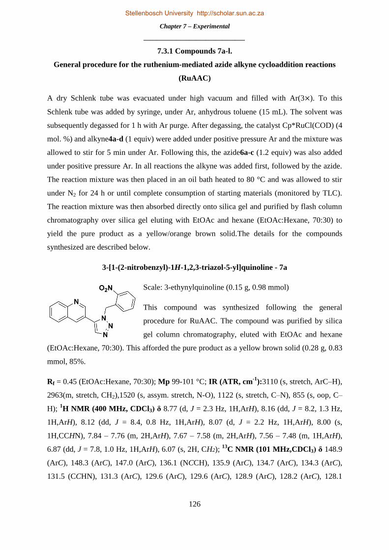
Chapter 7 – Experimental
_______________________________
126
7.3.1 Compounds 7a-l.
General procedure for the ruthenium-mediated azide alkyne cycloaddition reactions
(RuAAC)
A dry Schlenk tube was evacuated under high vacuum and filled with Ar(3×). To this
Schlenk tube was added by syringe, under Ar, anhydrous toluene (15 mL). The solvent was
subsequently degassed for 1 h with Ar purge. After degassing, the catalyst Cp*RuCl(COD) (4
mol. %) and alkyne4a-d (1 equiv) were added under positive pressure Ar and the mixture was
allowed to stir for 5 min under Ar. Following this, the azide6a-c (1.2 equiv) was also added
under positive pressure Ar. In all reactions the alkyne was added first, followed by the azide.
The reaction mixture was then placed in an oil bath heated to 80 °C and was allowed to stir
under N2 for 24 h or until complete consumption of starting materials (monitored by TLC).
The reaction mixture was then absorbed directly onto silica gel and purified by flash column
chromatography over silica gel eluting with EtOAc and hexane (EtOAc:Hexane, 70:30) to
yield the pure product as a yellow/orange brown solid.The details for the compounds
synthesized are described below.
3-[1-(2-nitrobenzyl)-1H-1,2,3-triazol-5-yl]quinoline - 7a
Scale: 3-ethynylquinoline (0.15 g, 0.98 mmol)
This compound was synthesized following the general
procedure for RuAAC. The compound was purified by silica
gel column chromatography, eluted with EtOAc and hexane
(EtOAc:Hexane, 70:30). This afforded the pure product as a yellow brown solid (0.28 g, 0.83
mmol, 85%.
Rf = 0.45 (EtOAc:Hexane, 70:30); Mp 99-101 °C; IR (ATR, cm-1
):3110 (s, stretch, ArC–H),
2963(m, stretch, CH2),1520 (s, assym. stretch, N-O), 1122 (s, stretch, C–N), 855 (s, oop, C–
H); 1H NMR (400 MHz, CDCl3) δ 8.77 (d, J = 2.3 Hz, 1H,ArH), 8.16 (dd, J = 8.2, 1.3 Hz,
1H,ArH), 8.12 (dd, J = 8.4, 0.8 Hz, 1H,ArH), 8.07 (d, J = 2.2 Hz, 1H,ArH), 8.00 (s,
1H,CCHN), 7.84 – 7.76 (m, 2H,ArH), 7.67 – 7.58 (m, 2H,ArH), 7.56 – 7.48 (m, 1H,ArH),
6.87 (dd, J = 7.8, 1.0 Hz, 1H,ArH), 6.07 (s, 2H, CH2); 13
C NMR (101 MHz,CDCl3) δ 148.9
(ArC), 148.3 (ArC), 147.0 (ArC), 136.1 (NCCH), 135.9 (ArC), 134.7 (ArC), 134.3 (ArC),
131.5 (CCHN), 131.3 (ArC), 129.6 (ArC), 129.6 (ArC), 128.9 (ArC), 128.2 (ArC), 128.1
Stellenbosch University http://scholar.sun.ac.za

Chapter 7 – Experimental
_______________________________
127
(ArC), 127.2 (ArC), 125.8 (ArC), 119.8 (ArC), 49.6 (CH2); HRMS-TOF MS ES+: m/z
[M+H]+ calcd for C18H14N5O2: 332.1147; found: 332.1156
3-[1-(3-nitrobenzyl)-1H-1,2,3-triazol-5-yl]quinoline - 7b
Scale: 3-ethynylquinoline (0.15 g, 0.98 mmol)
This compound was synthesized following the general
procedure for RuAAC. The compound was purified by silica
gel column chromatography, eluted with EtOAc and hexane
(EtOAc:Hexane, 70:30). This afforded the pure product as a
yellow brown solid (0.30 g, 0.9 mmol, 92%)
Rf = 0.45 (EtOAc:Hexane, 70:30); Mp 142-144 °C; IR (ATR, cm-1
):3070 (s, stretch, ArC–
H), 2901(m, stretch, CH2),1521 (s, assym. stretch, N-O), 1125 (s, stretch, C–N), 837 (s, oop,
C–H); 1H NMR (400 MHz, CDCl3) δ 8.78 (d, J = 2.2 Hz, 1H,ArH), 8.15 (d, J = 9.1 Hz,
2H,ArH), 8.05 (d, J = 2.1 Hz, 1H,ArH), 7.99 – 7.94 (m, 1H,ArH), 7.93 (s, 1H,CCHN), 7.82
(m, 2H,ArH), 7.69 – 7.61 (m, 1H,ArH), 7.55 – 7.42 (m, 2H,ArH), 5.72 (s, 2H, CH2); 13
C
NMR (101 MHz,CDCl3) δ 149.1 (ArC), 148.6 (ArC), 148.3 (ArC), 137.1 (NCCH), 136.4
(ArC), 135.4 (ArC), 134.5 (ArC), 133.3 (ArC), 131.4 (CCHN), 130.4 (ArC), 129.7 (ArC),
128.3 (ArC), 128.1 (ArC), 127.1 (ArC), 123.7 (ArC), 122.39 (ArC), 119.92 (ArC), 51.4
(CH2); HRMS-TOF MS ES+: m/z [M+H]+ calcd for C18H14N5O2: 332.1147; found:
332.1152
3-[1-(4-nitrobenzyl)-1H-1,2,3-triazol-5-yl]quinoline - 7c
Scale: 3-ethynylquinoline (0.15 g, 0.98 mmol)
This compound was synthesized following the general
procedure for RuAAC. The compound was purified by
silica gel column chromatography, eluted with EtOAc
and hexane (EtOAc:Hexane, 70:30). This afforded the pure product as a brown solid (0.27g,
0.82 mmol, 83%)
Rf = 0.43 (EtOAc:Hexane, 70:30); Mp 144-146 °C; IR (ATR, cm-1
):3060 (s, stretch, ArC–
H), 2912(m, stretch, CH2),1508 (s, assym. stretch, N-O), 1141 (s, stretch, C–N), 836 (s, oop,
C–H); 1H NMR (400 MHz, CDCl3) δ 8.77 (d, J = 2.3 Hz, 1H,ArH), 8.16 (dt, J = 9.1, 2.4 Hz,
Stellenbosch University http://scholar.sun.ac.za

Chapter 7 – Experimental
_______________________________
128
3H,ArH), 8.00 (t, J = 4.1 Hz, 1H,ArH), 7.94 (s, 1H,CCHN), 7.83 (ddd, J = 8.4, 6.9, 1.5 Hz,
1H,ArH), 7.78 (dd, J = 8.0, 0.6 Hz, 1H,ArH), 7.64 (ddd, J = 8.1, 6.9, 1.1 Hz, 1H,ArH), 7.29 –
7.28 (m, 1H,ArH), 7.27 – 7.25 (m, 1H,ArH), 5.72 (s, 2H, CH2); 13
C NMR (101 MHz,CDCl3)
δ 149.0 (ArC), 148.3 (ArC), 148.1 (ArC), 142.1 (ArC), 136.3 (NCCH), 135.5 (ArC), 134.5
(ArC), 131.4 (CCHN), 129.7 (ArC), 128.3 (ArC), 128.1 (ArC), 128.1 (ArC), 127.1 (ArC),
124.4 (ArC), 119.9 (ArC); 51.5 (CH2); HRMS-TOF MS ES+: m/z [M+H]+ calcd for
C18H14N5O2: 332.1147; found: 332.1150
7-chloro-4-[1-(2-nitrobenzyl)-1H-1,2,3-triazol-5-yl]quinoline - 7d
Scale: 7-chloro-4-ethynylquinoline (0.15 g, 0.8 mmol)
This compound was synthesized following the general
procedure for RuAAC. The compound was purified by silica
gel column chromatography, eluted with EtOAc and hexane
(EtOAc:Hexane, 70:30). This afforded the pure product as an
orange brown solid (0.23 g, 0.64 mmol, 81%)
Rf = 0.46 (EtOAc:Hexane, 70:30); Mp 173-175 °C; IR (ATR, cm-1
):3148 (s, stretch, ArC–
H), 2901(m, stretch, CH2),1524 (s, assym. stretch, N-O), 1244 (s, stretch, C–N), 882 (s, oop,
C–H);1H NMR (400 MHz, CDCl3) δ 8.93 (d, J = 4.1 Hz, 1H,ArH), 8.19 (s, 1H,ArH), 8.01
(d, J = 8.0 Hz, 1H,ArH), 7.96 (s, 1H,CCHN), 7.51 (m, 5H,ArH), 7.19 (d, J = 4.1 Hz,
1H,ArH), 6.94 (d, J = 7.6 Hz, 1H,ArH), 5.90 (d, J = 50.3 Hz, 2H, CH2); 13
C NMR (101
MHz,CDCl3) δ 150.9 (ArC), 149.1 (ArC), 147.2 (ArC), 136.7 (NCCH), 135.3 (ArC), 134.3
(ArC), 133.9 (ArC), 133.0 (ArC), 130.5 (CCHN), 129.7 (ArC), 129.7 (ArC), 129.5 (ArC),
129.3 (ArC), 125.7 (ArC), 125.6 (ArC), 124.8 (ArC), 122.3 (ArC), 49.6 (CH2); HRMS-TOF
MS ES+: m/z [M+H]+ calcd for C18H13ClN5O2: 366.0758; found: 366.0759
7-chloro-4-[1-(3-nitrobenzyl)-1H-1,2,3-triazol-5-yl]quinoline - 7e
Scale: 7-chloro-4-ethynylquinoline (0.15 g, 0.8 mmol)
This compound was synthesized following the general
procedure for RuAAC. The compound was purified by
silica gel column chromatography, eluted with EtOAc
and hexane (EtOAc:Hexane, 70:30). This afforded the
pure product as an orange brown solid (0.24 g, 0.66 mmol, 83%)
Stellenbosch University http://scholar.sun.ac.za

Chapter 7 – Experimental
_______________________________
129
Rf = 0.48 (EtOAc:Hexane, 70:30); Mp 176-178 °C; IR (ATR, cm-1
):3076 (s, stretch, ArC–
H), 2961(m, stretch, CH2),1524 (s, assym. stretch, N-O), 1259 (s, stretch, C–N), 819 (s, oop,
C–H); 1H NMR (400 MHz, CDCl3) δ 8.99 (d, J = 4.0 Hz, 1H,ArH), 8.22 (s, 1H,ArH), 8.09
(d, J = 7.9 Hz, 1H,ArH), 7.94 (s, 1H,CCHN), 7.71 (s, 1H,ArH), 7.47 (d, J = 8.6 Hz, 1H,ArH),
7.39 (t, J = 7.9 Hz, 1H,ArH), 7.33 (d, J = 8.9 Hz, 1H,ArH), 7.28 (d, J = 7.6 Hz, 1H,ArH),
7.21 (d, J = 4.0 Hz, 1H,ArH), 5.52 (s, 2H, CH2); 13
C NMR (101 MHz,CDCl3) δ 150.8
(ArC), 148.9 (ArC), 148.4 (ArC), 136.8 (NCCH), 136.2 (ArC), 135.3 (ArC), 133.6 (ArC),
133.2 (ArC), 130.1 (CCHN), 129.4 (ArC), 129.3 (ArC), 125.7 (ArC), 124.8 (ArC), 123.7
(ArC), 122.7 (ArC), 122.6 (ArC), 51.7 (CH2); HRMS-TOF MS ES+: m/z [M+H]+ calcd for
C18H13ClN5O2: 366.0758; found: 366.07.57
7-chloro-4-[1-(4-nitrobenzyl)-1H-1,2,3-triazol-5-yl]quinoline - 7f
Scale: 7-chloro-4-ethynylquinoline (0.15 g, 0.8 mmol)
This compound was synthesized following the general
procedure for RuAAC. The compound was purified by
silica gel column chromatography, eluted with EtOAc and
hexane (EtOAc:Hexane, 70:30). This afforded the pure
product as a brown solid (0.24 g, 0.65 mmol, 82%)
Rf = 0.46 (EtOAc:Hexane, 70:30); Mp 181-189 °C; IR (ATR, cm-1
):3115 (s, stretch, ArC–
H), 2903(m, stretch, CH2),1516 (s, assym. stretch, N-O), 1285 (s, stretch, C–N), 856 (s, oop,
C–H); 1H NMR (400 MHz, CDCl3) δ 8.94 (d, J = 4.1 Hz, 1H,ArH), 8.21 (s, 1H,ArH), 8.04
(d, J = 8.4 Hz, 2H,ArH), 7.92 (s, 1H,CCHN), 7.47 (d, J = 8.5 Hz, 1H,ArH), 7.33 (d, J = 8.9
Hz, 1H,ArH), 7.11 (t, J = 8.1 Hz, 1H,ArH), 7.05 (d, J = 8.3 Hz, 2H,ArH), 5.49 (s, 2H, CH2);
13C NMR (101 MHz,CDCl3) δ 150.8 (ArC), 149.0 (ArC), 148.0 (ArC), 141.2 (ArC), 136.8
(NCCH), 135.31 (ArC), 133.3 (ArC), 133.2 (ArC), 129.5 (CCHN), 129.4 (ArC), 128.5 (ArC),
125.7 (ArC), 124.8 (ArC), 124.2 (ArC), 122.5 (ArC), 51.7 (CH2); HRMS-TOF MS ES+:
m/z [M+H]+ calcd for C18H13ClN5O2: 366.0758; found: 366.0759
4-[1-(2-nitrobenzyl)-1H-1,2,3-triazol-5-yl]isoquinoline - 7g
Scale: 4-ethynylisoquinoline (0.15 g, 0.98 mmol)
This compound was synthesized following the general procedure
for RuAAC. The compound was purified by silica gel column
Stellenbosch University http://scholar.sun.ac.za

Chapter 7 – Experimental
_______________________________
130
chromatography, eluted with EtOAc and hexane (EtOAc:Hexane, 70:30). This afforded the
pure product as a yellow brown solid (0.16 g, 0.5 mmol, 54%)
Rf = 0.44 (EtOAc:Hexane, 70:30); Mp 129-131 °C; IR (ATR, cm-1
):3055 (s, stretch, ArC–
H), 2961(m, stretch, CH2),1523 (s, assym. stretch, N-O), 1260 (s, stretch, C–N), 861 (s, oop,
C–H); 1H NMR (400 MHz, CDCl3) δ 9.32 (s, 1H,ArH), 8.27 (s, 1H,ArH), 8.07 (dt, J = 7.1,
3.0 Hz, 1H,ArH), 8.01 – 7.93 (m, 2H,ArH,CCHN), 7.78 – 7.65 (m, 2H,ArH), 7.55 (td, J =
7.7, 1.2 Hz, 1H,ArH), 7.50 (dd, J = 8.1, 6.2 Hz, 1H,ArH), 7.46 – 7.37 (m, 1H,ArH), 6.89 (d, J
= 7.3 Hz, 1H,ArH), 5.83 (s, 2H, CH2); 13
C NMR (101 MHz,CDCl3) δ 154.9 (ArC), 147.1
(ArC), 143.0 (ArC), 135.7 (NCCH), 134.3 (ArC), 133.8 (ArC), 132.2 (ArC), 131.1 (CCHN),
129.4 (ArC), 129.3 (ArC), 128.6 (ArC), 128.5 (ArC), 125.5 (ArC), 123.4 (ArC), 118.1 (ArC),
49.3 (CH2); HRMS-TOF MS ES+: m/z [M+H]+ calcd for C18H14N5O2: 332.1147; found:
332.1150
4-[1-(3-nitrobenzyl)-1H-1,2,3-triazol-5-yl]isoquinoline - 7h
Scale: 4-ethynylisoquinoline (0.15 g, 0.98 mmol)
This compound was synthesized following the general
procedure for RuAAC. The compound was purified by silica
gel column chromatography, eluted with EtOAc and hexane
(EtOAc:Hexane, 70:30). This afforded the pure product as an
orange brown solid (0.17 g, 0.51 mmol, 55%)
Rf = 0.44 (EtOAc:Hexane, 70:30); Mp 131-133 °C;IR (ATR, cm-1
):3034 (s, stretch, ArC–
H), 2916(m, stretch, CH2),1525 (s, assym. stretch, N-O), 1161 (s, stretch, C–N), 907 (s, oop,
C–H); 1H NMR (400 MHz, CDCl3) δ 9.35 (s, 1H,ArH), 8.32 (s, 1H,ArH), 8.09 – 8.04 (m,
1H,ArH), 7.97 (ddd, J = 8.0, 2.1, 1.4 Hz, 1H,ArH), 7.90 (s, 1H,CCHN), 7.64 (m, 2H,ArH),
7.52 (t, J = 1.7 Hz, 1H,ArH), 7.33 (t, J = 7.8 Hz, 1H,ArH), 7.27 (ddd, J = 12.6, 7.0, 4.7 Hz,
2H,ArH), 5.50 (s, 2H, CH2);13
C NMR (101 MHz,CDCl3) δ 154.9 (ArC), 148.2 (ArC), 144.1
(ArC), 136.4 (NCCH), 135.8 (ArC), 134.4 (ArC), 133.8 (ArC), 133.0 (ArC), 132.0 (ArC),
130.0 (CCHN), 128.5 (ArC), 128.5 (ArC), 123.5 (ArC), 123.3 (ArC), 122.9 (ArC), 118.4
(ArC), 51.7 (CH2);HRMS-TOF MS ES+: m/z [M+H]+ calcd for C18H14N5O2: 332.1147;
found: 332.1145
Stellenbosch University http://scholar.sun.ac.za

Chapter 7 – Experimental
_______________________________
131
4-[1-(4-nitrobenzyl)-1H-1,2,3-triazol-5-yl]isoquinoline - 7i
Scale: 4-ethynylisoquinoline (0.15 g, 0.98 mmol)
This compound was synthesized following the general procedure for
RuAAC. The compound was purified by silica gel column
chromatography, eluted with EtOAc and hexane (EtOAc:Hexane,
70:30). This afforded the pure product as an orange brown solid
(0.18 g, 0.55 mmol, 60%)
Rf = 0.43 (EtOAc:Hexane, 70:30); Mp 214-216 °C; IR (ATR, cm-1
):3140 (s, stretch, ArC–
H), 2935(m, stretch, CH2),1517 (s, assym. stretch, N-O), 1163 (s, stretch, C–N), 928 (s, oop,
C–H); 1H NMR (400 MHz, CDCl3) δ 9.38 (s, 1H,ArH), 8.32 (s, 1H,ArH), 8.05 (t, J = 12.7
Hz, 1H,ArH), 7.96 (d, J = 8.7 Hz, 2H,ArH), 7.91 (s, 1H,CCHN), 7.66 (m, 2H,ArH), 7.29 (d,
J = 8.1 Hz, 1H,ArH), 7.02 (d, J = 8.7 Hz, 2H,ArH), 5.50 (s, 2H, CH2); 13
C NMR (101
MHz,CDCl3) δ 154.8 (ArC), 147.8 (ArC), 144.1 (ArC), 141.5 (ArC), 135.8 (NCCH), 134.36
(ArC), 133.1 (ArC), 132.0 (CCHN), 128.6 (ArC), 128.5 (ArC), 128.5 (ArC), 124.0 (ArC),
123.4 (ArC), 51.6 (CH2); HRMS-TOF MS ES+: m/z [M+H]+ calcd for C18H14N5O2:
332.1147; found: 332.1147
5-[1-(2-nitrobenzyl)-1H-1,2,3-triazol-5-yl]isoquinoline - 7j
Scale: 5-ethynylisoquinoline (0.1 g, 0.65 mmol)
This compound was synthesized following the general procedure for
RuAAC. The compound was purified by silica gel column
chromatography, eluted with EtOAc and hexane (EtOAc:Hexane,
70:30). This afforded the pure product as an orange brown solid (0.11
g, 0.34 mmol, 53%)
Rf = 0.29 (EtOAc:Hexane, 80:20); Mp 194-196 °C; IR (ATR, cm-1
):3128 (s, stretch, ArC–
H), 2922(m, stretch, CH2),1524 (s, assym. stretch, N-O), 1105 (s, stretch, C–N), 869 (s, oop,
C–H); 1H NMR (400 MHz, CDCl3) δ 9.35 (s, 1H,ArH), 8.52 (s, 1H,ArH), 8.13 (d, J = 8.2
Hz, 1H,ArH), 7.95 (d, J = 6.1 Hz, 2H,ArH,CCHN), 7.65 (t, J = 7.7 Hz, 1H,ArH), 7.61 – 7.50
(m, 2H,ArH), 7.43 (t, J = 7.8 Hz, 1H,ArH), 7.28 (s, 1H,ArH), 7.00 (d, J = 7.7 Hz, 1H,ArH),
5.83 (s, 2H, CH2); 13
C NMR (101 MHz,CDCl3) δ 153.2 (ArC), 147.3 (ArC), 144.5 (ArC),
135.3 (NCCH), 135.2 (ArC), 134.6 (ArC), 134.2 (ArC), 132.6 (ArC), 130.9 (ArC), 130.4
Stellenbosch University http://scholar.sun.ac.za
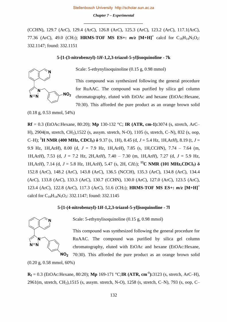
Chapter 7 – Experimental
_______________________________
132
(CCHN), 129.7 (ArC), 129.4 (ArC), 126.8 (ArC), 125.3 (ArC), 123.2 (ArC), 117.1(ArC),
77.36 (ArC), 49.0 (CH2); HRMS-TOF MS ES+: m/z [M+H]+ calcd for C18H14N5O2:
332.1147; found: 332.1151
5-[1-(3-nitrobenzyl)-1H-1,2,3-triazol-5-yl]isoquinoline - 7k
Scale: 5-ethynylisoquinoline (0.15 g, 0.98 mmol)
This compound was synthesized following the general procedure
for RuAAC. The compound was purified by silica gel column
chromatography, eluted with EtOAc and hexane (EtOAc:Hexane,
70:30). This afforded the pure product as an orange brown solid
(0.18 g, 0.53 mmol, 54%)
Rf = 0.3 (EtOAc:Hexane, 80:20); Mp 130-132 °C; IR (ATR, cm-1):3074 (s, stretch, ArC–
H), 2904(m, stretch, CH2),1522 (s, assym. stretch, N-O), 1105 (s, stretch, C–N), 832 (s, oop,
C–H); 1H NMR (400 MHz, CDCl3) δ 9.37 (s, 1H), 8.45 (d, J = 5.4 Hz, 1H,ArH), 8.19 (t, J =
9.9 Hz, 1H,ArH), 8.00 (d, J = 7.9 Hz, 1H,ArH), 7.85 (s, 1H,CCHN), 7.74 – 7.64 (m,
1H,ArH), 7.53 (d, J = 7.2 Hz, 2H,ArH), 7.40 – 7.30 (m, 1H,ArH), 7.27 (d, J = 5.9 Hz,
1H,ArH), 7.14 (d, J = 5.8 Hz, 1H,ArH), 5.47 (s, 2H, CH2); 13
C NMR (101 MHz,CDCl3) δ
152.8 (ArC), 148.2 (ArC), 143.8 (ArC), 136.5 (NCCH), 135.3 (ArC), 134.8 (ArC), 134.4
(ArC), 133.8 (ArC), 133.3 (ArC), 130.7 (CCHN), 130.0 (ArC), 127.0 (ArC), 123.5 (ArC),
123.4 (ArC), 122.8 (ArC), 117.3 (ArC), 51.6 (CH2); HRMS-TOF MS ES+: m/z [M+H]+
calcd for C18H14N5O2: 332.1147; found: 332.1145
5-[1-(4-nitrobenzyl)-1H-1,2,3-triazol-5-yl]isoquinoline - 7l
Scale: 5-ethynylisoquinoline (0.15 g, 0.98 mmol)
This compound was synthesized following the general procedure for
RuAAC. The compound was purified by silica gel column
chromatography, eluted with EtOAc and hexane (EtOAc:Hexane,
70:30). This afforded the pure product as an orange brown solid
(0.20 g, 0.58 mmol, 60%)
Rf = 0.3 (EtOAc:Hexane, 80:20); Mp 169-171 °C;IR (ATR, cm-1
):3123 (s, stretch, ArC–H),
2961(m, stretch, CH2),1515 (s, assym. stretch, N-O), 1258 (s, stretch, C–N), 793 (s, oop, C–
Stellenbosch University http://scholar.sun.ac.za

Chapter 7 – Experimental
_______________________________
133
H);1H NMR (400 MHz, CDCl3) δ 9.37 (s, 1H,ArH), 8.49 (d, J = 5.3 Hz, 1H,ArH), 8.16 (d, J
= 8.2 Hz, 1H,ArH), 7.98 (t, J = 9.3 Hz, 2H,ArH), 7.87 (s, 1H,CCHN), 7.65 (t, J = 7.7 Hz,
1H,ArH), 7.44 (t, J = 9.5 Hz, 1H,ArH), 7.19 (d, J = 5.8 Hz, 1H,ArH), 7.02 (d, J = 8.5 Hz,
2H,ArH), 5.46 (s, 2H, CH2);13
C NMR (101 MHz,CDCl3) δ 153.0 (ArC), 147.9 (ArC), 144.1
(ArC), 141.6 (ArC), 135.3 (NCCH), 134.7 (ArC), 134.6 (ArC), 133.2 (ArC), 130.5 (CCHN),
128.5 (ArC), 126.9 (ArC), 124.0 (ArC), 123.3 (ArC), 117.3 (ArC), 51.51 (CH2); HRMS-
TOF MS ES+: m/z [M+H]+ calcd for C18H14N5O2: 332.1147; found: 332.1146
7.3.2 Compounds 8a-l.
General Procedure for Reduction of Aromatic Nitro Groups and Acylation with
Acryloyl Chloride
To a 2 neck round bottom flask was added a solution of the aromatic nitro "clicked"
compound7a-l (1 equiv) in anhydrous DCM (15 mL) under N2. A hydrogen ballon was
attached to one neck and Pd/C (10 mol. % on C) (5 mol. %) was added to the reaction
mixture. The N2 was evacuated through a tap on the other neck, creating a hydrogen
atmosphere in the reaction vessel under atmospheric pressure. The resulting suspension was
then stirred at rt for 6 hours or until complete reduction of the nitro moiety to the amine
(monitored by TLC). The reaction mixture was then filtered through Celite and the solvent
was removed under reduced pressure to afford the product as a white foam, which was
sufficiently pure to use without further purification. To a dry round bottom flask was then
directly added a solution of the crude reduced aromatic amine (1 equiv) in anhydrous DCM
(15 mL) under N2. The reaction mixture was placed in an ice bath and cooled to 0 °C, where
after Et3N (12 equiv) was added slowly by syringe and allowed to stir for 5 min. Acryloyl
chloride (1.2 equiv) was then added drop-wise and a colour change from colourless to yellow
was seen immediately. The ice bath was then removed and the reaction mixture was allowed
to stir at rt for up to 4 hours or until complete consumption of the starting material (monitored
by TLC). After removal of the solvent in vacuo, the residue was purified by flash column
chromatography over silica gel, eluting first with EtOAc and hexane (EtOAc:Hexane, 90:10),
then EtOAc. This afforded the pure product as a white foam/solid.
N-{2-{[5-(quinolin-3-yl)-1H-1,2,3-triazol-1-yl]methyl}phenyl}acrylamide - 8a
Scale: 3-(1-(2-nitrobenzyl)-1H-1,2,3-triazol-5-yl)quinoline (200 mg,
0.604 mmol)
Stellenbosch University http://scholar.sun.ac.za

Chapter 7 – Experimental
_______________________________
134
This compound was synthesized following the general procedure for reduction of aromatic
nitro and acylation with acryloyl chloride. The compound was purified by silica gel column
chromatography, eluted first with EtOAc and hexane (EtOAc:Hexane, 90:10) and then
EtOAc. This afforded the pure product as a white foam (131 mg, 0.37 mmol, 61%)
Rf = 0.44 (EtOAc:Hexane, 90:10); Mp 185-187 °C; IR (ATR, cm-1
):3247 (m, stretch, N–H),
3120 (s, stretch, ArC–H), 1652 (s, stretch,–C=C–), 1208 (s, stretch, C–N), 803 (s, oop, C–H);
1H NMR (400 MHz, DMSO-d6) δ 9.84 (s, 1H, NH), 8.94 (d, J = 2.2 Hz, 1H, ArH), 8.40 (d,
J = 2.1 Hz, 1H, ArH), 8.24 (s, 1H,CCHN), 8.05 (d, J = 8.3 Hz, 1H, ArH), 7.96 (d, J = 8.4 Hz,
1H, ArH), 7.88 – 7.78 (m, 1H, ArH), 7.68 (t, J = 7.1 Hz, 1H, ArH), 7.39 – 7.22 (m, 2H, ArH),
7.11 (t, J = 7.5 Hz, 1H, ArH), 6.73 (d, J = 7.3 Hz, 1H, ArH), 6.28 (s, 1H, CCHC), 6.12 (dd, J
= 17.1, 2.0 Hz, 1H, CCH2), 5.77 (s, 2H, CH2), 5.70 – 5.59 (m, 1H, CCH2); 13
C NMR (101
MHz, DMSO-d6) δ 163.5 (HNCO), 149.3 (ArC), 147.1 (ArC), 135.2 (NCCH), 135.0 (ArC),
135.0 (ArC), 133.9 (ArC), 131.2 (CCCH2), 130.7 (CCHN), 130.5 (ArC), 128.7 (ArC), 128.5
(ArC), 128.3 (ArC), 127.5 (ArC), 127.4 (ArC), 126.8 (CCH2), 126.8 (ArC), 126.0 (ArC),
125.7 (ArC), 119.96 (ArC), 48.83 (CH2); HRMS-TOF MS ES+: m/z [M+H]+ calcd for
C21H18N5O: 356.1511; found: 356.1510
N-{3-{[5-(quinolin-3-yl)-1H-1,2,3-triazol-1-yl]methyl}phenyl}acrylamide - 8b
Scale: 3-(1-(3-nitrobenzyl)-1H-1,2,3-triazol-5-yl)quinoline (200 mg,
0.604 mmol)
This compound was synthesized following the general procedure for
reduction of aromatic nitro and acylation with acryloyl chloride. The
compound was purified by silica gel column chromatography, eluted
first with EtOAc and hexane (EtOAc:Hexane, 90:10) and then EtOAc.
This afforded the pure product as a white foam (132 mg, 0.37 mmol,
62%)
Rf = 0.44 (EtOAc:Hexane, 90:10); Mp 122-124 °C; IR (ATR, cm-1
):3259 (m, stretch, N–H),
1667 (s, stretch,–C=C–), 1202 (s, stretch, C–N), 747 (s, oop, C–H);1H NMR (400 MHz,
DMSO-d6) δ 10.12 (s, 1H, NH), 8.95 (d, J = 2.2 Hz, 1H, ArH), 8.51 (dd, J = 9.0, 1.9 Hz, 1H,
ArH), 8.21 (d, J = 8.7 Hz, 1H,CCHN), 8.05 (d, J = 8.5 Hz, 1H, ArH), 7.97 (t, J = 7.2 Hz, 1H,
ArH), 7.90 – 7.79 (m, 1H, ArH), 7.68 (t, J = 7.4 Hz, 1H, ArH), 7.55 (d, J = 8.3 Hz, 1H, ArH),
7.39 (s, 1H, ArH), 7.21 (t, J = 7.9 Hz, 1H, ArH), 6.94 – 6.72 (m, 1H, CCHC), 6.46 – 6.30 (m,
Stellenbosch University http://scholar.sun.ac.za

Chapter 7 – Experimental
_______________________________
135
1H, CCH2), 6.29 – 6.13 (m, 1H, CCH2), 5.88 – 5.62 (m, 3H, CH2); 13
C NMR (101 MHz,
DMSO-d6) δ 163.1 (HNCO), 149.3 (ArC), 147.2 (ArC), 139.5 (NCCH), 136.5 (ArC), 135.6
(ArC), 134.8 (ArC), 134.2 (ArC), 131.7 (CCCH2), 130.7 (CCHN), 129.2 (ArC), 128.8 (ArC),
128.5 (ArC), 127.6 (ArC), 127.0 (ArC), 126.8 (CCH2), 122.0 (ArC), 120.0 (ArC), 118.7
(ArC), 117.5 (ArC), 51.5 (CH2); HRMS-TOF MS ES+: m/z [M+H]+ calcd for C21H18N5O:
356.1511; found: 356.1520
N-{4-{[5-(quinolin-3-yl)-1H-1,2,3-triazol-1-yl]methyl}phenyl}acrylamide - 8c
Scale: 3-(1-(4-nitrobenzyl)-1H-1,2,3-triazol-5-yl)quinoline (200 mg,
0.604 mmol)
This compound was synthesized following the general procedure for
reduction of aromatic nitro and acylation with acryloyl chloride.
The compound was purified by silica gel column chromatography,
eluted first with EtOAc and hexane (EtOAc:Hexane, 90:10) and
then EtOAc. This afforded the pure product as a white foam (134
mg, 0.38 mmol, 62%)
Rf= 0.43 (EtOAc:Hexane, 90:10); Mp 178-180 °C; IR (ATR, cm-1
):3239 (m, stretch, N–H),
1679 (s, stretch,–C=C–), 1254 (s, stretch, C–N), 790 (s, oop, C–H); 1H NMR (400 MHz,
DMSO-d6) δ 10.12 (s, 1H, NH), 8.95 (d, J = 2.2 Hz, 1H, ArH), 8.54 (d, J = 2.1 Hz, 1H,
ArH), 8.19 (s, 1H,CCHN), 8.06 (d, J = 8.5 Hz, 1H, ArH), 8.00 (d, J = 8.1 Hz, 1H, ArH), 7.84
(t, J = 7.0 Hz, 1H, ArH), 7.69 (t, J = 7.5 Hz, 1H, ArH), 7.54 (d, J = 8.5 Hz, 2H, ArH), 7.01
(d, J = 8.5 Hz, 2H, ArH), 6.37 (dd, J = 17.0, 10.1 Hz, 1H, CCHC), 6.21 (dd, J = 17.0, 2.0 Hz,
1H, CCH2), 5.79 (s, 2H, CH2), 5.76 – 5.69 (m, 1H, CCH2); 13
C NMR (101 MHz, DMSO-d6)
δ 163.1 (HNCO), 149.4 (ArC), 147.2 (ArC), 138.6 (NCCH), 135.6 (ArC), 134.7 (ArC), 134.2
(ArC), 131.7 (CCCH2), 130.8 (ArC), 130.6 (CCHN), 128.8 (ArC), 128.5 (ArC), 127.7 (ArC),
127.6 (ArC), 127.0 (ArC), 126.8 (CCH2), 120.1 (ArC), 119.4 (ArC), 51.3 (CH2); HRMS-
TOF MS ES+: m/z [M+H]+ calcd for C21H18N5O: 356.1511; found: 356.1521
N-{2-{[5-(7-chloroquinolin-4-yl)-1H-1,2,3-triazol-1-yl]methyl}phenyl}acrylamide - 8d
Scale: 7-chloro-4-(1-(2-nitrobenzyl)-1H-1,2,3-triazol-5-
yl)quinoline (140 mg, 0.38 mmol)
Stellenbosch University http://scholar.sun.ac.za

Chapter 7 – Experimental
_______________________________
136
This compound was synthesized following the general procedure for reduction of aromatic
nitro and acylation with acryloyl chloride. The compound was purified by silica gel column
chromatography, eluted first with EtOAc and hexane (EtOAc:Hexane, 90:10) and then
EtOAc. This afforded the pure product as a white foam (90 mg, 0.23 mmol, 61%)
Rf = 0.41 (EtOAc:Hexane, 90:10); Mp 154-156 °C; IR (ATR, cm-1
):3249 (m, stretch, N–H),
1665 (s, stretch,–C=C–), 1186 (s, stretch, C–N), 876 (s, oop, C–H); 1H NMR (400 MHz,
CDCl3) δ 9.09 (s, 1H, NH), 9.03 (d, J = 4.3 Hz, 1H, ArH), 8.25 (d, J = 1.9 Hz, 1H, ArH),
7.89 (t, J = 8.3 Hz, 1H, ArH), 7.85 (s, 1H,CCHN), 7.46 (dd, J = 8.9, 2.0 Hz, 1H, ArH), 7.28 –
7.25 (m, 3H, ArH), 6.79 (t, J = 7.4 Hz, 1H, ArH), 6.47 (t, J = 3.7 Hz, 1H, ArH), 6.46 – 6.40
(m, 1H, CCHC), 6.31 (d, J = 7.4 Hz, 1H, CCH2), 5.85 (d, J = 10.8 Hz, 1H, CCH2), 5.30 (s,
2H, CH2); 13
C NMR (101 MHz,CDCl3) δ 164.3 (HNCO), 150.9 (ArC), 148.9 (ArC), 137.0
(NCCH), 136.9 (ArC), 135.1 (ArC), 133.5 (ArC), 133.2 (ArC), 131.4 (CCCH2), 130.4
(CCHN), 130.2 (ArC), 129.4 (ArC), 129.3 (ArC), 128.2 (ArC), 125.9 (CCH2), 125.1 (ArC),
124.94 (ArC), 123.05 (ArC), 50.1 (CH2); HRMS-TOF MS ES+: m/z [M+H]+ calcd for
C21H17ClN5O: 390.1122; found: 390.1120
N-{3-{[5-(7-chloroquinolin-4-yl)-1H-1,2,3-triazol-1-yl]methyl}phenyl}acrylamide - 8e
Scale: 7-chloro-4-(1-(3-nitrobenzyl)-1H-1,2,3-triazol-5-
yl)quinoline (190 mg, 0.52 mmol)
This compound was synthesized following the general
procedure for reduction of aromatic nitro and acylation with
acryloyl chloride. The compound was purified by silica gel
column chromatography, eluted first with EtOAc and hexane
(EtOAc:Hexane, 90:10) and then EtOAc. This afforded the
pure product as a white foam (113 mg, 0.29 mmol, 56%
Rf = 0.41 (EtOAc:Hexane, 90:10); Mp 128-131 °C;IR (ATR, cm-1
):3271 (m, stretch, N–H),
2920(m, stretch, CH2), 1668 (s, stretch,–C=C–), 1169 (s, stretch, C–N), 877 (s, oop, C–H);
1H NMR (400 MHz, CDCl3) δ 8.94 (s, 1H, NH), 8.16 (s, 1H, ArH), 7.88 (d, J = 24.5 Hz,
1H,CCHN), 7.65 (s, 1H, ArH), 7.48 – 7.36 (m, 2H, ArH), 7.35 – 7.27 (m, 1H, ArH), 7.18 (d,
J = 4.3 Hz, 1H, ArH), 7.15 – 6.99 (m, 2H, ArH), 6.56 (t, J = 7.9 Hz, 1H, ArH), 6.45 – 6.35
(m, 1H, CCHC), 6.29 – 6.15 (m, 1H, CCH2), 5.75 (d, J = 10.3 Hz, 1H, CCH2), 5.40 (d, J =
20.4 Hz, 2H, CH2);13
C NMR (101 MHz,CDCl3) δ 163.6 (HNCO), 150.8 (ArC), 149.6
Stellenbosch University http://scholar.sun.ac.za

Chapter 7 – Experimental
_______________________________
137
(ArC), 148.8 (ArC), 138.6 (NCCH), 136.5 (ArC), 135.3 (ArC), 135.1 (ArC), 133.6 (CCCH2),
131.0 (CCHN), 129.6 (ArC), 129.1 (ArC), 129.0 (ArC), 128.3 (ArC), 126.0 (CCH2), 125.1
(ArC), 123.4 (ArC), 122.9 (ArC), 120.0 (ArC), 119.0 (ArC), 52.6 (CH2); HRMS-TOF MS
ES+: m/z [M+H]+ calcd for C21H17ClN5O: 390.1122; found: 390.1118
N-{4-{[5-(7-chloroquinolin-4-yl)-1H-1,2,3-triazol-1-yl]methyl}phenyl}acrylamide - 8f
Scale: 7-chloro-4-(1-(4-nitrobenzyl)-1H-1,2,3-triazol-
5-yl)quinoline (190 mg, 0.52 mmol)
This compound was synthesized following the
general procedure for reduction of aromatic nitro and
acylation with acryloyl chloride. The compound was
purified by silica gel column chromatography, eluted
first with EtOAc and hexane (EtOAc:Hexane, 90:10) and then EtOAc. This afforded the pure
product as a white foam (116 mg, 0.30 mmol, 57%)
Rf = 0.4 (EtOAc:Hexane, 90:10); Mp 161-173 °C;IR (ATR, cm-1
):3287 (m, stretch, N–H),
2850(m, stretch, CH2), 1675 (s, stretch,–C=C–), 1242 (s, stretch, C–N), 877 (s, oop, C–H);
1H NMR (400 MHz, CDCl3) δ 8.92 (s, 1H, NH), 8.19 (t, J = 6.1 Hz, 1H, ArH), 7.85 (s,
1H,CCHN), 7.47 (d, J = 8.5 Hz, 1H, ArH), 7.43 (dd, J = 8.9, 1.6 Hz, 1H, ArH), 7.36 (d, J =
8.0 Hz, 2H, ArH), 7.30 (d, J = 8.9 Hz, 1H, ArH), 7.12 (d, J = 4.2 Hz, 1H, ArH), 6.79 (d, J =
8.3 Hz, 2H, ArH), 6.42 (d, J = 16.8 Hz, 1H, CCHC), 6.24 (dd, J = 16.8, 10.2 Hz, 1H, CCH2),
5.77 (d, J = 10.2 Hz, 1H, CCH2), 5.36 (s, 2H, CH2);13
C NMR (101 MHz,CDCl3) δ 163.6
(HNCO), 150.8 (ArC), 148.9 (ArC), 138.3 (NCCH), 136.5 (ArC), 135.2 (ArC), 133.7 (ArC),
133.0 (CCCH2), 131.0 (CCHN), 130.2 (ArC), 129.2 (ArC), 129.1 (ArC), 128.57 (ArC),
128.42 (ArC), 126.0 (CCH2), 125.1 (ArC), 122.7 (ArC), 120.1v, 52.4 (CH2);HRMS-TOF
MS ES+: m/z [M+H]+ calcd for C21H17ClN5O: 390.1122; found: 390.1111
N-{2-{[5-(isoquinolin-4-yl)-1H-1,2,3-triazol-1-yl]methyl}phenyl}acrylamide - 8g
Scale: 4-(1-(2-nitrobenzyl)-1H-1,2,3-triazol-5-yl)isoquinoline (108
mg, 0.33 mmol)
This compound was synthesized following the general procedure for
reduction of aromatic nitro and acylation with acryloyl chloride. The
compound was purified by silica gel column chromatography, eluted
Stellenbosch University http://scholar.sun.ac.za

Chapter 7 – Experimental
_______________________________
138
first with EtOAc and hexane (EtOAc:Hexane, 90:10) and then EtOAc. This afforded the pure
product as a white foam (68 mg, 0.19 mmol, 59%)
Rf = 0.24 (EtOAc:Hexane, 90:10); Mp 189-191 °C; IR (ATR, cm-1
):3224 (m, stretch, N–H),
2919(m, stretch, CH2), 1673 (s, stretch,–C=C–), 1205 (s, stretch, C–N), 749 (s, oop, C–H);
1H NMR (400 MHz, CDCl3) δ 9.43 (s, 1H, NH), 8.41 (s, 1H, ArH), 8.13 (d, J = 7.9 Hz, 1H,
ArH), 7.92 (d, J = 7.8 Hz, 1H, ArH), 7.84 (s, 1H,CCHN), 7.76 – 7.63 (m, 2H, ArH), 7.28 (d,
J = 8.3 Hz, 1H, ArH), 7.22 (d, J = 7.2 Hz, 1H, ArH), 6.76 (t, J = 7.4 Hz, 1H, CCHC), 6.55 –
6.44 (m, 2H, ArH), 6.34 (d, J = 7.4 Hz, 1H, CCH2), 5.84 (dd, J = 8.9, 2.3 Hz, 1H, CCH2),
5.28 (s, 2H, CH2); 13
C NMR (101 MHz,CDCl3) δ 164.3 (HNCO), 155.0 (ArC), 144.5 (ArC),
137.1 (NCCH), 135.5 (ArC), 134.6 (ArC), 133.4 (ArC), 132.2 (ArC), 131.6 (CCCH2), 130.4
(CCHN), 130.2 (ArC), 128.5 (ArC), 128.4 (ArC), 127.9 (ArC), 125.3 (CCH2), 124.9 (ArC),
124.8 (ArC), 123.6 (ArC), 118.2 (ArC), 49.7 (CH2); HRMS-TOF MS ES+: m/z [M+H]+
calcd for C21H18N5O: 356.1511; found: 356.1515
N-{3-{[5-(isoquinolin-4-yl)-1H-1,2,3-triazol-1-yl]methyl}phenyl}acrylamide - 8h
Scale: 4-(1-(3-nitrobenzyl)-1H-1,2,3-triazol-5-yl)isoquinoline (131
mg, 0.4 mmol)
This compound was synthesized following the general procedure
for reduction of aromatic nitro and acylation with acryloyl
chloride. The compound was purified by silica gel column
chromatography, eluted first with EtOAc and hexane
(EtOAc:Hexane, 90:10) and then EtOAc. This afforded the pure
product as a white foam (91 mg, 0.26 mmol, 64%)
Rf = 0.24 (EtOAc:Hexane, 90:10); Mp 126-128 °C;IR (ATR, cm-1
):3266 (m, stretch, N–
H),1667 (s, stretch,–C=C–), 1207 (s, stretch, C–N), 747 (s, oop, C–H); 1H NMR (400 MHz,
CDCl3) δ 9.32 (s, 1H, NH), 8.29 (d, J = 5.0 Hz, 1H, ArH), 8.08 – 8.02 (m, 2H, ArH), 7.85 (s,
1H,CCHN), 7.68 – 7.59 (m, 2H, ArH), 7.54 (d, J = 8.0 Hz, 1H, ArH), 7.35 – 7.30 (m, 1H,
ArH), 7.10 (s, 1H, ArH), 7.02 (t, J = 7.9 Hz, 1H, ArH), 6.52 (d, J = 7.7 Hz, 1H, ArH), 6.38
(dd, J = 16.9, 1.4 Hz, 1H, CCHC), 6.24 (dd, J = 16.9, 10.1 Hz, 1H, CCH2), 5.72 (dd, J = 10.1,
1.4 Hz, 1H, CCH2), 5.37 (s, 2H, CH2);13
C NMR (101 MHz,CDCl3) δ 163.7 (HNCO), 154.6
(ArC), 144.2 (ArC), 138.7 (NCCH), 135.5 (ArC), 135.4 (ArC), 134.6 (ArC), 133.1 (ArC),
131.9 (CCCH2), 131.2 (CCHN), 129.5 (ArC), 128.3 (ArC), 128.3 (ArC), 128.0 (ArC), 123.7
Stellenbosch University http://scholar.sun.ac.za

Chapter 7 – Experimental
_______________________________
139
(CCH2), 123.4 (ArC), 120.0 (ArC), 119.0 (ArC), 118.7 (ArC), 52.4 (CH2);HRMS-TOF MS
ES+: m/z [M+H]+ calcd for C21H18N5O: 356.1511; found: 356.1511
N-{4-{[5-(isoquinolin-4-yl)-1H-1,2,3-triazol-1-yl]methyl}phenyl}acrylamide - 8i
Scale: 4-(1-(4-nitrobenzyl)-1H-1,2,3-triazol-5-
yl)isoquinoline (155 mg, 0.47 mmol)
This compound was synthesized following the general
procedure for reduction of aromatic nitro and acylation
with acryloyl chloride. The compound was purified by
silica gel column chromatography, eluted first with
EtOAc and hexane (EtOAc:Hexane, 90:10) and then EtOAc. This afforded the pure product
as a white foam (95 mg, 0.27 mmol, 57%)
Rf = 0.26 (EtOAc:Hexane, 90:10); Mp 179-181 °C;IR (ATR, cm-1
):3224 (m, stretch, N–H),
2960(m, stretch, CH2), 1681 (s, stretch,–C=C–), 1250 (s, stretch, C–N), 752 (s, oop, C–H);
1H NMR (400 MHz, CDCl3) δ 9.36 (s, 1H, NH), 8.25 (s, 1H, ArH), 8.09 (dd, J = 17.8, 11.4
Hz, 2H, ArH), 7.86 (s, 1H,CCHN), 7.74 – 7.63 (m, 2H, ArH), 7.36 (d, J = 6.8 Hz, 3H, ArH),
6.76 (t, J = 13.9 Hz, 2H, ArH), 6.47 – 6.21 (m, 2H, CCHC, CCH2), 5.73 (d, J = 10.1 Hz, 1H,
CCH2), 5.42 – 5.28 (m, 2H, CH2);13
C NMR (101 MHz,CDCl3) δ 163.7 (HNCO), 154.6
(ArC), 144.17 (ArC), 138.3 (NCCH), 135.5 (ArC), 134.6 (ArC), 132.9 (ArC), 132.0
(CCCH2), 131.2 (CCHN), 130.3 (ArC), 128.6 (ArC), 128.4 (ArC), 128.3 (ArC), 128.2 (ArC),
128.1 (ArC), 123.7 (CCH2), 120.1 (ArC), 118.7 (ArC), 52.2 (CH2);HRMS-TOF MS ES+:
m/z [M+H]+ calcd for C21H18N5O: 356.1511; found: 356.1516
N-{2-{[5-(quinolin-5-yl)-1H-1,2,3-triazol-1-yl]methyl}phenyl}acrylamide - 8j
Scale: 5-(1-(2-nitrobenzyl)-1H-1,2,3-triazol-5-yl)isoquinoline (140 mg,
0.42mmol)
This compound was synthesized following the general procedure for
reduction of aromatic nitro and acylation with acryloyl chloride. The
compound was purified by silica gel column chromatography, eluted with
EtOAc and MeOH (EtOAc:MeOH, 95:5). This afforded the pure product
as a white foam (91 mg, 0.26 mmol, 61%)
Stellenbosch University http://scholar.sun.ac.za

Chapter 7 – Experimental
_______________________________
140
Rf = 0.38 (EtOAc:MeOHl, 95:5) ; Mp 193-195 °C;IR (ATR, cm-1
):3222 (m, stretch, N–H),
1673 (s, stretch,–C=C–), 1245 (s, stretch, C–N), 827 (s, oop, C–H); 1H NMR (400 MHz,
CDCl3) δ 9.39 (s, 2H, NH), 8.49 (s, 1H, ArH), 8.19 (dd, J = 22.3, 7.6 Hz, 1H, ArH), 7.97 (d,
J = 7.6 Hz, 1H, ArH), 7.83 (s, 1H,CCHN), 7.75 (t, J = 7.6 Hz, 1H, ArH), 7.66 – 7.57 (m, 1H,
ArH), 7.26 (t, J = 7.7 Hz, 1H, ArH), 7.08 (d, J = 5.6 Hz, 1H, ArH), 6.75 (t, J = 7.3 Hz, 1H,
CCHC), 6.49 (q, J = 17.2 Hz, 2H, ArH), 6.23 (d, J = 7.2 Hz, 1H, CCH2), 5.84 (t, J = 12.6 Hz,
1H, CCH2), 5.28 (d, J = 12.7 Hz, 2H, CH2);13
C NMR (101 MHz,CDCl3) δ 164.3 (HNCO),
153.2 (ArC), 144.7 (ArC), 137.3 (NCCH), 135.1 (ArC), 134.9 (ArC), 134.9 (ArC), 133.4
(ArC), 131.65 (CCCH2), 130.6 (CCHN), 130.3 (ArC), 130.3 (ArC), 128.0 (ArC), 126.8
(CCH2), 125.1 (ArC), 124.8 (ArC), 123.1 (ArC), 117.3 (ArC), 49.7 (CH2);HRMS-TOF MS
ES+: m/z [M+H]+ calcd for C21H18N5O: 356.1511; found: 356.1515
N-{2-{[5-(quinolin-5-yl)-1H-1,2,3-triazol-1-yl]methyl}phenyl}acrylamide - 8k
Scale: 5-(1-(3-nitrobenzyl)-1H-1,2,3-triazol-5-yl)isoquinoline (150 mg,
0.45 mmol)
This compound was synthesized following the general procedure for
reduction of aromatic nitro and acylation with acryloyl chloride. The
compound was purified by silica gel column chromatography, eluted
with EtOAc and MeOH (EtOAc:MeOH, 95:5). This afforded the pure
product as a white foam (93 mg, 0.26 mmol, 58%)
Rf = 0.37 (EtOAc:MeOH, 95:5); Mp 134-136 °C;IR (ATR, cm-1
):3262 (m, stretch, N–H),
2960(m, stretch, CH2), 1611 (s, stretch,–C=C–), 1257 (s, stretch, C–N), 792 (s, oop, C–H);
1H NMR (400 MHz, CDCl3) δ 9.30 (s, 1H, NH), 8.40 (s, 1H, ArH), 8.10 (d, J = 8.3 Hz, 1H),
7.83 (s, 1H,CCHN), 7.65 (t, J = 7.7 Hz, 1H, ArH), 7.57 – 7.47 (m, 2H, ArH), 7.36 (d, J = 8.0
Hz, 1H, ArH), 7.14 – 7.02 (m, 3H, ArH), 6.58 (d, J = 7.8 Hz, 1H, ArH), 6.40 (d, J = 16.9 Hz,
1H, CCHC), 6.21 (dd, J = 16.8, 10.2 Hz, 1H, CCH2), 5.76 (d, J = 10.2 Hz, 1H, CCH2), 5.34
(s, 2H, CH2);13
C NMR (101 MHz,CDCl3) δ 163.5 (HNCO), 152.9 (ArC), 144.1 (ArC),
138.4 (NCCH), 135.6 (ArC), 135.1 (ArC), 134.9 (ArC), 134.5 (ArC), 133.2 (ArC), 131.1
(CCCH2), 130.1 (CCHN), 129.5 (ArC), 128.6 (ArC), 128.2 (ArC), 126.9 (CCH2), 123.7
(ArC), 123.6 (ArC), 119.8 (ArC), 119.2 (ArC), 117.4 (ArC), 52.4 (CH2);HRMS-TOF MS
ES+: m/z [M+H]+ calcd for C21H18N5O: 356.1511; found: 356.1512
N-{2-{[5-(quinolin-5-yl)-1H-1,2,3-triazol-1-yl]methyl}phenyl}acrylamide - 8l
Stellenbosch University http://scholar.sun.ac.za

Chapter 7 – Experimental
_______________________________
141
Scale: 5-(1-(4-nitrobenzyl)-1H-1,2,3-triazol-5-yl)isoquinoline
(140 mg, 0.42 mmol)
This compound was synthesized following the general
procedure for reduction of aromatic nitro and acylation with
acryloyl chloride. The compound was purified by silica gel
column chromatography, eluted with EtOAc and MeOH
(EtOAc:MeOH, 95:5). This afforded the pure product as a white foam (99 mg, 0.28 mmol,
66%)
Rf = 0.36 (EtOAc:MeOH, 95:5); Mp 196-198 °C; IR (ATR, cm-1
):3274 (m, stretch, N–H),
2960(m, stretch, CH2), 1664 (s, stretch,–C=C–), 1256 (s, stretch, C–N), 760 (s, oop, C–H);
1H NMR (400 MHz, CDCl3) δ 9.33 (s, 1H, NH), 8.45 (d, J = 5.8 Hz, 1H, ArH), 8.12 (d, J =
8.3 Hz, 1H, ArH), 7.82 (s, 1H,CCHN), 7.65 (dd, J = 18.5, 11.2 Hz, 1H, ArH), 7.51 (s, 1H,
ArH), 7.46 (d, J = 6.6 Hz, 1H, ArH), 7.34 (d, J = 8.1 Hz, 2H, ArH), 7.13 (d, J = 5.9 Hz, 1H,
ArH), 6.78 (d, J = 8.4 Hz, 2H, ArH), 6.42 (d, J = 16.8 Hz, 1H, CCHC), 6.30 – 6.17 (m, 1H,
CCH2), 5.84 – 5.71 (m, 1H, CCH2), 5.32 (s, 2H, CH2); 13
C NMR (101 MHz,CDCl3) δ 153.1
(HNCO), 144.5 (ArC), 138.1 (NCCH), 135.1 (ArC), 134.8 (ArC), 134.4 (ArC), 133.0 (ArC),
131.1 (CCCH2), 130.6 (CCHN), 130.1 (ArC), 128.6 (ArC), 128.3 (ArC), 126.7 (CCH2),
123.7 (ArC), 120.0 (ArC), 117.4 (ArC), 52.1 (CH2); HRMS-TOF MS ES+: m/z [M+H]+
calcd for C21H18N5O: 356.1511; found: 356.151
Stellenbosch University http://scholar.sun.ac.za

Chapter 8 – References
_______________________________
142
CHAPTER 8 – REFERENCES
References for Chapters 1-7 follow. Figures, diagrams and schemes taken directly from the
source are referenced accordingly in the title of the corresponding figure, diagram or scheme.
1. Cancer Mortality and Morbidity Situation and Trends [Online], 2013.
http://www.who.int/gho/ncd/mortality_morbidity/cancer_text/en/ (accessed 14/01).
2. Parkin, D. M.; Bray, F.; Ferlay, J.; Pisani, P., Global cancer statistics, 2002. Ca-A
Cancer Journal for Clinicians 2005,55 (2), 74-108.
3. Global Cancer Facts & Figures 2nd Edition. American Cancer Society: Atlanta, 2011.
4. Parkin, D. M.; Pisani, P.; Ferlay, J., Global Cancer Statistics. Ca-A Cancer Journal
for Clinicians 1999,49 (1), 33-64.
5. World Cancer Factsheet. International Agency for Research on Cancer and Cancer
Research UK: London, 2012.
6. Ferlay, J., Soerjomataram, I., Ervik, M., Dikshit, R., Eser, S., Mathers, C., Rebelo, M.,
Parkin, D. M., Forman, D., Bray, F. GLOBOCAN 2012 v1.0, Cancer Incidence and Mortality
Worldwide 2013. http://globocan.iarc.fr (accessed 14/01/14).
7. Bray, F.; Ren, J. S.; Masuyer, E.; Ferlay, J., Global estimates of cancer prevalence for
27 sites in the adult population in 2008. International Journal of Cancer 2013,132 (5), 1133-
1145.
8. Ferlay, J.; Shin, H. R.; Bray, F.; Forman, D.; Mathers, C. D.; Parkin, D. GLOBOCAN
2008, Cancer Incidence and Mortality Worldwide 2010. http://globocan.iarc.fr (accessed
14/01/14).
9. Jemal, A.; Bray, F.; Forman, D.; O'Brien, M.; Ferlay, J.; Center, M.; Parkin, D. M.,
Cancer burden in Africa and opportunities for prevention. Cancer 2012,118 (18), 4372-4384.
10. Cancer Fact sheet N°297 2013. http://www.who.int/mediacentre/factsheets/fs297/en/
(accessed 15/01/14).
11. Dangoor, N. E. How many different types of cancer are there? 2013.
http://www.cancerresearchuk.org/cancer-help/about-cancer/cancer-questions/how-many-
different-types-of-cancer-are-there (accessed 15/01/14).
12. Hajdu, S. I., A note from history: Landmarks in history of cancer, part 1. Cancer
2011,117 (5), 1097-1102.
13. The History of Cancer 2012.
http://www.cancer.org/cancer/cancerbasics/thehistoryofcancer/ (accessed 15/01/14).
14. Hajdu, S. I., A note from history: Landmarks in history of cancer, part 3. Cancer
2012,118 (4), 1155-1168.
Stellenbosch University http://scholar.sun.ac.za

Chapter 8 – References
_______________________________
143
15. Robinson, D. H.; Toledo, A. H., Historical development of modern anesthesia.
Journal of Investigative Surgery 2012,25 (3), 141-149.
16. Miller, J. T.; Rahimi, S. Y.; Lee, M., History of infection control and its contributions
to the development and success of brain tumor operations. Neurosurgical focus 2005,18 (4),
1-5.
17. Young, R. E., The history of breast cancer surgery: Halsted‟s radical mastectomy and
beyond. Australian Medical Student Journal 2013,4 (1), 53-55.
18. Hajdu, S. I., A note from history: Landmarks in history of cancer, part 4. Cancer
2012,118 (20), 4914-4928.
19. DeVita Jr, V. T.; Rosenberg, S. A., Two hundred years of cancer research. New
England Journal of Medicine 2012,366 (23), 2207-2214.
20. Hajdu, S. I.; Darvishian, F., A note from history: Landmarks in history of cancer, part
5. Cancer 2013,119 (8), 1450-1466.
21. Rous, P., A transmissible avian neoplasm. (Sarcoma of the common fowl). Journal of
Experimental Medicine 1979,150 (4), 738-753.
22. Boveri, T., Concerning the origin of malignant tumours by Theodor Boveri.
Translated and annotated by Henry Harris. Journal of Cell Science 2008,121 (SUPPL. 1), 1-
84.
23. Wagener, D. J. T., The History of Oncology. Springer Uitgeverij: Netherlands, 2009;
pp. 19, 25.
24. Olson, J. S., Bathsheba's breast: women, cancer & history. The Johns Hopkins
University Press: Baltimore, 2002; pp. 32-33.
25. Hajdu, S. I., A note from history: Landmarks in history of cancer, part 2. Cancer
2011,117 (12), 2811-2820.
26. Fenster, J. M. Hazardous to Your Health: The Surgeon General Makes His Case
American Heritage [Online], 2006, p. 62-64.
27. Gordon, R., The Alarming History of Medicine. St Martin's Press: New York, 1993.
28. Hajdu, S. I.; Vadmal, M., A note from history: Landmarks in history of cancer, Part 6.
Cancer 2013,119 (23), 4058-4082.
29. Nordling, C. O., A New Theory on the Cancer-inducing Mechanism. British Journal
of Cancer 1953,7 (1), 68-72.
30. Knudson Jr, A. G., Mutation and cancer: statistical study of retinoblastoma.
Proceedings of the National Academy of Sciences of the United States of America 1971,68
(4), 820-823.
Stellenbosch University http://scholar.sun.ac.za

Chapter 8 – References
_______________________________
144
31. Huebner, R. J.; Todaro, G. J., Oncogenes of RNA tumor viruses as determinants of
cancer. Proceedings of the National Academy of Sciences of the United States of America
1969,64 (3), 1087-1094.
32. Martin, G. S., The hunting of the Src. Nature Reviews Molecular Cell Biology 2001,2
(6), 467-474.
33. Todd, R.; Wong, D. T. W., Oncogenes. Anticancer Research 1999,19 (6 A), 4729-
4746.
34. Barillot, E.; Calzone, L.; Hupe, P.; Vert, J.-P.; Zinovyev., A., Computational Systems
Biology of Cancer. Chapman & Hall/CRC Mathematical & Computational Biology: 2012.
35. Croce, C. M., Oncogenes and cancer. New England Journal of Medicine 2008,358
(5), 502-511.
36. Sabin, A. B., Suppression of malignancy in human cancer cells: issues and challenges.
Proceedings of the National Academy of Sciences of the United States of America 1981,78
(11), 7129-7133.
37. Sherr, C. J., Principles of Tumor Suppression. Cell 2004,116 (2), 235-246.
38. Hanahan, D.; Weinberg, R. A., Hallmarks of cancer: The next generation. Cell
2011,144 (5), 646-674.
39. Anand, P.; Kunnumakkara, A. B.; Sundaram, C.; Harikumar, K. B.; Tharakan, S. T.;
Lai, O. S.; Sung, B.; Aggarwal, B. B., Cancer is a preventable disease that requires major
lifestyle changes. Pharmaceutical research 2008,25 (9), 2097-2116.
40. Ribatti, D.; Mangialardi, G.; Vacca, A., Stephen Paget and the 'seed and soil' theory of
metastatic dissemination. Clinical and Experimental Medicine 2006,6 (4), 145-149.
41. Stockwell, S., Classics in oncology. George Thomas Beatson, M.D. (1848-1933). Ca-
A Cancer Journal for Clinicians 1983,33 (2), 105-121.
42. Classics in oncology. Charles Brenton Huggins. Ca-A Cancer Journal for Clinicians
1972,22 (4), 230-231.
43. Thomsen, V., Wilhelm Conrad Röntgen and the discovery of X-rays. Spectroscopy
(Santa Monica) 2008,23 (7), 31-34.
44. Pioneer in x-ray therapy. Science 1957,125 (3236), 18.
45. Thwaites, D. I.; Tuohy, J. B., Back to the future: The history and development of the
clinical linear accelerator. Physics in Medicine and Biology 2006,51 (13), R343-R362.
46. Ginzton, E. L.; Nunan, C. S., History of microwave electron linear accelerators for
radiotherapy. International Journal of Radiation Oncology Biology Physics 1985,11 (2), 205-
216.
47. Johnston, T. A cannon for oncologists Stanford Alumni [Online], 2011.
https://alumni.stanford.edu/get/page/magazine/article/?article_id=28139 (accessed 20/01/15).
Stellenbosch University http://scholar.sun.ac.za

Chapter 8 – References
_______________________________
145
48. Kaufmann, S. H. E., Immunology's foundation: The 100-year anniversary of the
Nobel Prize to Paul Ehrlich and Elie Metchnikoff. Nature Immunology 2008,9 (7), 705-712.
49. Chabner, B. A.; Roberts Jr, T. G., Chemotherapy and the war on cancer. Nature
Reviews Cancer 2005,5 (1), 65-72.
50. DeVita Jr, V. T.; Chu, E., A history of cancer chemotherapy. Cancer Research
2008,68 (21), 8643-8653.
51. Torchilin, V. P., Recent advances with liposomes as pharmaceutical carriers. Nature
Reviews Drug Discovery 2005,4 (2), 145-160.
52. Rabik, C. A.; Dolan, M. E., Molecular mechanisms of resistance and toxicity
associated with platinating agents. Cancer Treatment Reviews 2007,33 (1), 9-23.
53. Strebhardt, K.; Ullrich, A., Paul Ehrlich's magic bullet concept: 100 Years of
progress. Nature Reviews Cancer 2008,8 (6), 473-480.
54. Rosenberg, S. A.; Restifo, N. P.; Yang, J. C.; Morgan, R. A.; Dudley, M. E., Adoptive
cell transfer: A clinical path to effective cancer immunotherapy. Nature Reviews Cancer
2008,8 (4), 299-308.
55. Takayama, T.; Sekine, T.; Makuuchi, M.; Yamasaki, S.; Kosuge, T.; Yamamoto, J.;
Shimada, K.; Sakamoto, M.; Hirohashi, S.; Ohashi, Y.; Kakizoe, T., Adoptive
immunotherapy to lower postsurgical recurrence rates of hepatocellular carcinoma: A
randomised trial. Lancet 2000,356 (9232), 802-807.
56. Fujita, K.; Ikarashi, H.; Takakuwa, K.; Kodama, S.; Tokunaga, A.; Takahashi, T.;
Tanaka, K., Prolonged disease-free period in patients with advanced epithelial ovarian cancer
after adoptive transfer of tumor-infiltrating lymphocytes. Clinical Cancer Research 1995,1
(5), 501-507.
57. Kimura, H.; Yamaguchi, Y., A phase III randomized study of interleukin-2
lymphokine-activated killer cell immunotherapy combined with chemotherapy or
radiotherapy after curative or noncurative resection of primary lung carcinoma. Cancer
1997,80 (1), 42-49.
58. Kantoff, P. W.; Higano, C. S.; Shore, N. D.; Berger, E. R.; Small, E. J.; Penson, D. F.;
Redfern, C. H.; Ferrari, A. C.; Dreicer, R.; Sims, R. B.; Xu, Y.; Frohlich, M. W.;
Schellhammer, P. F., Sipuleucel-T immunotherapy for castration-resistant prostate cancer.
New England Journal of Medicine 2010,363 (5), 411-422.
59. Pfisterer, P. H.; Wolber, G.; Efferth, T.; Rollinger, J. M.; Stuppner, H., Natural
products in structure-assisted design of molecular cancer therapeutics. Current
Pharmaceutical Design 2010,16 (15), 1718-1741.
60. Siegel, R.; Ma, J.; Zou, Z.; Jemal, A., Cancer Statistics, 2014. CA-A Cancer Journal
for Clinicians 2014, 9-29.
61. Kidwai, M.; Venkataramanan, R.; Mohan, R.; Sapra, P., Cancer chemotherapy and
heterocyclic compounds. Current Medicinal Chemistry 2002,9 (12), 1209-1228.
Stellenbosch University http://scholar.sun.ac.za

Chapter 8 – References
_______________________________
146
62. Craig Venter, J.; Adams, M. D.; Myers, E. W.; Li, P. W.; Mural, R. J.; Sutton, G. G.;
Smith, H. O.; Yandell, M.; Evans, C. A.; Holt, R. A.; Gocayne, J. D.; Amanatides, P.;
Ballew, R. M.; Huson, D. H.; Wortman, J. R.; Zhang, Q.; Kodira, C. D.; Zheng, X. H.; Chen,
L.; Skupski, M.; Subramanian, G.; Thomas, P. D.; Zhang, J.; Gabor Miklos, G. L.; Nelson,
C.; Broder, S.; Clark, A. G.; Nadeau, J.; McKusick, V. A.; Zinder, N.; Levine, A. J.; Roberts,
R. J.; Simon, M.; Slayman, C.; Hunkapiller, M.; Bolanos, R.; Delcher, A.; Dew, I.; Fasulo,
D.; Flanigan, M.; Florea, L.; Halpern, A.; Hannenhalli, S.; Kravitz, S.; Levy, S.; Mobarry, C.;
Reinert, K.; Remington, K.; Abu-Threideh, J.; Beasley, E.; Biddick, K.; Bonazzi, V.;
Brandon, R.; Cargill, M.; Chandramouliswaran, I.; Charlab, R.; Chaturvedi, K.; Deng, Z.; di
Francesco, V.; Dunn, P.; Eilbeck, K.; Evangelista, C.; Gabrielian, A. E.; Gan, W.; Ge, W.;
Gong, F.; Gu, Z.; Guan, P.; Heiman, T. J.; Higgins, M. E.; Ji, R. R.; Ke, Z.; Ketchum, K. A.;
Lai, Z.; Lei, Y.; Li, Z.; Li, J.; Liang, Y.; Lin, X.; Lu, F.; Merkulov, G. V.; Milshina, N.;
Moore, H. M.; Naik, A. K.; Narayan, V. A.; Neelam, B.; Nusskern, D.; Rusch, D. B.;
Salzberg, S.; Shao, W.; Shue, B.; Sun, J.; Yuan Wang, Z.; Wang, A.; Wang, X.; Wang, J.;
Wei, M. H.; Wides, R.; Xiao, C.; Yan, C.; Yao, A.; Ye, J.; Zhan, M.; Zhang, W.; Zhang, H.;
Zhao, Q.; Zheng, L.; Zhong, F.; Zhong, W.; Zhu, S. C.; Zhao, S.; Gilbert, D.; Baumhueter, S.;
Spier, G.; Carter, C.; Cravchik, A.; Woodage, T.; Ali, F.; An, H.; Awe, A.; Baldwin, D.;
Baden, H.; Barnstead, M.; Barrow, I.; Beeson, K.; Busam, D.; Carver, A.; Center, A.; Lai
Cheng, M.; Curry, L.; Danaher, S.; Davenport, L.; Desilets, R.; Dietz, S.; Dodson, K.; Doup,
L.; Ferriera, S.; Garg, N.; Gluecksmann, A.; Hart, B.; Haynes, J.; Haynes, C.; Heiner, C.;
Hladun, S.; Hostin, D.; Houck, J.; Howland, T.; Ibegwam, C.; Johnson, J.; Kalush, F.; Kline,
L.; Koduru, S.; Love, A.; Mann, F.; May, D.; McCawley, S.; McIntosh, T.; McMullen, I.;
Moy, M.; Moy, L.; Murphy, B.; Nelson, K.; Pfannkoch, C.; Pratts, E.; Puri, V.; Qureshi, H.;
Reardon, M.; Rodriguez, R.; Rogers, Y.-H.; Romblad, D.; Ruhfel, B.; Scott, R.; Sitter, C.;
Smallwood, M.; Stewart, E.; Strong, R.; Suh, E.; Thomas, R.; Ni Tint, N.; Tse, S.; Vech, C.;
Wang, G.; Wetter, J.; Williams, S.; Williams, M.; Windsor, S.; Winn-Deen, E.; Wolfe, K.;
Zaveri, J.; Zaveri, K.; Abril, J. F.; Guigo, R.; Campbell, M. J.; Sjolander, K. V.; Karlak, B.;
Kejariwal, A.; Mi, H.; Lazareva, B.; Hatton, T.; Narechania, A.; Diemer, K.; Muruganujan,
A.; Guo, N.; Sato, S.; Bafna, V.; Istrail, S.; Lippert, R.; Schwartz, R.; Walenz, B.; Yooseph,
S.; Allen, D.; Basu, A.; Baxendale, J.; Blick, L.; Caminha, M.; Carnes-Stine, J.; Caulk, P.;
Chiang, Y. H.; Coyne, M.; Dahlke, C.; Deslattes Mays, A.; Dombroski, M.; Donnelly, M.;
Ely, D.; Esparham, S.; Fosler, C.; Gire, H.; Glanowski, S.; Glasser, K.; Glodek, A.;
Gorokhov, M.; Graham, K.; Gropman, B.; Harris, M.; Heil, J.; Henderson, S.; Hoover, J.;
Jennings, D.; Jordan, C.; Jordan, J.; Kasha, J.; Kagan, L.; Kraft, C.; Levitsky, A.; Lewis, M.;
Liu, X.; Lopez, J.; Ma, D.; Majoros, W.; McDaniel, J.; Murphy, S.; Newman, M.; Nguyen,
T.; Nguyen, N.; Nodell, M.; Pan, S.; Peck, J.; Peterson, M.; Rowe, W.; Sanders, R.; Scott, J.;
Simpson, M.; Smith, T.; Sprague, A.; Stockwell, T.; Turner, R.; Venter, E.; Wang, M.; Wen,
M.; Wu, D.; Wu, M.; Xia, A.; Zandieh, A.; Zhu, X., The sequence of the human genome.
Science 2001,291 (5507), 1304-1351.
63. Pierotti, M. A.; Negri, T.; Tamborini, E.; Perrone, F.; Pricl, S.; Pilotti, S., Targeted
Therapies: The Rare Cancer Paradigm. Molecular Oncology 2010,4 (1), 19-37.
64. Cancer Drugs & Treatments Market - Data, Analysis and Forecasts to 2023 2013.
http://www.reportlinker.com/p01889380-summary/The-Cancer-Drugs-Treatments-Market-
Data-Analysis-Forecasts-to-.html (accessed 23/01/14).
65. Bolten, B. M.; Drapeau, M.; Grubert, N.; Pickering, L.; Vora, P.; Mack, G. S.
Strategic Overview of the Targeted Cancer Therapies Marketplace 2010.
Stellenbosch University http://scholar.sun.ac.za

Chapter 8 – References
_______________________________
147
http://www.decisionresources.com/Products-and-Services/Report?r=spech60110 (accessed
23/01/14).
66. Aloe, L., Rita Levi-Montalcini: The discovery of nerve growth factor and modern
neurobiology. Trends in Cell Biology 2004,14 (7), 395-399.
67. Carpenter, G.; Cohen, S., Epidermal Growth Factor. Annual Review of Biochemistry
1979,48, 193-216.
68. Signal transduction as a drug-discovery platform. Nature biotechnology 2000,18
Suppl, IT37-39.
69. Abou-Jawde, R.; Choueiri, T.; Alemany, C.; Mekhail, T., An overview of targeted
treatments in cancer. Clinical Therapeutics 2003,25 (8), 2121-2137.
70. Jordan, V. C., Tamoxifen: Catalyst for the change to targeted therapy. European
Journal of Cancer 2008,44 (1), 30-38.
71. Jordan, V. C., Tamoxifen: A most unlikely pioneering medicine. Nature Reviews
Drug Discovery 2003,2 (3), 205-213.
72. Cole, M. P.; Jones, C. T.; Todd, I. D., A new anti-oestrogenic agent in late breast
cancer. An early clinical appraisal of ICI46474. British Journal of Cancer 1971,25 (2), 270-
275.
73. Wang, D. Y.; Fulthorpe, R.; Liss, S. N.; Edwards, E. A., Identification of Estrogen-
Responsive Genes by Complementary Deoxyribonucleic Acid Microarray and
Characterization of a Novel Early Estrogen-Induced Gene: EEIG1. Molecular Endocrinology
2004,18 (2), 402-411.
74. Bange, J.; Zwick, E.; Ullrich, A., Molecular targets for breast cancer therapy and
prevention. Nature Medicine 2001,7 (5), 548-552.
75. Hudis, C. A., Trastuzumab - Mechanism of action and use in clinical practice. New
England Journal of Medicine 2007,357 (1), 39-51.
76. Lemonick, M. D.; Park, A. New Hope for Cancer Time Magazine US Edition
[Online], 2001. http://content.time.com/time/magazine/article/0,9171,999978,00.html
(accessed 24/01/14).
77. Goldman, J. M.; Melo, J. V., Mechanisms of disease: Chronic myeloid leukemia -
Advances in biology and new approaches to treatment. New England Journal of Medicine
2003,349 (15), 1451-1464.
78. Capdeville, R.; Buchdunger, E.; Zimmermann, J.; Matter, A., Glivec (ST1571,
imatinib), a rationally developed, targeted anticancer drug. Nature Reviews Drug Discovery
2002,1 (7), 493-502.
79. Druker, B. J.; Guilhot, F.; O'Brien, S. G.; Gathmann, I.; Kantarjian, H.; Gattermann,
N.; Deininger, M. W. N.; Silver, R. T.; Goldman, J. M.; Stone, R. M.; Cervantes, F.;
Hochhaus, A.; Powell, B. L.; Gabrilove, J. L.; Rousselot, P.; Reiffers, J.; Cornelissen, J. J.;
Hughes, T.; Agis, H.; Fischer, T.; Verhoef, G.; Shepherd, J.; Saglio, G.; Gratwohl, A.;
Stellenbosch University http://scholar.sun.ac.za
Chapter 8 – References
_______________________________
148
Nielsen, J. L.; Radich, J. P.; Simonsson, B.; Taylor, K.; Baccarani, M.; So, C.; Letvak, L.;
Larson, R. A., Five-year follow-up of patients receiving imatinib for chronic myeloid
leukemia. New England Journal of Medicine 2006,355 (23), 2408-2417.
80. Folkman, J., Tumor angiogenesis: therapeutic implications. New England Journal of
Medicine 1971,285 (21), 1182-1186.
81. Ribatti, D., Judah Folkman, a pioneer in the study of angiogenesis. Angiogenesis
2008,11 (1), 3-10.
82. Zetter, B. R., The scientific contributions of M. Judah Folkman to cancer research.
Nature Reviews Cancer 2008,8 (8), 647-654.
83. Folkman, J., Angiogenesis. Annual Review of Medicine 2006,57, 1-18.
84. Weinstein, I. B., Cancer: Addiction to oncogenes - The Achilles heal of cancer.
Science 2002,297 (5578), 63-64.
85. Fernandez, A.; Udagawa, T.; Schwesinger, C.; Beecken, W. D.; Achilles-Gerte, E.;
McDonnell, T. J.; D'Amato, R. J., Angiogenic potential of prostate carcinoma cells
overexpressing bcl-2. Journal of the National Cancer Institute 2001,93 (3), 208-213.
86. Ferrara, N.; Henzel, W. J., Pituitary follicular cells secrete a novel heparin-binding
growth factor specific for vascular endothelial cells. Biochemical and Biophysical Research
Communications 1989,161 (2), 851-858.
87. Ferrara, N.; Hillan, K. J.; Gerber, H. P.; Novotny, W., Discovery and development of
bevacizumab, an anti-VEGF antibody for treating cancer. Nature Reviews Drug Discovery
2004,3 (5), 391-400.
88. Kerbel, R.; Folkman, J., Clinical translation of angiogenesis inhibitors. Nature
Reviews Cancer 2002,2 (10), 727-739.
89. Cao, Y.; Langer, R., A review of Judah Folkman's remarkable achievements in
biomedicine. Proceedings of the National Academy of Sciences of the United States of
America 2008,105 (36), 13203-13205.
90. Brown, J. M.; Attardi, L. D., The role of apoptosis in cancer development and
treatment response. Nature Reviews Cancer 2005,5 (3), 231-237.
91. Richardson, P. G.; Barlogie, B.; Berenson, J.; Singhal, S.; Jagannath, S.; Irwin, D.;
Rajkumar, S. V.; Srkalovic, G.; Alsina, M.; Alexanian, R.; Siegel, D.; Orlowski, R. Z.; Kuter,
D.; Limentani, S. A.; Lee, S.; Hideshima, T.; Esseltine, D. L.; Kauffman, M.; Adams, J.;
Schenkein, D. P.; Anderson, K. C., A phase 2 study of Bortezomib in relapsed, refractory
myeloma. New England Journal of Medicine 2003,348 (26), 2609-2617.
92. Richardson, P. G.; Sonneveld, P.; Schuster, M. W.; Irwin, D.; Stadtmauer, E. A.;
Facon, T.; Harousseau, J. L.; Ben-Yehuda, D.; Lonial, S.; Goldschmidt, H.; Reece, D.; San-
Miguel, J. F.; Bladé, J.; Boccadoro, M.; Cavenagh, J.; Dalton, W. S.; Boral, A. L.; Esseltine,
D. L.; Porter, J. B.; Schenkein, D.; Anderson, K. C., Bortezomib or high-dose dexamethasone
for relapsed multiple myeloma. New England Journal of Medicine 2005,352 (24), 2487-2498.
Stellenbosch University http://scholar.sun.ac.za

Chapter 8 – References
_______________________________
149
93. Adams, J.; Kauffman, M., Development of the proteasome inhibitor Velcade™
(Bortezomib). Cancer Investigation 2004,22 (2), 304-311.
94. Tabernero, J.; Shapiro, G. I.; LoRusso, P. M.; Cervantes, A.; Schwartz, G. K.; Weiss,
G. J.; Paz-Ares, L.; Cho, D. C.; Infante, J. R.; Alsina, M.; Gounder, M. M.; Falzone, R.;
Harrop, J.; White, A. C. S.; Toudjarska, I.; Bumcrot, D.; Meyers, R. E.; Hinkle, G.;
Svrzikapa, N.; Hutabarat, R. M.; Clausen, V. A.; Cehelsky, J.; Nochur, S. V.; Gamba-Vitalo,
C.; Vaishnaw, A. K.; Sah, D. W. Y.; Gollob, J. A.; Burris Iii, H. A., First-in-humans trial of
an RNA interference therapeutic targeting VEGF and KSP in cancer patients with liver
involvement. Cancer Discovery 2013,3 (4), 406-417.
95. Graziani, G.; Szabó, C., Clinical perspectives of PARP inhibitors. Pharmacological
Research 2005,52 (1 SPEC. ISS.), 109-118.
96. Sandhu, S. K.; Yap, T. A.; de Bono, J. S., Poly(ADP-ribose) polymerase inhibitors in
cancer treatment: A clinical perspective. European Journal of Cancer 2010,46 (1), 9-20.
97. Golub, T. R.; Slonim, D. K.; Tamayo, P.; Huard, C.; Gaasenbeek, M.; Mesirov, J. P.;
Coller, H.; Loh, M. L.; Downing, J. R.; Caligiuri, M. A.; Bloomfield, C. D.; Lander, E. S.,
Molecular classification of cancer: Class discovery and class prediction by gene expression
monitoring. Science 1999,286 (5439), 531-527.
98. Blackstock, W. P.; Weir, M. P., Proteomics: Quantitative and physical mapping of
cellular proteins. Trends in Biotechnology 1999,17 (3), 121-127.
99. Gerber, D. E., Targeted therapies: A new generation of cancer treatments. American
Family Physician 2008,77 (3), 311-319.
100. Society, A. C. Carl and Gerty Cori and Carbohydrate Metabolism.
http://www.acs.org/content/acs/en/education/whatischemistry/landmarks/carbohydratemetabo
lism.html (accessed 01/25/14).
101. Cohen, P., The origins of protein phosphorylation. Nature Cell Biology 2002,4 (5),
E127-E130.
102. Fischer, E. H.; Krebs, E. G., Conversion of phosphorylase B to phosphorylase A in
muscle extracts. The Journal of Biological Chemistry 1955,216 (1), 121-132.
103. Krebs, E. G.; Fischer, E. H., The phosphorylase b to a converting enzyme of rabbit
skeletal muscle. BBA - General Subjects 1956,20 (C), 150-157.
104. Fischer, E. H.; Graves, D. J.; Crittenden, E. R.; Krebs, E. G., Structure of the site
phosphorylated in the phosphorylase B to A reaction. The Journal of Biological Chemistry
1959,234 (7), 1698-1704.
105. Ingebritsen, T. S.; Cohen, P., The protein phosphatases involved in cellular
regulation: 1. Classification and substrate specificities. European Journal of Biochemistry
1983,132 (2), 255-261.
106. Shoji, S.; Parmelee, D. C.; Wade, R. D.; Kumar, S.; Ericsson, L. H.; Walsh, K. A.;
Neurath, H.; Long, G. L.; Demaille, J. G.; Fischer, E. H.; Titani, K., Complete amino acid
Stellenbosch University http://scholar.sun.ac.za

Chapter 8 – References
_______________________________
150
sequence of the catalytic subunit of bovine cardiac muscle cyclic AMP-dependent protein
kinase. Proceedings of the National Academy of Sciences of the United States of America
1981,78 (2 II), 848-851.
107. Collett, M. S.; Erikson, R. L., Protein kinase activity associated with the avian
sarcoma virus src gene product. Proceedings of the National Academy of Sciences of the
United States of America 1978,75 (4), 2021-2024.
108. Hunter, T.; Sefton, B. M., Transforming gene product of Rous sarcoma virus
phosphorylates tyrosine. Proceedings of the National Academy of Sciences of the United
States of America 1980,77 (3 I), 1311-1315.
109. Hunter, T., Tony Hunter: kinase king. Interview by Ruth Williams. The Journal of
Cell Biology 2008,181 (4), 572-573.
110. Ushiro, H.; Cohen, S., Identification of phosphotyrosine as a product of epidermal
growth factor-activated protein kinase in A-431 cell membranes. Journal of Biological
Chemistry 1980,255 (18), 8363-8365.
111. Ubersax, J. A.; Ferrell Jr, J. E., Mechanisms of specificity in protein phosphorylation.
Nature Reviews Molecular Cell Biology 2007,8 (7), 530-541.
112. Nobelprize.org. "The Nobel Prize in Physiology or Medicine 1992".
<http://www.nobelprize.org/nobel_prizes/medicine/laureates/1992/index.html> (accessed
28/01/14).
113. (a) Hanks, S. K., Genomic analysis of the eukaryotic protein kinase superfamily: A
perspective. Genome Biology 2003,4 (5);(b) Stout, T. J.; Foster, P. G.; Matthews, D. J., High-
throughput structural biology in drug discovery: Protein kinases. Current Pharmaceutical
Design 2004,10 (10), 1069-1082;(c) Hunter, T., Protein kinases and phosphatases: The yin
and yang of protein phosphorylation and signaling. Cell 1995,80 (2), 225-236;(d) Manning,
G.; Whyte, D. B.; Martinez, R.; Hunter, T.; Sudarsanam, S., The protein kinase complement
of the human genome. Science 2002,298 (5600), 1912-1934.
114. Hanks, S. K.; Hunter, T., The eukaryotic protein kinase superfamily: Kinase
(catalytic) domain structure and classification. FASEB Journal 1995,9 (8), 576-596.
115. Cohen, P., The regulation of protein function by multisite phosphorylation - A 25 year
update. Trends in Biochemical Sciences 2000,25 (12), 596-601.
116. Echols, N.; Harrison, P.; Balasubramanian, S.; Luscombe, N. M.; Bertone, P.; Zhang,
Z.; Gerstein, M., Comprehensive analysis of amino acid and nucleotide composition in
eukaryotic genomes, comparing genes and pseudogenes. Nucleic Acids Research 2002,30
(11), 2515-2523.
117. Human Protein Kinases Overview.
http://www.cellsignal.com/reference/kinase/overview.html (accessed 29/01/14).
118. Ghose, A. K.; Herbertz, T.; Pippin, D. A.; Salvino, J. M.; Mallamo, J. P., Knowledge
based prediction of ligand binding modes and rational inhibitor design for kinase drug
discovery. Journal of Medicinal Chemistry 2008,51 (17), 5149-5171.
Stellenbosch University http://scholar.sun.ac.za

Chapter 8 – References
_______________________________
151
119. Kritikou, E. Phosphorylate and Conquer Nature Milestones Cancer [Online], 2006, p.
S17. http://www.nature.com/milestones/milecancer/full/milecancer16.html (accessed
03/02/14).
120. Goymer, P. An Important Difference Nature Milestones Cancer [Online], 2006, p.
S18. http://www.nature.com/milestones/milecancer/full/milecancer17.html (accessed
03/01/02).
121. Zhang, J.; Yang, P. L.; Gray, N. S., Targeting cancer with small molecule kinase
inhibitors. Nature Reviews Cancer 2009,9 (1), 28-39.
122. Blume-Jensen, P.; Hunter, T., Oncogenic kinase signalling. Nature 2001,411 (6835),
355-365.
123. Paul, M. K.; Mukhopadhyay, A. K., Tyrosine kinase – Role and significance in
Cancer. International Journal of Medical Sciences 2004,1 (2), 101-115.
124. Krause, D. S.; Van Etten, R. A., Tyrosine kinases as targets for cancer therapy. New
England Journal of Medicine 2005,353 (2), 172-187.
125. Roskoski, R. USFDA Approved Protein Kinase Inhibitors 2013.
http://www.brimr.org/PKI/PKIs.htm (accessed 04/02/14).
126. Fedorov, O.; Müller, S.; Knapp, S., The (un)targeted cancer kinome. Nature Chemical
Biology 2010,6 (3), 166-169.
127. Davies, S. P.; Reddy, H.; Caivano, M.; Cohen, P., Specificity and mechanism of
action of some commonly used protein kinase inhibitors. Biochemical Journal 2000,351 (1),
95-105.
128. Shaw, A. T.; Kim, D. W.; Nakagawa, K.; Seto, T.; Crinó, L.; Ahn, M. J.; De Pas, T.;
Besse, B.; Solomon, B. J.; Blackhall, F.; Wu, Y. L.; Thomas, M.; O'Byrne, K. J.; Moro-
Sibilot, D.; Camidge, D. R.; Mok, T.; Hirsh, V.; Riely, G. J.; Iyer, S.; Tassell, V.; Polli, A.;
Wilner, K. D.; Jänne, P. A., Crizotinib versus chemotherapy in advanced ALK-positive lung
cancer. New England Journal of Medicine 2013,368 (25), 2385-2394.
129. Geyer, C. E.; Forster, J.; Lindquist, D.; Chan, S.; Romieu, C. G.; Pienkowski, T.;
Jagiello-Gruszfeld, A.; Crown, J.; Chan, A.; Kaufman, B.; Skarlos, D.; Campone, M.;
Davidson, N.; Berger, M.; Oliva, C.; Rubin, S. D.; Stein, S.; Cameron, D., Lapatinib plus
capecitabine for HER2-positive advanced breast cancer. New England Journal of Medicine
2006,355 (26), 2733-2743.
130. Cappuzzo, F., SATURN: A double-blind, randomized, phase III study of maintenance
erlotinib versus placebo following nonprogression with first-line platinum-based
chemotherapy in patients with advanced NSCLC. Journal of Clinical Oncology 2009,27, 15.
131. Lynch, T. J.; Bell, D. W.; Sordella, R.; Gurubhagavatula, S.; Okimoto, R. A.;
Brannigan, B. W.; Harris, P. L.; Haserlat, S. M.; Supko, J. G.; Haluska, F. G.; Louis, D. N.;
Christiani, D. C.; Settleman, J.; Haber, D. A., Activating Mutations in the Epidermal Growth
Factor Receptor Underlying Responsiveness of Non-Small-Cell Lung Cancer to Gefitinib.
New England Journal of Medicine 2004,350 (21), 2129-2139.
Stellenbosch University http://scholar.sun.ac.za

Chapter 8 – References
_______________________________
152
132. Kobayashi, S.; Boggon, T. J.; Dayaram, T.; Jänne, P. A.; Kocher, O.; Meyerson, M.;
Johnson, B. E.; Eck, M. J.; Tenen, D. G.; Halmos, B., EGFR mutation and resistance of non-
small-cell lung cancer to gefitinib. New England Journal of Medicine 2005,352 (8), 786-792.
133. Daub, H.; Specht, K.; Ullrich, A., Strategies to overcome resistance to targeted
protein kinase inhibitors. Nature Reviews Drug Discovery 2004,3 (12), 1001-1010.
134. (a) Bogoyevitch, M. A.; Fairlie, D. P., A new paradigm for protein kinase inhibition:
blocking phosphorylation without directly targeting ATP binding. Drug Discovery Today
2007,12 (15-16), 622-633;(b) Garuti, L.; Roberti, M.; Bottegoni, G., Irreversible protein
kinase inhibitors. Current Medicinal Chemistry 2011,18 (20), 2981-2994.
135. Thaimattam, R.; Banerjee, R.; Miglani, R.; Iqbal, J., Protein kinase inhibitors:
Structural insights into selectivity. Current Pharmaceutical Design 2007,13 (27), 2751-2765.
136. Carmi, C.; Mor, M.; Petronini, P. G.; Alfieri, R. R., Clinical perspectives for
irreversible tyrosine kinase inhibitors in cancer. Biochemical Pharmacology 2012,84 (11),
1388-1399.
137. Leproult, E.; Barluenga, S.; Moras, D.; Wurtz, J. M.; Winssinger, N., Cysteine
mapping in conformationally distinct kinase nucleotide binding sites: Application to the
design of selective covalent inhibitors. Journal of Medicinal Chemistry 2011,54 (5), 1347-
1355.
138. Merkul, E.; Klukas, F.; Dorsch, D.; Grädler, U.; Greiner, H. E.; Müller, T. J. J., Rapid
preparation of triazolyl substituted NH-heterocyclic kinase inhibitors via one-pot Sonogashira
coupling-TMS-deprotection-CuAAC sequence. Organic and Biomolecular Chemistry 2011,9
(14), 5129-5136.
139. Buckle, D. R.; Rockell, C. J. M.; Smith, H.; Spicer, B. A., Studies on 1,2,3-triazoles.
13. (Piperazinylalkoxy)[1]benzopyrano[2,3-d]-1,2,3-triazol-9(1H)-ones with combined H1-
antihistamine and mast cell stabilizing properties. Journal of Medicinal Chemistry 1986,29
(11), 2262-2267.
140. Bourne, Y.; Kolb, H. C.; Radić, Z.; Sharpless, K. B.; Taylor, P.; Marchot, P., Freeze-
frame inhibitor captures acetylcholinesterase in a unique conformation. Proceedings of the
National Academy of Sciences of the United States of America 2004,101 (6), 1449-1454.
141. Fan, W.-Q.; Katritzky, A. R., 4.01 - 1,2,3-Triazoles. In Comprehensive Heterocyclic
Chemistry II, Katritzky, A. R.; Rees, C. W.; Scriven, E. F. V., Eds. Pergamon: Oxford, 1996;
pp 1-126.
142. Potratz, S.; Mishra, A.; Bäuerle, P., Thiophene-based donor-acceptor co-oligomers by
copper-catalyzed 1,3-dipolar cycloaddition. Beilstein Journal of Organic Chemistry 2012,8,
683-692.
143. Collin, M. P.; Hobbie, S. N.; Böttger, E. C.; Vasella, A., Synthesis of 1,2,3-triazole
analogues of lincomycin. Helvetica Chimica Acta 2008,91 (10), 1838-1848.
Stellenbosch University http://scholar.sun.ac.za

Chapter 8 – References
_______________________________
153
144. Irie, T.; Fujii, I.; Sawa, M., Design and combinatorial synthesis of a novel kinase-
focused library using click chemistry-based fragment assembly. Bioorganic and Medicinal
Chemistry Letters 2012,22 (1), 591-596.
145. Dempke, W. C. M.; Suto, T.; Reck, M., Targeted therapies for non-small cell lung
cancer. Lung Cancer 2010,67 (3), 257-274.
146. Sharma, S. V.; Bell, D. W.; Settleman, J.; Haber, D. A., Epidermal growth factor
receptor mutations in lung cancer. Nature Reviews Cancer 2007,7 (3), 169-181.
147. (a) Mendelsohn, J.; Baselga, J., The EGF receptor family as targets for cancer
therapy. Oncogene 2000,19 (56), 6550-6565;(b) Olayioye, M. A.; Neve, R. M.; Lane, H. A.;
Hynes, N. E., The ErbB signaling network: Receptor heterodimerization in development and
cancer. EMBO Journal 2000,19 (13), 3159-3167;(c) Klapper, L. N.; Waterman, H.; Sela, M.;
Yarden, Y., Tumor-inhibitory antibodies to HER-2/ErbB-2 may act by recruiting c-Cbl and
enhancing ubiquitination of HER-2. Cancer Research 2000,60 (13), 3384-3388.
148. Normanno, N.; De Luca, A.; Bianco, C.; Strizzi, L.; Mancino, M.; Maiello, M. R.;
Carotenuto, A.; De Feo, G.; Caponigro, F.; Salomon, D. S., Epidermal growth factor receptor
(EGFR) signaling in cancer. Gene 2006,366 (1), 2-16.
149. Kumar, S.; Bawa, S.; Gupta, H., Biological activities of quinoline derivatives. Mini-
Reviews in Medicinal Chemistry 2009,9 (14), 1648-1654.
150. Karunakaran, C. S., A clinical trial of malaria prophylaxis using a single dose of
chloroquine at different intervals in an endemic malarious area. Journal of Tropical Medicine
and Hygiene 1980,83 (5), 195-201.
151. Rowland, M.; Durrani, N., Randomized controlled trials of 5- and 14-days primaquine
therapy against relapses of vivax malaria in an Afghan refugee settlement in Pakistan.
Transactions of the Royal Society of Tropical Medicine and Hygiene 1999,93 (6), 641-643.
152. Harinasuta, T.; Bunnag, D.; Wernsdorfer, W. H., A phase II clinical trial of
mefloquine in patients with chloroquine-resistant falciparum malaria in Thailand. Bulletin of
the World Health Organization 1983,61 (2), 299-305.
153. Senthilkumar, P.; Dinakaran, M.; Yogeeswari, P.; Sriram, D.; China, A.; Nagaraja, V.,
Synthesis and antimycobacterial activities of novel 6-nitroquinolone-3-carboxylic acids.
European Journal of Medicinal Chemistry 2009,44 (1), 345-358.
154. Sadana, A. K.; Mirza, Y.; Aneja, K. R.; Prakash, O., Hypervalent iodine mediated
synthesis of 1-aryl/hetryl-1,2,4-triazolo[4,3-a] pyridines and 1-aryl/hetryl 5-methyl-1,2,4-
triazolo[4,3-a]quinolines as antibacterial agents. European Journal of Medicinal Chemistry
2003,38 (5), 533-536.
155. Guo, L. J.; Wei, C. X.; Jia, J. H.; Zhao, L. M.; Quan, Z. S., Design and synthesis of 5-
alkoxy-[1,2,4]triazolo[4,3-a]quinoline derivatives with anticonvulsant activity. European
Journal of Medicinal Chemistry 2009,44 (3), 954-958.
Stellenbosch University http://scholar.sun.ac.za

Chapter 8 – References
_______________________________
154
156. Clemence, F.; Le Martret, O.; Delevallee, F.; Benzoni, J.; Jouanen, A.; Jouquey, S.;
Mouren, M.; Deraedt, R., 4-Hydroxy-3-quinolinecarboxamides with antiarthritic and
analgesic activities. Journal of Medicinal Chemistry 1988,31 (7), 1453-1462.
157. McCall, J. M.; TenBrink, R. E.; Kamdar, B. V.; Skaletzky, L. L.; Perricone, S. C.;
Piper, R. C.; Delehanty, P. J., 7-(Trifluoromethyl)-4-aminoquinoline hypotensives: Novel
peripheral sympatholytics. Journal of Medicinal Chemistry 1986,29 (1), 133-137.
158. (a) Wang, Y. D.; Miller, K.; Boschelli, D. H.; Ye, F.; Wu, B.; Floyd, M. B.; Powell,
D. W.; Wissner, A.; Weber, J. M.; Boschelli, F., Inhibitors of Src tyrosine kinase: The
preparation and structure-activity relationship of 4-anilino-3-cyanoquinolines and 4-
anilinoquinazolines. Bioorganic and Medicinal Chemistry Letters 2000,10 (21), 2477-
2480;(b) Mettey, Y.; Vierfond, J. M.; Baudry, M.; Cochet, C.; Sarrouilhe, D.,
Benzo[c]quinoliziniums: A new family of inhibitors for protein kinase CKII. Bioorganic and
Medicinal Chemistry Letters 1997,7 (8), 961-964.
159. (a) Gentry, C. L.; Lukas, R. J., Local anesthetics noncompetitively inhibit function of
four distinct nicotinic acetylcholine receptor subtypes. Journal of Pharmacology and
Experimental Therapeutics 2001,299 (3), 1038-1048;(b) Falkner, B.; Daniels, S. R.; Flynn, J.
T.; Gidding, S.; Green, L. A.; Ingelfinger, J. R.; Lauer, R. M.; Morgenstern, B. Z.; Portman,
R. J.; Prineas, R. J.; Rocchini, A. P.; Rosner, B.; Sinaiko, A. R.; Stettler, N.; Urbina, E.;
Roccella, E. J.; Hoke, T.; Hunt, C. E.; Pearson, G.; Karimbakas, J.; Horton, A., The fourth
report on the diagnosis, evaluation, and treatment of high blood pressure in children and
adolescents. Pediatrics 2004,114 (2 III), 555-576;(c) Liu, J. K.; Couldwell, W. T., Intra-
arterial papaverine infusions for the treatment of cerebral vasospasm induced by aneurysmal
subarachnoid hemorrhage. Neurocritical Care 2005,2 (2), 124-132.
160. Iwasa, K.; Moriyasu, M.; Tachibana, Y.; Kim, H. S.; Wataya, Y.; Wiegrebe, W.;
Bastow, K. F.; Cosentino, L. M.; Kozuka, M.; Lee, K. H., Simple isoquinoline and
benzylisoquinoline alkaloids as potential antimicrobial, antimalarial, cytotoxic, and anti-HIV
agents. Bioorganic and Medicinal Chemistry 2001,9 (11), 2871-2884.
161. Rode, H. B.; Sos, M. L.; Grütter, C.; Heynck, S.; Simard, J. R.; Rauh, D., Synthesis
and biological evaluation of 7-substituted-1-(3-bromophenylamino) isoquinoline-4-
carbonitriles as inhibitors of myosin light chain kinase and epidermal growth factor receptor.
Bioorganic and Medicinal Chemistry 2011,19 (1), 429-439.
162. Chinchilla, R.; Nájera, C., Recent advances in Sonogashira reactions. Chemical
Society Reviews 2011,40 (10), 5084-5121.
163. Sonogashira, K.; Tohda, Y.; Hagihara, N., A convenient synthesis of acetylenes:
catalytic substitutions of acetylenic hydrogen with bromoalkenes, iodoarenes and
bromopyridines. Tetrahedron Letters 1975,16 (50), 4467-4470.
164. Heravi, M. M.; Sadjadi, S., Recent advances in the application of the Sonogashira
method in the synthesis of heterocyclic compounds. Tetrahedron 2009,65 (37), 7761-7775.
165. Chinchilla, R.; Nájera, C., The Sonogashira reaction: A booming methodology in
synthetic organic chemistry. Chemical Reviews 2007,107 (3), 874-922.
Stellenbosch University http://scholar.sun.ac.za

Chapter 8 – References
_______________________________
155
166. Xue, L.; Lin, Z., Theoretical aspects of palladium-catalysed carbon-carbon cross-
coupling reactions. Chemical Society Reviews 2010,39 (5), 1692-1705.
167. Schelhaas, M.; Waldmann, H., Protecting group strategies in organic synthesis.
Angewandte Chemie - International Edition in English 1996,35 (18), 2056-2083.
168. Greene, T. W.; Wuts, P. G. M., Protective Groups in Organic Synthesis. John Wiley
& Sons: New York, 1991.
169. Palmer, C. J.; Casida, J. E., Selective catalytic hydrogenation of an olefin moiety in
the presence of a terminal alkyne function. Tetrahedron Letters 1990,31 (20), 2857-2860.
170. Freeze-Pump-Thaw Degassing of Liquids.
http://depts.washington.edu/eooptic/linkfiles/Freeze_Pump_Thaw.pdf (accessed 10/02/14).
171. Van Aeken, S.; Verbeeck, S.; Deblander, J.; Maes, B. U. W.; Abbaspour Tehrani, K.,
Synthesis of 3-substituted benzo[g]isoquinoline-5,10-diones: A convenient one-pot
Sonogashira coupling/iminoannulation procedure. Tetrahedron 2011,67 (12), 2269-2278.
172. Hosmane, R. S.; Liebman, J. F., Paradoxes and paradigms: Why is quinoline less
basic than pyridine or isoquinoline? A classical organic chemical perspective. Structural
Chemistry 2009,20 (4), 693-697.
173. Campbell-Verduyn, L. S.; Mirfeizi, L.; Dierckx, R. A.; Elsinga, P. H.; Feringa, B. L.,
Phosphoramidite accelerated copper(I)-catalyzed [3 + 2] cycloadditions of azides and
alkynes. Chemical Communications 2009, (16), 2139-2141.
174. Brotherton, W. S.; Michaels, H. A.; Tyler Simmons, J.; Clark, R. J.; Dalai, N. S.; Zhu,
L., Apparent copper(II)-accelerated azide - alkyne cycloaddition. Organic Letters 2009,11
(21), 4954-4957.
175. Steed, J. W.; Atwood, J. L., Supramolecular Chemistry. 2nd ed.; Wiley: West Sussex,
England.
176. Kolb, H. C.; Finn, M. G.; Sharpless, K. B., Click Chemistry: Diverse Chemical
Function from a Few Good Reactions. Angewandte Chemie - International Edition 2001,40
(11), 2004-2021.
177. Hoyle, C. E.; Bowman, C. N., Thiol-ene click chemistry. Angewandte Chemie -
International Edition 2010,49 (9), 1540-1573.
178. Blackman, M. L.; Royzen, M.; Fox, J. M., Tetrazine ligation: Fast bioconjugation
based on inverse-electron-demand Diels-Alder reactivity. Journal of the American Chemical
Society 2008,130 (41), 13518-13519.
179. Kashemirov, B. A.; Bala, J. L. F.; Chen, X.; Ebetino, F. H.; Xia, Z.; Russell, R. G. G.;
Coxon, F. P.; Roelofs, A. J.; Rogers, M. J.; McKenna, C. E., Fluorescently labeled
risedronate and related analogues: "Magic linker" synthesis. Bioconjugate Chemistry 2008,19
(12), 2308-2310.
Stellenbosch University http://scholar.sun.ac.za

Chapter 8 – References
_______________________________
156
180. Stöckmann, H.; Neves, A. A.; Stairs, S.; Brindle, K. M.; Leeper, F. J., Exploring
isonitrile-based click chemistry for ligation with biomolecules. Organic and Biomolecular
Chemistry 2011,9 (21), 7303-7305.
181. Spiteri, C.; Moses, J. E., Copper-catalyzed azide-alkyne cycloaddition: Regioselective
synthesis of 1,4,5-trisubstituted 1,2,3-triazoles. Angewandte Chemie - International Edition
2010,49 (1), 31-33.
182. Boren, B. C.; Narayan, S.; Rasmussen, L. K.; Zhang, L.; Zhao, H.; Lin, Z.; Jia, G.;
Fokin, V. V., Ruthenium-catalyzed azide-alkyne cycloaddition: Scope and mechanism.
Journal of the American Chemical Society 2008,130 (28), 8923-8930.
183. Moses, J. E.; Moorhouse, A. D., The growing applications of click chemistry.
Chemical Society Reviews 2007,36 (8), 1249-1262.
184. Iha, R. K.; Wooley, K. L.; Nyström, A. M.; Burked, D. J.; Kade, M. J.; Hawker, C. J.,
Applications of orthogonal "Click" Chemistries in the synthesis of functional soft materials.
Chemical Reviews 2009,109 (11), 5620-5686.
185. Angell, Y. L.; Burgess, K., Peptidomimetics via copper-catalyzed azide-alkyne
cycloadditions. Chemical Society Reviews 2007,36 (10), 1674-1689.
186. Chouhan, G.; James, K., Efficient construction of proline-containing β-turn mimetic
cyclic tetrapeptides via CuAAC macrocyclization. Organic Letters 2013,15 (6), 1206-1209.
187. Wu, P.; Feldman, A. K.; Nugent, A. K.; Hawker, C. J.; Scheel, A.; Voit, B.; Pyun, J.;
Fréchet, J. M. J.; Sharpless, K. B.; Fokin, V. V., Efficiency and fidelity in a click-chemistry
route to triazole dendrimers by the copper(I)-catalyzed ligation of azides and alkynes.
Angewandte Chemie - International Edition 2004,43 (30), 3928-3932.
188. Kolb, H. C.; Sharpless, K. B., The growing impact of click chemistry on drug
discovery. Drug Discovery Today 2003,8 (24), 1128-1137.
189. (a) Baskin, J. M.; Prescher, J. A.; Laughlin, S. T.; Agard, N. J.; Chang, P. V.; Miller,
I. A.; Lo, A.; Codelli, J. A.; Bertozzi, C. R., Copper-free click chemistry for dynamic in vivo
imaging. Proceedings of the National Academy of Sciences of the United States of America
2007,104 (43), 16793-16797;(b) Jewett, J. C.; Bertozzi, C. R., Cu-free click cycloaddition
reactions in chemical biology. Chemical Society Reviews 2010,39 (4), 1272-1279.
190. Rostovtsev, V. V.; Green, L. G.; Fokin, V. V.; Sharpless, K. B., A stepwise huisgen
cycloaddition process: Copper(I)-catalyzed regioselective "ligation" of azides and terminal
alkynes. Angewandte Chemie - International Edition 2002,41 (14), 2596-2599.
191. Tornøe, C. W.; Christensen, C.; Meldal, M., Peptidotriazoles on solid phase: [1,2,3]-
Triazoles by regiospecific copper(I)-catalyzed 1,3-dipolar cycloadditions of terminal alkynes
to azides. Journal of Organic Chemistry 2002,67 (9), 3057-3064.
192. Himo, F.; Lovell, T.; Hilgraf, R.; Rostovtsev, V. V.; Noodleman, L.; Sharpless, K. B.;
Fokin, V. V., Copper(I)-catalyzed synthesis of azoles. DFT study predicts unprecedented
reactivity and intermediates. Journal of the American Chemical Society 2005,127 (1), 210-
216.
Stellenbosch University http://scholar.sun.ac.za

Chapter 8 – References
_______________________________
157
193. Hein, J. E.; Fokin, V. V., Copper-catalyzed azide-alkyne cycloaddition (CuAAC) and
beyond: New reactivity of copper(i) acetylides. Chemical Society Reviews 2010,39 (4), 1302-
1315.
194. Bernard, S.; Defoy, D.; Dory, Y. L.; Klarskov, K., Efficient synthesis of nevirapine
analogs to study its metabolic profile by click fishing. Bioorganic and Medicinal Chemistry
Letters 2009,19 (21), 6127-6130.
195. Kamata, K.; Nakagawa, Y.; Yamaguchi, K.; Mizuno, N., 1,3-Dipolar cycloaddition of
organic azides to alkynes by a dicopper-substituted silicotungstate. Journal of the American
Chemical Society 2008,130 (46), 15304-15310.
196. Katayama, T.; Kamata, K.; Yamaguchi, K.; Mizuno, N., A supported copper
hydroxide as an efficient, ligand-free, and heterogeneous precatalyst for 1,3-dipolar
cycloadditions of organic azides to terminal alkynes. ChemSusChem 2009,2 (1), 59-62.
197. Singh, J.; Petter, R. C.; Kluge, A. F., Targeted covalent drugs of the kinase family.
Current Opinion in Chemical Biology 2010,14 (4), 475-480.
198. Singh, J.; Petter, R. C.; Baillie, T. A.; Whitty, A., The resurgence of covalent drugs.
Nature Reviews Drug Discovery 2011,10 (4), 307-317.
199. Barf, T.; Kaptein, A., Irreversible protein kinase inhibitors: Balancing the benefits and
risks. Journal of Medicinal Chemistry 2012,55 (14), 6243-6262.
200. Ou, S. H. I., Second-generation irreversible epidermal growth factor receptor (EGFR)
tyrosine kinase inhibitors (TKIs): A better mousetrap? A review of the clinical evidence.
Critical Reviews in Oncology/Hematology 2012,83 (3), 407-421.
201. (a) Wissner, A.; Fraser, H. L.; Ingalls, C. L.; Dushin, R. G.; Floyd, M. B.; Cheung,
K.; Nittoli, T.; Ravi, M. R.; Tan, X.; Loganzo, F., Dual irreversible kinase inhibitors:
Quinazoline-based inhibitors incorporating two independent reactive centers with each
targeting different cysteine residues in the kinase domains of EGFR and VEGFR-2.
Bioorganic and Medicinal Chemistry 2007,15 (11), 3635-3648;(b) Mi, Y. C.; Lee, K. O.;
Jong, W. K.; Chang, G. L.; Ji, Y. S.; Young, H. K.; Gwan, S. L.; Seung, B. P.; Maeng, S. K.,
Discovery of a novel Her-1/Her-2 dual tyrosine kinase inhibitor for the treatment of Her-1
selective inhibitor-resistant non-small cell lung cancer. Journal of Medicinal Chemistry
2009,52 (21), 6880-6888;(c) Zhou, W.; Ercan, D.; Chen, L.; Yun, C. H.; Li, D.; Capelletti,
M.; Cortot, A. B.; Chirieac, L.; Iacob, R. E.; Padera, R.; Engen, J. R.; Wong, K. K.; Eck, M.
J.; Gray, N. S.; Jänne, P. A., Novel mutant-selective EGFR kinase inhibitors against EGFR
T790M. Nature 2009,462 (7276), 1070-1074;(d) Chang, S.; Zhang, L.; Xu, S.; Luo, J.; Lu,
X.; Zhang, Z.; Xu, T.; Liu, Y.; Tu, Z.; Xu, Y.; Ren, X.; Geng, M.; Ding, J.; Pei, D.; Ding, K.,
Design, synthesis, and biological evaluation of novel conformationally constrained inhibitors
targeting epidermal growth factor receptor threonine 790 → methionine 790 mutant. Journal
of Medicinal Chemistry 2012,55 (6), 2711-2723;(e) Zhou, W.; Ercan, D.; Jänne, P. A.; Gray,
N. S., Discovery of selective irreversible inhibitors for EGFR-T790M. Bioorganic and
Medicinal Chemistry Letters 2011,21 (2), 638-643.
202. Carmi, C.; Cavazzoni, A.; Vezzosi, S.; Bordi, F.; Vacondio, F.; Silva, C.; Rivara, S.;
Lodola, A.; Alfieri, R. R.; Monica, S. L.; Galetti, M.; Ardizzoni, A.; Petronini, P. G.; Mor,
Stellenbosch University http://scholar.sun.ac.za

Chapter 8 – References
_______________________________
158
M., Novel irreversible epidermal growth factor receptor inhibitors by chemical modulation of
the cysteine-trap portion. Journal of Medicinal Chemistry 2010,53 (5), 2038-2050.
203. Zhang, L.; Chen, X.; Xue, P.; Sun, H. H. Y.; Williams, I. D.; Sharpless, K. B.; Fokin,
V. V.; Jia, G., Ruthenium-catalyzed cycloaddition of alkynes and organic azides. Journal of
the American Chemical Society 2005,127 (46), 15998-15999.
204. Ykman, P.; L'Abbé, G.; Smets, G., Reactions of α-keto- and α-ester phosphorus ylides
with carbonyl azides. Isomerization of the reaction products. Tetrahedron Letters 1970,11
(60), 5225-5228.
205. Krasiński, A.; Fokin, V. V.; Sharpless, K. B., Direct synthesis of 1,5-disubstituted-4-
magnesio-1,2,3-triazoles, revisited. Organic Letters 2004,6 (8), 1237-1240.
206. Fioravanti, S.; Pellacani, L.; Ricci, D.; Tardella, P. A., Stereoselective azide
cycloaddition to chiral cyclopentanone enamines. Tetrahedron Asymmetry 1997,8 (13), 2261-
2266.
207. Hou, D. R.; Kuan, T. C.; Li, Y. K.; Lee, R.; Huang, K. W., Electronic effects of
ruthenium-catalyzed [3+2]-cycloaddition of alkynes and azides. Tetrahedron 2010,66 (48),
9415-9420.
208. Majireck, M. M.; Weinreb, S. M., A study of the scope and regioselectivity of the
ruthenium-catalyzed [3 + 2]-cycloaddition of azides with internal alkynes. Journal of
Organic Chemistry 2006,71 (22), 8680-8683.
209. Salmon, A. J.; Williams, M. L.; Maresca, A.; Supuran, C. T.; Poulsen, S. A.,
Synthesis of glycoconjugate carbonic anhydrase inhibitors by ruthenium-catalysed azide-
alkyne 1,3-dipolar cycloaddition. Bioorganic and Medicinal Chemistry Letters 2011,21 (20),
6058-6061.
210. Zhang, J.; Kemmink, J.; Rijkers, D. T. S.; Liskamp, R. M. J., Synthesis of 1,5-triazole
bridged vancomycin CDE-ring bicyclic mimics using RuAAC macrocyclization. Chemical
Communications 2013,49 (40), 4498-4500.
211. Johansson, J. R.; Lincoln, P.; Nordén, B.; Kann, N., Sequential one-pot ruthenium-
catalyzed azide-alkyne cycloaddition from primary alkyl halides and sodium azide. Journal
of Organic Chemistry 2011,76 (7), 2355-2359.
212. Naota, T.; Takaya, H.; Murahashi, S. L., Ruthenium-catalyzed reactions for organic
synthesis. Chemical Reviews 1998,98 (7), 2599-2660.
213. Bruneau, C.; Dixneuf, P. H., Metal vinylidenes in catalysis. Accounts of Chemical
Research 1999,32 (4), 311-323.
214. Trost, B. M.; Toste, F. D.; Pinkerton, A. B., Non-metathesis ruthenium-catalyzed C-C
bond formation. Chemical Reviews 2001,101 (7), 2067-2096.
215. Boz, E.; Tüzün, N. Ş., Reaction mechanism of ruthenium-catalyzed azide-alkyne
cycloaddition reaction: A DFT study. Journal of Organometallic Chemistry 2013,724, 167-
176.
Stellenbosch University http://scholar.sun.ac.za

Chapter 8 – References
_______________________________
159
216. Vanier, G. S., Simple and efficient microwave-assisted hydrogenation reactions at
moderate temperature and pressure. Synlett 2007, (1), 131-135.
217. Chandrasekhar, S.; Prakash, S. J.; Rao, C. L., Poly(ethylene glycol) (400) as superior
solvent medium against ionic liquids for catalytic hydrogenations with PtO2. Journal of
Organic Chemistry 2006,71 (5), 2196-2199.
218. Xu, L.; Li, X.; Zhu, Y.; Xiang, Y., One-pot synthesis of N,N-dimethylaniline from
nitrobenzene and methanol. New Journal of Chemistry 2009,33 (10), 2051-2054.
219. Xing, W. K.; Ogata, Y., Effect of meta and para substituents on the stannous chloride
reduction of nitrobenzenes in aqueous ethanol. Journal of Organic Chemistry 1983,48 (15),
2515-2520.
220. Porzelle, A.; Woodrow, M. D.; Tomkinson, N. C. O., Facile procedure for the
synthesis of N-aryl-N-hydroxy carbamates. Synlett 2009, (5), 798-802.
221. Korich, A. L.; Hughes, T. S., A facile, one-pot procedure for forming diarylimines
from nitroarenes and benzaldehydes. Synlett 2007, (16), 2602-2604.
222. Pogorelić, I.; Filipan-Litvić, M.; Merkaš, S.; Ljubić, G.; Cepanec, I.; Litvić, M.,
Rapid, efficient and selective reduction of aromatic nitro compounds with sodium
borohydride and Raney nickel. Journal of Molecular Catalysis A: Chemical 2007,274 (1-2),
202-207.
223. Mozingo, R. Catalyst, Raney Nicekl, W-2 Organic Synthesis Collections [Online],
1955, p. 181. http://www.orgsyn.org/demo.aspx?prep=cv3p0181 (accessed 08/02/14).
224. Simard, J. R.; Getlik, M.; Grütter, C.; Pawar, V.; Wulfert, S.; Rabiller, M.; Rauh, D.,
Development of a fluorescent-tagged kinase assay system for the detection and
characterization of allosteric kinase inhibitors. Journal of the American Chemical Society
2009,131 (37), 13286-13296.
225. Schneider, R.; Beumer, C.; Simard, J. R.; Grütter, C.; Rauh, D., Selective detection of
allosteric phosphatase inhibitors. Journal of the American Chemical Society 2013,135 (18),
6838-6841.
226. Schneider, R.; Gohla, A.; Simard, J. R.; Yadav, D. B.; Fang, Z.; van Otterlo, W. A. L.;
Rauh, D., Overcoming compound fluorescence in the FLiK screening assay with red-shifted
fluorophores. Journal of the American Chemical Society 2013,135 (22), 8400-8408.
227. Getlik, M.; Simard, J. R.; Termathe, M.; Grütter, C.; Rabiller, M.; van Otterlo, W. A.
L.; Rauh, D., Fluorophore labeled kinase detects ligands that bind within the MAPK insert of
p38α kinase. PLoS ONE 2012,7 (7).
228. Schneider, R.; Becker, C.; Simard, J. R.; Getlik, M.; Bohlke, N.; Janning, P.; Rauh,
D., Direct binding assay for the detection of type IV allosteric inhibitors of Abl. Journal of
the American Chemical Society 2012,134 (22), 9138-9141.
Stellenbosch University http://scholar.sun.ac.za

Chapter 8 – References
_______________________________
160
229. Simard, J. R.; Klüter, S.; Grütter, C.; Getlik, M.; Rabiller, M.; Rode, H. B.; Rauh, D.,
A new screening assay for allosteric inhibitors of cSrc. Nature Chemical Biology 2009,5 (6),
394-396.
230. Pawar, V. G.; Sos, M. L.; Rode, H. B.; Rabiller, M.; Heynck, S.; van Otterlo, W. A.
L.; Thomas, R. K.; Rauh, D., Synthesis and biological evaluation of 4-anilinoquinolines as
potent inhibitors of epidermal growth factor receptor. Journal of Medicinal Chemistry
2010,53 (7), 2892-2901.
Stellenbosch University http://scholar.sun.ac.za