The Significance of Tc-MAA SPECT/CT Liver Perfusion...
8
The Significance of 99m Tc-MAA SPECT/CT Liver Perfusion Imaging in Treatment Planning for 90 Y-Microsphere Selective Internal Radiation Treatment Hojjat Ahmadzadehfar 1 , Amir Sabet 1 , Kim Biermann 1 , Marianne Muckle 1 , Holger Brockmann 1 , Christiane Kuhl 2 , Kai Wilhelm 2 , Hans-Ju ¨rgen Biersack 1 , and Samer Ezziddin 1 1 Department of Nuclear Medicine, University Hospital Bonn, Bonn, Germany; and 2 Department of Radiology, University Hospital Bonn, Bonn, Germany Selective internal radiation therapy (SIRT), a catheter-based liver-directed modality for treating primary and metastatic liver cancer, requires appropriate planning to maximize its thera- peutic response and minimize its side effects. 99m Tc-macroag- gregated albumin (MAA) scanning should precede the therapy to detect any extrahepatic shunting to the lung or gastrointes- tinal tract. Our aim was to compare the ability of SPECT/CT with that of planar imaging and SPECT in the detection and local- ization of extrahepatic 99m Tc-MAA accumulation and to evalu- ate the impact of SPECT/CT on SIRT treatment planning and its added value to angiography in this setting. Methods: Ninety diagnostic hepatic angiograms with 99m Tc-MAA were obtained for 76 patients with different types of cancer. All images were reviewed retrospectively for extrahepatic MAA deposition in the following order: planar, non–attenuation-corrected SPECT, and SPECT/CT. Review of angiograms and follow-up of patients with abdominal shunting served as reference standards. Results: Extrahepatic accumulation was detected by planar imaging, SPECT, and SPECT/CT in 12%, 17%, and 42% of examinations, respectively. The sensitivity for detecting extra- hepatic shunting with planar imaging, SPECT, and SPECT/CT was 32%, 41%, and 100%, respectively; specificity was 98%, 98%, and 93%, respectively. The respective positive predictive values were 92%, 93%, and 89%, and the respective negative predictive values were 71%, 73%, and 100%. The therapy plan was changed according to the results of planar imaging, SPECT, and SPECT/CT in 7.8%, 8.9%, and 29% of patients, respectively. Conclusion: In pre-SIRT planning, 99m Tc-MAA SPECT/CT is valuable for identifying extrahepatic visceral sites at risk for postradioembolization complications. Key Words: selective internal radiation therapy; 99m Tc- macroaggregated albumin; SPECT/CT J Nucl Med 2010; 51:1206–1212 DOI: 10.2967/jnumed.109.074559 In addition to being at risk for the development of primary carcinoma, the liver is a predominant site of metastasis from a wide variety of neoplasms, and 60%–80% of patients with a history of colorectal carcinoma, pancreatic carcinoma, breast cancer, and other tumor types will develop liver metastases (1). Radiation is tumoricidal if sufficient doses can be deliv- ered selectively to the tumor without damaging adjacent nor- mal tissue, considering that normal hepatocytes have a lower tolerance to the effects of radiation than does neoplastic tis- sue. The dose required to destroy a solid tumor, estimated at 70 Gy or more, is far greater than the liver tolerance dose of 35 Gy delivered to the whole liver in 1.8 Gy/d fractions (2). 90 Y radioembolization, or selective internal radiation therapy (SIRT), is a promising catheter-based liver-directed modality approved by the Food and Drug Administration for patients with primary and metastatic liver cancer. Liver- directed SIRT provides several advantages over traditional treatment methods because of its low toxicity profile (3,4). Two microsphere products are commercially available. TheraSphere (glass microspheres; MDS Nordion) and SIR-Spheres (resin microspheres; Sirtex Medical) were approved by the Food and Drug Administration in 1999 and 2002, respectively (2). Overall, the incidence of com- plications after SIRT for appropriately selected patients and meticulously targeted delivery is low (5). Serious compli- cations have been reported when microspheres were inad- vertently deposited in excessive amounts in organs other than the liver. Reported complications include gastrointes- tinal ulceration or bleeding, gastritis or duodenitis, chole- cystitis, pancreatitis, and radiation pneumonitis (1,2,6–11). Generally, for any selected candidate for SIRT, an angio- graphic evaluation combined with 99m Tc-macroaggregated albumin (MAA) scanning should precede the therapy to detect any extrahepatic shunting to the lung or gastroin- testinal tract. Prophylactic embolization of all extrahepatic vessels, including gastroduodenal, right gastric, and other Received Dec. 30, 2009; revision accepted Apr. 19, 2010. For correspondence or reprints contact: Hojjat Ahmadzadehfar, Department of Nuclear Medicine, University Hospital Bonn, Sigmund- Freud-Strasse 25, 53127 Bonn, Germany E-mail: [email protected] or nuclearmedicine@ gmail.com COPYRIGHT ª 2010 by the Society of Nuclear Medicine, Inc. 1206 THE JOURNAL OF NUCLEAR MEDICINE • Vol. 51 • No. 8 • August 2010 by on June 25, 2018. For personal use only. jnm.snmjournals.org Downloaded from
Transcript of The Significance of Tc-MAA SPECT/CT Liver Perfusion...
The Significance of 99mTc-MAA SPECT/CTLiver Perfusion Imaging in TreatmentPlanning for 90Y-Microsphere SelectiveInternal Radiation Treatment
Hojjat Ahmadzadehfar1, Amir Sabet1, Kim Biermann1, Marianne Muckle1, Holger Brockmann1, Christiane Kuhl2,Kai Wilhelm2, Hans-Jurgen Biersack1, and Samer Ezziddin1
1Department of Nuclear Medicine, University Hospital Bonn, Bonn, Germany; and 2Department of Radiology, University HospitalBonn, Bonn, Germany
Selective internal radiation therapy (SIRT), a catheter-basedliver-directed modality for treating primary and metastatic livercancer, requires appropriate planning to maximize its thera-peutic response and minimize its side effects. 99mTc-macroag-gregated albumin (MAA) scanning should precede the therapyto detect any extrahepatic shunting to the lung or gastrointes-tinal tract. Our aim was to compare the ability of SPECT/CT withthat of planar imaging and SPECT in the detection and local-ization of extrahepatic 99mTc-MAA accumulation and to evalu-ate the impact of SPECT/CT on SIRT treatment planning and itsadded value to angiography in this setting. Methods: Ninetydiagnostic hepatic angiograms with 99mTc-MAA were obtainedfor 76 patients with different types of cancer. All images werereviewed retrospectively for extrahepatic MAA deposition in thefollowing order: planar, non–attenuation-corrected SPECT, andSPECT/CT. Review of angiograms and follow-up of patientswith abdominal shunting served as reference standards.Results: Extrahepatic accumulation was detected by planarimaging, SPECT, and SPECT/CT in 12%, 17%, and 42% ofexaminations, respectively. The sensitivity for detecting extra-hepatic shunting with planar imaging, SPECT, and SPECT/CTwas 32%, 41%, and 100%, respectively; specificity was 98%,98%, and 93%, respectively. The respective positive predictivevalues were 92%, 93%, and 89%, and the respective negativepredictive values were 71%, 73%, and 100%. The therapy planwas changed according to the results of planar imaging,SPECT, and SPECT/CT in 7.8%, 8.9%, and 29% of patients,respectively. Conclusion: In pre-SIRT planning, 99mTc-MAASPECT/CT is valuable for identifying extrahepatic visceral sitesat risk for postradioembolization complications.
Key Words: selective internal radiation therapy; 99mTc-macroaggregated albumin; SPECT/CT
J Nucl Med 2010; 51:1206–1212DOI: 10.2967/jnumed.109.074559
In addition to being at risk for the development of primary
carcinoma, the liver is a predominant site of metastasis froma wide variety of neoplasms, and 60%–80% of patients with ahistory of colorectal carcinoma, pancreatic carcinoma, breastcancer, and other tumor types will develop liver metastases(1). Radiation is tumoricidal if sufficient doses can be deliv-ered selectively to the tumor without damaging adjacent nor-mal tissue, considering that normal hepatocytes have a lowertolerance to the effects of radiation than does neoplastic tis-sue. The dose required to destroy a solid tumor, estimated at70 Gy or more, is far greater than the liver tolerance dose of35 Gy delivered to the whole liver in 1.8 Gy/d fractions (2).
90Y radioembolization, or selective internal radiation
therapy (SIRT), is a promising catheter-based liver-directed
modality approved by the Food and Drug Administration
for patients with primary and metastatic liver cancer. Liver-
directed SIRT provides several advantages over traditional
treatment methods because of its low toxicity profile (3,4).
Two microsphere products are commercially available.
TheraSphere (glass microspheres; MDS Nordion) and
SIR-Spheres (resin microspheres; Sirtex Medical) were
approved by the Food and Drug Administration in 1999
and 2002, respectively (2). Overall, the incidence of com-
plications after SIRT for appropriately selected patients and
meticulously targeted delivery is low (5). Serious compli-
cations have been reported when microspheres were inad-
vertently deposited in excessive amounts in organs other
than the liver. Reported complications include gastrointes-
tinal ulceration or bleeding, gastritis or duodenitis, chole-
cystitis, pancreatitis, and radiation pneumonitis (1,2,6–11).Generally, for any selected candidate for SIRT, an angio-
graphic evaluation combined with 99mTc-macroaggregated
albumin (MAA) scanning should precede the therapy todetect any extrahepatic shunting to the lung or gastroin-testinal tract. Prophylactic embolization of all extrahepaticvessels, including gastroduodenal, right gastric, and other
Received Dec. 30, 2009; revision accepted Apr. 19, 2010.For correspondence or reprints contact: Hojjat Ahmadzadehfar,
Department of Nuclear Medicine, University Hospital Bonn, Sigmund-Freud-Strasse 25, 53127 Bonn, GermanyE-mail: [email protected] or nuclearmedicine@
gmail.comCOPYRIGHT ª 2010 by the Society of Nuclear Medicine, Inc.
1206 THE JOURNAL OF NUCLEAR MEDICINE • Vol. 51 • No. 8 • August 2010
by on June 25, 2018. For personal use only. jnm.snmjournals.org Downloaded from

extrahepatic vessels, at the time of 99mTc-MAA assessmentis recommended to avoid extrahepatic deposition of micro-spheres (2). Normally, only planar imaging and SPECT ofthe upper abdomen are performed to assess 99mTc-MAAdistribution in the liver and to rule out extrahepatic shunts.Recently, hybrid SPECT/CT installations consisting of ahigh-end multislice CT scanner that allows for isotropicimaging by thin collimation (0.5 mm) and a dual-headg-camera have become available, enabling high diagnosticaccuracy. SPECT/CT allows direct correlation of anatomicand functional information, resulting in a better localizationand definition of scintigraphic findings. In addition to ana-tomic referencing, the benefit of CT coregistration is basedon the attenuation correction capabilities of CT (12). Thepositive impact of SPECT/CT in diagnosing benign andmalignant diseases has been shown in different studies(13–20).Our aim was to compare the ability of SPECT/CT with
that of planar imaging and SPECT in the detection andlocalization of extrahepatic 99mTc-MAA accumulation andto evaluate the impact of SPECT/CT on SIRT treatmentplanning and its added value to angiography in this setting.The significance of SPECT/CT with regard to intrahepaticdistribution of MAA was not addressed in this study.
MATERIALS AND METHODS
PatientsNinety diagnostic hepatic angiograms combined with 99mTc-
MAA scanning were obtained for 76 patients with different typesof cancer (Table 1). The mean and median ages of the patientswere 64 y and 65 y, respectively (range, 34–83 y; 27 women[36%], 49 men [64%]). Three patients, all with hepatocellularcarcinoma, had single liver tumors, whereas the others had multi-ple tumors or metastases. Sixty-six patients (87%) had tumors ormetastases in both liver lobes, 9 patients had tumors only in theright lobe, and tumors or metastases were limited to the left lobe in1 patient.
Angiogram with Selective Visceral Catheterizationand Therapy Simulation with 99mTc-MAA(Test Angiogram)
Angiography was performed in the Department of Radiology todefine the vascular anatomy of the liver and to assess thevascularity of hepatic tumors and metastases before SIRT. Sixty-two patients (82%) had normal vascular anatomy. To avoidextrahepatic deposition of microspheres, all visible nonhepaticarteries were embolized prophylactically at the time of MAAassessment, including arterial flow to the pulmonary, gastro-duodenal, right gastric, and other extrahepatic vessels. Duringthe angiogram and depending on the anatomy of the liver, 200–400 MBq of 99mTc-MAA (Bonn protocol) were administeredintraarterially into either the hepatic artery proper or the left andright hepatic arteries separately to simulate the distributionof microspheres to the liver, lungs, and possibly extrahepaticabdominal organs. In this study, 99mTc-MAA was injected intothe artery proper, the right and left hepatic arteries in 1 session,or only into the right or left hepatic artery in 39%, 42%, 13%, and4% of cases, respectively. In 2 cases, the 99mTc-MAAwas injected
superselectively into segmental arteries. To avoid nonspecifictracer uptake in the abdomen due to free 99mTc, 86 angiogramswere performed after oral administration of 600 mg of perchlorate.Planar, whole-body, and SPECT/CT scans were performed in theDepartment of Nuclear Medicine within 1 h of 99mTc-MAA injec-tion.
Image AcquisitionWhole-body scans in anterior and posterior projections were
obtained to calculate the percentage of liver-to-lung shunting and,consequently, the possibility of pulmonary side effects. Further-more, planar scintigraphic images of the upper abdomen followedby SPECT/CT images were obtained using a dual-detector g-cam-era with a mounted 2-row CT scanner (Symbia T2; SiemensHealthcare) to allow better evaluation of tracer accumulation inthe liver and to rule out extrahepatic hot spots in other organs.
The acquisition parameters for SPECTwere a 128 · 128 matrixwith 64 frames (20 s/frame). The scan parameters for CTwere 130kV, 60 mAs, and 5-mm slices. SPECT images were corrected forattenuation and scatter. Reconstructed data were visualized in sag-ittal, coronal, and axial slices. Fused images were generated fromthe coregistered SPECT and diagnostic CT images using SyngoMMWP VE25A software 2007 (Siemens Healthcare).
Image InterpretationAll images were reviewed retrospectively for extrahepatic
99mTc-MAA deposition by 2 experienced nuclear medicine physi-cians using a Syngo workstation (VE25A; Siemens Healthcare).Images were read in the following order: planar, SPECT, andSPECT/CT.
Reference StandardRetrospective review of the SPECT/CT images revealed over-
looked extrahepatic accumulation of 99mTc-MAA in 6 patients,who had received the test angiogram in the first weeks of usingSPECT/CT in our department. In these patients, either the respon-sible vessel had been found in the routine angiographic reexami-nation just before the commencement of the therapy or clinicalside effects developed. In other patients with diagnosed extrahe-patic accumulations, the angiographic scans had been reviewed inconsultation with the radiologists in an effort to find the respon-sible aberrant vessels. Follow-ups including physical examinationand laboratory testing were performed on all patients receivingSIRT on days 2, 14, 30, 60, 90, and up to 1 y (if the patients were
TABLE 1. Seventy-Six Patients with Different Types ofCancer Evaluated for Radioembolization Therapy
Type of cancer Number of patients
Colorectal cancer 20
Neuroendocrine tumors 16
Hepatocellular carcinoma 15
Cholangiocellular carcinoma 9Breast cancer 5
Pancreatic carcinoma 3
Gastric cancer 2
Lung cancer 1Ovarian cancer 1
Leiomyosarcoma 1
Ocular melanoma 1Malignant melanoma 1
Cancer of unknown origin 1
99MTC-MAA SPECT/CT PLANNING FOR SIRT • Ahmadzadehfar et al. 1207
by on June 25, 2018. For personal use only. jnm.snmjournals.org Downloaded from

alive) after intervention. All patients with gastrointestinal com-plaints underwent gastroduodenoscopy.
Contrast-enhanced MRI and metabolic imaging with PET/CTor SPECT/CT were performed for response evaluation andassessment of extrahepatic metastases on postinterventional days30, 90, and 180.
Data AnalysisBased on the reference standard, potential gastrointestinal
accumulations detected on planar imaging, SPECT, and SPECT/CT were first rated retrospectively as true-positive, true-negative,false-positive, or false-negative. Six overlooked patients (testangiogram) were rated for a second time in a separate so-calledreal-life condition (RLC) as false-negative. The sensitivity,specificity, positive predictive value, and negative predictive valueof all imaging modalities were calculated for both conditions. Theform and intensity of intrahepatic 99mTc-MAA accumulation werenot considered in this study.
RESULTS
Extrahepatic accumulation was detected by planar imag-ing, SPECT, and SPECT/CT in 12%, 17%, and 42% ofexaminations, respectively. False-positive accumulation inthe stomach on SPECT/CT images was detected in 4 patients(6%) who received no perchlorate before 99mTc-MAAinjection, which was always in concordance with thyroidgland uptake (Supplemental Fig. 1; supplemental materialsare available online only at http://jnm.snmjournals.org), andwas detected in only 1 patient on SPECT and planar imag-ing. The number of false-negative results was 23 for planarimaging, 20 for SPECT, and in retrospective reviews zerofor SPECT/CT. However, in RLC the number of false-neg-ative results was 6 for SPECT/CT simply because of the 6previously mentioned patients with overlooked extrahepaticaccumulation of 99mTc-MAA. On the basis of these find-ings, the sensitivity for detecting extrahepatic shunting withplanar imaging, SPECT, and SPECT/CT was 32%, 41%,and 100% (82% RLC), respectively, whereas the specificitywas 98%, 98%, and 93%, respectively. The positive predic-tive values were 92%, 93%, and 89% (88% RLC), respec-tively, and the negative predictive values were 71%, 73%,and 100% (90% RLC), respectively (Table 2).Altogether, the number of abnormal findings detected by
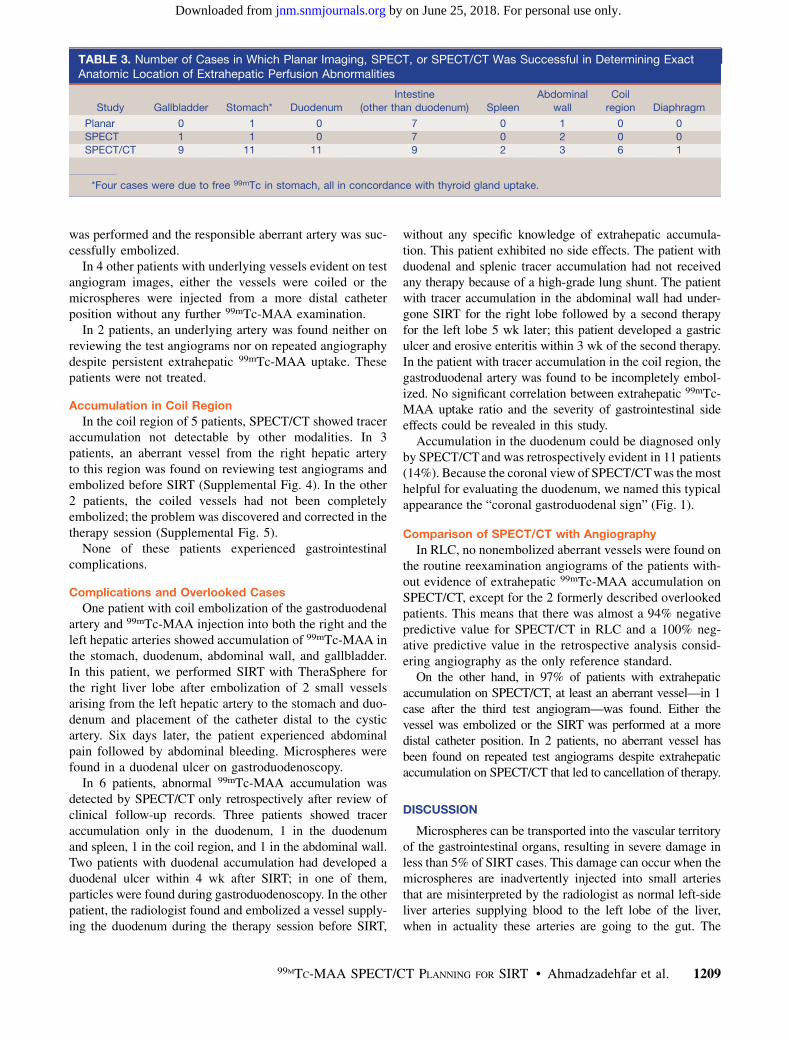
planar imaging, SPECT, and SPECT/CT was 13, 18, and52, respectively. However, exact anatomic classificationwas possible for only 9 and 11 abnormal findings on planarimaging and SPECT, respectively (Table 3). For 36% of the
abnormal findings detected on planar imaging, the anatomicregion could be determined only with SPECT/CT, corre-sponding to 33% in the case of SPECT. SPECT/CTdetected 50% and 20% additional abnormalities in casesfor which other extrahepatic shunting had already beendetected by planar imaging and SPECT, respectively.
Of 4 patients with MAA deposition in the stomach onSPECT/CT images, interpreted as free 99mTc, 1 patienttreated with SIR-Spheres developed a gastric ulcer despiteno noticeable problems such as reflux of resin microspheresduring therapy. A surgical finding of resin spheres in theulcer disputed the possibility that the accumulation of tracerin stomach was due to free 99mTc. This result emphasizesthe importance of perchlorate administration before 99mTc-MAA injection to avoid nonspecific tracer uptake in thestomach due to free 99mTc, the finding of which can bemisleading.
The therapy plan was changed in RLC according to theresults of planar imaging, SPECT, or SPECT/CT in 7.8%,8.9%, and 29% of examinations, respectively, as follows.
Accumulation in Gallbladder
Tracer deposition in the gallbladder wall was evidencedby SPECT in 1 patient and by SPECT/CT in 9 patients(12%) (Supplemental Fig. 2). To prevent the spheres fromreaching the gallbladder and to avoid possible cholecystitis(1,5,21), the catheter was placed distal to the cystic artery in6 patients, and in 1 case it was embolized. None of thesepatients developed cholecystitis during follow-up. Onepatient was not treated because of increasing bilirubin,and the other patient with a neuroendocrine tumor had anadditional abnormal accumulation in the duodenum,detected only by SPECT/CT. In this patient, the aberrantvessel could not be found on review of test angiograms.Therefore, the therapy plan was changed to radioreceptortherapy to avoid gastrointestinal side effects.
Extrahepatic Abdominal Accumulation
Reviewing the test angiograms revealed the possibleaberrant vessels in 5 patients with tracer deposition in thegastrointestinal system followed by a second 99mTc-MAAexamination after embolization of suspected arteries (Sup-plemental Fig. 3). Four of these patients no longer showedabnormal extrahepatic tracer accumulation. In the otherpatient, who still showed some shunting in the bowel inthe second examination, a third 99mTc-MAA examination
TABLE 2. Sensitivity, Specificity, Positive Predictive Value (PPV), and Negative Predictive Value (NPV) of 99mTc-MAAPlanar, SPECT, and SPECT/CT Studies in Retrospective Analysis and RLC
Study Sensitivity (%) Specificity (%) PPV (%) NPV (%)
Planar 32 (18–51) 98 92 71
SPECT 41 (25–59) 98 93 73
SPECT/CT 100 (87–100); 82% in RLC 93 89; 88 in RLC 100; 90 in RLC
Data in parentheses are 95% confidence intervals.
1208 THE JOURNAL OF NUCLEAR MEDICINE • Vol. 51 • No. 8 • August 2010
by on June 25, 2018. For personal use only. jnm.snmjournals.org Downloaded from
was performed and the responsible aberrant artery was suc-cessfully embolized.In 4 other patients with underlying vessels evident on test
angiogram images, either the vessels were coiled or themicrospheres were injected from a more distal catheterposition without any further 99mTc-MAA examination.In 2 patients, an underlying artery was found neither on
reviewing the test angiograms nor on repeated angiographydespite persistent extrahepatic 99mTc-MAA uptake. Thesepatients were not treated.
Accumulation in Coil Region
In the coil region of 5 patients, SPECT/CT showed traceraccumulation not detectable by other modalities. In 3patients, an aberrant vessel from the right hepatic arteryto this region was found on reviewing test angiograms andembolized before SIRT (Supplemental Fig. 4). In the other2 patients, the coiled vessels had not been completelyembolized; the problem was discovered and corrected in thetherapy session (Supplemental Fig. 5).None of these patients experienced gastrointestinal
complications.
Complications and Overlooked Cases
One patient with coil embolization of the gastroduodenalartery and 99mTc-MAA injection into both the right and theleft hepatic arteries showed accumulation of 99mTc-MAA inthe stomach, duodenum, abdominal wall, and gallbladder.In this patient, we performed SIRT with TheraSphere forthe right liver lobe after embolization of 2 small vesselsarising from the left hepatic artery to the stomach and duo-denum and placement of the catheter distal to the cysticartery. Six days later, the patient experienced abdominalpain followed by abdominal bleeding. Microspheres werefound in a duodenal ulcer on gastroduodenoscopy.In 6 patients, abnormal 99mTc-MAA accumulation was
detected by SPECT/CT only retrospectively after review ofclinical follow-up records. Three patients showed traceraccumulation only in the duodenum, 1 in the duodenumand spleen, 1 in the coil region, and 1 in the abdominal wall.Two patients with duodenal accumulation had developed aduodenal ulcer within 4 wk after SIRT; in one of them,particles were found during gastroduodenoscopy. In the otherpatient, the radiologist found and embolized a vessel supply-ing the duodenum during the therapy session before SIRT,
without any specific knowledge of extrahepatic accumula-tion. This patient exhibited no side effects. The patient withduodenal and splenic tracer accumulation had not receivedany therapy because of a high-grade lung shunt. The patientwith tracer accumulation in the abdominal wall had under-gone SIRT for the right lobe followed by a second therapyfor the left lobe 5 wk later; this patient developed a gastriculcer and erosive enteritis within 3 wk of the second therapy.In the patient with tracer accumulation in the coil region, thegastroduodenal artery was found to be incompletely embol-ized. No significant correlation between extrahepatic 99mTc-MAA uptake ratio and the severity of gastrointestinal sideeffects could be revealed in this study.
Accumulation in the duodenum could be diagnosed onlyby SPECT/CTand was retrospectively evident in 11 patients(14%). Because the coronal view of SPECT/CTwas themosthelpful for evaluating the duodenum, we named this typicalappearance the “coronal gastroduodenal sign” (Fig. 1).
Comparison of SPECT/CT with Angiography
In RLC, no nonembolized aberrant vessels were found onthe routine reexamination angiograms of the patients with-out evidence of extrahepatic 99mTc-MAA accumulation onSPECT/CT, except for the 2 formerly described overlookedpatients. This means that there was almost a 94% negativepredictive value for SPECT/CT in RLC and a 100% neg-ative predictive value in the retrospective analysis consid-ering angiography as the only reference standard.
On the other hand, in 97% of patients with extrahepaticaccumulation on SPECT/CT, at least an aberrant vessel—in 1case after the third test angiogram—was found. Either thevessel was embolized or the SIRT was performed at a moredistal catheter position. In 2 patients, no aberrant vessel hasbeen found on repeated test angiograms despite extrahepaticaccumulation on SPECT/CT that led to cancellation of therapy.
DISCUSSION
Microspheres can be transported into the vascular territoryof the gastrointestinal organs, resulting in severe damage inless than 5% of SIRT cases. This damage can occur when themicrospheres are inadvertently injected into small arteriesthat are misinterpreted by the radiologist as normal left-sideliver arteries supplying blood to the left lobe of the liver,when in actuality these arteries are going to the gut. The
TABLE 3. Number of Cases in Which Planar Imaging, SPECT, or SPECT/CT Was Successful in Determining ExactAnatomic Location of Extrahepatic Perfusion Abnormalities
Study Gallbladder Stomach* DuodenumIntestine
(other than duodenum) SpleenAbdominal
wallCoil
region Diaphragm
Planar 0 1 0 7 0 1 0 0
SPECT 1 1 0 7 0 2 0 0
SPECT/CT 9 11 11 9 2 3 6 1
*Four cases were due to free 99mTc in stomach, all in concordance with thyroid gland uptake.
99MTC-MAA SPECT/CT PLANNING FOR SIRT • Ahmadzadehfar et al. 1209
by on June 25, 2018. For personal use only. jnm.snmjournals.org Downloaded from

main culprit is a right gastric artery originating from theleft hepatic artery that is not recognized as or considered tobe a small artery supplying the left lobe of the liver.Alternatively, these arteries may not have been detected bythe radiologists. In about 15% of patients, vessels too smallto be detected on an angiogram may pass from the liver tothe gut (22). Radiation and diminished blood supply due toembolization by the spheres and subsequent hypoxia mayresult in ulceration and even perforation of the stomachand duodenum (10,23). 90Y-induced ulceration of the stom-ach or duodenum can be resistant to medical therapy, andsurgery may be required (8).SIRT requires accurate planning to ensure a good
therapeutic response with as few side effects as possible.99mTc-MAA must be administered during the preliminaryexaminations to evaluate a potential shunt from the liver tothe lung or abdominal organs. The prophylactic emboliza-tion of all extrahepatic vessels, including gastroduodenal,right gastric, and other extrahepatic vessels, at the time of99mTc-MAA assessment is recommended to avoid extrahe-patic deposition of microspheres. These vessels and organscan revascularize quickly; therefore, the embolizationshould be performed close to the intended time of SIRT.In addition, a reassessment arteriogram is required justbefore SIRT to ensure that such revascularization has notoccurred (2). Scintigraphy should be performed within 1 hof 99mTc-MAA injection to prevent false-positive extrahe-patic activity due to free 99mTc. In such cases, pathologicuptake in the stomach should be ruled out before treatment.Free 99mTc is normally observed in SPECT/CT as diffuseuptake in the gastric mucosa, often in concordance withthe thyroid gland on planar imaging, whereas pathologic
uptake is seen as a focally increased accumulation. For thepast 2 y, all patients in our department have received 600 mgof perchlorate by mouth 30 min before angiography to pre-vent nonspecific uptake of 99mTc-pertechnetate in the thyroidand stomach. During these 2 y, we have not observed anyfree 99mTc uptake in the stomach, as affirmed by SPECT/CT.
Obtaining a whole-body scan to predict possible lungdamage due to liver-to-lung shunting is adequate. Detectinghot spots in other organs besides the liver with planarimages is not always possible, and problems arise whenthese 2-dimensional images are used for more preciseevaluations. Extrahepatic spots indirectly mark the possiblelocations of microsphere misplacement during therapy;however, planar image analysis can be difficult and leadto misinterpretation of possible extrahepatic locationsbecause of the low spatial resolution of planar scintigraphicimages. Furthermore, especially in the upper abdomen, thelocalization of several different organs within a relativelysmall region demands the analysis of tomographic imagesto accurately distinguish whether the 99mTc-MAA has accu-mulated in the liver or in some adjacent organ (24,25).Planar images cannot always make this distinction becauseof organ superposition. SPECT provides valuable addi-tional information. SPECT images not only allow thelesions to be identified more accurately, and their arterialperfusion to be evaluated without superimposition, but alsoform a basis for image fusion with other pre- and postther-apeutic SPECT, PET, CT, and MR images. This processcan be improved using SPECT/CT cameras, which avoidproblems related to the interval between different studies,different positioning in different tomographs, and time-consuming software-based fusion as compared with hard-ware-based procedures (26).
Although SPECT or planar scanning is widely usedbefore SIRT (2,5,27,28), the value of preradioembolizationSPECT/CT has been investigated only in patients with col-orectal cancer and hepatocellular carcinoma (28,29).
Denecke et al. (28) performed SPECT/CT on a smallgroup of patients (n 5 13) with colorectal cancer and foundmore gastrointestinal 99mTc-MAA uptake in SPECT/CT(31%) than in SPECT alone (15%). In a recently publishedprospective study, Hamami et al. (29) showed a sensitivityof 100% for SPECT/CT. They performed sixty-eight 99mTc-MAA examinations on 58 patients with hepatocellular car-cinoma using clinical follow-up as the reference standardfor proof of extrahepatic gastrointestinal perfusion abnor-malities. None of the treated patients experienced gastro-intestinal complications. In our study, we had moreexaminations and patients with different types of tumors.We found no significant difference in abdominal shuntingbetween tumor types. Hamami et al. (29) mentioned thattheir study had a possible limitation in that all patients withgastrointestinal 99mTc-MAA deposition were excludedfrom treatment with SIRT if no causative gastrointestinalvessel was identified. Some of our patients were subject tothe same limitation as those in the Hamami study. However,
FIGURE 1. Duodenalaccumulation (arrows) ina patient with colorectalcancer, not definable onplanar images: planarscan (A), SPECT/CTcoronal view (B), andCT coronal view (C).
1210 THE JOURNAL OF NUCLEAR MEDICINE • Vol. 51 • No. 8 • August 2010
by on June 25, 2018. For personal use only. jnm.snmjournals.org Downloaded from
retrospective review of SPECT/CT images in our studyrevealed previously overlooked extrahepatic tracer accumu-lation in 3 patients who developed posttherapeutic gastro-intestinal ulcers; this finding intensifies the significance andimportance of this modality.We demonstrated the different anatomic regions of
extrahepatic 99mTc-MAA accumulation in detail (Table3). An important and frequently overlooked anatomicregion is the duodenum, which could be evaluated onlyby SPECT/CT; reviewing the coronal views of fusion scanswas especially helpful. Duodenal 99mTc-MAA accumula-tion was present in 11 patients (14.5%), being identifiedas the only abnormality in 5 (Fig. 1).Six patients (8%) showed an abnormal accumulation in
the coil region, detected only on SPECT/CT images. Tworeasons for this type of accumulation were small aberrantvessels ending in this region and an incompletely embol-ized vessel. Knowledge of the anatomic region of extra-hepatic organ perfusion may help the radiologist to estimatethe origin of the aberrant vessel.SPECT/CT showed a high negative predictive value in our
study. On the other hand, in 97% of patients with extra-hepatic 99mTc-MAA accumulation, at least 1 aberrant vesselcould be found on repeated angiograms. But as described inthe “Results” section, the patients with overlooked 99mTc-MAA accumulation developed gastrointestinal ulcers despiteno evident nonembolized aberrant vessels on routine angio-graphic reexamination, as also did 1 patient who did notundergo a second MAA examination after repeated angiog-raphy before SIRT. Thus, to increase the therapy, we recom-
mend performing repeated test angiography and coilembolization of aberrant arteries until no extrahepatic accu-mulation can be detected in 99mTc-MAA SPECT/CT.
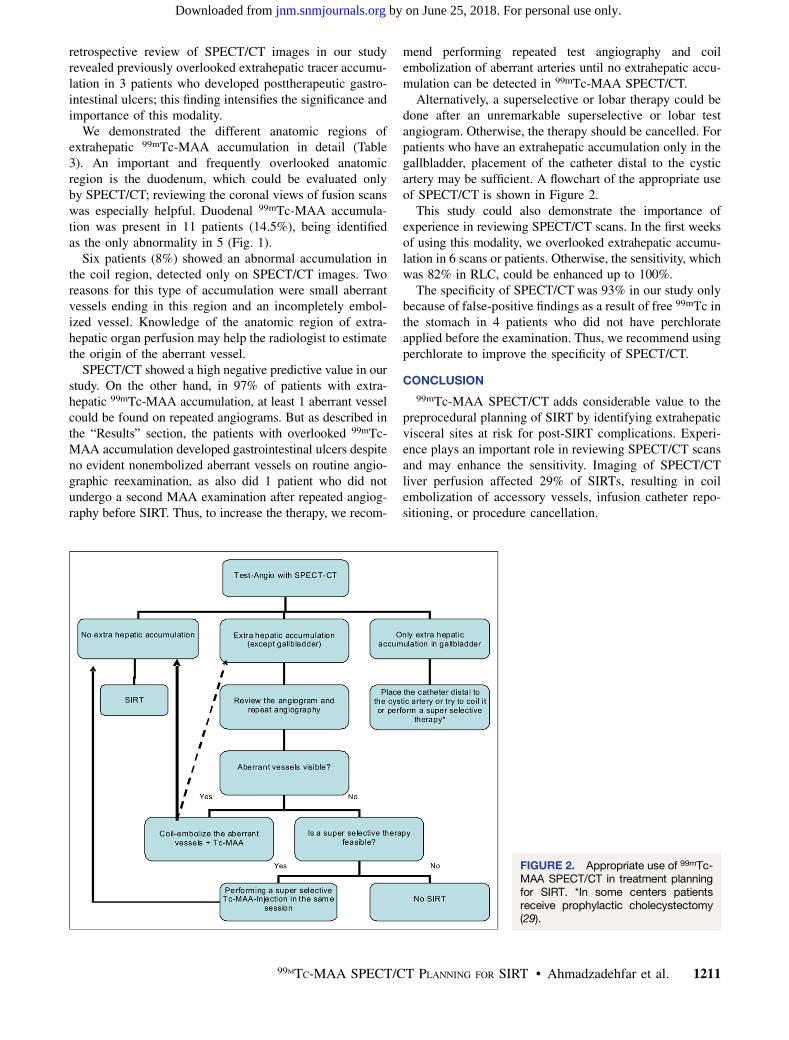
Alternatively, a superselective or lobar therapy could bedone after an unremarkable superselective or lobar testangiogram. Otherwise, the therapy should be cancelled. Forpatients who have an extrahepatic accumulation only in thegallbladder, placement of the catheter distal to the cysticartery may be sufficient. A flowchart of the appropriate useof SPECT/CT is shown in Figure 2.
This study could also demonstrate the importance ofexperience in reviewing SPECT/CT scans. In the first weeksof using this modality, we overlooked extrahepatic accumu-lation in 6 scans or patients. Otherwise, the sensitivity, whichwas 82% in RLC, could be enhanced up to 100%.
The specificity of SPECT/CT was 93% in our study onlybecause of false-positive findings as a result of free 99mTc inthe stomach in 4 patients who did not have perchlorateapplied before the examination. Thus, we recommend usingperchlorate to improve the specificity of SPECT/CT.
CONCLUSION
99mTc-MAA SPECT/CT adds considerable value to thepreprocedural planning of SIRT by identifying extrahepaticvisceral sites at risk for post-SIRT complications. Experi-ence plays an important role in reviewing SPECT/CT scansand may enhance the sensitivity. Imaging of SPECT/CTliver perfusion affected 29% of SIRTs, resulting in coilembolization of accessory vessels, infusion catheter repo-sitioning, or procedure cancellation.
FIGURE 2. Appropriate use of 99mTc-MAA SPECT/CT in treatment planningfor SIRT. *In some centers patientsreceive prophylactic cholecystectomy(29).
99MTC-MAA SPECT/CT PLANNING FOR SIRT • Ahmadzadehfar et al. 1211
by on June 25, 2018. For personal use only. jnm.snmjournals.org Downloaded from

REFERENCES
1. Salem R, Thurston KG. Radioembolization with 90yttrium microspheres: a state-
of-the-art brachytherapy treatment for primary and secondary liver malignancies.
Part 1: technical and methodologic considerations. J Vasc Interv Radiol.
2006;17:1251–1278.
2. Kennedy A, Nag S, Salem R, et al. Recommendations for radioembolization of
hepatic malignancies using yttrium-90 microsphere brachytherapy: a consensus
panel report from the radioembolization brachytherapy oncology consortium. Int
J Radiat Oncol Biol Phys. 2007;68:13–23.
3. Goin JE, Salem R, Carr BI, et al. Treatment of unresectable hepatocellular
carcinoma with intrahepatic yttrium 90 microspheres: factors associated with
liver toxicities. J Vasc Interv Radiol. 2005;16:205–213.
4. Salem R, Lewandowski RJ, Atassi B, et al. Treatment of unresectable
hepatocellular carcinoma with use of 90Y microspheres (TheraSphere): safety,
tumor response, and survival. J Vasc Interv Radiol. 2005;16:1627–1639.
5. Murthy R, Nunez R, Szklaruk J, et al. Yttrium-90 microsphere therapy for
hepatic malignancy: devices, indications, technical considerations, and
potential complications. Radiographics. 2005;25(suppl 1):S41–S55.
6. Leung TW, Lau WY, Ho SK, et al. Radiation pneumonitis after selective internal
radiation treatment with intraarterial 90yttrium-microspheres for inoperable
hepatic tumors. Int J Radiat Oncol Biol Phys. 1995;33:919–924.
7. Murthy R, Brown DB, Salem R, et al. Gastrointestinal complications associated
with hepatic arterial yttrium-90 microsphere therapy. J Vasc Interv Radiol.
2007;18:553–562.
8. Carretero C, Munoz-Navas M, Betes M, et al. Gastroduodenal injury after
radioembolization of hepatic tumors. Am J Gastroenterol. 2007;102:1216–1220.
9. Salem R, Parikh P, Atassi B, et al. Incidence of radiation pneumonitis after
hepatic intra-arterial radiotherapy with yttrium-90 microspheres assuming
uniform lung distribution. Am J Clin Oncol. 2008;31:431–438.
10. Yip D, Allen R, Ashton C, Jain S. Radiation-induced ulceration of the stomach
secondary to hepatic embolization with radioactive yttrium microspheres in the
treatment of metastatic colon cancer. J Gastroenterol Hepatol. 2004;19:347–349.
11. Atassi B, Bangash AK, Lewandowski RJ, et al. Biliary sequelae following
radioembolization with yttrium-90 microspheres. J Vasc Interv Radiol.
2008;19:691–697.
12. Buck AK, Nekolla S, Ziegler S, et al. SPECT/CT. J Nucl Med. 2008;49:1305–
1319.
13. Slart RH, Tio RA, Zijlstra F, Dierckx RA. Diagnostic pathway of integrated
SPECT/CT for coronary artery disease. Eur J Nucl Med Mol Imaging. 2009;
36:1829–1834.
14. Bockisch A, Freudenberg LS, Schmidt D, Kuwert T. Hybrid imaging by SPECT/
CT and PET/CT: proven outcomes in cancer imaging. Semin Nucl Med.
2009;39:276–289.
15. Even-Sapir E, Keidar Z, Bar-Shalom R. Hybrid imaging (SPECT/CT and PET/
CT): improving the diagnostic accuracy of functional/metabolic and anatomic
imaging. Semin Nucl Med. 2009;39:264–275.
16. Vermeeren L, Valdes Olmos RA, Meinhardt W, et al. Value of SPECT/CT for
detection and anatomic localization of sentinel lymph nodes before laparoscopic
sentinel node lymphadenectomy in prostate carcinoma. J Nucl Med. 2009;50:
865–870.
17. Schmidt D, Szikszai A, Linke R, Bautz W, Kuwert T. Impact of 131I SPECT/
spiral CT on nodal staging of differentiated thyroid carcinoma at the first
radioablation. J Nucl Med. 2009;50:18–23.
18. Chen L, Luo Q, Shen Y, et al. Incremental value of 131I SPECT/CT in the
management of patients with differentiated thyroid carcinoma. J Nucl Med.
2008;49:1952–1957.
19. Rozovsky K, Koplewitz BZ, Krausz Y, et al. Added value of SPECT/CT for
correlation of MIBG scintigraphy and diagnostic CT in neuroblastoma and
pheochromocytoma. AJR. 2008;190:1085–1090.
20. Patel CN, Chowdhury FU, Scarsbrook AF. Clinical utility of hybrid SPECT-CT
in endocrine neoplasia. AJR. 2008;190:815–824.
21. Salem R, Lewandowski RJ, Sato KT, et al. Technical aspects of radioembolization
with 90Y microspheres. Tech Vasc Interv Radiol. 2007;10:12–29.
22. Sirtex Medical Training Manual: Training Program Physicians and Institutions.
Lane Cove, New South Wales, Australia: Sirtex Medical.
23. Lau WY, Ho S, Leung TW, et al. Selective internal radiation therapy for
nonresectable hepatocellular carcinoma with intraarterial infusion of 90yttrium
microspheres. Int J Radiat Oncol Biol Phys. 1998;40:583–592.
24. Boan J, Valero M, Arbizu J. Improving treatment design by image
fusion techniques. In: Bilbao JI, Reiser MF, eds. Liver Radioembolization with90Y Microspheres: New York, NY: Springer; 2008:62.
25. Ahmadzadehfar H, Biersack HJ, Ezziddin S. Radioembolization of liver tumors
with yttrium-90 microspheres. Semin Nucl Med. 2010;40:105–121.
26. Koch W, Tatsch K. Nuclear medicine procedures for treatment evaluation. In:
Bilbao JI, Reiser MF, eds. Liver Radioembolization with 90Y Microspheres: New
York, NY: Springer; 2008:78–79.
27. Salem R, Thurston KG. Radioembolization with yttrium-90 microspheres: a
state-of-the-art brachytherapy treatment for primary and secondary liver
malignancies. Part 3: comprehensive literature review and future direction.
J Vasc Interv Radiol. 2006;17:1571–1593.
28. Denecke T, Ruhl R, Hildebrandt B, et al. Planning transarterial
radioembolization of colorectal liver metastases with yttrium 90 microspheres:
evaluation of a sequential diagnostic approach using radiologic and nuclear
medicine imaging techniques. Eur Radiol. 2008;18:892–902.
29. Hamami ME, Poeppel TD, Muller S, et al. SPECT/CT with 99mTc-MAA in
radioembolization with 90Y microspheres in patients with hepatocellular
cancer. J Nucl Med. 2009;50:688–692.
1212 THE JOURNAL OF NUCLEAR MEDICINE • Vol. 51 • No. 8 • August 2010
by on June 25, 2018. For personal use only. jnm.snmjournals.org Downloaded from

Doi: 10.2967/jnumed.109.074559Published online: July 21, 2010.
2010;51:1206-1212.J Nucl Med. Wilhelm, Hans-Jürgen Biersack and Samer EzziddinHojjat Ahmadzadehfar, Amir Sabet, Kim Biermann, Marianne Muckle, Holger Brockmann, Christiane Kuhl, Kai
Y-Microsphere Selective Internal Radiation Treatment90Planning for Tc-MAA SPECT/CT Liver Perfusion Imaging in Treatment99mThe Significance of
http://jnm.snmjournals.org/content/51/8/1206This article and updated information are available at:
http://jnm.snmjournals.org/site/subscriptions/online.xhtml
Information about subscriptions to JNM can be found at:
http://jnm.snmjournals.org/site/misc/permission.xhtmlInformation about reproducing figures, tables, or other portions of this article can be found online at:
(Print ISSN: 0161-5505, Online ISSN: 2159-662X)1850 Samuel Morse Drive, Reston, VA 20190.SNMMI | Society of Nuclear Medicine and Molecular Imaging
is published monthly.The Journal of Nuclear Medicine
© Copyright 2010 SNMMI; all rights reserved.
by on June 25, 2018. For personal use only. jnm.snmjournals.org Downloaded from