The Senses General senses of touch –Temperature –Pressure –Pain Special senses –Smell...
26
The Senses • General senses of touch – Temperature – Pressure – Pain • Special senses – Smell – Taste – Sight – Hearing – Equilibrium
-
Upload
sibyl-ward -
Category
Documents
-
view
227 -
download
0
Transcript of The Senses General senses of touch –Temperature –Pressure –Pain Special senses –Smell...
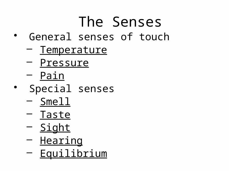
The Senses• General senses of touch
– Temperature– Pressure– Pain
• Special senses– Smell– Taste– Sight– Hearing– Equilibrium

The Eye and Vision
• 70 percent of all sensory receptors are in the eyes
• Each eye has over a million nerve fibers• The eye is a sphere about 1 inch in
diameter…like a ping pong• Only 1/6 of the eye is seen
– Most of the eye is surrounded by bone & cushioned by fat

Accessory Structures of the Eye
• Eyelids• Eyelashes• Muscles
Figure 8.1b

Accessory Structures of the Eye
• Conjunctiva– Membrane that lines the eyelids– Connects to the surface of the eye & secretes
mucus for lubrication

• Homeo Imbalance– Conjunctivitis =
reddened irritated eyes. “Pinkeye” is the HIGHLY infectious form caused by bacteria or virus

Accessory Structures of the Eye
• Lacrimal apparatus– Lacrimal gland –
produces diluted salt solution (tears)
– Lacrimal canals – drains tears from eyes
– Lacrimal sac – provides passage of tears towards nasal cavity
Figure 8.1a

Properties of lacrimal fluid (AKA TEARS!)– Dilute salt solution which contains lysozyme an
anti-bacterial protein• Protects, moistens, and lubricates the eye• Empties into the nasal cavity

• Nasolacrimal duct – empties tears into the nasal cavity (connects eye with nose)– Crying makes you
sniffle

• Homeo Imbalance– A cold or allergies can cause the lacrimal duct
to swell shut. This stops drainage of tears and you get watery eyes.

Structure of the Eye
• The wall is composed of three tunics (layers)– Fibrous tunic –
outside layer– Choroid –
middle layer
– Sensory tunic – inside layer
Figure 8.3a
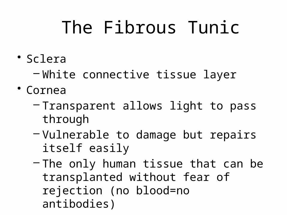
The Fibrous Tunic
• Sclera– White connective tissue layer
• Cornea– Transparent allows light to pass through– Vulnerable to damage but repairs itself easily– The only human tissue that can be
transplanted without fear of rejection (no blood=no antibodies)

Choroid Layer• Blood-rich nutritive tunic• Pigment dark in color prevents light from
scattering• Modified interiorly into two structures
– Cilliary body – smooth muscle, focuses lens for clear vision
– Iris- smooth muscle, regulates amount of light that enters• Pigmented layer that gives eye color• Pupil – rounded opening in the iris

Sensory Tunic (Retina)
• Contains millions of receptor cells called photoreceptors– Rods & Cones
• Signals pass from photoreceptors to retina• Signals leave the retina toward the brain
through the optic nerve
• Homeo Imbalance– Retinal Detachment: Retina separates from
choroid. Retina cannot get nutrients and can die. Easily fixed with laser surgery.
– Caused by violent motion of the head, genetics

Lens• Biconvex crystal-like structure• Held in place by a ligament attached to the
ciliary body
Figure 8.3a

• The lens divides the eye into 2 segments or chambers
• Homeo Imbalance– Cataracts: Occur as we age. The lens
becomes hard and opaque

Internal Eye Chamber Fluids
• Aqueous humor– Watery fluid found in chamber between the
lens and cornea– Maintains intraocular pressure– Provides nutrients for the lens and cornea
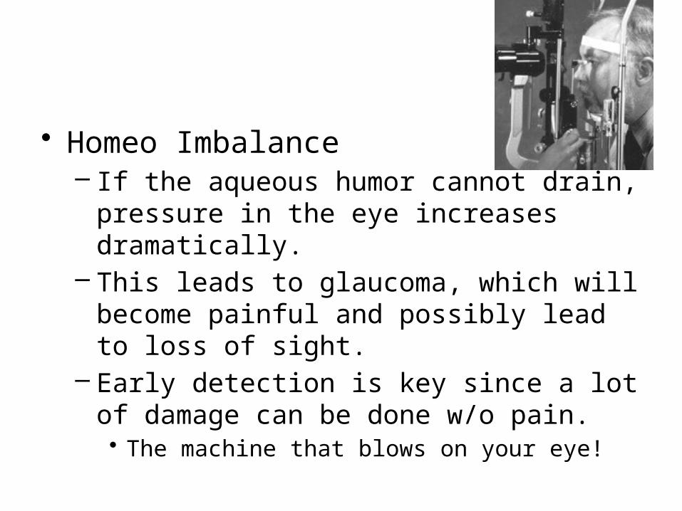
• Homeo Imbalance– If the aqueous humor cannot drain, pressure
in the eye increases dramatically.– This leads to glaucoma, which will become
painful and possibly lead to loss of sight.– Early detection is key since a lot of damage
can be done w/o pain.• The machine that blows on your eye!

Internal Eye Chamber Fluids
• Vitreous humor– Gel-like substance behind the lens that fills
the eyeball– Lasts a lifetime and is not replaced (can be
floaters)

Neurons of the Retina and Vision
• Rods– Most are found towards the edges of the
retina– Allow dim light & peripheral vision– all in gray tones
• Cones– Allow for detailed color vision– Densest in the center of the retina

Cone Sensitivity• There are 3 types of
cones each sensitive to different wavelengths
• Total Color blindness is the result of lack all cone types. Partial is due to lack of 1 or 2 types.
Figure 8.6
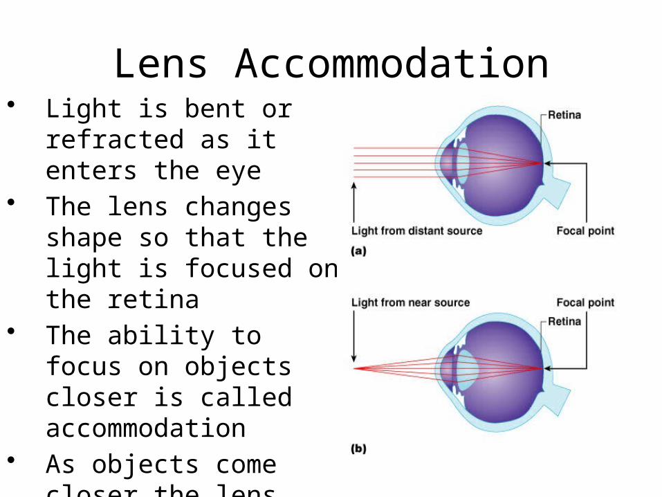
Lens Accommodation• Light is bent or refracted
as it enters the eye• The lens changes shape
so that the light is focused on the retina
• The ability to focus on objects closer is called accommodation
• As objects come closer the lens bulges

Vision Problems
• Perfect vision is called emmetropia or “harmonious vision”
• Nearsightedness- you can see up close but not far away. Picture focuses in front of retina
• Farsightedness- you can see far away but not close up. Picture focuses behind retina.

Images Formed on the Retina
• The image on the retina is reversed, upside-down and smaller
Figure 8.10

Visual Pathway• Optic nerve: carry
impulses from retina to brain,
• Part of each optic nerve crosses at the optic chiasma– Each side of brain
receives info from both eyes
– Allows for binocular vision & depth perception
Figure 8.11

• http://www.youtube.com/watch?v=38PGB9dcr4c
• Perspective on the 5 senses