The SAD1/RAD53 protein kinase controls multiple ...
16
The SAD1/RAD53 protein kinase controls multiple checkpoints and DNA damage- induced transcription in yeast -3 James B. Allen/'^ Zheng Zhou/-^ Wolfram Siede/ Errol C. Friedberg/ and Stephen J. Elledge^ 'Howard Hughes Medical Institute, ^Vema &. Marrs McLean Department of Biochemistry, ^histitute for Molecular Genetics, Baylor College of Medicine, Houston, Texas 77030 USA; "^Department of Pathology, University of Texas, Southwestern Medical Center, Dallas, Texas 75235 USA Inhibition of DNA synthesis prevents mitotic entry through the action of the S-phase checkpoint. We have isolated S-phase arrest-defective {sad) mutants that show lethality in the presence of the DNA synthesis inhibitor hydroxyurea (HU). Several of these mutants show phenotypes consistent with inappropriate mitotic entry in the presence of unreplicated DNA, indicating a defect in the S-phase checkpoint, sadl mutants are additionally defective for the G^ and Gj DNA damage checkpoints, and for DNA damage-induced transcription of RNR2 and RNR3. The transcriptional response to DNA damage requires activation of the Dunl protein kinase. Activation of Dunl in response to replication blocks or DNA damage is blocked in sadl mutants. The HU sensitivity of sadl mutants is suppressed by mutations in CKSl, a subunit of the p34^"*^^* kinase, further establishing a link between cell cycle progression and lethality, sadl mutants are allelic to rad53, a radiation-sensitive mutant. SADl encodes an essential protein kinase. The observation that SADl controls three distinct checkpoints suggests a common mechanism for cell cycle arrest at these points. Together, these observations implicate protein phosphorylation in the cellular response to DNA damage and replication blocks. [Key Words: Protein kinase; DNA damage; transcription; yeast; protein phosphorylation] Received July 14, 1994; revised version accepted August 19, 1994. A successful eukaryotic cell division requires that cer- tain cellular processes occur in a defined order and are coupled so that the initiation of one event is dependent on the completion of another. In most cell types, entry into mitosis is dependent on the completion of DNA synthesis. Cells blocked for DNA replication arrest in S phase and delay mitotic initiation. Cell cycle arrest in response to S-phase inhibition is attributable to the pres- ence of a feedback control or checkpoint mechanism (for reviews on cell cycle checkpoints, see Hartwell and Weinert 1989; Enoch and Nurse 1991; Murray 1992; Roberge 1992; Li and Deshaies 1993). Checkpoint con- trols also provide regulation of cell cycle progression in response to environmental challenges such as DNA damage. Cellular responses to DNA damage and blocks in DNA replication are similar: Cells arrest progression through the cell cycle at distinct points and induce the transcription of genes whose products facilitate DNA re- pair. In response to DNA damage, eukaryotic cells arrest either in G^ to prevent replication of damaged genetic templates or in G2 to avoid segregation of defective chro- mosomes. A current point of interest is whether feed- back controls also order events in a normal cell cycle in the absence of DNA damage or replication blocks. In this regard it has been proposed that relative timing mecha- nisms may order events in a normal cell cycle and that cell cycle arrest checkpoints are activated to ensure de- pendencies in cases when relative timing is insufficient (Murray 1992; Li and Deshaies 1993). Defects in cell cycle checkpoints can result in geno- mic instability and increase the rate that cells accumu- late heritable genetic damage. In budding yeasty muta- tions exist that exhibit elevated levels of chromosome loss and are unable to arrest the cell cycle in G^ (Siede et al. 1993) or G2 (Weinert and Hartwell 1988; Weinert et al. 1994) in response to DNA damage. Among these, the mecl and mec2 mutants have been implicated in the S-phase checkpoint because of sensitivity to conditions resulting in replication blocks and abnormal spindle structures in the presence of hydroxyurea (HU) (Weinert et al. 1994). However, a definitive role in the S-phase checkpoint has not yet been established for these mu- tants because cells can leak through a HU block, and it was not demonstrated that these mutants entered mito- sis with DNA that was less than fully replicated. Strains lacking feedback controls that couple spindle assembly and exit from mitosis also lose chromosomes at a higher rate than wild-type cells (Hoyt et al. 1991; Li and Murray 1991). Furthermore, it has been suggested that check- point control deficiency in mammalian cells may play a GENES & DEVELOPMENT 8:2416-2428 © 1994 by Cold Spring Harbor Laboratory Press ISSN 0890-9369/94 $5.00 2401 Cold Spring Harbor Laboratory Press on January 17, 2022 - Published by genesdev.cshlp.org Downloaded from
Transcript of The SAD1/RAD53 protein kinase controls multiple ...
The SAD1/RAD53 protein kinase controls multiple checkpoints and DNA damage-induced transcription in yeast
- 3 James B. Allen/'^ Zheng Zhou/-^ Wolfram Siede/ Errol C. Friedberg/ and Stephen J. Elledge^ 'Howard Hughes Medical Institute, ^Vema &. Marrs McLean Department of Biochemistry, ^histitute for Molecular Genetics, Baylor College of Medicine, Houston, Texas 77030 USA; "^Department of Pathology, University of Texas, Southwestern Medical Center, Dallas, Texas 75235 USA
Inhibition of DNA synthesis prevents mitotic entry through the action of the S-phase checkpoint. We have isolated S-phase arrest-defective {sad) mutants that show lethality in the presence of the DNA synthesis inhibitor hydroxyurea (HU). Several of these mutants show phenotypes consistent with inappropriate mitotic entry in the presence of unreplicated DNA, indicating a defect in the S-phase checkpoint, sadl mutants are additionally defective for the G^ and Gj DNA damage checkpoints, and for DNA damage-induced transcription of RNR2 and RNR3. The transcriptional response to DNA damage requires activation of the Dunl protein kinase. Activation of Dunl in response to replication blocks or DNA damage is blocked in sadl mutants. The HU sensitivity of sadl mutants is suppressed by mutations in CKSl, a subunit of the p34^"*^^* kinase, further establishing a link between cell cycle progression and lethality, sadl mutants are allelic to rad53, a radiation-sensitive mutant. SADl encodes an essential protein kinase. The observation that SADl controls three distinct checkpoints suggests a common mechanism for cell cycle arrest at these points. Together, these observations implicate protein phosphorylation in the cellular response to DNA damage and replication blocks.
[Key Words: Protein kinase; DNA damage; transcription; yeast; protein phosphorylation]
Received July 14, 1994; revised version accepted August 19, 1994.
A successful eukaryotic cell division requires that certain cellular processes occur in a defined order and are coupled so that the initiation of one event is dependent on the completion of another. In most cell types, entry into mitosis is dependent on the completion of DNA synthesis. Cells blocked for DNA replication arrest in S phase and delay mitotic initiation. Cell cycle arrest in response to S-phase inhibition is attributable to the presence of a feedback control or checkpoint mechanism (for reviews on cell cycle checkpoints, see Hartwell and Weinert 1989; Enoch and Nurse 1991; Murray 1992; Roberge 1992; Li and Deshaies 1993). Checkpoint controls also provide regulation of cell cycle progression in response to environmental challenges such as DNA damage. Cellular responses to DNA damage and blocks in DNA replication are similar: Cells arrest progression through the cell cycle at distinct points and induce the transcription of genes whose products facilitate DNA repair. In response to DNA damage, eukaryotic cells arrest either in G^ to prevent replication of damaged genetic templates or in G2 to avoid segregation of defective chromosomes. A current point of interest is whether feedback controls also order events in a normal cell cycle in the absence of DNA damage or replication blocks. In this regard it has been proposed that relative timing mecha
nisms may order events in a normal cell cycle and that cell cycle arrest checkpoints are activated to ensure dependencies in cases when relative timing is insufficient (Murray 1992; Li and Deshaies 1993).
Defects in cell cycle checkpoints can result in genomic instability and increase the rate that cells accumulate heritable genetic damage. In budding yeasty mutations exist that exhibit elevated levels of chromosome loss and are unable to arrest the cell cycle in G^ (Siede et al. 1993) or G2 (Weinert and Hartwell 1988; Weinert et al. 1994) in response to DNA damage. Among these, the mecl and mec2 mutants have been implicated in the S-phase checkpoint because of sensitivity to conditions resulting in replication blocks and abnormal spindle structures in the presence of hydroxyurea (HU) (Weinert et al. 1994). However, a definitive role in the S-phase checkpoint has not yet been established for these mutants because cells can leak through a HU block, and it was not demonstrated that these mutants entered mitosis with DNA that was less than fully replicated. Strains lacking feedback controls that couple spindle assembly and exit from mitosis also lose chromosomes at a higher rate than wild-type cells (Hoyt et al. 1991; Li and Murray 1991). Furthermore, it has been suggested that checkpoint control deficiency in mammalian cells may play a
GENES & DEVELOPMENT 8:2416-2428 © 1994 by Cold Spring Harbor Laboratory Press ISSN 0890-9369/94 $5.00 2401
Cold Spring Harbor Laboratory Press on January 17, 2022 - Published by genesdev.cshlp.orgDownloaded from

Allen et al.
role in cellular transformation (Hartwell 1992; Livingstone et al. 1992; Kastan et al. 1992; Murray 1992; Yin et al. 1992). Ataxia-telangiectasia (AT) is a human recessive disorder characterized by a number of phenotypes including a high incidence of cancer. Cell lines derived from AT patients are defective for both the G^ and G2 DNA damage checkpoints (Painter and Young 1980; Zambetti-Bosseler and Scott 1981; Nagasawa et al. 1985; Cohen and Levy 1989; Rudolph and Latt 1989). hi addition, primary murine fibroblasts that carry null or mutant alleles of the tumor suppressor gene p53 fail to arrest the cell cycle in Gi in response to ionizing radiation jKuerbitz et al. 1992; Yin et al. 1992) and are defective for the DNA damage-induced transcription of CIPl/WAFl (El Diery et al. 1993, 1994), an inhibitor of Cdk2 and Cdk4 (Gu et al. 1993; Harper et al. 1993; Xiong et al. 1993). Taken together, these observations suggest that the inability to respond to DNA damage by cell cycle arrest or to induce transcription of genes that facilitate DNA repair may potentiate cellular transformation.
A number of checkpoint mutants have been identified in a variety of species; however, the biochemical nature of the cell's ability to coordinate cell cycle progression with the replicational status and integrity of the genome remains unclear. The eukaryotic cell cycle is composed of two major transitions, one at the G^/S boundary and another at G2/M. These transitions are controlled by the p34 protein kinase and associated regulatory subunits called cyclins. The p34/cyclin complex is an attractive target for checkpoint inhibition. In Schizosacchaiomy-ces pombe, cells treated with the chemical HU are blocked for DNA replication and maintain high levels of inhibitory Tyr-15 phosphorylation on p34'" '' (Gould and Nurse 1989; Nurse 1990). Cells that contain an unphos-phorylatable mutant of p34'''^''^, Y15F, bypass the cell cycle arrest imposed by unreplicated DNA and enter a lethal mitosis (Gould and Nurse 1989). It is not yet clear whether the dominant cdc2:Y15F mutant functions indirectly by bypassing the checkpoint, or directly by blocking its target, phosphorylation of Tyr-15. In contrast, Saccharomyces cerevisiae strains that contain an analogous mutation on p34^^*^^^ are competent to respond to S-phase inhibition and progress normally through the cell cycle (Amon et al. 1992; Sorger and Murray 1992). This suggests that budding yeast possess a mode of checkpoint control distinct from inhibitory tyrosine phosphorylation of p34.
Although progress has been made in understanding cell cycle checkpoints and the cellular response to DNA damage, several outstanding questions remain. First, what is the molecular nature of the S-phase checkpoint? Second, does it operate during an unperturbed cell cycle or only in response to stress? Third, is there overlap between the DNA damage checkpoint pathways in Gj and G2 and the S-phase checkpoint pathway? Fourth, to what extent do the cell cycle arrest and DNA damage transcriptional induction pathways overlap? To address these questions and elucidate the signaling pathway coordinating S phase and mitosis, we have focused on the isolation of S-phase arrest-defective (sad) mutants that
disrupt cell cycle arrest in response to blocks in DNA replication.
Results
Isolation of sad mutants HU inhibits the enzyme ribonucleotide reductase (Rnr) by quenching the tyrosyl free radical required for catalysis. Inhibition of Rnr results in the inability to synthesize deoxyribonucleotides^ which blocks DNA synthesis. Wild-type cells respond to HU treatment by revers-ibly arresting cell cycle progression through activation of the S-phase checkpoint. Mutations in the S-phase checkpoint pathway should confer sensitivity to HU because the inability to delay cell cycle progression may result in mitosis in the absence of DNA replication. EMS-muta-genized colonies (250,000) were replica-plated to plates containing 100 mM HU, and —700 HU-sensitive mutants were isolated. To specifically identify sad mutants, two secondary screens were employed. First, HU-sensitive mutants were screened for loss of viability in the presence of HU (HU reversibility) by replica-plating and liquid survival assays, and second, for rescue by prior arrest with a-factor as described in Materials and methods, sad mutants should exhibit rapid loss of viability in the presence of HU because of their inability to delay cell cycle progression when DNA synthesis is inhibited. In addition, HU-induced lethality should be suppressed by prior treatment with a-factor, which arrests cells in G^ and prevents inappropriate cell cycle progression. Six recessive mutants representing five different complementation groups, sadl~sad5, were isolated that exhibit rapid loss of viability with HU that is a-factor suppressible. sad mutants display significant killing after 6 hr of HU treatment compared with wild-type controls (Fig. lA). In addition, sad mutants are significantly more sensitive to UV-irradiation than wild-type strains (Fig. IB). This suggests that sad mutants may be additionally defective for DNA repair or the DNA damage cell cycle checkpoints. sadl -1 mutants exhibit the most rapid lethality and were selected for further analysis. a-Factor suppression of sadl-1 HU lethality is shown in Figure IC.
Screen of rad mutants for S-phase feedback control deficiency Mutants defective for DNA damage-induced cell cycle arrest have been identified among preexisting collections of mutations that confer radiation sensitivity in budding yeast and fission yeast {rad mutants) (Weinert and Hart-well 1988; Al-Khodairy and Carr 1992; Enoch et al. 1992; Rowley et al. 1992; Weinert 1992). It is also likely that the DNA damage and the S-phase checkpoint pathways share common components (Al-Khodairy and Carr 1992; Enoch et al. 1992; Weinert 1992). In this regard, a bank of budding yeast rad mutants were screened for the sad phenotype. As shown in Table 1, radSO, radSl, and rad53 mutants exhibit some sad phenotypes. sad mutants
2402 GENES &. DEVELOPMENT
Cold Spring Harbor Laboratory Press on January 17, 2022 - Published by genesdev.cshlp.orgDownloaded from

SADl controls multiple cell cycle checkpoints
• 2 4 Time in HU (hrs)
D WT ^ uv — ^ * +
UV Dose (J/m^) Time in HU (hrs)
sadl UV — +
• 1 2 3 4 Time at 37'C (hrs)
a factor release
30 min.
50 min.
70 min.
Time after Benomy] Release (min)
Figure 1. Checkpoint deficiency and radiation sensitivity of sad mutants. [A] HU lethality of wild-type and sad strains. Wild-type strain Y300 (■) and sad mutant cells (Y301, sadl-1, • ; Y305, sad2-l, □; Y306, sad3-l, O; Y307, sad4-l, A, and Y308, sad5-l, A) were grown to log phase in YPD (pH 3.9); and HU was added to 0.2 M. Aliquots were removed at timed intervals to determine cell number and to score for viable colony-forming units on YPD plates. (S) Radiation sensitivity of sad mutants. Wild-type (Y300, SADl, ■) and sad mutant cells (Y301, sadl-1, • ; Y305, sad2-l, D; Y306, sad3-l, O; Y307, sad4-l, k, and Y308, sad5-l, A) were irradiated with the indicated doses of UV light, and percent survival relative to unirradiated controls was determined. (C) a-Factor rescue of sadl-1 cells. Log-phase cultures were treated with 10 [j-g/ml of a-factor for 1 hr. Samples were washed with YPD, split, and resuspended in YPD containing 0.2 M HU (Y300, wild-type, ■; Y301, sadl-1, • ) or 0.2 M HU plus 10 M-g/ml of a-factor (Y300, wild-type, D; Y301, sadl-1, O). a-Factor (2 |xg/ml) was subsequently added every 2 hr to a-factor-treated samples. (D) DNA damage-induced G^ cell cycle delay in SAD and sadl-1. Wild-type Y300 {left] and sadl-1 Y301 [right] strains were synchronized with a-factor and UV-irradiated. Unirradiated j - ) and irradiated (-I-) samples were resuspended into fresh YPD media, and cell cycle progression was monitored by FACS analysis. Numbers in the center refer to the time in minutes after release from a-factor block. (£) Survival of cdc9 sadl double mutants at the restrictive temperature. Wild-type strain Y300 (■), Y301 sadl-1 (•), Y309 cdc9 (D), and Y310 cdc9 sadl double mutants (O) were grown to log phase at 23°C and shifted to 37°C for 4 hr. Dilutions were plated at timed intervals and incubated at 23°C to determine cell viability. (P) DNA damage-induced Gj cell cycle delay in wild-type and sadl-1 strains. Wild-type strain Y300 (D, ■) and Y301 sadl-1 cells (O, #) were arrested with 100 jig/ml of benomyl and UV-irradiated. Unirradiated (■, #) and UV-irradiated (40 J/m^, O, D) samples were resupended into fresh media, and cell cycle progression was monitored by direct visualization of nuclear division using DAPI staining.
v^ere crossed with ladSO, ladSl, and ladSS to test for complementation and linkage. iad53 is allelic to sadl
while ladSO and ladSl are not allelic to any of the sad mutants.
GENES & DEVELOPMENT 2403
Cold Spring Harbor Laboratory Press on January 17, 2022 - Published by genesdev.cshlp.orgDownloaded from
Allen et al.
Table 1. Scieen of rad mutants for sad phenotype
HU
Mutant sensitive reversible HU lethality suppressed by a-factor
radS Tads mdl4 radl6 iad23 iad24 ladSO ladSl iad52 ladSS rad54 ladSS ladSS iad57
no no no no no no yes yes yes yes yes yes yes yes
no no yes no yes yes yes yes
yes yes
yes
A collection of preexisting lad mutants were screened for HU sensitivity, HU reversibility, and a-factor suppression of HU lethality as described in Materials and methods.
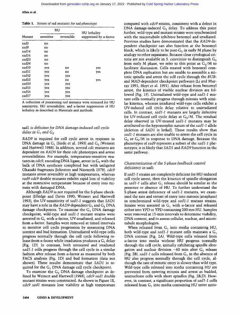
sadl is deficient for DNA damage-induced cell cycle delay in G^ and G2
RAD9 is required for cell cycle arrest in response to DNA damage in Gj (Siede et al. 1993) and G2 (Weinert and Hartwell 1988). In addition, several cdc mutants are dependent on RAD9 for their cdc phenotypes and arrest reversibilities. For example, temperature-sensitive mutants in cdc9, encoding DNA ligase, arrest in Gj with the bulk of DNA synthesis completed but with unligated Okazaki fragments (Johnston and Nasmyth 1978). cdc9 mutants arrest reversibly at high temperatures, whereas iad9 cdc9 double mutants exhibit rapid loss of viability at the restrictive temperature because of entry into mitosis with damaged DNA.
Although RAD9 is not required for the S-phase checkpoint (Elledge and Davis 1990; Weinert and Hartwell 1993), the UV-sensitivity of sadl-1 suggests that SADl may have a role in the RAD9-dependent Gi and G2 DNA damage checkpoints. To examine the Gi DNA damage checkpoint, wild-type and sadl-1 mutant strains were arrested in G^ with a-factor, UV-irradiated, and released from a-factor. Samples were removed at timed intervals to monitor cell cycle progression by measuring DNA content and bud formation. Unirradiated wild-type cells progress normally through the cell cycle following release from a-factor while irradiation produces a G^ delay (Fig. ID). In contrast, both untreated and irradiated sadl -1 cells progress through the cell cycle in a similar fashion after release from a-factor as measured by both FACS analysis (Fig. ID) and bud formation (data not shown). These results demonstrate that SADl is required for the Gi DNA damage cell cycle checkpoint.
To examine the G2 DNA damage checkpoint as defined by Weinert and Hartwell (1988), cdc9 sadl double mutant strains were constructed. As shown in Figure IE, cdc9 sadl mutants lose viability at high temperature
compared with cdc9 strains, consistent with a defect in DNA damage-induced G2 delay. To address this point further, wild-type and mutant strains were synchronized with the microtubule inhibitor benomyl and irradiated. Previous studies have demonstrated that the RAD9-de-pendent checkpoint can also fimction at the benomyl block, which is likely to be post-G2 in early M phase by analogy to other organisms. Because clear cytological criteria are not available in S. ceievisiae to distinguish G2 from early M phase, we refer to this point as G2/M to facilitate discussion. Cells treated with benomyl complete DNA replication but are unable to assemble a mitotic spindle and arrest the cell cycle through the BUB-and MAD-dependent checkpoint pathways (Li and Murray 1991; Hoyt et al. 1991). After release from benomyl arrest, the kinetics of visible nuclear division are followed (Fig. IF). Unirradiated wild-type and sadl-1 mutant cells normally progress through mitosis with similar kinetics, whereas irradiated wild-type cells exhibit a UV-induced cell cycle delay relative to unirradiated cells. In contrast, sadl-1 mutants are largely defective for UV-induced cell cycle delay at G2/M. The residual delay observed in UV-treated sadl-1 mutants may be attributed to the hypomorphic nature of the sadl-1 allele (deletion of SADl is lethal). These results show that sadl-1 mutants are also unable to arrest the cell cycle in G2 or G2/M in response to DNA damage. Because the phenotypes of iad9 represent a subset of the sadl-1 phenotypes, it is likely that SADl and RAD9 function in the same pathway.
Characterization of the S-phase feedback control deficiency in sadl
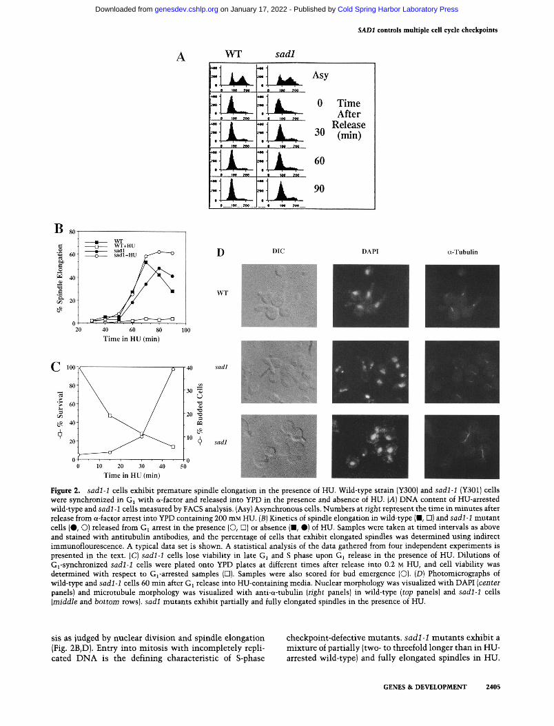
If sadl-1 strains are completely deficient for HU-induced cell cycle arrest, then the kinetics of spindle elongation in sadl-1 cells after Gi release should be similar in the presence or absence of HU. To further understand the S-phase arrest deficiency of sadl-1 mutants, we examined the rate and extent of entry into mitosis (anaphase) in synchronized wild-type and sadl-1 mutant strains. Strains were arrested in Gj with a-factor and released either into YPD or YPD containing 200 mM HU. Samples were removed at 15-min intervals to determine viability, DNA content, and to assess cellular, nuclear, and microtubule morphologies.
When released from G^ into media containing HU, both wild-type and sadl-1 mutant cells maintain a G^ DNA content (Fig. 2A). Wild-type cells released from a-factor into media without HU progress normally through the cell cycle, initially exhibiting spindle elongation and nuclear division —60 min after Gi release (Fig. 2B). sadl-1 cells released from G^ in the absence of HU also progress normally through the cell cycle, although the rate of mitotic entry is slower than wild type. Wild-type cells released into media containing HU are prevented from entering mitosis and arrest as budded, uninucleate cells with short spindles (Fig. 2B,D). However, in contrast, a significant proportion of sadl-1 cells released from G^ into media containing HU enter mito-
2404 GENES & DEVELOPMENT
Cold Spring Harbor Laboratory Press on January 17, 2022 - Published by genesdev.cshlp.orgDownloaded from
SADl controls multiple cell cycle checkpoints
A WT *m4
a iM Me
» 1M J «
• IN » •
1 IM 7M
sadl
"LjUk- ^^ Z] A 0 Time « t ^ ' " " After
• "«.. res.
• IM._ »fl....
~ U ^ 60
DAPI a-Tubulin
10 20 30 40 Time in HU (min)
Figure 2. sadl-1 cells exhibit premature spindle elongation in the presence of HU. Wild-type strain (Y300) and sadl-1 (Y301) cells were synchronized in Gi with a-factor and released into YPD in the presence and absence of HU. [A] DNA content of HU-arrested wild-type and sadl-1 cells measured by FACS analysis. (Asy) Asynchronous cells. Numbers at right represent the time in minutes after release from a-factor arrest into YPD containing 200 mM HU. (5) Kinetics of spindle elongation in wild-type (■, D) and sadl-1 mutant cells (•, O) released from Gi arrest in the presence (O, D) or absence (■, #) of HU. Samples were taken at timed intervals as above and stained with antitubulin antibodies, and the percentage of cells that exhibit elongated spindles was determined using indirect immunoflourescence. A typical data set is shown. A statistical analysis of the data gathered from four independent experiments is presented in the text. (C) sadl-1 cells lose viability in late Gj and S phase upon Gj release in the presence of HU. Dilutions of Gi-synchronized sadl-1 cells were plated onto YPD plates at different times after release into 0.2 M HU, and cell viability was determined with respect to Gj-arrested samples (□). Samples were also scored for bud emergence (O). [D] Photomicrographs of wild-type and sadl-1 cells 60 min after Gj release into HU-containing media. Nuclear morphology was visualized with DAPI [centei panels) and microtubule morphology was visualized with anti-a-tubulin {right panels) in wild-type (top panels) and sadl-1 cells [middle and bottom rows), sadl mutants exhibit partially and fully elongated spindles in the presence of HU.
sis as judged by nuclear division and spindle elongation (Fig. 2B,D). Entry into mitosis with incompletely replicated DNA is the defining characteristic of S-phase
checkpoint-defective mutants, sadl-1 mutants exhibit a mixture of partially (two- to threefold longer than in HU-arrested wild-type) and fully elongated spindles in HU.
GENES & DEVELOPMENT 2405
Cold Spring Harbor Laboratory Press on January 17, 2022 - Published by genesdev.cshlp.orgDownloaded from

Allen et al.
Interestingly, HU-treated sadl-1 cultures accumulate cells with elongated spindles at a rate faster than sadl-1 cells released from Gj in the absence of HU (Fig. 2B). The mean time for 30% of the cells to exhibit spindle elongation in sadl mutants in HU was 51.2±2.4 min, compared with 61.8±2.5 min without HU (averaged over four data sets). Thus, HU accelerates mitotic onset by 10.5±2.4 min. In addition, sadl-1 cells released in the presence of HU contain a significant proportion of un-budded and small budded cells that possess mitotic spindles (data not shown). Unbudded or small budded cells that contain elongated spindles were not observed in wild-type cultures and were seen only rarely in sadl-1 strains not treated with HU.
sadl-1 cells released from G^ in the presence of HU quickly lose viability following Gi release (Fig. 2C). Correlation of sadl-1 lethality with cellular morphology indicates that sadl-1 cells lose viability coincident with entry and progression through S phase. These kinetics are consistent with a commitment to lethality once replication is delayed for a short period of time.
SADl is lequiied for the DNA damage inducibility of RNR2 and RNR3
Both prokaryotic and eukaryotic cells respond to DNA damage by arresting the cell cycle and inducing transcription of genes whose products facilitate DNA repair. We were interested in whether these pathways share common components. To address this, sadl-1 mutants were examined for regulation of the DNA damage-induc-ible RNR2, RNR3, and UBI4 genes. RNR2 and RNR3 encode the catalytic and an alternative regulatory sub-unit of ribonucleotide reductase, respectively. UBI4 encodes polyubiquitin. As shown in Figure 3, sadl-1 mutants are defective for methylmethane sulfonate (MMS)-induced transcription of RNR2 and RNR3 but are competent for induction of UBI4. This phenotype is similar to that observed for dunl mutants and supports the observation that multiple pathways exist for transcription induced by DNA damaging agents (Zhou and EUedge 1993).
Sadl controls the kinase activity of Dunl in response to DNA damage
The Dxml protein kinase is required for the DNA damage-induced expression of RNR2 and RNR3 (Zhou and Elledge 1993). Dunl is activated in response to DNA damage as evidenced by the DNA damage-dependent generation of slower migrating forms of Dunl in in vitro autophosphorylation assays and an increase in DUNl-dependent autophosphorylation in vivo. While DUNl is required for the transcriptional response to DNA damage, it is not required for the G -, G2-, or S-phase checkpoints, sadl mutants are defective for both DNA damage-induced cell cycle arrest and transcription of RNR2 and RNR3. It is therefore likely that SADl functions in the same pathway as DUNl. Because SADl also controls cell cycle checkpoints and DUNl does not, it is possible
WT sadl-1 MMS - + - +
RNR2
RNR3
UBI4
URA3
Figure 3. sadl-1 is defective for DNA damage inducibility of RNR2 and RNR3. Cultures at OD^QO of 0.05 in YPD were treated with or without 0.1% MMS for 1 hr at 30°C. RNA was extracted, fractionated on a formaldehyde-1% agarose gel, transferred to nitrocellulose, and probed with " P-labeled DNA probes derived from RNR2, RNR3, UBI4, and URA3 as described in Materials and methods.
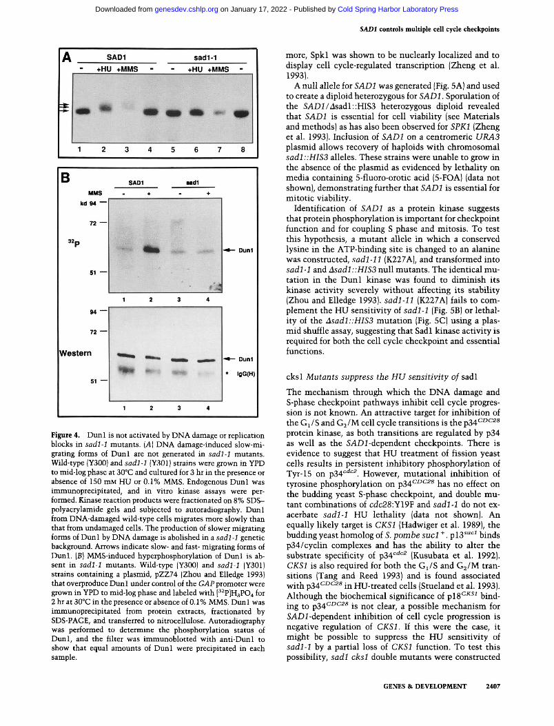
that SADl is upstream of DUNl and regulates its activity in response to DNA damage. To test this hypothesis, DNA damage-induced activation of Dunl was assayed in sadl mutant strains. Wild-type cells treated with either HU or MMS exhibit slower migrating forms of Diml (Fig. 4A). Neither treatment in sadl mutants resulted in the Dunl mobility shift observed in wild-type cells, indicating that activation of Dunl by DNA damaging agents is SADl-dependent.
To further investigate the relationship between SADl and DUNl, the DNA damage-induced hyperphosphory-lation of Dunl in vivo was examined in wild-type and sadl mutant cells. As shown in Figure 4B, MMS treatment of wild-type cells overexpressing Dunl results in an increase of the phosphorylation state of Dunl. However, this response is absent in sadl mutants, further strengthening the correlation of the mobility shift and autophosphorylation in response to DNA damage. These results suggest further that SADl functions upstream of DUNl in the signaling pathway that activates DNA damage-induced transcription.
Cloning and disruption of SADl
A wild-type SADl genomic clone was isolated by complementation of sadl-1 HU sensitivity and sequenced. This clone complements all of the checkpoint and transcriptional defects of the sadl-1 mutant (data not shown). A GenBank data base search revealed that the open reading frame encodes a previously cloned gene, SPKl, a serine/threonine protein kinase. SPKl was isolated in a screen to identify yeast protein kinases (Stem et al. 1991) using anti-phosphotyrosine antibodies, and was shown to possess kinase activity in vitro. Futher-
2406 GENES &. DEVELOPMENT
Cold Spring Harbor Laboratory Press on January 17, 2022 - Published by genesdev.cshlp.orgDownloaded from
SADl controls multiple cell cycle checkpoints
SAD1 sad1-1 +HU -i-MMS +HU -i MMS -
IMP v p •» fp i
1 2 3 4 5 6 7 8
B MMS
kd94 —
72 —
«P
51 —
94 —
72 —
Western
51 —
SADl
+
'" ^ mn
1 2
^ ^ ^IPIP mmf: :..:
1 2
Mdl ♦
t^KW*- • » ^ ' '
.
9'M 3 4
« B i » M ^ M
Wi«»^
3 4
-^— Duni
- • — Duni
• igG(H)
Figute 4. Duni is not activated by DNA damage or replication blocks in sadl-1 mutants. {A] DNA damage-induced slow-migrating forms of Duni are not generated in sadl-1 mutants. Wild-type |Y300) and sadl-1 (Y301) strains were grown in YPD to mid-log phase at 30°C and cultured for 3 hr in the presence or absence of 150 mM HU or 0.1% MMS. Endogenous Duni was immunoprecipitated, and in vitro kinase assays were performed. Kinase reaction products were fractionated on 8% SDS-polyacrylamide gels and subjected to autoradiography. Duni from DNA-damaged wild-type cells migrates more slowly than that from undamaged cells. The production of slower migrating forms of Duni by DNA damage is abolished in a sadl -1 genetic background. Arrows indicate slow- and fast- migrating forms of Duni. [B] MMS-induced hyperphosphorylation of Duni is absent in sadl-1 mutants. Wild-type (Y300) and sadl-1 (Y301) strains containing a plasmid, pZZ74 (Zhou and Elledge 1993) that overproduce Duni under control of the GAP promoter were grown in YPD to mid-log phase and labeled with [' ^P]H3P04 for 2 hr at 30°C in the presence or absence of 0.1 % MMS. Dun 1 was immunoprecipitated from protein extracts, fractionated by SDS-PAGE, and transferred to nitrocellulose. Autoradiography was performed to determine the phosphorylation status of Duni, and the filter was immunoblotted with anti-Dun 1 to show that equal amounts of Duni were precipitated in each sample.
more, Spkl w as show^n to be nuclearly localized and to display cell cycle-regulated transcription (Zheng et al. 1993).
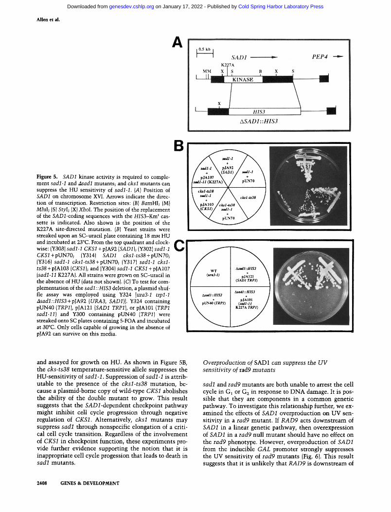
A null allele for SADl was generated (Fig. 5A) and used to create a diploid heterozygous for SADl. Sporulation of the SADl/^sadl: :HIS3 heterozygous diploid revealed that SADl is essential for cell viability (see Materials and methods) as has also been observed for SPKl (Zheng et al. 1993). Inclusion of SADl on a centromeric URA3 plasmid allows recovery of haploids with chromosomal sadl::HIS3 alleles. These strains were unable to grow in the absence of the plasmid as evidenced by lethality on media containing 5-fluoro-orotic acid (5-FOA) (data not shown), demonstrating further that SADl is essential for mitotic viability.
Identification of SADl as a protein kinase suggests that protein phosphorylation is important for checkpoint function and for coupling S phase and mitosis. To test this hypothesis, a mutant allele in which a conserved lysine in the ATP-binding site is changed to an alanine was constructed, sadl-11 (K227A), and transformed into sadl-1 and Asadl::HIS3 null mutants . The identical mutation in the Dun i kinase was found to diminish its kinase activity severely without affecting its stability (Zhou and Elledge 1993). sadl-11 (K227A) fails to complement the HU sensitivity of sadl-1 (Fig. 5B) or lethality of the Asadl::HIS3 mutation (Fig. 5C) using a plasmid shuffle assay, suggesting that Sadl kinase activity is required for both the cell cycle checkpoint and essential functions.
cksl Mutants suppress the HU sensitivity of sadl
The mechanism through which the DNA damage and S-phase checkpoint pathways inhibit cell cycle progression is not known. An attractive target for inhibition of the Gi /S and G^/M cell cycle transitions is the p34^^^^^ protein kinase, as both transitions are regulated by p34 as well as the SADl-dependent checkpoints. There is evidence to suggest that HU treatment of fission yeast cells results in persistent inhibitory phosphorylation of Tyr-15 on p34'=' ' . However, mutational inhibition of tyrosine phosphorylation on p34^^*^^^ has no effect on the budding yeast S-phase checkpoint, and double mutant combinations of cdc28:Y19F and sadl-1 do not exacerbate sadl-1 HU lethality (data not shown). An equally likely target is CKSl (Hadwiger et al. 1989), the budding yeast homolog of S. pombe sucl " . plS^"*^^ binds p34/cyclin complexes and has the ability to alter the substrate specificity of p34'''^''^ (Kusubata et al. 1992). CKSl is also required for both the G^/S and G2/M transitions (Tang and Reed 1993) and is found associated with p34^^^2« in HU-treated cells (Stueland et al. 1993). Although the biochemical significance of pi8^^^^ binding to p34'^^^^* is not clear, a possible mechanism for SADl-dependent inhibition of cell cycle progression is negative regulation of CKSl. U this were the case, it might be possible to suppress the HU sensitivity of sadl-1 by a partial loss of CKSl function. To test this possibility, sadl cksl double mutants were constructed
GENES & DEVELOPMENT 2407
Cold Spring Harbor Laboratory Press on January 17, 2022 - Published by genesdev.cshlp.orgDownloaded from
Allen et al.
Figure 5. SADl kinase activity is required to complement sadl-1 and Asadl mutants; and cksl mutants can suppress the HU sensitivity of sadl-1. {A) Position of SADl on chromosome XVI. Arrows indicate the direction of transcription. Restriction sites: (B) BamHl; (M) Miul; (S) Styl; (X) Xhol. The position of the replacement of the SAD2-coding sequences with the HIS3-Km^ cassette is indicated. Also shown is the position of the K227A site-directed mutation. [B] Yeast strains were streaked upon an SC-uracil plate containing 18 mM HU and incubated at 23°C. From the top quadrant and clockwise: (Y303) sadl-1 CKSl +pJA92 [SADl]-, (Y302) sadl-1 CXSi+pUN70; (Y314) SADl cksl-ts38-\-pVN70; (Y316) sadl-1 cksl-ts38-\-pUN70; (Y317) sadl-1 cksl-ts38 + pJA103 [CKSl]; and (Y304) sadl-1 CKSl-\-p]Al07 {sadl-11 K227A). All strains were grown on SC-uracil in the absence of HU (data not shown). (C) To test for complementation of the sadl::HIS3 deletion, a plasmid shuffle assay was employed using Y324 [uiaS-l trpl-1 Asadl::HISS-^p]A92 {URA3, SADl]]. Y324 containing pUN40 [TRPl], PJA121 {SADl TRPl], or pJAlOl (TRPl sadl-11] and Y300 containing pUN40 [TRPl] were streaked onto SC plates containing 5-FOA and incubated at 30°C. Only cells capable of growing in the absence of pJA92 can survive on this media.
B
0.5 kb J
I SADl * -K227A
MM X S B X S
PEP4
KINA.SK ^ ^ ^ ^ » ■
I \ HIS3 1 1
ASAD1::H!S3
sadll
/ u^ll \ pJA92 / + \(SAD1) J
1 pJA107 \ / iMadl-ll (K227A)\ /
I cksl-ts38 / \ \ udl-l / \ \ pJA103 /ekil-itlS
MCKSl)/ u^fl
' sadl-1 \ + \
pUN70 \
ckil-tiM 1
pUN70
and assayed for growth on HU. As shown in Figure 5B, the cks-ts38 temperature-sensitive allele suppresses the HU-sensitivity of sadl-1. Suppression of sadl-1 is attributable to the presence of the cksl-ts38 mutation, because a plasmid-bome copy of wild-type CKSl abolishes the ability of the double mutant to grow. This result suggests that the SADi-dependent checkpoint pathway might inhibit cell cycle progression through negative regulation of CKSl. Alternatively, cksl mutants may suppress sadl through nonspecific elongation of a critical cell cycle transition. Regardless of the involvement of CKSl in checkpoint function, these experiments provide further evidence supporting the notion that it is inappropriate cell cycle progession that leads to death in sadl mutants .
Oveiproduction of SADl can suppress the UV sensitivity of rad9 mutants
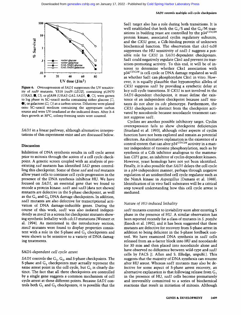
sadl and iad9 mutants are both unable to arrest the cell cycle in Gi or G2 in response to DNA damage. It is possible that they are components in a common genetic pathway. To investigate this relationship further, we examined the effects of SADl overproduction on UV sensitivity in a iad9 mutant. If RAD9 acts downstream of SADl in a linear genetic pathway, then overexpression of SADl in a rad9 null mutant should have no effect on the rad9 phenotype. However, overproduction of SADl from the inducible GAL promoter strongly suppresses the UV sensitivity of rad9 mutants (Fig. 6). This result suggests that it is imlikely that RAD9 is downstream of
2408 GENES & DEVELOPMENT
Cold Spring Harbor Laboratory Press on January 17, 2022 - Published by genesdev.cshlp.orgDownloaded from
SADl controls multiple cell cycle checkpoints
100 V
> u 3
20 40 60 80 UV(lose(J/m2)
Figure 6. Overexpression of SADl suppresses the UV sensitivity of Tad9 mutants. Y318 {iad9::LEU2), containing pUN70 [URA3, m, U], or pJA98 {URA3 GAL:SADl, %, O), were grown to log phase in SC-uracil media containing either glucose (D, •) , or galactose (D, O) as a carbon source. Dilutions were plated onto SC-uracil medium containing the appropriate carbon source and were UV-irradiated at the indicated doses. After 3-4 days growth at 30°C, colony-forming units were counted.
SADl in a. linear pathway, although alternative interpretations of this experiment exist and are discussed below.
Discussion
Inhibition of DNA synthesis results in cell cycle arrest prior to mitosis through the action of a cell cycle checkpoint. A genetic screen coupled with an analysis of preexisting lad mutants has identified SAD genes controlling this checkpoint. Some of these sad and rad mutants allow yeast cells to continue cell cycle progression in the presence of the DNA synthesis inhibitor HU. We have focused on SADl, an essential gene that we found to encode a protein kinase, sadl and sad3 (data not shown) mutants are defective in the S-phase checkpoint, as well as the Gi and G2 DNA damage checkpoints. In addition, sadl mutants are also defective for transcriptional activation of DNA damage-inducible genes. During the course of this work, sadl was also isolated independently as mec2 in a screen for checkpoint mutants showing synthetic lethality with cdcl3 mutations (Weinert et al. 1994). As mentioned in the introductory section, mec2 mutants were found to display properties consistent with a role in the S-phase and G2 checkpoints and were shown to be sensitive to a variety of DNA damaging treatments.
SADl-dependent cell cycle aiiest
SADl controls the Gi, G2, and S-phase checkpoints. The S-phase and G2 checkpoints may actually represent the same arrest point in the cell cycle, but G^ is clearly distinct. The fact that all three checkpoints are controlled by a single gene suggests a common mechanism of cell cycle arrest at these different points. Because SADl controls both Gj and G2 checkpoints, it is possible that the
Sadl target also has a role during both transitions. It is well established that both the G^/S and the G2/M transitions in budding yeast are controlled by the p34' ^*^^* protein kinase, associated cyclin regulatory subunits, and the CKSl gene, a Cdk-binding protein of unknown biochemical function. The observation that cksl-ts38 suppresses the HU sensitivity of sadl -1 suggests a possible role for CKSl in SADl-dependent checkpoints. Sadl could negatively regulate Cksl and prevent its transition-promoting activity. To this end, it will be of interest to determine whether Cksl association with p34CDC28 jg ^QY\ cycle or DNA damage regulated as well as whether Sadl can phosphorylate Cksl in vitro. However, it is equally plausible that hypomorphic alleles of CKSl suppress sadl by providing a synthetic delay at key cell cycle transitions. If CKSl is not involved in the SADi-dependent checkpoint, it must define the existence of an independent checkpoint because sadl mutants do not alter its cdc phenotype. Furthermore, the CKSl checkpoint is distinct from the checkpoint activated by nocodozole because nocodazole treatment cannot suppress sadl.
Cyclins are another possible inhibitory target. Cyclin overexpression fails to show checkpoint deficiencies (Stueland et al. 1993), although other aspects of cyclin function have not been explored and remain as potential effectors. An alternative explanation is the existence of a control system that can alter p34* °*- * activity in a manner independent of tyrosine phosphorylation, such as by synthesis of a Cdk inhibitor analogous to the mammalian CIPl gene, an inhibitor of cyclin-dependent kinases. However, yeast homologs have not yet been identified. Finally, it is also possible that SADl delays the cell cycle in a p34-independent manner, perhaps through negative regulation of an unidentified cell cycle regulator such as the NimA kinase in Aspergillus [Osmani et al. 1988). Identification of in vivo Sadl substrates will be a critical step toward understanding how this cell cycle arrest is mediated.
Nature of HU-induced lethality sadl mutants commit to inviability soon after entering S phase in the presence of HU. A similar observation has been reported recently for a class of mutants in 5. pombe (Enoch et al. 1992), and it has been suggested that these mutants are defective for recovery from S-phase arrest in addition to being deficient in the S-phase feedback control. We have examined DNA synthesis in sadl cells released from an a-factor block into HU and nocodazole for 30 min and then placed into nocodazole alone and have observed no difference between wild-type and sadl cells by FACS (J, Allen and S. Elledge, unpubl.). This suggests that the majority of DNA synthesis can resume after HU arrest. Whereas sadl mutants may also be defective for some aspect of S-phase arrest recovery, an altemative explanation is that following release from Gi in the presence of HU, sadl cells become prematurely and irreversibly committed to a series of biochemical reactions that result in initiation of mitosis. Although
GENES & DEVELOPMENT 2409
Cold Spring Harbor Laboratory Press on January 17, 2022 - Published by genesdev.cshlp.orgDownloaded from

Allen et al.
segregation of unreplicated chromosomes would certainly be lethal, it is unlikely to be the sole explanation of lethality in these cells because nocodazole, which blocks anaphase, is unable to suppress killing when present during and after HU treatment (data not shown). This is based on the assumption that cells are still competent to replicate DNA at the nocodazole block, which has not been tested. Thus, the nature of HU-induced lethality is not clear but may result from other aspects of premature mitotic commitment such as chromosome condensation, maturation of kinetechores, or alterations in nuclear architecture.
Does SADl coordinate S phase and mitosis in a normal cell cycle}
The S-phase and DNA damage checkpoints allow cells to integrate cell cycle progression with the replicational status and integrity of the genome. However, it is not known whether these checkpoints also order events in a normal cell cycle in the absence of DNA damage or replication blocks, or whether S-phase and mitotic coordination is normally ensured by relative timing. The interpretation of an active mitotic inhibitory signal produced from S-phase nuclei is supported by the experiments of Rao and Johnson (1970), who observed that fusion of unperturbed mammalian S-phase cells to G2 cells caused the G2 nucleus to arrest entry into mitosis until the S-phase nucleus completed DNA replication. The data presented here do not provide a definitive answer for yeast, although several properties of sadl mutants are germane to this issue. First, SADl is a member of the Mlul cell cycle box (MCB) group of cell cycle-regulated genes expressed in S phase that encode many enzymes essential for S phase (Zheng et al. 1993; J. Allen and S. Elledge, unpubl.). Second, unlike RAD9, SADl is essential for viability, suggesting a role in every cell cycle. It is likely that this essential function is during S phase, although SADl could be essential for reasons unrelated to feedback control. Finally, sadl mutants accelerate mitotic entry in the presence of HU, although not significantly faster than wild-type cells. It is difficult to imagine why a treatment that normally blocks cell cycle progression would have the opposite effect in a sadl mutant. Perhaps the signal to inhibit mitosis emanates from active replication forks that are diminished in the presence of HU. Normally, a dampened but persistant signal would be sufficient to activate the checkpoint, but in a sadl-1 mutant the reduced signal and diminished transducer together would be insufficient and cells would commit to mitosis faster. Without a conditional lethal allele of SADl, it is not possible to determine definitively whether Sadl controls normal timing of mitosis.
SADl controls the cell cycle arrest response to DNA damage
sadl mutants are defective for the lMD9-dependent Gi and G2 DNA damage checkpoints. If RAD9 functions downstream of SADl, then overexpression of SADl in a
Arad9 background should have no effect with respect to the damage sensitivity of Arad9 mutants. However, overexpression of SADl partially suppresses the UV sensitivity of Arad9 strains. If SADl overproduction enhances its role in checkpoint-mediated cell cycle arrest, then RAD9 cannot function after SADl in a linear pathway. However, overexpression of SADl slightly slows down the cell cycle in wild-type cells. If this slow growth is not a manifestation of the checkpoint function of SADl, suppression of rad9 mutants may be attributable to nonspecifically lengthening the cycle. Because we cannot distinguish between these alternatives, we cannot definitively order SADl and RAD9.
SADl controls the transcriptional response to DNA damage
In addition to multiple checkpoint deficiencies, sadl mutants also exhibit a DNA damage-uninducible pheno-type similar to that of the dun mutants (Zhou and Elledge 1993), defective for RNR2 and RNR3 induction but proficient for UBI4 induction. Mutants in DUNl, a protein kinase whose activity is increased in response to DNA damage, are proficient in their cell cycle response to DNA damage and have only a subset of sadl pheno-types, suggesting that DUNl is downstream of SADl. In addition, Dun I is not activated in sadl mutants. Based on these observations coupled with the DNA damage checkpoint role, it is likely that the Sadl kinase is positioned very close to the signal sensor in the DNA damage signaling pathway.
The relationship between the checkpoint and transcriptional roles of SADl is not clear. The absence of RNR3 expression during a normal S phase in which SADl is likely to function presents a puzzle. If Sadl is functioning during a normal S phase to activate the checkpoint, why does it not also activate expression of RNR3 at that time? The presence of DNA damage or a replication block must provide information distinct from normal DNA replication to SADl or its effectors. Sadl does contain potential regulatory domains on either side of the kinase domain that could respond to or send different signals. Activation of RNR3 transcription could require higher levels of Sadl function than is necessary to activate the checkpoint. Alternatively, the activation of a checkpoint might lengthen the period of the cell cycle during which a factor accumulates that facilitates transcriptional activation. This factor could be active SADl or a substrate. Another possibility is that a SADl-independent pathway is additionally required to provide DNA damage information necessary for RNR3 induction. These complexities can only be resolved by rigorous biochemical and genetic analysis of the SADl kinase.
A model depicting the genetic pathway regulated by Sadl is shown in Figure 7. SADl is placed downstream from the direct sensors for DNA damage and DNA replication blocks. Although no formal evidence has been presented that Sadl is regulated in response to these stimuli, the fact that Sadl is a protein kinase makes it
2410 GENES & DEVELOPMENT
Cold Spring Harbor Laboratory Press on January 17, 2022 - Published by genesdev.cshlp.orgDownloaded from
SADl controls multiple cell cycle checkpoints
REPLICATION BLOCKS
Dunl
Ceil Cycle Arrest
Transcriptional Response
RNR2 RNR3
Gl S G2 M
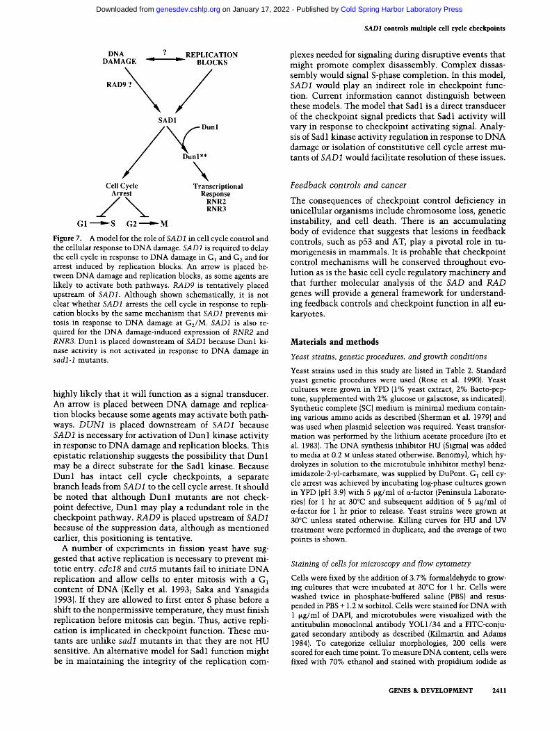
Figure 7. A model for the role of SADl in cell cycle control and the cellular response to DNA damage. SADl is required to delay the cell cycle in response to DNA damage in Gi and G2 and for arrest induced by replication blocks. An arrow is placed between DNA damage and replication blocks, as some agents are likely to activate both pathways. RAD9 is tentatively placed upstream of SADl. Although shown schematically, it is not clear whether SADl arrests the cell cycle in response to replication blocks by the same mechanism that SADl prevents mitosis in response to DNA damage at Gj/M. SADl is also required for the DNA damage-induced expression of RNR2 and RNR3. Dunl is placed downstream of SADl because Dunl kinase activity is not activated in response to DNA damage in sadl-1 mutants.
highly Hkely that it will function as a signal transducer. An arrow is placed between DNA damage and replication blocks because some agents may activate both pathways. DUNl is placed downstream of SADl because SADl is necessary for activation of Dun l kinase activity in response to DNA damage and replication blocks. This epistatic relationship suggests the possibility that Dun l may be a direct substrate for the Sadl kinase. Because Dunl has intact cell cycle checkpoints, a separate branch leads from SADl to the cell cycle arrest. It should be noted that although Dunl mutants are not checkpoint defective, Dun l may play a redundant role in the checkpoint pathway. RAD9 is placed upstream of SADl because of the suppression data, although as mentioned earlier, this positioning is tentative.
A number of experiments in fission yeast have suggested that active replication is necessary to prevent mitotic entry, cdcl 8 and cut5 mutants fail to initiate DNA replication and allow cells to enter mitosis with a G^ content of DNA (Kelly et al. 1993; Saka and Yanagida 1993). If they are allowed to first enter S phase before a shift to the nonpermissive temperature, they must finish replication before mitosis can begin. Thus, active replication is implicated in checkpoint function. These mutants are unlike sadl mutants in that they are not HU sensitive. An alternative model for Sadl function might be in maintaining the integrity of the replication com
plexes needed for signaling during disruptive events that might promote complex disassembly. Complex dissas-sembly would signal S-phase completion. In this model, SADl would play an indirect role in checkpoint function. Current information cannot distinguish between these models. The model that Sadl is a direct transducer of the checkpoint signal predicts that Sadl activity will vary in response to checkpoint activating signal. Analysis of Sadl kinase activity regulation in response to DNA damage or isolation of constitutive cell cycle arrest mutants of SADl would facilitate resolution of these issues.
Feedback controls and cancer
The consequences of checkpoint control deficiency in unicellular organisms include chromosome loss, genetic instability, and cell death. There is an accumulating body of evidence that suggests that lesions in feedback controls, such as p53 and AT, play a pivotal role in tu-morigenesis in mammals. It is probable that checkpoint control mechanisms will be conserved throughout evolution as is the basic cell cycle regulatory machinery and that further molecular analysis of the SAD and RAD genes will provide a general framework for understanding feedback controls and checkpoint function in all eu-karyotes.
Materials and methods
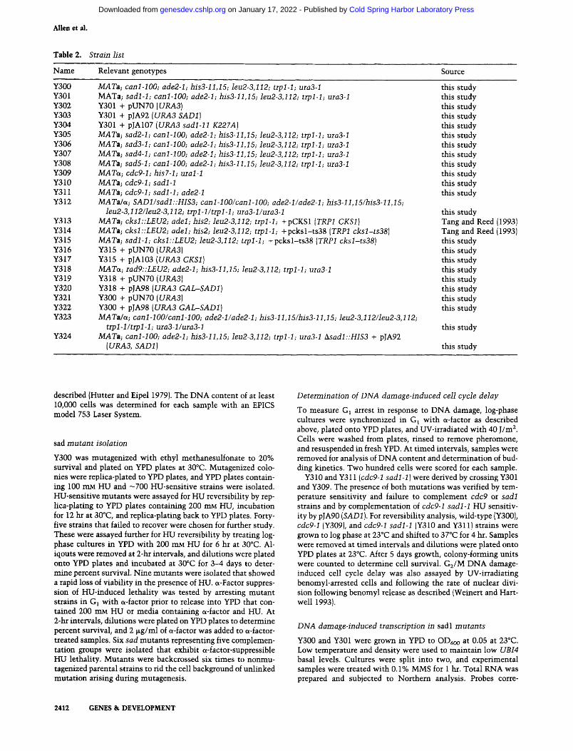
Yeast strains, genetic procedures, and growth conditions
Yeast strains used in this study are listed in Table 2. Standard yeast genetic procedures were used (Rose et al. 1990). Yeast cultures were grown in YPD (1% yeast extract, 2% Bacto-pep-tone, supplemented with 2% glucose or galactose, as indicated). Synthetic complete (SC) medium is minimal medium containing various amino acids as described (Sherman et al. 1979) and was used when plasmid selection was required. Yeast transformation was performed by the lithium acetate procedure (Ito et al. 1983). The DNA synthesis inhibitor HU (Sigma) was added to media at 0.2 M unless stated otherwise. Benomyl, which hy-drolyzes in solution to the microtubule inhibitor methyl benz-imidazole-2-yl-carbamate, was supplied by DuPont. Gi cell cycle arrest was achieved by incubating log-phase cultures grown in YPD (pH 3.9) with 5 |xg/ml of a-factor (Peninsula Laboratories) for 1 hr at 30°C and subsequent addition of 5 M-g/ml of a-factor for 1 hr prior to release. Yeast strains were grown at 30°C unless stated otherwise. Killing curves for HU and UV treatment were performed in duplicate, and the average of two points is shown.
Staining of cells for microscopy and flow cytometry
Cells were fixed by the addition of 3.7% formaldehyde to growing cultures that were incubated at 30°C for 1 hr. Cells were washed twice in phosphate-buffered saline (PBS) and resus-pended in PBS -I-1.2 M sorbitol. Cells were stained for DNA with 1 |xg/ml of DAPI, and microtubules were visualized with the antitubulin monoclonal antibody YOLl/34 and a FITC-conju-gated secondary antibody as described (Kilmartin and Adams 1984). To categorize cellular morphologies, 200 cells were scored for each time point. To measure DNA content, cells were fixed with 70% ethanol and stained with propidium iodide as
GENES & DEVELOPMENT 2411
Cold Spring Harbor Laboratory Press on January 17, 2022 - Published by genesdev.cshlp.orgDownloaded from
Allen et al.
Table 2. Strain list
Name Relevant genotypes Source
Y300 MATa; canl-100; ade2-l; his3-ll,15; leu2-3,112; trpl-1; ura3-l Y301 MATa; sadl-1; canl-100; ade2-l; his3-ll,15; leu2-3,112; trpl-h ura3-l Y302 Y301 + pUN70 {URA3] Y303 Y301 + pJA92 iURA3 SADl] Y304 Y301 + PJA107 {URA3 sadl-11 K227A] Y305 MATa; sad2-l; canl-100; ade2-l; his3-ll,15; leu2-3,112; trpl-l; maS-l Y306 MATa; sad3-l; canl-100; ade2-l; his3-ll,15; leu2-3,112; trpl-h ura3-l Y307 MATa; sad4-l; canl-100; ade2-l; his3-llJ5; leu2-3,112; trpl-1; ura3-l Y308 MATa; sad5-l; canl-100; ade2-l; his3-ll,15; leu2-3,112; trpl-h ura3-l Y309 MATa; cdc9-l; his7-l; ural-1 Y310 MATa; cdc9-l; sadl-1 Y311 MATa; cdc9-l; sadl-1; ade2-l Y312 MATa/a; SADl/sadl::HIS3; canl-lOO/canl-100; ade2-l/ade2-h his3-ll,15/his3-ll,15;
Ieu2-3,112/leu2-3,112; trpl-l/trpl-1; ura3-l/ura3-l Y313 MATa; cksl::LEU2; adel; his2; leu2-3,112; trpl-1; +pCKSl {TRPl CKSl) Y314 MATa; cksl::LEU2; adel; his2; leu2-3,112; trpl-1; +pcksl-ts38 [TRPl cksl-ts38] Y315 MATa; sadl-1; cksl::LEU2; leu2-3,112; trpl-1; +pcksl-ts38 {TRPl cksl-ts38) Y316 Y315 + pUN70 {URA3] Y317 Y315 + PJA103 (URA3 CKSl ] Y318 MATa; rad9::LEU2; ade2-l; his3-ll,15; leu2-3,112; trpl-1; ma3-l Y319 Y318 + pUN70 {URA3] Y320 Y318 + pJA98 {URA3 GAI^SADl) Y321 Y300 + pUN70 iURA3] Y322 Y300 + pJA98(t7i?A3GAZ^SADJ) Y323 MATa/a; canl-lOO/canl-100; ade2-l/ade2-l; his3-ll,15/his3-ll,15; Ieu2-3,112/leu2-3,112;
trpl-1/trpl-1; ura3-l/ura3-l Y324 MATa; canl-100; ade2-l; his3-ll,15; leu2-3,112; trpl-1; ura3-l ^sadl::HIS3 + pJA92
{URA3, SADl]
this study this study this study this study this study this study this study this study this study this study this study this study
this study Tang and Reed (1993) Tang and Reed (1993) this study this study this study this study this study this study this study this study
this study
this study
described (Hutter and Eipel 1979). The DNA content of at least 10,000 cells was determined for each sample with an EPICS model 753 Laser System.
sad mutant isolation
Y300 was mutagenized with ethyl methanesulfonate to 20% survival and plated on YPD plates at 30°C. Mutagenized colonies were replica-plated to YPD plates, and YPD plates containing 100 mM HU and —700 HU-sensitive strains were isolated. HU-sensitive mutants were assayed for HU reversibility by replica-plating to YPD plates containing 200 mM HU, incubation for 12 hr at 30°C, and replica-plating back to YPD plates. Forty-five strains that failed to recover were chosen for further study. These were assayed further for HU reversibility by treating log-phase cultures in YPD with 200 mM HU for 6 hr at 30°C. Al-iqouts were removed at 2-hr intervals, and dilutions were plated onto YPD plates and incubated at 30°C for 3-4 days to determine percent survival. Nine mutants were isolated that showed a rapid loss of viability in the presence of HU. a-Factor suppression of HU-induced lethality was tested by arresting mutant strains in Gj with a-factor prior to release into YPD that contained 200 mM HU or media containing ct-factor and HU. At 2-hr intervals, dilutions were plated on YPD plates to determine percent survival, and 2 (xg/ml of a-factor was added to a-factor-treated samples. Six sad mutants representing five complementation groups were isolated that exhibit a-factor-suppressible HU lethality. Mutants were backcrossed six times to nonmu-tagenized parental strains to rid the cell background of unlinked mutation arising during mutagenesis.
Determination of DNA damage-induced cell cycle delay
To measure Gj arrest in response to DNA damage, log-phase cultures were synchronized in Gi with a-factor as described above, plated onto YPD plates, and UV-irradiated with 40 J/m^. Cells were washed from plates, rinsed to remove pheromone, and resuspended in fresh YPD. At timed intervals, samples were removed for analysis of DNA content and determination of budding kinetics. Two hundred cells were scored for each sample.
Y310 and Y311 {cdc9-l sadl-1) were derived by crossing Y301 and Y309. The presence of both mutations was verified by temperature sensitivity and failure to complement cdc9 or sadl strains and by complementation of cdc9-l sadl-1 HU sensitivity by pJA90 {SADl ]. For reversibiHty analysis, wild-type (Y300), cdc9-l (Y309), and cdc9-l sadl-1 (Y310 and Y311) strains were grown to log phase at 23°C and shifted to 37°C for 4 hr. Samples were removed at timed intervals and dilutions were plated onto YPD plates at 23°C. After 5 days growth, colony-forming units were coxmted to determine cell survival. Gj/M DNA damage-induced cell cycle delay was also assayed by UV-irradiating benomyl-arrested cells and following the rate of nuclear division following benomyl release as described (Weinert and Hart-well 1993).
DNA damage-induced transcription in sadl mutants
Y300 and Y301 were grown in YPD to OD^oo at 0.05 at 23°C. Low temperature and density were used to maintain low UBI4 basal levels. Cultures were split into two, and experimental samples were treated with 0.1% MMS for 1 hr. Total RNA was prepared and subjected to Northern analysis. Probes corre-
2412 GENES & DEVELOPMENT
Cold Spring Harbor Laboratory Press on January 17, 2022 - Published by genesdev.cshlp.orgDownloaded from

SADl controls multiple cell cycle checkpoints
sponded to the 0.9-kb Hindlil fragment of RNR2 from pSE310, the 2.5-kb Mlul-Hindlll fragment of RNR3 from pSE734, and the 2.2-kb £coRI fragment of UBI4 from pUB200.
Immunopiecipitations and kinase assays
Ueast extract (100 i.g) was diluted in 500 (JLI of DB buffer (20 mM Tris-HCl at pH 7.9, 100 mM NaCl, 2 mM EDTA, 0.05% Tween 20,1 mM PMSF, 0.1 |xg/ml of leupeptin, 0.1 mM benzymidine, 1 fjLM aprotinin, 0.1 |xg/ml of pepstatin, 0.1 mM Na3V04, and 30 mM NaF) and incubated with 10-20 |xl of affinity-purified anti-Dvml antibodies for 1 hr at 4°C. Protein A-Sepharose CL-4B beads were used to precipitate immunocomplexes as described (Zhou and EUedge 1993). Immunocomplexes were incubated in 25 |xl of kinase buffer with 2 piM cold ATP and 2.5 nCi of ( .32p]ATP for 30 min at 30°C. Reactions were terminated by the addition of 25 |xl of 2x SDS sample buffer. Phosphorylated products were visualized by 8% SDS-polyacrylamide gels and autoradiography.
Determination of the in vivo phosphorylation state of Dunl
Y300 and Y301 were grown to ODeoo of 0.2 in 100 ml of YPD, without inorganic phosphate (YPD-PO4) (Salah-ud-Din et al. 1990). Cells were pelleted, resuspended in 10 ml of YPD-PO4 containing 1 mCi of [^^P]H3P04, and cultured for 2 hr at 30°C in the presence or absence of 0.1% MMS. Protein extraction and immunoprecipitation of Dunl were performed as described above except that 100 fig of RNase A was added to each immunoprecipitation reaction prior to electrophoresis. Samples were subjected to Western blot analysis and autoradiography.
Cloning of SADl
A yeast genomic centromeric library carrying the Ui?A3-select-able marker (Ramer et al. 1993) was transformed into sadl-1. Ura" transformants (5x 10 ) were plated onto SC plates lacking uracil containing 100 mM HU. Three plasmids, pJA90, pJA91, and pJA92, complemented sadl-1 upon retransformation and contained overlapping genomic inserts. A common 1.5-kb Xhol fragment was subcloned into pBS KSIH- (Stratagene) and sequenced (Sanger et al. 1977). A null allele for SADl was generated by transplacement of the 3.9-kb H/S3-containing Xhol fragment from pJAlOO into Y323 and selection for histidine pro-totrophy. Diploids (Y312) heterozygous for SADl were confirmed by Southern blotting. pJA93 {URA3 SADl] rescues viability of His^ haploids from tetrads. His^ Ura* spores are also 5-FOA sensitive, demonstrating the requirement for wild-type SADl.
Plasmid constructions
The 4.2-kb Xhol partial fragment from pJA90 was subcloned into ZAoI-digested pBS KSII-i- (Stratagene) to give pJA93. The SADl open reading frame was amplified from pJA93 by polymerase chain reaction (PCR) with the 5' primer, 5'-CAGGCATATGGAAAATATTACACAACCC-3'. and the 3' primer, 5' -TCTTAGCGGCCGCCCATGGGCGAAAATTGC-AAATTCTCGGG-3'. Ndel and NotI sites are underhned, respectively. The PCR-derived product was digested with BgTlI, and the resulting 2.0-kb fragment representing the 3' region of SADl was subcloned as a Bglll blunt-ended fragment into BglU.-Smal-digested pSE386 to give pJA95.
To place SADl under T7 control, a three-way ligation was performed using the 0.5-kb fragment from the Ndel-BglU-di-gested SADl PCR-derived product described above, the 3.2-kb
BgRl-Notl fragment from pJA95, and the Ncfel-Notl-digested pETHAX vector to give pJA97. The PCR-derived fragment was sequenced after cloning and was not mutant. To place SADl under control of the GAL promoter on a CEN plasmid, the 2.7-kb NcoI(blunt)-NotI fragment from pJA97 was cloned into XfaaI(blunt)-NotI-digested pSE556 to give p}A98.
A transplacement vector, pJAlOO, containing a SADl null allele was constructed by replacing the 1.7-kb Styl fragment of pJA93 with the 2.3-kb Smal fragment from pJA50 (Allen and Elledge 1994), which contains the HISS and Tn5 neo-selectable markers.
Site-directed mutagenesis was performed to generate a ki-nase-deficient mutant of sadl. pJA93 was used as a template for PCR to mutate nucleotides 712-714 from AAG (Lys) to GCC (Ala). First, two PCR reactions were carried out to produce overlapping amplified fragments. In reaction 1, wild-type forward primer (5') CATGCCATGGGTAGAAACCCAGCCTGTGAC (3') and mutagenic reverse primer (5') ACTTATAATGGCCAC-CGCGAATGTTTTC (3') were used. In reaction 2, mutagenic forward primer (5') GGGAAAACATTCGCGGTGGCCATT-ATAAGTAAA (3') and wild-type reverse primer (5') GCTCTA-GATTATTGAGCATCGTCCATATTTTC (3') were used. PCR-derived products were recovered from agarose gels, and 10 ng of each were combined and amplified using wild-type forward and reverse primers. The full length PCR-derived fragment was digested with Xhol and BamHl, and the resulting 1.2-kb fragment was subcloned into Xhol-BamHl-cleived pBS KSII-f- for sequencing to confirm the presence of the mutant codon and the absence of other PCR-induced mutations. The 1.2-kb BglU-BamHl fragment from PCR-mutagenized product was subcloned into Bgill-BflmHI-digested pJA93 to give pJA104. The 1.7-kb Styl fragment from p}A93 was subcloned into the Styl site of pJA104 to give pJAlOS. In addition, the 2.4-kb Sad fragment from pyA93 was subcloned into Sacl-digested pJAlOS to give pJA106. A site-directed mutagenized sadl allele {sadl-11 K227A) under control of the endogenous SADl promoter was generated in a three-way ligation using the 0.5-kb Kpnl-Bglll fragment from pJA93, the 3.2-kb BglH-Xbal fragment from PJA106, and Kpnl-Xbal-digested pUN70 (Elledge and Davis 1988).
PJA103 (CKSl URA3] was made by ligating the 2.2-kb Smal URA3 neo fragment from pJA53 (Allen and Elledge 1994) into £coRV-cleaved pSE271:CK:Sl (Tang and Reed 1993).
Southern and Northern blot analysis
DNA probes were labeled by the hexamer primer method (Fein-berg and Vogelstein 1983). Hybridizations for Southern blots were carried out as described previously (Elledge and Davis 1987). Yeast RNA was isolated using the hot phenol method (Kohrer and Domdey 1991). RNA was resolved on formalde-hyde-1.2% agarose gels (Sambrook et al. 1989), and hybridizations were carried out as described for Southern analysis.
Acknowledgments
We thank S. Reed, M. Kuroda, S. Sazer, W. Harper, V. Lundblad, G. May, T. Navas, T. Weinert, L. Hartwell, and A. Carr for comments, helpful discussions, and/or reagents. We thank D. Achille for DNA sequencing and Sonal Amin for assistance in the preliminary screen. This work was supported by a National Institutes of Health grant GM44664 and a Welch Foundation grant to S.J.E. Z.Z. is a Welch Foundation predoctoral fellow. S.J.E. is a Pew scholar in the biomedical sciences and an investigator of the Howard Hughes Medical Institute.
GENES & DEVELOPMENT 2413
Cold Spring Harbor Laboratory Press on January 17, 2022 - Published by genesdev.cshlp.orgDownloaded from

Allen et al.
The publication costs of this article were defrayed in part by payment of page charges. This article must therefore be hereby marked "advertisement" in accordance with 18 USC section 1734 solely to indicate this fact.
References
Al-Khodairy, F. and A.M. Carr. 1992. DNA repair mutants defining G2 checkpoint pathways in Schizosaccharomyces pombe. EMBO /. 11: 1343-1350.
Allen, J.B. and S.J. EUedge. 1994. A family of vectors that facilitate transposon and insertional mutagenesis of cloned genes in yeast. Yeast (in press).
Amon, A., U. Surana, L. Muroff, and K. Nasmyth. 1992. Regul-atin of p34*^°^^* phosphorylation is not required for entry into mitosis in S. cerevisiae. Nature 355: 368-371.
Cohen, M.M. and H.P. Levy. 1989. Chromosome instability syndromes. Adv. Hum. Genet. 18: 43-149.
El-Deiry, W.S., T. Tokino, V.E. Velculescu, D.B. Levy, R. Parsons, J.M. Trent. D. Lin, W.E. Mercer, K.W. Kinzler, and B. Vogelstein. 1993. WAFl, a potential mediator of p53 tumor suppression. Cell 75: 817-825.
El-Deiry, W.S., J.W. Harper, P.M. O'Connor, V. Velculescu, C.E. Canman, J. Jackman, J. Pietenpol, M. Burell, D.E. Hill, W.E. Mercer, M.B. Kastan, K.W. Kohn, S.J. EUedge, K.W. Kinzler, and B. Vogelstein. 1994. WAFl/CIPl is induced in p53-me-diated Gl arrest and apoptosis. Cancer Res. 54: 1169-1174.
EUedge, S.J. and R.W. Davis. 1987. Identification and isolation of the gene encoding the small subunit of ribonucleotide reductase from Saccharomyces cerevisiae: A DNA damage inducible gene required for mitotic viability. Mol. Cell. Biol. 7: 2783-2793.
. 1988. A family of versatile centromeric vectors designed for use in the sectoring-shuffle mutagenesis assay in Saccharomyces cerevisiae. Gene 70: 303-312.
1990. Two genes, differentially regulated by DNA damage and the cell cycle, encode alternate regulatory subunits of ribonucleotide reductase. Genes 8k Dev. 4: 740-751.
Enoch, T. and P. Nurse. 1991. Coupling M phase and S phase: Controls maintaining the dependence of mitosis on chromosome rephcation. Cell 65: 921-923.
Enoch, T., A. Carr, and P. Nurse. 1992. Fission yeast genes involved in coupling mitosis to completion of DNA replication. Genes & Dev. 6: 2035-2046.
Feinberg, A.P. and B. Vogelstein. 1983. A technique for radiola-beling DNA restriction endonuclease fragments to high specific activity. Anal. Biochem. 132: 6-13.
Gould, K.L. and P. Nurse. 1989. Tyrosine phosphorylation of the fission yeast cdc2'^ protein kinase regulates entry into mitosis. Nature 342: 39-45.
Gu, Y., C.W. Turck, and D.O. Morgan. 1993. Inhibition of CDK2 activity in vivo by an associated 20K regulatory sub-unit. Nature 366: 707-710.
Hadwiger, J.A., C. Wittenberg, M.D. Mendenhall, and S.I. Reed. 1989. The Saccharomyces cerevisiae CKSl gene, a homolog of the Schizosaccharomyces pombe sucl " gene, encodes a subunit of the Cdc28 protein kinase complex. Mol. Cell. Biol. 9:2034-2041.
Harper, J.W., G.R. Adami, N. Wei, K. Keyomarsi, and S.J. EUedge. 1993. The p21 Cdk-interacting protein Cipl is a potent inhibitor of Gl cyclin-dependent kinases. Cell 75:805-816.
Hartwell, L. 1992. Defects in a cell cycle checkpoint may be responsible for the genomic instability of cancer cells. Cell 71: 543-546.
Hartwell, L.H. and T.A. Weinert. 1989. Checkpoints: Controls that ensure the order of cell cycle events. Science 246: 229-234.
Hoyt, M.A., L. Totis, andB.T. Roberts. 1991. S. cerevisiae genes required for cell cycle arrest in response to loss of microtubule hinction. Cell 66: 507-517.
Hutter, K.J. and H.E. Eipel. 1979. DNA determination of yeast by flow cytometry. /. Gen. Microbiol. 113: 369-375.
Ito, H., Y. Fukada, K. Murata, and A. Kimura. 1983. Transformation of intact yeast cells with alkali cations, f. Bacteriol. 153: 163-168.
Johnston, L.H. and K.A. Nasmyth. 1978. Saccharomyces cere-' visiae cell cycle mutant cdc9 is defective in DNA ligase.
Nature 274: 891-893. Kastan, M.B., Q. Zhan, W.S. El-Deiry, F. Carrier, T. Jacks, W.V.
Walsh, B.S. Plunkett, B. Vogelstein, and A.J. Foumace Jr. 1992. A mammalian cell cycle checkpoint pathway utilizing p53 and GADD45 is defective in Ataxia-Telangiectasia. Cell 71: 587-597.
Kelly, T.J., G.S. Martin, S.L. Forsburg, R.J. Stephen, A. Russo, and P. Nurse. 1993. The fission yeast cdclS'^ gene product couples S phase to START and mitosis. Cell 74: 371-382.
Kilmartin, J.V. and A.E.M. Adams. 1984. Structural rearrangements of tubulin and actin during the cell cycle of the yeast Saccharomyces. /. Cell Biol. 98: 922-933.
Kohrer, K. and H. Domdey. 1991. Preparation of high molecular weight RNA. Methods Enzymol. 194: 398-405.
Kuerbitz, S.J., B.S. Plunkett, W.V. Walsh, and M.B. Kastan. 1992. Wild type p53 is a cell cycle checkpoint determinant following irradiation. Proc. Natl. Acad. Sci. 89: 7491-7495.
Kusubata, M., T. Tokui, Y. Matsuoka, E. Okumura, K. Ta-chibana, S. Hisanaga, T. Kishimoto, H. Yasuda, M. Kamijo, Y. Ohba, K. Tsujimura, R. Yatani, and M. Inagaki. 1992. pl3*"''' suppresses the catalytic function of p34' ' ' ^ kinase for intermediate filament proteins, in vitro. /. Biol. Chem. 267: 20937-20942.
Li, J.J. and R.J. Deshaies. 1993. Exercising self-restraint: Discouraging illicit acts of S and M in eukaryotes. Cell 74: 223-226.
Li, R. and A.W. Murray. 1991. Feedback control of mitosis in budding yeast. Cell 66: 519-531.
Livingstone, L.R., A. White, J. Sprouse, E. Livanos, T. Jacks, and T.D. Tisty. 1992. Altered cell cycle arrest and gene amplification potential accompany loss of wild-type p53. Cell 70: 923-935.
Murray, A.W. 1992. Creative blocks: Cell-cycle checkpoints and feedback controls. Nature 359: 599-604.
Nagasawa, H., S.A. Latt, M.E. Lalande, and J.B. Little. 1985. Effects of X-irradiation on cell cycle progression, induction of chromosomal aberrations and cell killing in Ataxia Telangiectasia. Mutat. Res. 148: 71-82.
Nurse, P. 1990. Universal control mechanism regulating onset of M-phase. Nature 344: 503-508.
Osmani, S.A., R.T. Pu, and N.R. Morris. 1988. Mitotic induction and maintenance by overexpression of a G2-specific gene that encodes a potential protein kinase. Cell 53: 237-244.
Painter, R.B. and B.R. Young. 1980. Radiosensitivity in Ataxia Telangiectasia: A new explanation. Proc. Natl. Acad. Sci. 77: 7315-7317.
Ramer, S.W., S.J. EUedge, and R.W. Davis. 1993. Dominant genetics using a yeast genomic library under the control of a strong inducible promoter. Proc. Natl. Acad. Sci. 89: 11589-11593.
Rao, P.N. and R.T. Johnson. 1970. Mammalian cell fusion studies on the regulation of DNA synthesis and mitosis. Nature
2414 GENES & DEVELOPMENT
Cold Spring Harbor Laboratory Press on January 17, 2022 - Published by genesdev.cshlp.orgDownloaded from

SADl controls multiple cell cycle checkpoints
225:159-164. Roberge, M. 1992. Checkpoint controls that couple mitosis to
completion of DNA replication. T.I.C.B. 2: 277-281. Rose, M.D., F. Winston, and P. Heiter. 1990. Methods in yeast
genetics, a laboratory course manual. Cold Spring Harbor Laboratory Press, Cold Spring Harbor, New York.
Rowley, R., S. Subramani, and P.G. Young. 1992. Checkpoint controls in Schizosaccharomyces pombe: radl. EMBO f. 10: 1335-1342.
Rudolph, N.S. and S.A. Latt. 1989. Flow cytometric analysis of X-ray sensitivity in Ataxia Telangiectasia. Mutat. Res. 211:31-41.
Saka, Y. and M. Yanagida. 1993. Fission yeast cutS^, required for S phase onset and M phase restraint, is identical to the radiation-damage repair gene rad4*. Cell 74: 383-393.
Salah-ud-Din, S.J. Brill, M.P. Fairman, and B. Stillman. 1990. Cell-cycle-regulated phosphorylation of DNA replication factor A from human and yeast cells. Genes &. Dev. 4: 968-977.
Sambrook, J., E.F. Fritsch, and T. Maniatis. 1989. Molecular cloning: A laboratory manual, 2nd ed. Cold Spring Harbor Laboratory Press, Cold Spring Harbor, New York.
Sanger, F., S. Nicklen, and A.R. Coulson. 1977. DNA sequencing with chain-terminating inhibitors. Proc. Natl. Acad. Sci. 87: 2916-2920.
Siede, W., A.S. Friedberg, and E.C. Friedberg. 1993. RAD9-de-pendent Gl arrest defines a second checkpoint for damaged DNA in the cell cycle of Saccharomyces cerevisiae. Proc. Natl. Acad. Sci. 90: 7985-7989.
Sherman, F., G.R. Fink, and C.W. Lawrence. 1979. Methods in yeast genetics. Cold Spring Harbor Laboratory, Cold Spring Harbor, New York.
Shinohara, A., H. Ogawa, and T. Ogawa. 1992. RadSl protein involved in repair and recombination in S. cerevisiae is a RecA-like protein. Cell 69: 457-470.
Sorger, P.K. and A.W. Murray. 1992. S-phase feedback control in budding yeast independent of tyrosine phosphorylation of p34CDC28 feature 355: 365-368.
Stem, D.F., P. Zheng, D.R. Beidler, and C. Zerillo. 1991. Spkl, a new kinase from Saccharomyces cerevisiae, phosphorylates proteins on serine, threonine, and tyrosine. Mol. Cell. Biol. 11: 987-1001.
Stueland, C.S., D.J. Lew, M.J. Cismowski, and S.I. Reed. 1993. Full activation of p34' ' ^* histone HI kinase activity is unable to promote entry into mitosis in checkpoint-arrested cells of the yeast Saccharomyces cerevisiae. Mol. Cell. Biol. 13: 3744-3755.
Tang, Y. and S.I. Reed. 1993. The Cdk-associated protein Cksl fvmctions both in Gj and G2 in Sacchromyces cerevisiae. Genes Si Dev. 7: 822-832.
Walker, G.C. 1987. The SOS response of E. coH. In Escherichia coli and Salmonella typhimurium: Cellular and molecular biology (ed. F.C. Neidhart), pp. 1346-1357. American Society for Microbiology, Washington, D.C.
Weinert, T.A. 1992. Dual cell cycle checkpoints sensitive to chromosome replication and DNA damage in a budding yeast Saccharomyces cerevisiae. Radiat. Res. 132: 141-143.
Weinert, T.A. and L.H. Hartwell. 1988. The iMD9 gene controls the cell cycle response to DNA damage in Saccharomyces cerevisiae. Science 241: 317-322.
. 1993. Cell cycle arrest of cdc mutants and specificity of the RAD9 checkpoint. Genetics 134: 63-80.
Weinert, T.A., G.L. Kiser, and L.H. Hartwell. 1994. Mitotic checkpoints in budding yeast and the dependence of mitosis on DNA replication and repair. Genes & Dev. 8: 652-665.
Xiong, Y., G. Harmon, H. Zhang, D. Casso, R.E. Kobayashi, and
D. Beach. 1993. p21 is a universal inhibitor of cyclin-depen-dent kinases. Nature 366: 701-704.
Yin, Y., M.A. Tainsky, F.Z. Bischoff, L.C. Strong, and G.M. Wahl. 1992. Wild-type p53 restores cell cycle control and inhibits gene amplification in cells with mutant p53 alleles. Cell 70: 937-948.
Zambetti-Bosseler, F. and D. Scott. 1981. Cell death, chromosome damage and mitotic delay in normal human. Ataxia Telangiectasia, and Retinoblastoma fibroblasts after X-irra-diation. Int. ]. Radiat. Biol. 39: 547-558.
Zheng, P., D.S. Fay, J. Burton, H. Xiao, J.L. Pinkham, and D.F. Stem. 1993. SPKl is an essential S-phase-specific gene of Saccharomyces cerevisiae that encodes a nuclear serine/ threonine/tyrosine kinase. Mol. Cell. Biol. 13: 5829-5842.
Zhou, Z. and S.J. Elledge. 1993. DUNl encodes a protein kinase that controls the DNA damage response in yeast. Cell 75:1119-1127.
GENES & DEVELOPMENT 2415
Cold Spring Harbor Laboratory Press on January 17, 2022 - Published by genesdev.cshlp.orgDownloaded from

10.1101/gad.8.20.2401Access the most recent version at doi: 8:1994, Genes Dev.
J B Allen, Z Zhou, W Siede, et al. DNA damage-induced transcription in yeast.The SAD1/RAD53 protein kinase controls multiple checkpoints and
References
http://genesdev.cshlp.org/content/8/20/2401.full.html#ref-list-1
This article cites 58 articles, 19 of which can be accessed free at:
License
ServiceEmail Alerting
click here.right corner of the article or
Receive free email alerts when new articles cite this article - sign up in the box at the top
Copyright © Cold Spring Harbor Laboratory Press
Cold Spring Harbor Laboratory Press on January 17, 2022 - Published by genesdev.cshlp.orgDownloaded from