The Respiratory System - Nutley Public · PDF file · 2016-02-17I. Functions of the...
32
The Respiratory System Chapter 13 Bania NHS - SCIENCE
Transcript of The Respiratory System - Nutley Public · PDF file · 2016-02-17I. Functions of the...

The Respiratory System Chapter 13
Bania NHS - SCIENCE

I. Functions of the Respiratory System
A. Distribution of air/diffusion of oxygen and carbon dioxide 1. Between air and blood external respiration 2. Between blood and cells – internal respiration 3. Excretory organ – removes CO2 from blood – makes less alkaline
B. movement of air into and out of the lungs - ventilation C. conditions air - warms, moistens, and cleans incoming air D. sound production E. detection of odors F. RESPIRATION - Exchange of gases between the atmosphere, the
blood, and the cells

II. Organs of the Respiratory System A. Nose - warms, cleans and
conditions air B. sinuses - not pictured -
resonates sound C. pharynx - muscular junction
of oral/nose passage D. larynx - voice box - separates
pipes (epiglottis E. trachea - wind pipe F. LUNGS
1. bronchi (bronchioles) 2. Alveoli – grapes for gas exchange

III. Respiratory Tracts
A. Upper respiratory tract (URT) 1. located outside the chest cavity 2. nose, pharynx, larynx, and associated
structures B. lower respiratory tract (LRT)
1. located within chest cavity 2. trachea, bronchi, lungs

IV. Respiratory Mucosa A. Membrane that lines most of the air distribution
tubes in the system B. covered with blanket of mucus C. contains hair-like projections called cilia D. Functions:
1. lines distribution tubes and lungs 2. cleanses, warms and humidifies the air
The image cannot be displayed. Your computer may not have enough memory to open the image, or the image may have been corrupted. Restart your computer, and then open the file again. If the red x still appears, you may have to delete the image and then insert it again.

V. Nose A. Passageway for air going into
the lungs- air enters - external nares (nostrils)
B. Function: 1. Warms – very vascular – heat leaves blood vessels and warms air
2. Moistens – by water evaporating from the mucous lining
3. filters air – mucous and cilia

Nose (Paranasal sinuses C. Four paranasal sinuses drain into the nasal cavities
1. Maxillary 2. Frontal 3. Ethmoid 4. Sphenoid
D. Reduce the weight of skull, clean air, and resonate sound
E. Sinus infections - sinusitis

VI. Pharynx A. Commonly called the throat - 5 inches B. nasopharynx - upper most tube behind nose C. oropharynx - portion behind the mouth D. laryngopharynx - lower portion E. used in respiration and digestion F. Connected to auditory tube - throat /ear infections G. tonsils - lymphatic tissues - tonsillitis = infection;
tonsillectomy 1. pharyngeal tonsils (adenoids) - located in upper portion 2. palatine tonsils - located in oropharynx

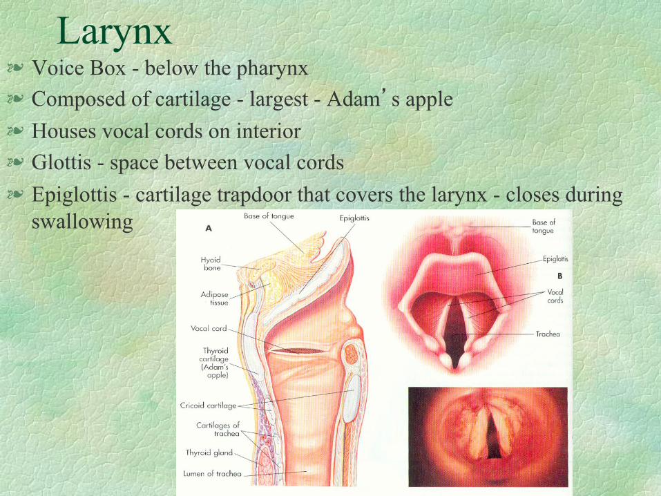
Larynx Voice Box - below the pharynx Composed of cartilage - largest - Adam’s apple Houses vocal cords on interior Glottis - space between vocal cords Epiglottis - cartilage trapdoor that covers the larynx - closes during
swallowing

Trachea
Windpipe - 11 cm long - made of nocollapsable material
Connects air to lungs - lined by cilliated epithelia - produces mucous

Cilia
Trap dust and foreign particles keep dust from reaching lungs sweep dust toward nostrils move only in one direction
cigarette smoke paralyzes cilia, which causes accumulations of mucus
( smokers’ cough)
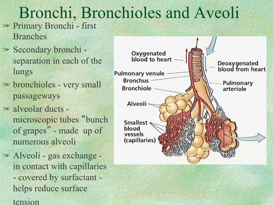
Bronchi, Bronchioles and Aveoli Primary Bronchi - first
Branches Secondary bronchi -
separation in each of the lungs
bronchioles - very small passageways
alveolar ducts - microscopic tubes “bunch of grapes” - made up of numerous alveoli
Alveoli - gas exchange - in contact with capillaries - covered by surfactant - helps reduce surface tension

Lungs and Pleura The right side of the lung
has 3 lobes and the left side has 2 lobes
Fits inside the ribs Pleura - cover outside of
lungs - thin, moist, slippery membrane; Pleurisy - inflammation of the pleura visceral pleura - covers
the lungs partietal pleura - lines
the thoracic cavity Pneumothorax - air in
interpleural space

Respiration
Exchange of gases - organs provide a space for gas and blood to come in contact with each other
pulmonary ventilation (breathing) - moves air in and out of lungs
exchange of air between lungs and blood - external
exchange of air between blood and cells - internal respiration (cellular)

Mechanisms
Inspiration - moves air into the lungs Expiration - moves air out of the lungs hiccup - spontaneous spasm of the diaphram

Inspiration Muscles of inspiration = diaphragm Flattening makes chest longer - air moves in phrenic nerve stimulate diaphragm to
contract

Expiration Normal expiration involves relaxation of the
diaphragm. During forceful expiration, accessory
expiration muscles are used (abdominal)


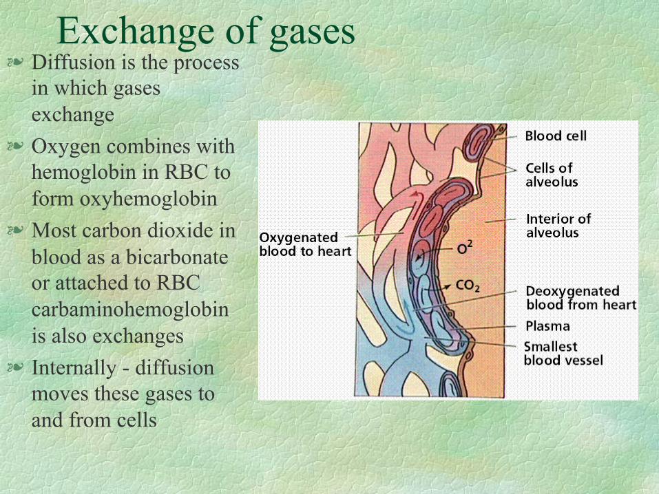
Exchange of gases Diffusion is the process
in which gases exchange
Oxygen combines with hemoglobin in RBC to form oxyhemoglobin
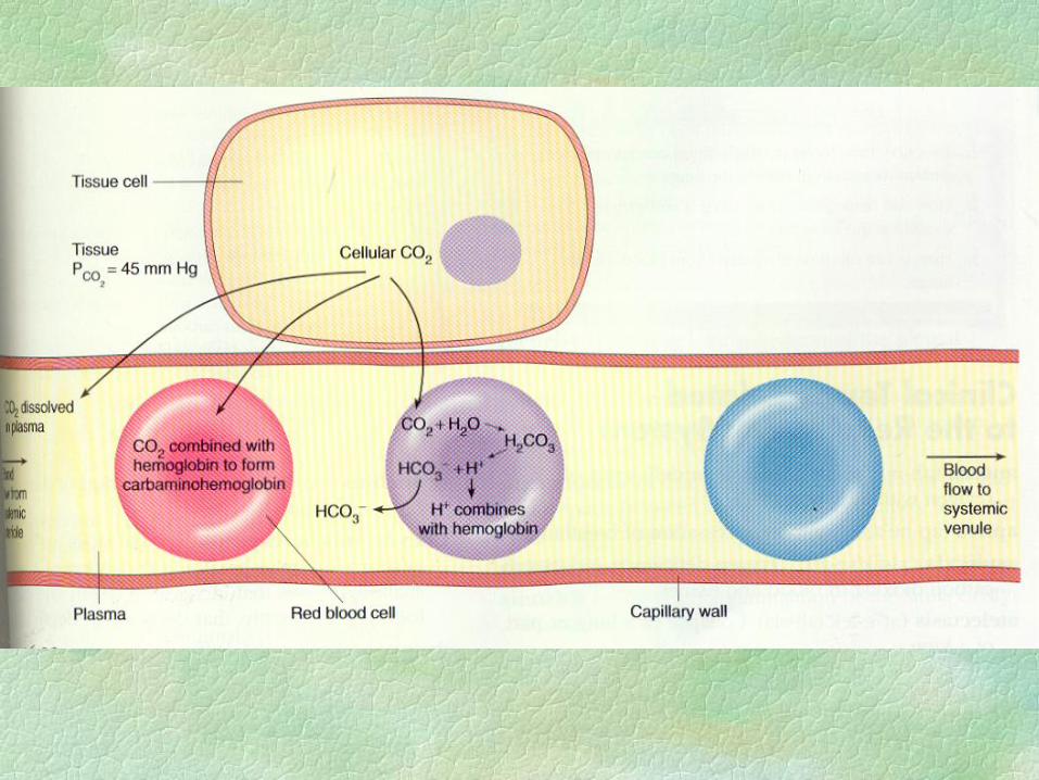
Most carbon dioxide in blood as a bicarbonate or attached to RBC carbaminohemoglobin is also exchanges
Internally - diffusion moves these gases to and from cells


Volume of air exchanged Spirometer - measure the amount of air used Air that comes and goes on a regular basis
(like the tides) is known as a tidal volume vital capacity - largest amount of air that
can be taken in expiratory reserve volume (erv)- amount of
air that can be forcefully exhaled inspiratory reserve volume (irv)- amount of
air that can be forcefully inhaled Residual Volume - amount of air left over
after forceful exhalation


Regulation of Respiration
Normal 12 to 18 breathes a minute - more when we exercise
respiratory control center - located in medulla and pons of the brain
receptors sense oxygen and carbon dioxide medulla has inspiratory control centers and
expiratory control centers

Cerebral Cortex Influence respiration by influencing
stimulating neurons Speed up/Slow down breathing rate

Receptors that Influence respiration
Chemoreceptors - located in corotid and aortic arteries sense O, CO, blood acidity
Pulmonary Stretch receptors - located through the pulmonary alveoli and airways - detects excess stretch