The Respiratory System - · PDF fileHuman Anatomy & Physiology ... •Palatine tonsils lie...
34
Human Anatomy & Physiology FIFTH EDITION Elaine N. Marieb PowerPoint ® Lecture Slide Presentation by Vince Austin Copyright © 2003 Pearson Education, Inc. publishing as Benjamin Cummings The Respiratory System Dr Nabil Khouri. MD, Ph.D
Transcript of The Respiratory System - · PDF fileHuman Anatomy & Physiology ... •Palatine tonsils lie...

Human Anatomy & Physiology FIFTH EDITION
Elaine N. Marieb
PowerPoint® Lecture Slide Presentation by Vince Austin
Copyright © 2003 Pearson Education, Inc. publishing as Benjamin Cummings
The Respiratory System
Dr Nabil Khouri. MD, Ph.D

Copyright © 2003 Pearson Education, Inc. publishing as Benjamin Cummings
Respiratory System
• Consists of a conducting zone and respiratory zone
• Conducting zone (Upper):
• Is part of the respiratory system lying outside of the thorax or above the sternal angle
• Provides tube–like conduction system for air
• Facilitate the air to reach the sites of gas exchange.
• Includes all other respiratory structures (e.g., nose, nasal cavity, pharynx, and the upper part of the trachea).
• Respiratory zone (Lower)
• The lung
• Site of gas exchange
• Consists of bronchioles, alveolar ducts, and alveoli
• Prime respiratory muscles – diaphragm and other inter-costal muscles that promote ventilation

Copyright © 2003 Pearson Education, Inc. publishing as Benjamin Cummings
Respiratory System
Copyright © 2003 Pearson Education, Inc. publishing as Benjamin Cummings
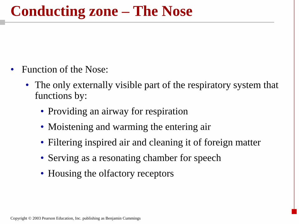
Conducting zone – The Nose
• Function of the Nose:
• The only externally visible part of the respiratory system that functions by:
• Providing an airway for respiration
• Moistening and warming the entering air
• Filtering inspired air and cleaning it of foreign matter
• Serving as a resonating chamber for speech
• Housing the olfactory receptors

Copyright © 2003 Pearson Education, Inc. publishing as Benjamin Cummings
Structure of the Nose
• The nose is divided into two regions
• The external nose, including the root, bridge, dorsum nasi, and apex
• The internal nasal cavity
• Philtrum – a shallow vertical groove inferior to the apex
• The external nares (nostrils) are bounded laterally by the alae
Copyright © 2003 Pearson Education, Inc. publishing as Benjamin Cummings
Copyright © 2003 Pearson Education, Inc. publishing as Benjamin Cummings
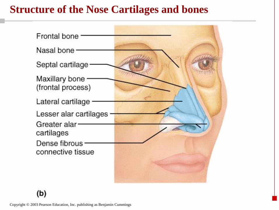
Structure of the Nose Cartilages and bones

Copyright © 2003 Pearson Education, Inc. publishing as Benjamin Cummings
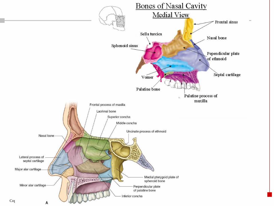
Nasal Cavity
Copyright © 2003 Pearson Education, Inc. publishing as Benjamin Cummings
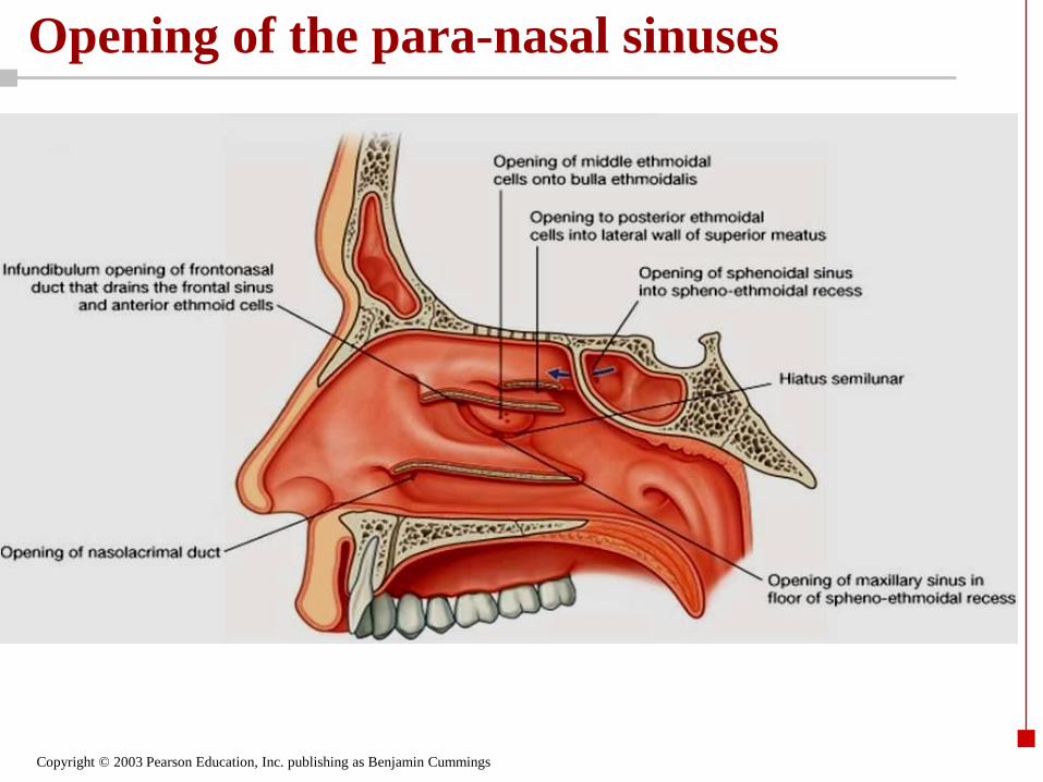
Opening of the para-nasal sinuses

Copyright © 2003 Pearson Education, Inc. publishing as Benjamin Cummings
The Pharynx
• Funnel-shaped tube of skeletal muscle that connects to the:
• Nasal cavity and mouth superiorly
• Larynx and esophagus inferiorly
• Extends from the base of the skull to the level of the sixth cervical vertebra
• It is divided into three regions:
• Nasopharynx
• Oropharynx
• Laryngopharynx

Copyright © 2003 Pearson Education, Inc. publishing as Benjamin Cummings
Nasopharynx
• Lies posterior to the nasal cavity, inferior to the sphenoid, and superior to the level of the soft palate
• Strictly an air passageway
• Lined with pseudo-stratified columnar epithelium
• The pharyngeal tonsil lies high on the posterior wall
• Pharyngotympanic (auditory) tubes open into the lateral walls

Copyright © 2003 Pearson Education, Inc. publishing as Benjamin Cummings

Copyright © 2003 Pearson Education, Inc. publishing as Benjamin Cummings
Oropharynx
• Extends inferiorly from the level of the soft palate to the epiglottis
• Opens to the oral cavity via an archway called the fauces
• Serves as a common passageway for food and air
• The epithelial lining is a protective epithelium:
• stratified Squamous epithelium
• Palatine tonsils lie in the lateral walls of the fauces
• Lingual tonsil covers the base of the tongue

Copyright © 2003 Pearson Education, Inc. publishing as Benjamin Cummings
Laryngopharynx
• Serves as a common passageway for food and air
• Lies posterior to the upright epiglottis
• Extends to the larynx, where the respiratory and digestive pathways diverge

Copyright © 2003 Pearson Education, Inc. publishing as Benjamin Cummings
The Larynx (Voice Box) • Attaches to the hyoid bone and opens into the laryngo-pharynx
superiorly
• Continue inferior with the trachea
• The three functions of the larynx are:
• To provide a patent airway
• To act as a switching mechanism to route air and food into the proper channels
• To function in voice
production

Copyright © 2003 Pearson Education, Inc. publishing as Benjamin Cummings
Framework of the Larynx
• Cartilages (hyaline cartelage) of the larynx are:
• Unpaired
• Shield-shaped antero-superior is the
– Thyroid cartilage with a midline laryngeal prominence (Adam’s apple)
• Signet ring–shaped anteroinferior
– Cricoid cartilage
• The Unpaired Epiglottis is made of elastic cartilage
• Covers the laryngeal inlet during swallowing
• Paired
• Three pairs of cartilages of small diameters:
– Arytenoid
– Cuneiform
– Corniculate

Copyright © 2003 Pearson Education, Inc. publishing as Benjamin Cummings
Framework of the Larynx

Copyright © 2003 Pearson Education, Inc. publishing as Benjamin Cummings

Copyright © 2003 Pearson Education, Inc. publishing as Benjamin Cummings
Lower Respiratory Tract
• Functions:
• Larynx: maintains an open airway, routes food and air appropriately, assists in sound production
• Trachea: transports air to and from lungs
• Bronchi: branch into lungs
• Lungs: transport air to alveoli for gas exchange

Copyright © 2003 Pearson Education, Inc. publishing as Benjamin Cummings
The Trachea • Flexible and mobile tube extending from the larynx into the
mediastinum
• Composed of three layers
• Mucosa:
• Pseudo-stratifief ciliated epithelium.
• Goblet cells
• Submucosa:
• Connective tissue deep to the mucosa
• Adventitia:
• Outermost layer made of C-shaped rings of hyaline cartilage.
• Covered with dense irregular CT

Copyright © 2003 Pearson Education, Inc. publishing as Benjamin Cummings
Trachea

Copyright © 2003 Pearson Education, Inc. publishing as Benjamin Cummings
Coverings of the Lungs: The Pleurae
• Thin, double-layered serosa
• Parietal pleura
• Covers the thoracic wall and superior face of the diaphragm
• Continues around heart and between lungs
• Visceral, or pulmonary, pleura
• Covers the external lung surface
• Divides the thoracic cavity into three chambers
• The central mediastinum
• Two lateral compartments, each containing a lung

Copyright © 2003 Pearson Education, Inc. publishing as Benjamin Cummings
Pleural cavity
Copyright © 2003 Pearson Education, Inc. publishing as Benjamin Cummings
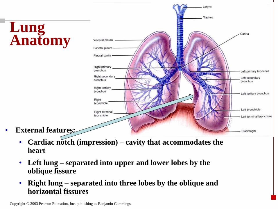
Lung Anatomy
• External features:
• Cardiac notch (impression) – cavity that accommodates the heart
• Left lung – separated into upper and lower lobes by the oblique fissure
• Right lung – separated into three lobes by the oblique and horizontal fissures

Copyright © 2003 Pearson Education, Inc. publishing as Benjamin Cummings
Lobes and Fissures of the Right Lung
Oblique fissure
Horizontal fissure

Copyright © 2003 Pearson Education, Inc. publishing as Benjamin Cummings
Left Lung medial surface Hillum of the lung
Copyright © 2003 Pearson Education, Inc. publishing as Benjamin Cummings
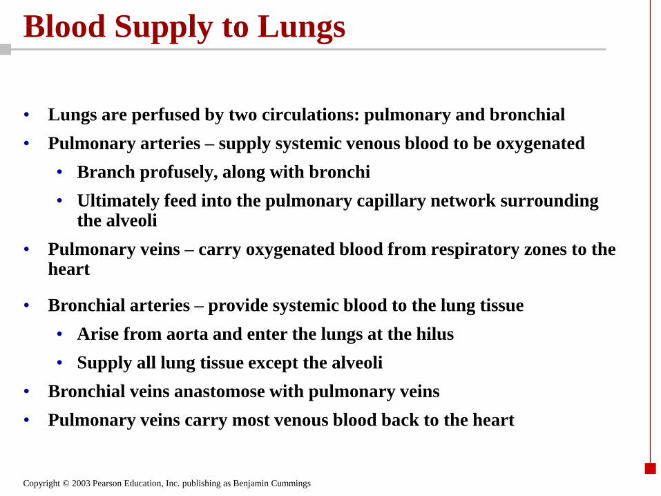
Blood Supply to Lungs
• Lungs are perfused by two circulations: pulmonary and bronchial
• Pulmonary arteries – supply systemic venous blood to be oxygenated
• Branch profusely, along with bronchi
• Ultimately feed into the pulmonary capillary network surrounding the alveoli
• Pulmonary veins – carry oxygenated blood from respiratory zones to the heart
• Bronchial arteries – provide systemic blood to the lung tissue
• Arise from aorta and enter the lungs at the hilus
• Supply all lung tissue except the alveoli
• Bronchial veins anastomose with pulmonary veins
• Pulmonary veins carry most venous blood back to the heart

Copyright © 2003 Pearson Education, Inc. publishing as Benjamin Cummings
Components of the Lower Respiratory Tract

Copyright © 2003 Pearson Education, Inc. publishing as Benjamin Cummings
Bronchial Tree
• Cartilage support structures change
• Epithelium types change
• Amount of smooth muscle increases
Wall layers are mucosa, submucosa and muscularis
• Trachea
• The lining (mucosa) is pseudostratified ciliated columnar
• "C" shaped hyaline cartilage rings keeps tracea open
• As conducting tubes become smaller, the names of the tubes are changing
• Bronchi
• Bronchioles

Copyright © 2003 Pearson Education, Inc. publishing as Benjamin Cummings
Primary Bronchi
• At the site of tracheal division “The carina”
• marks the end of the trachea
• Two in number:
• right and left bronchi
• Air reaching the bronchi is:
• Warm and cleansed of impurities
• Saturated with water vapor
• Bronchi (primary) are subdivide into secondary bronchi, each supplying one lobe of the lungs

Copyright © 2003 Pearson Education, Inc. publishing as Benjamin Cummings
Respiratory Zone
• Defined by the presence of alveoli.
• Begins as terminal bronchioles became Respiratory bronchioles
• Respiratory bronchioles contenue as Alveolar ducts, then to terminal clusters of alveolar sacs which is composed of alveoli
• Approximately 300 million alveoli:
• Gas exchange accure

Copyright © 2003 Pearson Education, Inc. publishing as Benjamin Cummings
Respiratory Zone

Copyright © 2003 Pearson Education, Inc. publishing as Benjamin Cummings
Blood supply : Gas Exchange Between the Blood and Alveoli

Copyright © 2003 Pearson Education, Inc. publishing as Benjamin Cummings
Respiratory Membrane: Alveolar walls:
• Are a single layer of type I epithelial cells
• Permit gas exchange by simple diffusion
• Type II cells secrete surfactant