The preventive effect of grape seeds ethanolic extract as ... · extract, a polyphenolic ......
18
Issues in Biological Sciences and Pharmaceutical Research Vol.4(4),pp.17-34, July 2016 Available online at http://www.journalissues.org/IBSPR/ http://dx.doi.org/10.15739/ibspr.16.004 Copyright © 2016 Author(s) retain the copyright of this article ISSN 2350-1588 Original Research Article The preventive effect of grape seeds ethanolic extract as antihyperglycemia against streptozotocin-induced diabetic rats Received 25 May, 2016 Revised 11 July, 2016 Accepted 14 July, 2016 Published 28 July, 2016 1* Nermien Z. Ahmed 2 Fouad A. Ahmed, 2 Moustasfa A. A., and 1 Dalia A. Hashim 1 National Organization for Drug Control & Research "NODCAR"; Department of Molecular Drug Evaluation, Giza, Egypt. 2 Faculty of Agriculture, Cairo University, Department of Biochemistry, Giza, Egypt. *Corresponding Author Email: [email protected] Tel.: (020) 01223740700 01069194681. This study aim to determine the protective effect of Grape seeds (Vitis vinifera) ethanolic extract against streptocotozin-induced diabetic rats. Sterptozotocin was used by (50mg/kg b.w. i.p.); Vitis vinifera seeds were used at (100 & 200 mg/kg b.w. oral) daily/ three weeks. The result indicated that Sterptozotocin increased the levels of serum glucose; uric acid; creatinine; ALP; ALT and AST, blood urea, and decreased the activities of SOD and GSH-Px, after treated with grape seeds by the high dose show a significantly decreased in levels of serum glucose; uric acid; creatinine, ALP; ALT and AST, urea. On the other hand, SOD and GSH-Px activities exerted a highly increase in diabetic rats given with the extract. Also, it can be speculated that Grape seed (Vitis vinifera) ethanolic extract has a hypoglycemic effect possibly related to the phytochemical compounds of Vitis vinifera plant seeds include Gallic acids, Catechin, Ρ-hydroxy bezoic acid, Vanillin, Ferulic acid, Ρ-Coumaric acid, Euganol , as well as, 5,7- dihydroxy 4-methoxy isoflavone which can optimize sugar uptake in the liver. These feats could induce insulin secretion and release from cells, as well as stimulates the tissue's insulin sensitivity leading to an increase of the tissues' glucose uptake, storage, and oxidation. Key words: DPPH, Antioxidant, Lysosomal enzymes, Grape seeds (Vitis vinifera) extracts, Sterptozotocin. INTRODUCTION Many herbs are good source of natural antioxidants and usually their consumption either fresh or dried in the diet. Therefore herbs are contributed to the daily antioxidant sources in-take (Justes and Knuthsen. 2001; Nevin and Vijayammal, 2005; Arabshahi-D et al., 2007). Phenolic compounds and flavonoids present in most fruits, vegetables and other products and hence it is expected that diseases caused by cholesterol oxidation such as cardiovascular, and cancers may be protected by eating fruits and vegetables owing to their contents of phenolic compounds, it has shown that vegetables and fruit protects against a variety of diseases, including cancer (Graham et al., 2000; Tiwari, 2001). Administration of antioxidants exerted a greatly reduces or eliminates the biochemical and pathological changes by lipid peroxidation, a large number of plants have been screened as a viable source of natural antioxidants including tocopherols, vitamin C, carotenoids and phenolic compounds which are responsible for maintenance of health and protection from coronary heart diseases and cancer (Castenmiller et al., 2002; Javanmardi et al., 2003). In recent years, there has been a growing interest in the use of grape seed extracts as a dietary antioxidant supplements (Santos-Buelgo and Scalbert, 2000; Jia et al., 2011). The skins and seeds of grapes are known to be rich sources of phenolic compounds, both flavonoids and non-flavonoids (Pinelo et al., 2006; Arnous and Meyer, 2008; Poudel et al., 2008). Grape seed extract (GSE), a well-known dietary supplement, contains
Transcript of The preventive effect of grape seeds ethanolic extract as ... · extract, a polyphenolic ......

Issues in Biological Sciences and Pharmaceutical Research Vol.4(4),pp.17-34, July 2016 Available online at http://www.journalissues.org/IBSPR/ http://dx.doi.org/10.15739/ibspr.16.004 Copyright © 2016 Author(s) retain the copyright of this article ISSN 2350-1588
Original Research Article
The preventive effect of grape seeds ethanolic extract as antihyperglycemia against streptozotocin-induced
diabetic rats
Received 25 May, 2016 Revised 11 July, 2016 Accepted 14 July, 2016 Published 28 July, 2016
1* Nermien Z. Ahmed 2Fouad A. Ahmed, 2
Moustasfa A. A., and
1 Dalia A. Hashim
1National Organization for Drug Control & Research "NODCAR"; Department of Molecular Drug
Evaluation, Giza, Egypt. 2Faculty of Agriculture, Cairo
University, Department of Biochemistry, Giza, Egypt.
*Corresponding Author Email:
[email protected] Tel.: (020) 01223740700 01069194681.
This study aim to determine the protective effect of Grape seeds (Vitis vinifera) ethanolic extract against streptocotozin-induced diabetic rats. Sterptozotocin was used by (50mg/kg b.w. i.p.); Vitis vinifera seeds were used at (100 & 200 mg/kg b.w. oral) daily/ three weeks. The result indicated that Sterptozotocin increased the levels of serum glucose; uric acid; creatinine; ALP; ALT and AST, blood urea, and decreased the activities of SOD and GSH-Px, after treated with grape seeds by the high dose show a significantly decreased in levels of serum glucose; uric acid; creatinine, ALP; ALT and AST, urea. On the other hand, SOD and GSH-Px activities exerted a highly increase in diabetic rats given with the extract. Also, it can be speculated that Grape seed (Vitis vinifera) ethanolic extract has a hypoglycemic effect possibly related to the phytochemical compounds of Vitis vinifera plant seeds include Gallic acids, Catechin, Ρ-hydroxy bezoic acid, Vanillin, Ferulic acid, Ρ-Coumaric acid, Euganol , as well as, 5,7- dihydroxy 4-methoxy isoflavone which can optimize sugar uptake in the liver. These feats could induce insulin secretion and release from cells, as well as stimulates the tissue's insulin sensitivity leading to an increase of the tissues' glucose uptake, storage, and oxidation. Key words: DPPH, Antioxidant, Lysosomal enzymes, Grape seeds (Vitis vinifera) extracts, Sterptozotocin.
INTRODUCTION Many herbs are good source of natural antioxidants and usually their consumption either fresh or dried in the diet. Therefore herbs are contributed to the daily antioxidant sources in-take (Justes and Knuthsen. 2001; Nevin and Vijayammal, 2005; Arabshahi-D et al., 2007). Phenolic compounds and flavonoids present in most fruits, vegetables and other products and hence it is expected that diseases caused by cholesterol oxidation such as cardiovascular, and cancers may be protected by eating fruits and vegetables owing to their contents of phenolic compounds, it has shown that vegetables and fruit protects against a variety of diseases, including cancer (Graham et al., 2000; Tiwari, 2001). Administration of antioxidants exerted a greatly reduces or eliminates the biochemical and
pathological changes by lipid peroxidation, a large number of plants have been screened as a viable source of natural antioxidants including tocopherols, vitamin C, carotenoids and phenolic compounds which are responsible for maintenance of health and protection from coronary heart diseases and cancer (Castenmiller et al., 2002; Javanmardi et al., 2003). In recent years, there has been a growing interest in the use of grape seed extracts as a dietary antioxidant supplements (Santos-Buelgo and Scalbert, 2000; Jia et al., 2011). The skins and seeds of grapes are known to be rich sources of phenolic compounds, both flavonoids and non-flavonoids (Pinelo et al., 2006; Arnous and Meyer, 2008; Poudel et al., 2008). Grape seed extract (GSE), a well-known dietary supplement, contains
Issues Biol. Sci. Pharma. Res. 18
Table 1. The family, Latin name, Arabic name, English name part used and source of the plant
important vitamins, minerals, and poly phenols including flavonoids, proanthocyanidins and procyanidins (Weber et al., 2007). It has recently become clear that GSE has shown various beneficial pharmacological effects such as its chemo-protective properties against reactive oxygen species (ROS) (Nandakumar et al., 2008) and oxidative stress as well as being anti-inflammatory (Terra et al., 2009), anti-cancer (Kaur et al., 2006), and anti-diabetic (Pinent et al., 2004). Grape seed extract (GSE) is widely used as nutritional supplement affording tremendous preventing and healing effects (Suwannaphet et al., 2010). GSE contains polyphenols, such as resveratrol, which is at the basis of the “French Paradox”, has a wide effect ranging as cardio protective (Charradi et al., 2011) or neuroprotective (Narita et al., 2011) mainly due to its antioxidant and anti inflammatory properties (Jia et al., 2011). Moreover, GSE also exerts antineoplastic effects by inducing cytotoxicity toward some cancer cells (Kaur et al., 2011) by cell cycle arrest and apoptosis (Dinicola et al., 2010).
Ghafoor et al. (2011) studied that Grape peel and seed are good sources of important bioactive components such as phenolics, anthocyanins and antioxidants, the phenolic content and antioxidant activity of grape seed (Vitis vinifera) powder extracted by in-vitro physiological procedure and chemical procedure were investigated by Hua et al. (2008). The antioxidant potential of the extract was assessed by employing different in-vitro assays such as DPPH, OH• radical scavenging capacity, and peroxidation inhibiting activity. The in-vitro physiological procedure yielded a higher phenolic content and antioxidant capacity than the chemical procedure.
Diabetes mellitus impairs glucose homeostasis causing neurological disorders due to perturbation in utilization of glucose. The mechanisms responsible for failure of glycaemic control in diabetes need to be thoroughly elucidated and hence the present study was initiated Ahmed (2009). Diabetes mellitus is a chronic metabolic disease which has significant effects on health, quality of life and also on health care system (Sharma and Kumar, 2011).
Landrault et al. (2003) demonstrated the active biological properties of polyphenols (P) extracted from the seeds of black grapes (Vitis vinifera) on glycaemia and pain perception (nociception) disorders in experimental diabetes mellitus. Chis et al. (2011) investigated the anti-hyperglycaemic and antioxidant effect of grape seed extract, a polyphenolic flavonoid, in normal and streptozotocin-induced diabetic Wister rats.
The aim of this study was to determine the protective effect of ethanolic extract of grape seeds (Vitis vinifera) as
antihyperglycemic on blood antioxidant; the liver and kidney functions enzymes on Streptozotocin-induced diabetic rats. Also, the histopathological studies were performed under the effect of plant extract in rat liver and pancreas. MATERIALS AND METHODS plants The families; Latin name; Arabic name; English name; part used Bedevian (1936), and source of plant samples are listed in Table (1). All plant samples were authenticated via plant herbarium of Orman Garden, Ministry of Agriculture. Preparation of ethanolic extracts All collected plant samples were cleaned, dried in Lypholyzer, and then grinded to fine powder before extraction. Such powdered samples were kept in dark glass bottles ready for further investigation Lee et al. (2002).
10 g plant powder + 100 ml ethanol 70%
Percolation over night
Wash twice each 300ml with ethanol 70% and centrifugation at
(5000g/10 min)
Filtration
Lypholyzation
Extract soluble material (Et OH 70%)
Chemicals All solvents used throughout the present work were obtained from different companies Rutin, DPPH˙ were
Latin Name Family English name Arabic name part used sources Vitis vinifera Vitaceae Black grape العنب االسود Seeds Market

purchased from Sigma Chemical Co., St. Louis, USA. Animal diet The basal diet, Salt mixture and vitamin mixture were formulated according to A.O.A.C (2000). Experimental Animals Thirty-two male albino rats (150-160g) Sprague - Dawley strain were obtained from Helwan breeding farm, Cairo, Egypt. The animals were housed in stainless steel cages with wire mesh bottoms in a room maintained at 25-30. The animals were adapted on free access of water and fed basal diet for week before the initiation of the experiments. All animals received humane care in compliance with the National Institutes of Health and the Research Ethics Committee Criteria for Care of Laboratory Animals at Cairo University. Preparation of diabetic rats Diabetic rats were induced in normal healthy male albino rats by subcutaneous injection of Streptozotocin (STZ) (50mg / kg b.w.) (i.p.) according to the method described by (Szkudelski, 2001). Experimental Design After periods of adaptation were performed using 32 rats which were divided into four groups, all groups were divided as follows: The first group (negative control) (8 rats) served as the non- diabetic rats were receiving saline only (vehicle) for three weeks daily. The second group (Positive control) (8 rats) diabetic rats injected i.p with streptozotocin solution in a single dose of (50 mg/kg b.w.) according to Szkudelski (2001) to elevate serum glucose and other blood analysis in order to compare with those of the Vitis vinifera seeds treated rats. The third group (Treated groups) (16 rats) was rendered diabetic by i.p. of streptozotocin (50 mg/kg b.w.) and it was then divided into two subgroups administered via intragastric tube at a dose of 100 and 200 mg/kg for 21 consecutive days after the induction of diabetes mellitus (Pinent et al., 2004; Chis et al., 2011):
The first subgroup (8 diabetic rats): streptozotocin-induced diabetic rats after overnight fast rat received (100 mg/kg b.w.) by oral administration of ethanolic extract of Vitis vinifera seed daily for three weeks.
The second subgroup (8 diabetic rats) streptozotocin-
Nermien et al. 19 induced diabetic rats after overnight fast rat received (200 mg/kg b.w.) by oral administration of ethanolic extract of Vitis vinifera seed daily for three weeks. Blood Sampling
At the end of experiment blood samples were collected from the orbital plexus by means of a fine capillary glass tubes in accordance with the method of Schermer (1967), each sample was placed in a dry clean centrifuge tube. The blood was centrifuged at 3000 r.p.m/15 min. to separate the sera and kept frozen until analysis. Serum glucose, blood urea, serum uric acid, serum creatinine, serum transaminases, serum alkaline phosphatase, glutathione peroxidase activity and superoxide dismutase were determined. Biochemical Analysis Determination of serum glucose Blood glucose was determined according to the procedure of Trinder (1969). The produced quinoneimine was colorimetrically determined at wavelength 505 nm; the glucose concentration was calculated using the following equation:
Serum glucose (mg/dl) = A sample / A standard X 100, whereas A = Absorbance at 505 nm Determination of serum uric acid Serum uric acid was determined according to the procedure of Barham and Trinder (1972). The produced quinoneimine was colourimetrically determined at wavelength 505 nm. The uric acid concentration was calculated by using the following equation:
Serum Uric acid (mg/dl) = A sample / A standard X 5, whereas A = Absorbance at 510 nm
Determination of serum creatinine Serum creatinine was determined according to the method described by Faulkner and King (1976). Excess of picric acid used for deproteinization, this technique avoids preparation of conventional tungestic acid protein-free filtrate or supernatant. Creatinine in a picric acid protein-free solution reacts with added alkali to form a reddish-brown complex. The absorbance of this compound at 520 nm is proportional to creatinine concentration. The creatinine concentration was calculated by using the following equation:
Serum creatinine (mg/dl) = A sample / A standard X 2.5, whereas A = Absorbance at 520 nm
Determination of serum transaminases Aspartate amino transferease (AST) or glutamate oxaloacetate transaminase (GOT) and alanine amino
Issues Biol. Sci. Pharma. Res. 20 transferease (ALT) or glutamate pyruvate transaminase (GPT) activities were measured in serum according to the method described by Reitman and Frankel (1957). The produced pyruvate or oxaloacetate reacted with 2,4 dinitrophenylhydrazine and the obtained color was colorimetricalily measured at 505 nm, AST and ALT activities were calculated using a standard curve. Determination of serum alkaline phosphatase (ALP) The activity of alkaline phosphatase in serum was determined according to the method of Kind and King (1954). The liberated phenol was measured in the presence of 4-aminophenazone and potassium ferricyanide, sodium arsenate was added to stop the enzymatic reaction. Activity of serum alkaline phosphastase (IU/L) = A serum
sample – A serum blank / A standard X 75, Whereas A = Absorbance at 510 nm. Determination of blood urea Blood urea was estimated by the enzymatic method of Patton and Couch (1977), the intensity of colour was read at 630 nm. The concentration of blood urea was calculated by using the following equation: Blood Urea (mg/dl) = A sample / A standard X 30, whereas A = Absorbance at 630 nm
Determination of glutathione peroxidase (GsPx) activity in whole blood. Glutathione peroxidase activity was determined in whole blood and liver homogenate according to Paglia and Valentine (1970) using randox kit (UK). The decrease in absorbance at 340 nm is measured. U/L of Haemolysate activity = 8412 * A 340 nm/min. Determination of superoxide dismutase activity Superoxide dismutase activity was determined in erythrocytes and liver homogenate according to Wolliams et al. (1983) using Randox laboratory kits. The colour intensity is read at wavelength 505 nm. The percentage of inhibition was plotted against log10 of the standard concentrations, in SOD units/ml. The percentage inhibition was used to obtain units of SOD from the standard curve. SOD U/ml of whole blood=SOD U/ml from std. curve * dil. Factor. Determination of phenolic compounds by high performance liquid chromatography (HPLC) Phenolic compounds of Vitis vinifera seeds were extracted according to the method outlined by Fernandez de Semon et al. (1990) and Dimitrios et al. (2000). Statistical analysis of the results All values are mean ± S. E. obtained from eight animals.
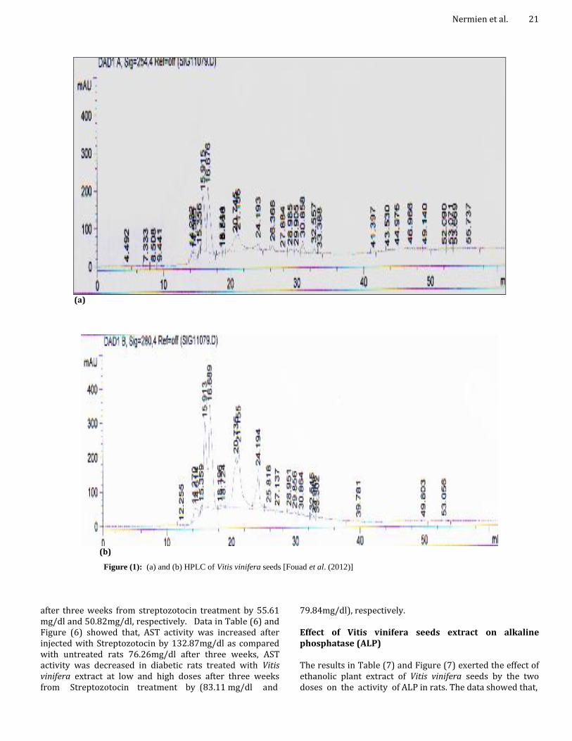
For statistical analysis, one way ANOVA (Steal and Torry, 1980) with Duncan's variance (SPSS 10) was used to compare groups. In all the cases a difference was considered significant when ρ was < 0.05. RESULTS HPLC Chromatogram of Vitis vinifera seeds Figure (1) shows the HPLC Chromatogram of Vitis vinifera seeds revealed the presence of the following compounds: Gallic acid represents (37.241 mg; highest), followed by 5, 7- dihydroxy 4-methoxy isoflavone (1.217 mg; lowest), these results are in agreement with those obtained by British Pharmacopoeia (2007) and European Pharmacopoeia (2010). Effect of Vitis vinifera seeds extract on serum glucose Data in Table (2) and Figure (2) represent the effect of Vitis vinifera seeds extract at a dose of (100 and 200 mg/kg b.w.) on rat serum glucose level. The data showed that, the extract possesses a significant hypoglycemic effect on serum glucose when compared with diabetic group (positive control). The best decrease of glucose concentration was recorded by the high dose of the extract. Effect of Vitis vinifera seeds extract on uric acid Data in Table (3) and Figure (3) indicated the levels of uric acid due to the treated of Vitis vinifera seeds extract by two doses. The results for the control rat, experiments showed no significant changes in the levels of uric acid during the entire experimental period. Serum uric acid was increased in diabetic rats from 7.93mg/dl (in normal rats) to 18.93mg/dl after three weeks of the experiment. Effect of Vitis vinifera seeds extract on serum creatinine Table (4) and Figure (4) illustrates the influence of Vitis vinifera treatment at various levels on rat serum creatinine content. The data indicated that there was slightly significant increase of serum creatinine levels due to treatment with Vitis vinifera extracts, addition of Vitis vinifera seeds extract significant decreased serum creatinine when compared with diabetic group (positive control). Effects of Vitis vinifera seeds extract on serum transaminases "ALT& AST" Data in Table (5) and Figure (5) showed that, ALT activity was increased after injected with Streptozotocin by (76.52 mg/dl) as compared with control rats (50.11mg/dl) after 3rd week, ALT activity was decreased in diabetic rats treated with Vitis vinifera extract at low and high doses
Nermien et al. 21
(a)
(b)
Figure (1): (a) and (b) HPLC of Vitis vinifera seeds [Fouad et al. (2012)]
after three weeks from streptozotocin treatment by 55.61 mg/dl and 50.82mg/dl, respectively. Data in Table (6) and Figure (6) showed that, AST activity was increased after injected with Streptozotocin by 132.87mg/dl as compared with untreated rats 76.26mg/dl after three weeks, AST activity was decreased in diabetic rats treated with Vitis vinifera extract at low and high doses after three weeks from Streptozotocin treatment by (83.11 mg/dl and
79.84mg/dl), respectively. Effect of Vitis vinifera seeds extract on alkaline phosphatase (ALP) The results in Table (7) and Figure (7) exerted the effect of ethanolic plant extract of Vitis vinifera seeds by the two doses on the activity of ALP in rats. The data showed that,

Issues Biol. Sci. Pharma. Res. 22
Table (2): Effect of Vitis vinifera seeds extract on serum glucose of normal and diabetic rats
Treatments
1st week 2nd week 3rd week mg/dl mg/dl mg/dl
Negative control (saline) 76.14d ± 3.61 -----------
75.93d ± 2.95 ------------
79.83d ± 2.47 ----------
Streptozotocin (STZ) (50mg / kg b.w.)
% Change
196.14a ± 6.76
157.60%
231.76 a ± 5.96
205.22%
272.18 a ± 6.32
240.94 % Streptozotocin (STZ)
+ 1ml/100gm b.w. of Vitis vinifera
(Subgroup 1) % Change
184a ± 6.16
141.66%
163.18b ± 6.01
114.90%
154.26b ± 5.31
93.23 %
Streptozotocin (STZ) +
2ml/100gm b.w. of Vitis vinifera (Subgroup 2)
% Change
176.51a ± 5.62
131.82%
149.46 c ± 5.31
96.83 %
136.14c ± 4.67
70.53%
a,b,c,d Values of 8 rats means ± S.E., % change is relative to normal control group Number in the same column followed by the same letters are not significant at P<0.01
0
50
100
150
200
250
300
Negative control Diabetic
group(positive
cntrol)
Subgroup 1 Subgroup 2
Treatment
Ser
um g
luco
se m
g/dl
week 1
week 2
week 3
Figure (2): Effect of Vitis vinifera seeds extract on Serum glucose
Table 3. Effect of Vitis vinifera seeds extract on uric acid of normal and diabetic rat
Treatments
1st week 2nd week 3rd week mg/dl mg/dl mg/dl
Negative control (saline)
7.93d ±0.43 ----------
8.13d ±0.42 ---------
8.08d ± 0.38 --------------
Streptozotocin (STZ) (50mg / kg b.w.)
% Change
13.88a ± 0.49
75.03%
15.06a ±0.81
85.23%
18.93a ± 0.63
134.2%
Streptozotocin (STZ) +
1ml/100gm b.w. of Vitis vinifera (Subgroup 1)
% Change
13.06a ± 0.26
64.69%
12.01c ± 0.19
47.72%
11.22c ± 0.21
38.86%
Streptozotocin (STZ) +
2ml/100gm b.w. of Vitis vinifera (Subgroup 2)
% Change
12.87a ± 0.87
62.29%
11.53c ± 0.19
41.82%
10.99 c ± 0.16
36.01%
a,b,c,d Values of 8 rats means ± S.E., % change is relative to normal control group Number in the same column followed by the same letters are not significant at P<0.01

Nermien et al. 23
0
2
4
6
8
10
12
14
16
18
20
Negative control Diabetic
group(positive cntrol)
Subgroup 1 Subgroup 2
Treatment
uri
c a
cid
mg
/dl
week 1
week 2
week 3
Figure 3: Effect of Vitis vnifera seeds extract on uric acid
Table 4. Effect of Vitis vinifera seeds extract on serum creatinine of normal and diabetic rats
Treatments
1st week 2nd week 3rd week mg/dl mg/dl mg/dl
Negative control (saline)
1.51a ± 0.01 -------------
1.49a ± 0.02
----------- 1.53a ± 0.01
------------ Streptozotocin (STZ)
(50mg / kg b.w.) % Change
2.01d ± 0.02
33.11%
2.47d ± 0.011
65.77%
2.69 d ± 0.011
75.81% Streptozotocin (STZ)
+ 1ml/100gm b.w. of Vitis vinifera
(Subgroup 1) % Change
2.39d ± 0.03
58.27%
2.08 c ± 0.02
39.59%
1.94 c ± 0.05
26.79% Streptozotocin (STZ)
+ 2ml/100gm b.w. of Vitis vinifera
(Subgroup 2) % Change
2.31d ± 0.01
52.98%
1.89 c ± 0.02
26.84%
1.76 b ± 0.04
15.03%
a,b,c,d Values of 8 rats means ± S.E., % change is relative to normal control group Number in the same column followed by the same letters are not significant at P<0.01
0
10
20
30
40
50
60
70
80
90
100
Negative control Diabetic
group(positive
control)
Subgroup 1 Subgroup 2
Treatment
AL
T m
g/d
l
week 1
week 2
week 3
Figure 5: Effect of Vitis vnifera seeds extract on serum ALT

Issues Biol. Sci. Pharma. Res. 24
Table 5. Effect of Vitis vnifera seeds extract on serum transaminases "ALT” of normal and diabetic rats
Treatments
1st week 2nd week 3rd week mg/dl mg/dl mg/dl
Negative control (saline)
50.11a ± 3.01 -----------
47.39a ± 2.73 ------------
46.18a ± 2.86 ------------
Streptozotocin (STZ) (50mg / kg b.w.)
% Change
76.52d ±2.93
52.70%
58.31d± 3.81
80.01%
93.82d ± 4.67
103.16% Streptozotocin (STZ)
+ 1ml/100gm b.w. of Vitis vinifera
(Subgroup 1) % Change
72.36 b±3.88
44.40%
63.84b ± 3.01
34.71%
55.61b ± 3.26
20.42% Streptozotocin (STZ)
+ 2ml/100gm b.w. of Vitis vinifera
(Subgroup 2) % Change
64.11b ±3.01
27.93%
57.14b ± 2.46
20.57%
50.82a ± 2.11
10.04%
a,b,c,d Values of 8 rats means ± S.E., % change is relative to normal control group Number in the same column followed by the same letters are not significant at P<0.01
Table 6. Effect of Vitis vinifera seeds extract on serum transaminases" AST of normal and diabetic rats
Treatments
1st week 2nd week 3rd week mg/dl mg/dl mg/dl
Negative control (saline) 78.11a ± 4.31 ----------
75.86a ± 3.26 ---------
76.26 a ± 3.01 ----------
Streptozotocin (STZ) (50mg / kg b.w.)
% Change
93.62d ± 5.83
19.85 %
108.87d ± 6.21
34.51%
132.87 d ±7.11
74.23 % Streptozotocin (STZ)
+ 1ml/100gm b.w. of Vitis vinifera
(Subgroup 1) % Change
98.14c ± 4.61
25.64 %
88.86c ± 4.32
17.13 %
83.11c ± 3.91
8.98 % Streptozotocin (STZ)
+ 2ml/100gm b.w. of Vitis vinifera
(Subgroup 2) % Change
86.36c ± 5.11
10.56 %
82.61c ± 4.92
8.89 %
79.84b ± 5.01
4.69%
a,b,c,d Values of 8 rats means ± S.E., % change is relative to normal control group Number in the same column followed by the same letters are not significant at P<0.01
0
20
40
60
80
100
120
140
Negative control Diabetic
group(positive
control)
Subgroup 1 Subgroup 2
Treatment
AS
T m
g/d
l
week 1
week 2
week 3
Figure 6: Effect of Vitis vinifera seeds extract on serum AST
Nermien et al. 25
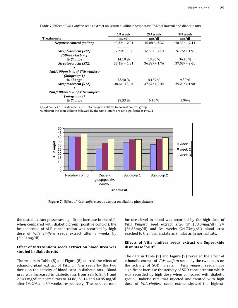
Table 7. Effect of Vitis vnifera seeds extract on serum alkaline phosphatase "ALP of normal and diabetic rats
Treatments
1st week 2nd week 3rd week mg/dl mg/dl mg/dl
Negative control (saline)
43.32a ± 2.01 ------------
40.08 a ±2.32 ------------
40.83 a ± 2.14 ------------
Streptozotocin (STZ) (50mg / kg b.w.)
% Change
37.21d ± 1.83
14.10 %
32.36 d ± 2.01
19.26 %
26.76d ± 1.91
34.45 % Streptozotocin (STZ)
+ 1ml/100gm b.w. of Vitis vinifera
(Subgroup 1) % Change
33.18c ± 1.81
23.40 %
36.82b ± 1.76
8.139 %
37.03b ± 2.61
9.30 % Streptozotocin (STZ)
+ 2ml/100gm b.w. of Vitis vinifera
(Subgroup 2) % Change
30.61c ±2.14
29.33 %
37.62b ± 2.44
6.13 %
39.21a ± 1.98
3.96%
a,b,c,d Values of 8 rats means ± S % change is relative to normal control group Number in the same column followed by the same letters are not significant at P<0.01
0
5
10
15
20
25
30
35
40
45
50
Negative control Diabetic
group(positive
control)
Subgroup 1 Subgroup 2
Treatment
AL
P m
g/d
l
week 1
week 2
week 3
Figure 7: Effect of Vitis vinifera seeds extract on alkaline phosphatase
the tested extract possesses significant increase in the ALP, when compared with diabetic group (positive control), the best increase of ALP concentration was recorded by high dose of Vitis vinifera seeds extract after 3 weeks by (39.21mg/dl). Effect of Vitis vinifera seeds extract on blood urea was studied in diabetic rats The results in Table (8) and Figure (8) exerted the effect of ethanolic plant extract of Vitis vinifera seeds by the two doses on the activity of blood urea in diabetic rats. Blood urea was increased in diabetic rats from 22.36; 20.81 and 21.43 mg/dl in normal rats to 34.86; 38.14 and 40.85 mg/dl after 1st; 2nd, and 3rd weeks, respectively. The best decrease
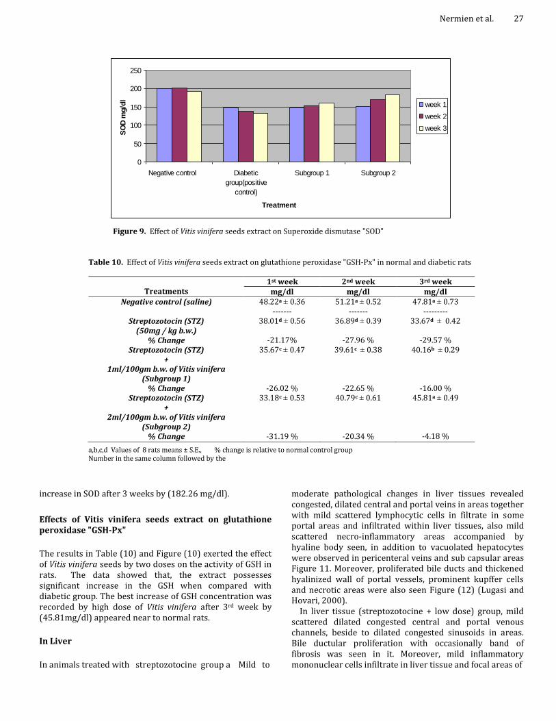
for urea level in blood was recorded by the high dose of Vitis Vinifera seed extract after 1st (30.04mg/dl), 2nd (26.85mg/dl) and 3rd weeks (24.73mg/dl) blood urea reached to the normal state as similar as in normal rats. Effects of Vitis vinifera seeds extract on Superoxide dismutase "SOD" The data in Table (9) and Figure (9) revealed the effect of ethanolic extract of Vitis vinifera seeds by the two doses on the activity of SOD in rats. Vitis vinifera seeds have significant increase the activity of SOD concentration which was recorded by high dose when compared with diabetic group. Diabetic rats that injected and treated with high dose of Vitis vinifera seeds extract showed the highest

Issues Biol. Sci. Pharma. Res. 26
Table 8. Effect of Vitis vinifera seeds extract on blood urea of normal and diabetic rats
Treatments
1st week 2nd week 3rd week mg/dl mg/dl mg/dl
Negative control (saline)
22.36 d ± 0.73
------------------ 20.81d ± 0.59
----------------- 21.43 d ± 0.92 ------------------
Streptozotocin (STZ) (50mg / kg b.w.)
% Change
34.86 a ± 1.36 55.9%
38.14a ± 1.87
83.27%
40.85 a ± 1.52
90.62 % Streptozotocin (STZ)
+ 1ml/100gm b.w. of Vitis vinifera
(Subgroup 1) % Change
32.86a ± 2.14
46.9%
29.13 b ± 1.93
39.98 %
27.84 c ± 1.68
29.91 % Streptozotocin (STZ)
+ 2ml/100gm b.w. of Vitis vinifera
(Subgroup 2) % Change
30.04 a ± 2.01
34.34%
26.85b ± 1.36
29.02 %
24.73 c ± 1.05
15.39%
a,b,c,d Values of 8 rats means ± S.E., % change is relative to normal control group Number in the same column followed by the same letters are not significant at P<0.01
0
5
10
15
20
25
30
35
40
45
Negative control Diabetic
group(positive
cntrol)
Subgroup 1 Subgroup 2
Treatments
Blo
od
ure
a m
g/d
l
week 1
week 2
week 3
Figure 8: Effect of Vitis vinifera seeds extract on blood urea
Table 9. Effect of Vitis vinifera seeds extract on Superoxide dismutase "SOD" in normal and diabetic rats
Treatments 1st week 2nd week 3rd week
mg/dl mg/dl mg/dl Negative control (saline)
198.36a ± 3.62
--------- 202.18 a ± 4.11
---------- 192.88 a ± 5.01
------------- Streptozotocin (STZ)
(50mg / kg b.w.) % Change
146.18d ± 3.16
-26.30 %
138.11d ± 2.99
-31.68 %
132.16d ± 2.67
-31.48 % Streptozotocin (STZ)
+ 1ml/100gm b.w. of Vitis vinifera
(Subgroup 1) % Change
147.06 d ± 2.11
-25.86 %
152.66 d ± 2.61
-24.49 %
160.81 c ± 3.11
-16.62 % Streptozotocin (STZ)
+ 2ml/100gm b.w. of Vitis vinifera
(Subgroup 2) % Change
150.81 d ± 2.62
-23.97 %
169.76 c ± 3.61
-16.03 %
182.26 b ±2.85
-5.50 %
a,b,c,d Values of 8 rats means ± S.E., % change is relative to normal control group Number in the same column followed by the same letters are not significant at P<0.01
Nermien et al. 27
0
50
100
150
200
250
Negative control Diabetic
group(positive
control)
Subgroup 1 Subgroup 2
Treatment
SO
D m
g/d
l
week 1
week 2
week 3
Figure 9. Effect of Vitis vinifera seeds extract on Superoxide dismutase "SOD"
Table 10. Effect of Vitis vinifera seeds extract on glutathione peroxidase "GSH-Px" in normal and diabetic rats
Treatments
1st week 2nd week 3rd week mg/dl mg/dl mg/dl
Negative control (saline)
48.22a ± 0.36 -------
51.21a ± 0.52 -------
47.81a ± 0.73 ---------
Streptozotocin (STZ) (50mg / kg b.w.)
% Change
38.01d ± 0.56
-21.17%
36.89d ± 0.39
-27.96 %
33.67d ± 0.42
-29.57 % Streptozotocin (STZ)
+ 1ml/100gm b.w. of Vitis vinifera
(Subgroup 1) % Change
35.67c ± 0.47
-26.02 %
39.61c ± 0.38
-22.65 %
40.16b ± 0.29
-16.00 % Streptozotocin (STZ)
+ 2ml/100gm b.w. of Vitis vinifera
(Subgroup 2) % Change
33.18c ± 0.53
-31.19 %
40.79c ± 0.61
-20.34 %
45.81a ± 0.49
-4.18 %
a,b,c,d Values of 8 rats means ± S.E., % change is relative to normal control group Number in the same column followed by the
increase in SOD after 3 weeks by (182.26 mg/dl).
Effects of Vitis vinifera seeds extract on glutathione peroxidase "GSH-Px"
The results in Table (10) and Figure (10) exerted the effect of Vitis vinifera seeds by two doses on the activity of GSH in rats. The data showed that, the extract possesses significant increase in the GSH when compared with diabetic group. The best increase of GSH concentration was recorded by high dose of Vitis vinifera after 3rd week by (45.81mg/dl) appeared near to normal rats.
In Liver
In animals treated with streptozotocine group a Mild to
moderate pathological changes in liver tissues revealed congested, dilated central and portal veins in areas together with mild scattered lymphocytic cells in filtrate in some portal areas and infiltrated within liver tissues, also mild scattered necro-inflammatory areas accompanied by hyaline body seen, in addition to vacuolated hepatocytes were observed in pericenteral veins and sub capsular areas Figure 11. Moreover, proliferated bile ducts and thickened hyalinized wall of portal vessels, prominent kupffer cells and necrotic areas were also seen Figure (12) (Lugasi and Hovari, 2000).
In liver tissue (streptozotocine + low dose) group, mild scattered dilated congested central and portal venous channels, beside to dilated congested sinusoids in areas. Bile ductular proliferation with occasionally band of fibrosis was seen in it. Moreover, mild inflammatory mononuclear cells infiltrate in liver tissue and focal areas of

Issues Biol. Sci. Pharma. Res. 28
0
10
20
30
40
50
60
Negative control Diabetic
group(positive
control)
Subgroup 1 Subgroup 2
Treatment
GS
H-P
x m
g/d
l
week 1
week 2
week 3
Figure 10: Effect of Vitis vinifera seeds extract on glutathione peroxidase "GSH-Px"Histopathological examination
Figure 11: Control liver section showed central vein with radiating cords of liver cells, liver cells with had vesicular nuclei and granular cytoplasm. Blood sinusoids were present between the cords of liver cells (H&E 150).
degeneration (hydrolytic changes). Focal area of vascular degenerative changes was noticed. Individual apoptic cells were seen Figure (13).
In liver tissue (streptozotocine + high dose) group showed moderate to marked vascular degenerative changes of hepatocytes was noticed in pericenteral zone of lobules and sub capsular area, mild lymphocytic aggregates seen in some portal veins accompanied by mild scattered dilated congested central and portal veins accompanied by mild scattered diluted congested central and portal veins
with hyalinzed wall and portal veins seen in areas, dilated congested sinusoid were seen in some areas Figure (14).
In Pancreatic
In animal treated with Streptozotocine showed moderate pathological changes in form of reduction in number, size of pancreatic islets, this were accompanied with necrotic changes of pancreatic islets (pyknosis , karyorrhexis and

Figure 12: A photomicrograph of section of treated rat streptozotocin showing dilated congested blood vessels (arrow) and few band of fibrous tissue (arrow head) (H&E 150)
Figure 13: A photomicrograph of section of rat treated with Streptozotocin (STZ) + Low dose showing congestion and dilation of blood Sinusoids (arrow) (H&E 150)
karyolysis) and vascular degeneration in the cells of islets langerhans (group C) Figures (19, 20 and 21). In addition to, dilated congested blood vessels. On the other hand, the pathological changes detected moderate improvement in pancreases of treated animals with (Streptozotocin (STZ) + High dose) (group A) Figure (17), the changes showed reduction in size in pancreatic islets accompanied by vascular degenerative changes in some cells of islets, Langerhan and nuclear changes (pyknotic nuclei). Besides to, moderate to marked dilated congested vascular channels, together with mild fibrous tissue around them. While in animals treated with (Streptozotocin (STZ) + Low
Nermien et al. 29
Figure 14: A photomicrograph of section of treated with Streptozotocin (STZ) + High dose revealed dilated congested blood vessels (arrow)(H&E 150
Figure 15: Control pancreas section showed normal glandular acinus (exocrine portion) and langerhans islet (endocrine portion).
dose)(group B) Figure (18), the improvement were mild revealed vascular degenerated in moderate cells of langerhan and nuclear changes (pyknotic nuclei), moreover, dilated congested blood vessels were seen.
DISCUSSION
Streptozotocin (STZ) is synthesized by Streptomycetes achromogenes and is used to induce both insulin-dependent and non insulin-dependent diabetes mellitus, injection STZ induced toxic events in β-cells rat pancreas was according to (szkudelski, 2001). Diabetic rats injected by Vitis vinifera

Issues Biol. Sci. Pharma. Res. 30
Figure 16: Section of pancreatic tissue of control rat showing normal pancreatic acini and normal islets langerhans (arrow) (H&E 600).
Figure 17: Section of pancreatic tissue in treated rat group (A) showed some islets cells with vacuolated cytoplasm (arrow) and pyknotic nucleoli (arrow head)(H&E 600).
seeds extract of high dose which had the highest decrease in serum glucose at the 3rd week due to the presences of procyanidins in GSE exhibit insulin by stimulating glucose uptake in insulin sensitive cell lines and decrease hyperglycemia in streptozotocin (STZ)-diabetic rats (Pinent et al., 2004).
A recent study from our laboratory has demonstrated the inhibitory activity of Grape seeds extracts (GSE) against intestinal α-glucosidase and pancreatic α-amylase, resulting in delayed carbohydrate digestion of absorbable
Figure 18: Section of pancreatic tissue in treated rat group (B) showing reduction in size, vacuolated cytoplasm (arrow head) (H&E 1500).
Figure 19: section of pancreatic tissue in treated rat group (c) showing reduction in size of islets langerhans (arrow) (H&E 600)
monosaccharide (Adisa kwattana et al., 2010), the hypoglycemic effects may be exerted through the inhibition of glucose absorption, increase sensitivity of receptors to insulin stimulation of β-cells of pancreas to secret insulin or stimulation of peripheral tissues uptake of glucose. Uric acid is the main end-product of nucleic acid and purine catabolism in human liver (Baron, 1987), there were moderate changes in levels of uric acid during the whole experiment due to treatments with Vitis vinifera extracts, our results are in agreement with (Nermien,

Figure 19: section of pancreatic tissue in treated rat group (c) showing reduction in size of islets langerhans (arrow) (H&E 600)
Figure 20: Section of pancreatic tissue in treated rat group (c) showing the islets cells with vacuolated cytoplasm (arrow), pyknotic nuclei (arrow head) and necrotic cells (without nucleus) (H&E 1500)
2003); Consequently, the determination of creatinine level in serum can be considered as a good index for renal impairment rather than serum urea Baron, 1987), the best decrease of serum creatinine concentration was recorded by the high concentration of Vitis vinifera seeds extract, our findings are in agreement with (Nermien, 2003); The liver enzymes 'AST and ALT ' are concerned with intra cellular metabolism, they are released from the liver when cells become necrotic as in viral or toxic hepatic or cirrhosis(Price and Steven, 2000), this means that Vitis
Nermien et al. 31
Figure 21: A photomicrograph of section of rat group (c) showing necrotic area (necro-inflammatory area) (arrow), hyaline body formation (douple-arrow) and pyknotic nuclei (arrow head) (H&E 600).
Vitis vinifera extract has reducing effect on ALT and AST activities in serum depend on Vitis vinifera concentration, our results are in agreement with those of (Nermien, 2003 and Mansouri et al., 2014). ALP is secreted into the serum at high levels in the presence of liver cell damage (Price and Steven, 2000) the results appear to be near to the normal rats, our results are in agreement with (Mansouri et al., 2014). Urea is the major end product of nitrogen catabolism in humans; urea is synthesized in the liver, released to the blood and cleared (excreted) by the kidney. The changes in serum urea level are due to alteration of renal function (diseased kidney) (Baron, 1987), these results are confirmed with those obtained by Jassim et al. (2010) who stated that, the tested extract possesses significant lowering effect on blood urea. SOD is the first enzyme involved in the antioxidant defense by lowering the steady state O 2ˉ, it̕ s a member of a mutually supportive team of defense against reactive oxygen species (ROS) (Nevin and Vijayammal, 2005) and it is appeared near to normal rats, these results are in agreement with those of (Mansouri et al., 2014). SUMMARY The hypo glycemic effect of Vitis vinifera seeds extract on rats Diabetic was induced in normal healthy male albino rats by subcutaneous injection of Streptozotocin (STZ) (50mg / kg b.w.) not receiving any treatment and other received treatment from Vitis vinifera seeds extract by oral intubation daily for three weeks.

Issues Biol. Sci. Pharma. Res. 32 Effect of Vitis vinifera seeds extract on serum: Glucose; Uric acid; Creatinine; transaminases "ALT&AST" and ALP, blood urea; Superoxide dismutase "SOD", and glutathione peroxidase "GSH-Px"
The effect of Vitis vinifera seed extract by two doses on rat serum glucose level. The best decrease of glucose concentration was recorded at high dose of Vitis vinifera seeds extract after 2 weeks.
Vitis vinifera seeds extract significant decreased kidney functions: Uric acid; the best decrease of uric acid concentration was recorded at the high dose of Vitis vinifera seeds extract after 2 weeks. Vitis vinifera seeds extract significant decreased serum creatinine and the best decrease of serum creatinine concentration was recorded at high dose of Vitis vinifera seeds extract after 2 weeks. Vitis vinifera decreased blood urea at high dose of Vitis vinifera seeds extract seeds after 2 weeks.
Treatment with Vitis vinifera seeds extract by two concentrations revealed an inhibitory effect for liver functions: ALT and AST, the best inhibitory of ALT and AST activity was recorded at high dose of Vitis vinifera seeds extract after 2 weeks. Vitis vinifera seeds extract significant increased inhibitory alkaline phosphatase activity and the best increase inhibitory of alkaline phosphatase activity was recorded at high dose of Vitis vinifera seeds extract after 2 weeks.
Vitis vinifera seeds extract has significantly effects on antioxidant enzymes as: Increased the activity of SOD and was recorded at the high dose of extract when compared with diabetic group. Also, increased glutathione peroxidase activity, the best increase of glutathione peroxidase activity was recorded by high dose of Vitis vinifera seeds extract after 2 weeks.
Histopatholoical examination
The histopathology of liver sections in Streptozotocin (STZ) treated group exerted an abnormal lobular pattern and marked loss of lobular architecture which shows diffuse moderate to marked affection, hepatic necrosis. The hepatic cells show variable degree of vascular degenerative changes and portal area fibrosis between liver tissues. Also, the liver section of Streptozotocin (STZ)-induced hepatotoxicity showed variable sized of micro vacuolation with cytoplasm of hepatocytes. On the other hand, after treatment with plant extracts by different concentration revealed an amelioration effect on different liver section showing mild pathological lesions "diffuse monocyte inflammatory cell aggregates and dilated congested venous channel fibrosis areas".
This improvement depend on the effect of active ingredients of poly phenolic compounds and the flavonoids which have the antioxidant activity in these plant extracts which could be inhibited. The cellular reactive oxygen species generation, this effect was dose-dependent. These antioxidant effects of the plant extracts might be useful for
the prevention of the inflammatory process as indicated by many references in this thesis. Conclusion It was concluded that Grape seeds (Vitis vinifera) ethanolic extract enhanced the antioxidant defense against reactive oxygen species produced under hyperglycemic conditions, hence protecting the liver cells and also, an ameliorated effect on diabetic management and it is recommended as a dietary supplement and even after cancer protocol is stopped. Financial support This work was all done at National Organization for Drug Control and Research (NODCAR), but HPLC for the plant extract was done at Agricultural Research center at Giza, Egypt. Competing interests The authors declare that they have no competing interests REFERENCES Adisakwattana S, Jiphimai P, Prutanopajai P, Chanathong B,
Sapwarobol S, Ariyapitipan T (2010). Evaluation of α-glucosidase, α- amylase and protein glycation inhibitory activities of edible plants. Int. J. Food. Sci. Nutr., 61:295-305.
Ahmed N (2009). Alloxan diabetes-induced oxidative stress and impairment of oxidative defense system in rat brain: neuroprotective effects of cichorium intybus . Int. J. Diab. Metabol., 17:105-109.
AOAC (2000). Official Methods of Analysis of the Association of official Agricultural chemists, 17 th ed, Published by A.O.A.C.
Arabshahi-DS, Devi DV, Urooj A (2007). Evaluation of antioxidant activity of some plant extracts and their heat, pH, and storage stability. Food Chem., 100:1100-1105.
Arnous A, Meyer AS (2008). Comparison of methods for compositional characterization of grape (Vitis vinifera L.) and apple (Malus domestica) skins. Food Bioprod. Proces., 86(7): 9–86.
Barham D, Trinder P (1972). A colorimetric method for the determination of serum uric acid. Analyst., 97: 142.
Baron DN (1987). A short textbook of chemical pathology, 4th edition- English Language Book Society/Hodder and Stoughton Ltd, Mill Road, Dunton Green, Sevenoaks, Kent, Great Britain, pp. 188–228.
Bedevian AK (1936). Illustrated pyglottic dictionary of plant names (W. Lawrence Balls, ed.), pp. 148-524.
British Pharmacopoeia (2007). Published on the recommendation on the medicine commission. Printed in

England for her Majesty's stationary office at university press; Cambridge, U.K, vol. (1).
Castenmiller JJM, Linssen JPH, Heinonen IM, Hopia AI, Schwartz K, Hollmann PCH (2002). Antioxidant properties of differently processed spinach products. Nahrung/Food 46: 290-293.
Charradi K, Sebai H, Elkahoui S, Ben-Hassine F, Limam F, Aouani E (2011). Grape seed extract alleviates high-fat diet-induced obesity and heart dysfunction by preventing cardiac siderosis. Cardiovasc.Toxicol., 11: 28-37.
Chis IC, Ungureanu MI, Marton A, Simedrea R, Muresan A, Postescu I, Decea N (2011). Antioxidant effects of a grape seed extract in a rat model of diabetes mellitus. Diab. Vasc. Dise. Res., 6(3): 200–204.
Dimitrios KP, Constantin E, Harrala C (2000). Achemometric comparison of three taxa of Scabiosa L.S. 1. Plant Biosys., 134(1): 67-70.
Dinicola S, Cucina A, Pasqualato A, Proietti S, D'Anselmi F, Pasqua G, Santamaria AR, Coluccia P, Laganà A, Antonacci D (2010). Apoptosis-inducing factor and caspase-dependent apoptotic pathways triggered by different grape seed extracts on human colon cancer cell line Caco-2. Br. J. Nutr., 104: 824-32.
European Pharmacopoeia (2010). Published by the direct-torate for the quality of medicine of council of Euro. Printed in Germany by Druckerei C.H. Beck, (EDQM).
Faulkner NR, King JW (1976). Fundamental of clinical chemistry, 2nd ed. pp. 994-008. Tietz (ed), Sanders Philadelphia.
Fernandez de Simon B, Perez-Jlzarbe J, Heranadez TC, Estrella I (1990). HPLC study of the efficiency of extraction phenolic compounds. Chromatographia,30: 35-37.
Fouad AA, Nermien ZA, Dalia AH (2012). Biochemical Studies of some natural antioxidants on diabetic rats. Advances in Food Sci., 34(1): 6-13.
Ghafoor K, Juhaimi F, Choi YH (2011). Effects of grape (Vitis Labrusca B.) peel and seed extracts on phenolics, antioxidants and anthocyanins IN grape juice. Pak. J. Bot., 43(3): 1581-1586.
Graham JG, Quinn ML, Fabricant DS, Farnsworth NR (2000). Plants used against cancer an extension of the work of Jonathan Harwell. Ethnopharmacol., 73(3): 357.
Hua LI, Wang X, Peihong LI, Yong LI, Wang H (2008). Comparative Study of Antioxidant Activity of Grape (Vitis vinifera) Seed Powder Assessed by Different Methods. J. Food Drug Ana., 16(6):8.
Javanmardi J, Stushnoff C, Locke E, Vivoanco JM (2003). Antioxidant activity and total phenolic content of Iranian Ocimum accessions. Food Chem., 83: 547-550.
Jia Z, Song Z, Zhao Y, Wang X, Liu P (2011). Grape seed proanthocyanidin extract protects human lens epithelial cells from oxidative stress via reducing NF-кB and MAPK protein expression. Mol. Vis., 17: 210-17.
Justesen U, Knuthsen P (2001). Composition of flavonoids in fresh herbs and calculation of flavonoid intake by use of herbs in traditional Danmish dishes. Food chem., 73: 245-250.
Nermien et al. 33 Kaur M, Singh RP, Gu M, Agarwal R, Agarwal C (2006).
Grape seed extract 374 inhibits in-vitro and in-vivo growth of human colorectal carcinoma cells. Clin. 375 Cancer Res., 12: 6194–6202.
Kaur M, Tyagi A, Singh RP, Sclafani RA, Agarwal R, Agarwal C (2011). Grape seed extract upregulates p21 (Cip1) through redox-mediated activation of ERK1/2 and posttranscriptional regulation leading to cell cycle arrest in colon carcinoma HT29 cells. Mol. Carcinog., 50: 553- 62.
Kind PRN, King GEG (1954). A colorimetric method for the determination of alkaline phosphatase, J. Clin. Path 7: 322.
Landrault N, Poucheret P, Azay J (2003). Effect of the Polyphenols- Enriched Chardonnay White Wine in Diabetic Rats, J. of Agric. and Food Chem., 51: 311-318.
Lee JC, Kim HR, Kim J, Jang YS (2002). Antioxidant property of an Ethanol extract of the stem of Opuntia Ficusindica var. Saboten. J. Agric. and Food Chem., 50:6490-6496.
Lugasi A, Hovari J (2000). Flavonoid aglycons in foods of plant origin I. Vegetables Acta Alimentari. 29:345-352.
Mansouri E, Khorsani L, Abedi HA (2014). Antioxidant effects of proanthocyanidin from grape seed on hepatic tissue injury in diabetic rats. Iran J. Basic Med. Sci., 17(6): 460–464.
Nandakumar V, Singh T, Katiyar SK (2008). Multi-targeted prevention and 390 therapy of cancer by proanthocyanidins. Cancer Lett., 269:378–387.
Narita K, Hisamoto M, Okuda T, Takeda S (2011). Differential neuroprotective activity of two different grape seed extracts. Plus One, 6(1): 24- 14575.
Nermien ZA (2003). Biochemical Studies of Some Antioxidant compounds on Subcellular Level, M.Sc. thesis in Biochemistry, Faculty of Agriculture, Cairo University.
Nevin KG, Vijayammal PL ( 2005). Effect of Avera lanata against hepatotoxicity of CCl4 in rats. Enviro. Toxicol. And Pharmacol., 792-799.
Paglia DE, valentine VW (1970). Studies on the qualitative and quantitative characterization of erthyrocyte glutathione peroxiodase. J. Lab. Clin. Med. 70 : 158- 178.
Patton CJ, Couch SP (1977). Spectrophotometic and kinetics investigation of the bertha lot reaction for determination of ammonia. Anal. Cherm., 49: 464-469.
Pinelo M, Arnous A, Meyer AS (2006). Upgrading of grape skins: Significance of plant cell-wall structural components and extraction techniques for phenol release. Trends in Food Sci. and Technol., 17: 579–590.
Pinent M, Blay M, Bladé MC, Salvadó MJ, Arola L, Ardévol A (2004). Grape seed-derived procyanidins have an antihyperglycemic effect in streptozotocin induced diabetic rats and insulin mimetic activity in insulin-sensitive cell lines. Endocrinol., 145: 4985–4990.
Poudel PR, Tamura H, Kataoka I, Mochioka R (2008). Phenolic compounds and antioxidant activities of skin and seeds of five wild grapes and two hybrids native to Japan. J. of Food Composition and Anal., 21:622–625.
Price A, Stevens P (2000). Experimental of clinical enzmology. Academic Press, N.Y., p: 404.
Issues Biol. Sci. Pharma. Res. 34 Reitman S, Frankel S (1957). A colorimetric method for the
determination of serum glutamic oxaloacetic and glutamic pyruvic transaminases. Anal. J. Clin. Path, 28 (56): 56.
Santos-Buelgo C, Scalbert A (2000). Proanthocyanidins and tannin like compounds-nature occurrence dietary intake and effects on nutrition and health. J. of the Sci. of Food and Agric., 80: 1094–1117.
Schermer S (1967). The blood morphology of lab. Davis FA, eds. Animals, 3rd ed, company Philadelphia, U.S.A., pp. 42.
Sharma US, Kumar A (2011). Anti-diabetic effect of Rubus ellipticus fruit extracts in alloxan induced diabetic rats. J. Diabet. 2(4): 1-6.
Steal RJ, Torry JW (1980). Principles and procedures of statistics. A biochemical approach, 2nd MC. Graw Hill Inc., London.
Suwannaphet W, Meeprom A, Yibchok-Anun S, Adisakwattana S (2010). Preventive effect of GSE against high fructose diet-induced insulin resistance and oxidative stress in rats. Food Chem. Toxicol., 48: 7-1853.
Szkudelski T (2001). The mechanism of alloxan and streptozotocin action in β-cell of the rat pancreas. Physiol. Res.50:536-546.
Terra X, Montagut G, Busto, M, Llopiz N, Ardèvol A, Bladé C,
Larrea J, Pujadas G, Salvadó J, Arola L, Blay M (2009). Grape-seed 420 procyanidins prevent low-grade inflammation by modulating cytokine 421 expression in rats fed a high-fat diet. J. Nutr. Biochem., 20: 210–218.
Tiwari AK (2001). Imbalance in antioxidant defense and human diseases: multiple approach of natural antioxidants therapy. Cur. Sci., 81: 1179-1187.
Trinder P (1969). Determination of blood glucose using an oxidation peroxidase system with a non carcinogenic chromogene. Ann. Clin. Biochem., 6: 24.
Weber HA, Hodges AE, Guthrie JR, O’Brien BM, Robaugh D, Clark R.K, Algaier JW, Smith CS (2007). Comparison of proanthocyanidins in 427 commercial antioxidants: grape seed and pine bark extracts. J. Agric. Food 428 Chem., 55: 148–156.
Wolliams JA, wiener G, Anderson P, Hand MC, Murray CH ( 1 9 8 3 ) . Variation in the activity of glutathione peroxdase and supperoxide dismutase in the blood in various breed crosses of sheep. Res. in Veter. Sci., 34:253-256.