The Pancreas Cancer...
12
The Pancreas Cancer Microenvironment Christine Feig 1 , Aarthi Gopinathan 1 , Albrecht Neesse 1 , Derek S. Chan 1 , Natalie Cook 1,2 , and David A. Tuveson 1,2,3 Abstract Pancreatic ductal adenocarcinoma (PDA) is a common and lethal malignancy resulting in more than 250,000 deaths per year worldwide. Despite extensive efforts, cytotoxic and targeted therapies have provided only limited efficacy for patients with PDA to date. One contributing factor to the failure of systemic therapies may be the abundant tumor stromal content that is the characteristic of PDA. The PDA stroma, aptly termed the tumor microenvironment, occupies the majority of the tumor mass, and consists of a dynamic assortment of extracellular matrix components and nonneoplastic cells including fibroblastic, vascular, and immune cells. Recent work has revealed that the PDA stroma supports tumor growth and promotes metastasis and simultaneously serves as a physical barrier to drug delivery. Accordingly, methods that alter stromal composition or function, for instance interference with the vasculature via Notch/Hedgehog pathway inhibition or relief of vascular compression by hyaluronidase, are under active investigation. Here, we will review our current understanding of the PDA tumor microenvironment, and highlight opportunities for further exploration that may benefit patients. Clin Cancer Res; 18(16); 4266–76. Ó2012 AACR. Tumor Microenvironment—Achilles' Heel of Pancreatic Cancer? Pancreatic ductal adenocarcinoma (PDA) is an aggressive malignant disease of the exocrine pancreas with a 5-year survival rate of less than 5% (1). In the United States, it represents the fourth-leading cause of cancer-related deaths with an estimated 43,920 new cases and 37,390 deaths in 2012 (2). The majority of patients initially present with advanced and metastatic disease with only 10% to 15% of patients being candidates for surgical resection. Unfortu- nately, postsurgically, most patients still relapse despite adjuvant systemic therapies (3). This dismal prognosis is a result of the late diagnosis of the disease, the lack of biomarkers allowing early screening, the early metastatic dissemination, and ultimately the resistance to systemic therapies. Recent years have seen significant advances in the treat- ment for many tumor types, including melanoma, lung, and colorectal cancer based on the rational design of tar- geted therapies directed at molecular alterations arising in cancer cells (4). Unfortunately, similar success has not occurred in PDA, which remains a lethal disease. Gemcita- bine, the current standard-of-care chemotherapeutic, was approved mainly on the basis of patient benefit and pro- duced only a modest increase in survival (5). Even targeted therapy approaches have had limited success so far. Indeed, the only other drug approved is for the EGF recep- tor (EGFR) tyrosine kinase inhibitor erlotinib (Tarceva; Genentech), which, when combined with gemcitabine, increased overall the survival from 5.91 to 6.24 months (6). A promising classical combination chemotherapy approach recently reported is FOLFIRINOX (oxaliplatin, irinotecan, leucovorin, and 5-fluorouracil), which achiev- ed a significant survival benefit for patients with metastatic PDA compared with gemcitabine (11.1 vs. 6.8 months; ref. 7). Unfortunately, FOLFIRINOX is only suitable for patients with a good performance status due to increased toxicity. Therefore, new approaches are sorely needed for the vast majority of patients with PDA. What are the reasons that most conventional and tar- geted therapies fail to provide substantial response rates in pancreatic cancer? The challenges faced by oncologists in the treatment of pancreatic cancer may in part be explained by the diverse influences exerted by the microenvironment on the cancer cells. Intriguingly, there is a huge discrepancy between the relative success and effectiveness of therapies, including gemcitabine, reported in preclinical assays (cell culture and xenograft mouse models) and subsequent failure in human PDA (8). Revealing the underlying molecular mechanisms of the microenvironment–tumor cell cross-talk is challenging due to the heterogeneous nature of the PDA stroma. Importantly, the generation of genetically engineered mouse models (GEMM) for pan- creatic cancer that faithfully recapitulate the human dis- ease, including resistance to gemcitabine, has enabled new approaches to understand the importance of the Authors' Affiliations: 1 Li Ka Shing Centre, Cambridge Research Institute, Cancer Research UK; 2 Department of Oncology, Cambridge University Hospitals NHS Foundation Trust, Cambridge, United Kingdom; and 3 The Lustgarten Foundation Pancreatic Cancer Laboratory at Cold Spring Har- bor, Cold Spring Harbor, New York Corresponding Author: David Tuveson, Li Ka Shing Centre, Cambridge Research Institute, Cancer Research UK, Robinson Way, Cambridge CB2 0RE, United Kingdom. Phone: 44-1223-404190; Fax: 44-1223-404199; E-mail: [email protected] doi: 10.1158/1078-0432.CCR-11-3114 Ó2012 American Association for Cancer Research. CCR FOCUS Clin Cancer Res; 18(16) August 15, 2012 4266 on October 12, 2018. © 2012 American Association for Cancer Research. clincancerres.aacrjournals.org Downloaded from
Transcript of The Pancreas Cancer...

The Pancreas Cancer Microenvironment
Christine Feig1, Aarthi Gopinathan1, Albrecht Neesse1, Derek S. Chan1, Natalie Cook1,2, andDavid A. Tuveson1,2,3
AbstractPancreatic ductal adenocarcinoma (PDA) is a common and lethal malignancy resulting in more than
250,000deaths per yearworldwide.Despite extensive efforts, cytotoxic and targeted therapies have provided
only limited efficacy for patients with PDA to date. One contributing factor to the failure of systemic
therapies may be the abundant tumor stromal content that is the characteristic of PDA. The PDA stroma,
aptly termed the tumor microenvironment, occupies the majority of the tumor mass, and consists of a
dynamic assortment of extracellular matrix components and nonneoplastic cells including fibroblastic,
vascular, and immune cells. Recent work has revealed that the PDA stroma supports tumor growth and
promotes metastasis and simultaneously serves as a physical barrier to drug delivery. Accordingly,
methods that alter stromal composition or function, for instance interference with the vasculature via
Notch/Hedgehog pathway inhibition or relief of vascular compression by hyaluronidase, are under active
investigation. Here, we will review our current understanding of the PDA tumor microenvironment, and
highlight opportunities for further exploration that may benefit patients. Clin Cancer Res; 18(16); 4266–76.
�2012 AACR.
Tumor Microenvironment—Achilles' Heel ofPancreatic Cancer?
Pancreatic ductal adenocarcinoma (PDA) is an aggressivemalignant disease of the exocrine pancreas with a 5-yearsurvival rate of less than 5% (1). In the United States, itrepresents the fourth-leading cause of cancer-related deathswith an estimated 43,920 new cases and 37,390 deaths in2012 (2). The majority of patients initially present withadvanced and metastatic disease with only 10% to 15% ofpatients being candidates for surgical resection. Unfortu-nately, postsurgically, most patients still relapse despiteadjuvant systemic therapies (3). This dismal prognosis isa result of the late diagnosis of the disease, the lack ofbiomarkers allowing early screening, the early metastaticdissemination, and ultimately the resistance to systemictherapies.
Recent years have seen significant advances in the treat-ment for many tumor types, including melanoma, lung,and colorectal cancer based on the rational design of tar-geted therapies directed at molecular alterations arising incancer cells (4). Unfortunately, similar success has notoccurred in PDA, which remains a lethal disease. Gemcita-
bine, the current standard-of-care chemotherapeutic, wasapproved mainly on the basis of patient benefit and pro-duced only a modest increase in survival (5). Even targetedtherapy approaches have had limited success so far.Indeed, the only other drug approved is for the EGF recep-tor (EGFR) tyrosine kinase inhibitor erlotinib (Tarceva;Genentech), which, when combined with gemcitabine,increased overall the survival from 5.91 to 6.24 months(6). A promising classical combination chemotherapyapproach recently reported is FOLFIRINOX (oxaliplatin,irinotecan, leucovorin, and 5-fluorouracil), which achiev-ed a significant survival benefit for patients with metastaticPDA compared with gemcitabine (11.1 vs. 6.8 months;ref. 7). Unfortunately, FOLFIRINOX is only suitable forpatients with a good performance status due to increasedtoxicity. Therefore, new approaches are sorely needed forthe vast majority of patients with PDA.
What are the reasons that most conventional and tar-geted therapies fail to provide substantial response rates inpancreatic cancer? The challenges faced by oncologists inthe treatment of pancreatic cancermay in part be explainedby the diverse influences exerted by the microenvironmenton the cancer cells. Intriguingly, there is a huge discrepancybetween the relative success and effectiveness of therapies,including gemcitabine, reported in preclinical assays (cellculture and xenograft mouse models) and subsequentfailure in human PDA (8). Revealing the underlyingmolecular mechanisms of the microenvironment–tumorcell cross-talk is challenging due to the heterogeneousnature of the PDA stroma. Importantly, the generation ofgenetically engineered mouse models (GEMM) for pan-creatic cancer that faithfully recapitulate the human dis-ease, including resistance to gemcitabine, has enablednew approaches to understand the importance of the
Authors' Affiliations: 1Li Ka Shing Centre, Cambridge Research Institute,Cancer Research UK; 2Department of Oncology, Cambridge UniversityHospitals NHS Foundation Trust, Cambridge, United Kingdom; and 3TheLustgarten Foundation Pancreatic Cancer Laboratory at Cold Spring Har-bor, Cold Spring Harbor, New York
Corresponding Author: David Tuveson, Li Ka Shing Centre, CambridgeResearch Institute, Cancer Research UK, Robinson Way, Cambridge CB20RE, United Kingdom. Phone: 44-1223-404190; Fax: 44-1223-404199;E-mail: [email protected]
doi: 10.1158/1078-0432.CCR-11-3114
�2012 American Association for Cancer Research.
CCRFOCUS
Clin Cancer Res; 18(16) August 15, 20124266
on October 12, 2018. © 2012 American Association for Cancer Research. clincancerres.aacrjournals.org Downloaded from

tumor microenvironment (TME) in disease pathogenesisand therapeutic response (9–11). These GEMMs arefounded on early genetic analyses that revealed the pres-ence of a single point mutation in the KRAS oncogene inmore than 90% of the human PDA specimens (12).Subsequent genetic manipulation of the orthologousgene in mice showed that this mutation was sufficientto initiate the formation of premalignant ductal transfor-mation (pancreatic intraepithelial neoplasia, PanIN). Fur-ther studies showed that the loss or mutation of tumorsuppressor genes commonly acquired during human dis-ease progression (Tp53 and Ink4a/Arf) cooperates withKras in mice to promote invasive cancer (13–15). Moreinsight into the underlying genetic alterations in pancre-atic cancer is provided by Iacobuzio-Donahue and col-leagues in this CCR Focus section (16).
Pancreatic ductal adenocarcinoma is one of the moststroma-rich cancers. It is not uncommon for stromal com-ponents to outnumber cancer cells, as illustrated in Fig. 1.PDA stroma is very heterogeneous and comprises cellularand acellular components, such as fibroblasts, myofibro-blasts, pancreatic stellate cells, immune cells, blood vessels,extracellular matrix (ECM), and soluble proteins such ascytokines and growth factors. The TME is not a static entity;rather, it is constantly changing in composition, especiallyin the progression from preneoplastic PanIN to invasivePDA. We aim to outline the current evidence for TMEinfluences on multiple aspects of PDA. These include pro-liferation and survival,metastasis, resistance to therapy, andescape from immune control. We have limited our analysisto those studies carried out in the most relevant GEMMs ororthotopic tumor models as well as clinical data because
© 2012 American Association for Cancer Research
B
DC
A
Figure 1. PDA contains an abundant stroma. A,mouse PDA tissuewas stainedwith picrosirius red and imaged under polarized light to visualize collagen fibers(�20 magnification). B, Masson trichrome staining highlights connective tissue distribution in an example of mouse PDA. Collagen fibers are staining in bluewith the cytoplasm appearing in red (�40 magnification). C, immunohistochemistry for SPARC on murine PDA (a-SPARC polyclonal antibody, proteintech catalog number: 15274-1-AP,�10magnification). D, histochemical staining of hyaluronan (biotinylated hyaluronan binding protein, Calbiochem385911,�10 magnification) reveals pan-stromal deposition in murine PDA.
TME in Pancreatic Ductal Cancer
www.aacrjournals.org Clin Cancer Res; 18(16) August 15, 2012 4267
on October 12, 2018. © 2012 American Association for Cancer Research. clincancerres.aacrjournals.org Downloaded from

xenograft/allograft models have a fundamentally differentcomposition and functionality of the microenvironmentincluding dramatic differences in the ECM components,immune cells, and tumor vasculature. This fact must beconsidered in the design of future research and interpreta-tion of published data.
Stromal Fibroblasts—The Pancreatic StellateCell
Pancreatic stellate cells (PaSC) were identified in 1998 asa rare stromal cell type in the healthy pancreas (17, 18).Their periacinar star-shaped morphology, characteristicmarker protein expression, and storage of fat droplets richin vitamin A resembled hepatic stellate cells and inspiredthe name. Under homeostatic conditions, PaSCs are qui-escent, and their physiologic role has yet to be delineated.Acute and chronic inflammatory conditions cause acti-vation of PaSCs, which is characterized by morphologicchanges, increased proliferation, deposition of ECM, andexpression of a-smooth muscle actin (a-SMA) as well asthe loss of the fat droplets (19). On the basis of theobservation that activated PaSCs are detected in areas withhigh collagen content, it was postulated that PaSCs may becausally involved in the pathogenesis of pancreatic fibrosis(20). Although the embryonic origin of PaSCs has not yetbeen addressed, mesenchymal cells in the bonemarrow area likely source for PaSCs in adult mice after injury (pancre-atitis and partial pancreatectomy) and in 7,12-dimethyl-benz(a)anthracene (DMBA)-initiated sarcomatoid pancre-atic tumors (21–23).
The scarcity of PaSCs and their limited life span in culturehas prompted the generation of immortalized PaSC linesfrom human, rat, and mouse pancreata (24–30; Table 1).Such immortalized PaSCs have enabled the dissection ofimportant cross-talk pathways between PaSCs and neoplas-tic PDA cells by coculturing inmonolayers or 3-dimension-al models. Indeed, it was recently reported that all-transretinoic acid induced the quiescence of PaSCs, and this ledto decreased proliferation and survival of pancreatic cancercells in 3-dimensional coculture and in a GEMM (31).Therefore, PaSCs represent a resource thatmaybeharnessedto explore the tumor-promoting aspects of tumor fibro-blasts in PDA.
Cocultures of PaSCs and PDA cells have generally shownan enhancement of pancreatic cancer cell proliferation andmigration by release of growth factors and cytokines (32).In vivo studies corroborate those findings, revealing that thecoinjection of PaSCs with tumor cells in orthotopic modelsof PDA increases tumor size and causes ahigher incidence ofmetastasis (25, 33). In a subsequent study, Xu and collea-gues investigated the role of PaSCs in the metastatic processand found that theyorchestratemetastatic dissemination bycomigratingwithneoplastic cells to potentially establish theappropriate metastatic niche or "soil" (34). Two recentpublications may provide an additional explanation for theenhanced tumorigenicity of tumor cell/PaSC cotransplants.In vitro experiments showed that PaSCs increase the stemcell phenotype of pancreatic cancer cells and suggest thatpharmacologic targeting of PaSCs could have unrecognizedadditional benefits (35, 36). The identification of majorsignaling pathways activated inPaSCs in response to contactwith cancer cells will be an interesting platform onwhich todevelop therapies targeting PaSCs. For instance, the mito-gen-activated protein kinase pathway plays a prominentrole in the response to mitogenic stimuli of which platelet-derived growth factor (PDGF) seems most potent (37, 38).Other potential targets include stimulators of the fibrino-genic program such as fibroblast growth factor, downstreameffectors generated by transforming growth factor b, con-nective tissue growth factor, and EGF. Additional insightsmay be gleaned through investigation of pathways knowntobe relevant forhepatic stellate cell activation (39). Finally,targeting specific pathways germane for PaSC–neoplasticcell cross-talk may also modulate other aspects of the TME,including the vascular and immune system.
Extracellular Matrix as an Obstacle to TherapyPancreatic ductal adenocarcinoma is histologically char-
acterized by the abundance of ECM, commonly alsoreferred to as desmoplasia (Fig. 1A–D). ECM componentsinclude collagen, fibronectin, proteoglycans, and hyaluro-nic acid, as well as catalytically active enzymes and protei-nases. The accumulation of ECM components distorts thenormal architecture of pancreatic tissue inducing an abnor-mal configuration of blood and lymphatic vessels (40–42).One factor potentially contributing to therapeutic resistance
Table 1. Overview of published pancreatic stellate cell lines of human, mouse, or rat origin
Name of cell line Source Method of immortalization Reference
RLT-PaSC Primary human from chronic pancreatitis SV40 large T antigen þ hTERT 2625HPaSC Human PDA SV40 large T antigen þ hTERT 25PS-1 Human pancreas (transplantation tissue) hTERT 24irPaSC Sprague–Dawley rat SV40 large T antigen 28SAM-K Male Wistar rat SV40 large T antigen 29SIPS Male Wistar rat Spontaneous immortalization 27LTC-7 and LTC-14 Male LEW.1W SV40 large T antigen 30imPaSC C57BL/6 mouse SV40 large T antigen 28
CCRFOCUS
Clin Cancer Res; 18(16) August 15, 2012 Clinical Cancer Research4268
on October 12, 2018. © 2012 American Association for Cancer Research. clincancerres.aacrjournals.org Downloaded from
in PDA may be the rigidity of the ECM that compressesblood vessels, leading to reduced perfusion that ultimatelyimpedes the delivery of drugs to neoplastic cells. Indeed, wepreviously reported that the concentration of an activeintracellular metabolite of gemcitabine, 20,2-difluorodeox-ycytidine triphosphate (dFdCTP), was high in stroma-poorsubcutaneous or orthotopic xenografts/syngeneics buthardly detectable in stroma-rich PDA tumors in a GEMM(11). Further analysis revealed that transplanted tumorsexhibited an increased vascular content and function ascompared with primary GEMM tumors and human PDA.Because sonic hedgehog (SHH) signaling has been shownto be restricted to the stromal compartment and to enhancethe desmoplastic reaction (43, 44), our laboratory reasonedthat pharmacologic inhibition of the Shh pathway mayhave a positive impact on gemcitabine delivery. As pre-dicted, the combination of a smoothened inhibitor (IPI-926) with gemcitabine caused depletion of tumor stromaand resulted in increased microvessel density and patency(11). This alteration of the TME was paralleled by signifi-cantly enhanced intratumoral concentrations of dFdCTP,transient disease stabilization, and a survival benefit (11).Several clinical trials have been initiated as a result of thisandare recruitingpatients to investigate themechanismandtreatment effect of pharmacologic SHH inhibitors inpatients with pancreatic cancer (http://clinicaltrials.gov;NCT01195415, NCT01064622, NCT01130142, andNCT01096732). Unfortunately, Infinity Pharmaceuticalsannounced in January 2012 that it was halting its phase IItrial of the smoothened inhibitor IPI-926 plus gemcitabine(NCT01130142). This is very surprising in light of theencouraging results of 31% partial response rate (10% forgemcitabine only) reported in a previous phase Ib trial(ASCO 2011, abstract 4114). An analysis of this trial isunder way by the investigators conducting this trial.Secreted protein acidic and rich in cysteine (SPARC)
represents another proposed target to facilitate depletionof the tumor stroma in pancreatic cancer. SPARC is over-expressed by fibroblasts in the TME of human and murinePDA (Fig. 1C) and has been shown to inversely correlatewith survival (45, 46). A novel drug formulation consistingof paclitaxel associated with albumin [Abraxane (Celgene)or nab-paclitaxel] has been hypothesized to accumulatein and potentially deplete PDA tumor stroma via bindingof albumin to SPARC-positive fibroblasts, thus represent-ing a mechanism for targeting a specific cell type withinthe PDA TME (47). The first clinical trial of gemcitabinein combination with nab-paclitaxel showed a promisingoverall survival of 12.2 months, and the subset of pati-ents with elevated SPARC expression was correlated withincreased survival in this study. The potential role of SPARCas a predictive biomarker for positive responsive to nab-paclitaxel and gemcitabine contrasts with a separate reportthat showed poor prognosis for patients who had SPARC-enriched tumors resected and received standard adjuvanttherapy (45, 46). In this trial, patients with high SPARClevels had a mean overall survival of 17.8 months ascompared with 8.1 for low SPARC (48). A preclinical study
in a GEMM from our laboratory confirmed the remarkableefficacy of nab-paclitaxel in combination with gemcitabine,although in contrast to the data originating from patient-derived xenografts, we did not observe stromal depletion inour preclinical setting. Instead, we reported a mechanisminvolving impaired gemcitabinemetabolismdue to reactiveoxygen species–mediated degradation of cytidine deami-nase (49). Furthermore, in depth investigations are requiredto elucidate the exact role of SPARC as a novel biomarker forpatients with PDA, in particular, whether treatment withnab-paclitaxel represents the sole determinant for its prog-nostic impact.
Another possible strategy to relieve vessel compressionand aid drug delivery is to enzymatically break down theECM scaffold. Many cancers are rich in hyaluronan (HA), amegadalton glycosaminoglycan that retains water due toits high colloid osmotic pressure (Fig. 1D; ref. 50). Thisprovides elasticity to connective tissue in healthy organs,but excessive HA accumulation in solid tumors raises inter-stitial fluid pressure and compresses blood vessels. Weand others have recently shown in a GEMM of PDA thatenzymatic remodeling of the ECM is indeed a promis-ing avenue. Hyaluronan degradation by hyaluronidasePEGPH20 decreased interstitial fluid pressure in murinePDA tumors (51), as shown previously in a prostate cancerxenograft model (52). Consequently, increased vesselpatency, drug delivery, and survival were observed (51, 53).It will be interesting to see if this concept holds true inhuman pancreatic cancer when the results of an ongoingphase I/II trial of hyaluronidase (PEGPH20) plus gemci-tabine will be released (NCT01453153).
TheConundrumofHypovascularity in PancreaticCancer
As alluded to in theprevious paragraph, the vasculature inPDA is profoundly affected by the excessive desmoplasia. Asa consequence, vascular dysfunction represents a majorobstacle to pharmacodelivery. Moreover, how cancer cellsmaintain their nutrient demands to fuel the rapid growthdespite the lack of adequate perfusion is elaborated uponin the accompanying article by Le and colleagues in thisCCR Focus section (54). The discovery of the hypovascular-ity and perfusion impairment has broken with the generalassumption of an "angiogenic switch" required for tumorprogression (55, 56). Unlike pancreatic neuroendocrinetumors, which are clearly dependent on angiogenic factors,their exocrine counterparts seem to thrive without therequirement for excessive angiogenesis (11, 57). In fact,hypovascularity and perfusion impairment have longserved as diagnostic tools in the imaging of pancreaticmasses (58, 59), but mechanisms behind these histopath-ologic features have not been fully elucidated.
Transcriptional analysis has previously shown a gradientof angiogenic activation fromnormal pancreas to PDA (60).Consistent with this finding, components of the prototyp-ically angiogenic VEGF pathway are highly expressed intumor cells and associated endothelia (61, 62).Despite this,
TME in Pancreatic Ductal Cancer
www.aacrjournals.org Clin Cancer Res; 18(16) August 15, 2012 4269
on October 12, 2018. © 2012 American Association for Cancer Research. clincancerres.aacrjournals.org Downloaded from

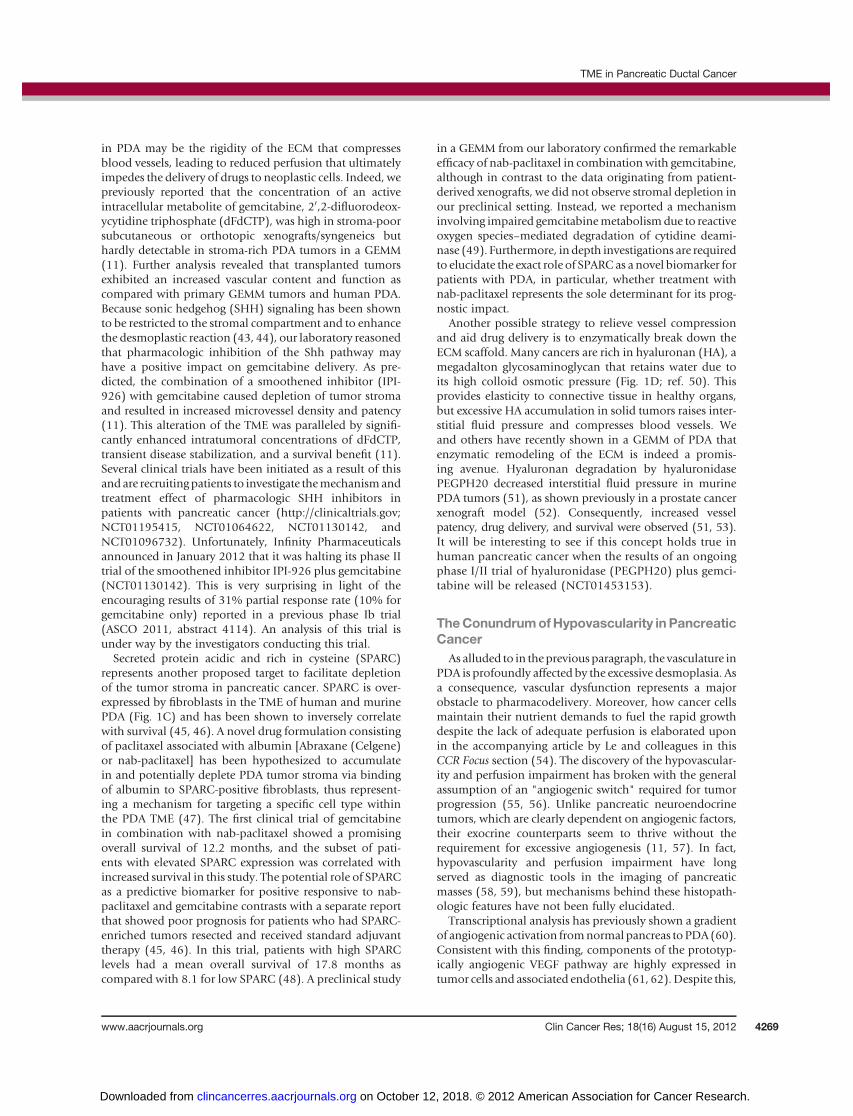
tumor samples show substantially lower microvessel den-sities (MVD) than those of the normal pancreas (Fig. 2;ref. 11, 57). Although VEGF immunostaining is positivelycorrelated with MVDs, it has limited association withpatient survival (63, 64). Despite these contradictory find-ings, antiangiogenic therapy was tested in PDA. Initialapproaches targeted matrix metalloproteinases using mar-imastat and BAY 12-9566 as well as the integrins aVb3 andaVb5 using cilengitide. These compounds did not provideany clinical benefit in trials (65–67).More recently, targetedagents such as bevacizumab, an anti-VEGFA monoclonalantibody, have been investigated in advanced pancreaticcancer in combination with gemcitabine and did notimprove survival compared with gemcitabine plus placeboin a randomized phase III trial (5.8 vs. 5.9 months; ref. 68).In addition, there was no significant benefit to overallsurvival in combining bevacizumab with erlotinib andgemcitabine compared with the combination of the latter2 compounds (7.1 vs. 6.0months; ref. 69).Bevacizumabhasalso been evaluated in other combinations including withdocetaxel and with concurrent capecitabine and radiationwithout any proven benefit, although certain studies arestill under way (70, 71). VEGF receptor inhibition usingaxitinib in combination with gemcitabine also had nobeneficial effect on overall survival (72). In addition, thekinase inhibitor sorafenib, which targets VEGFR as well asPDGFR, c-KIT, Raf1, and FLT3, was found to be inactive inadvanced pancreatic cancer (73). Likewise, discouragingresults testing sunitinib in a preclinical trial in theLSL-KrasG12D;p53R172H/þ;Ptf1a-Cre model add to mountingevidence of angiogenic independence and dominance oftumor-driven angiostasis of PDA (57). This phenotypesuggests that endogenous inhibitors in the microenviron-ment might exert an overriding angiostatic effect during the
natural history of PDA. Many of these factors are generatedfrom plasma and ECM proteins by proteases, which arefrequently upregulated in tumor and stellate cells (PaSCs;ref. 74). For example, angiostatin and endostatin are pro-duced by PDA and detected at high concentrations inpatients’ circulation (75, 76). Moreover, whereas activatedPaSCs are ostensibly proangiogenic, their coculture withtumor cells robustly increases endostatin levels, showing theangiostatic potential of such heterotypic interactions (77).
On the whole, antiangiogenic therapies have proved notto be a viable option for pancreatic cancer, and althoughtherapeutic delivery may contribute to these failures, alter-native approaches targeting the PDA vasculature remainattractive and potentially feasible. Two scenarios are possi-ble: If PDA maintains itself on frugal use of restrictedresources as a consequence of limited perfusion, it may beconceivable that impairing perfusion even more could tipthe balance toward widespread hypoxic necrosis (78). Incontrast, increasing tumor perfusion may seem counter-intuitive but could synergize with cytotoxic therapy toincrease the intratumoral drug delivery and response.
Is PDA Hypoxic?The previous paragraph laid the groundwork for this
question.Most solid tumors contain areas of below-optimaloxygen concentration (hypoxia). This occurs as a result ofinefficient tumor vascular supply and a highmetabolic needfor oxygen (79). Many studies have provided evidence thathypoxic cells are more resistant to both chemotherapy andradiotherapy and can increase their invasive and metastaticpotential, ultimately creating a more aggressive disease (80,81). The ability of cancer cells to surviveunder thesehypoxicconditions results from the ability to co-opt pathwaysnecessary for embryonic development under hypoxic con-ditions. Themainpathway involved in thehypoxic responseis the hypoxia-inducible factor (HIF) pathway (82). HIF caninduce a wide range of gene products controlling cellularmetabolism and energetics, cell survival, migration, and pH(83). The HIF transcription factors also direct the transcrip-tion of many angiogenic growth factors (84).
Considering that the hypovascular nature of PDA has asignificant impact on perfusion and drug delivery (11), itwould be reasonable to assume a hypoxic state. However,direct evidence is sparse and the majority of publicationshave used surrogatemarkers formeasuring hypoxia, such asnecrosis or expression of HIF target genes (85–87). Onlyone small study, involving 7patientswithpancreatic cancer,has directly measured the oxygen pressure during pancrea-ticoduodenectomy by inserting needle electrodes. Thisstudy revealed a dramatic reduction in oxygenation oftumor tissue versus normal pancreas (88). Interestingly,preclinical work in an orthotopic model of pancreaticcancer has shown a lack of correlation between MVD andhypoxia, perhaps suggesting that a hypovascular pancreatictumor is not directly linked to hypoxia (89). Nevertheless,this work did predict more aggressive behavior, including amore metastatic phenotype in hypoxic tumors. Clinical
© 2012 American Association for Cancer Research
T
PT
Figure 2. Murine PDA is characterized by hypovasularity. In this exampleof CD31 immunohistochemistry, the dotted line denotes the boundarybetween tumor (T) and peritumoral diseased pancreas (PT; �20magnification).
CCRFOCUS
Clin Cancer Res; 18(16) August 15, 2012 Clinical Cancer Research4270
on October 12, 2018. © 2012 American Association for Cancer Research. clincancerres.aacrjournals.org Downloaded from

work is under way to assess the prognostic significance ofthese results in patients with pancreatic cancer, using thehypoxia probe, pimonidazole, administered 24 hoursbefore surgery (NCT01248637).The reason hypoxia poses a challenge to the field of
anticancer therapeutics is that it provides a niche forslow-cycling, highly drug-resistant cells, which may beidentical to the proposed cancer stem cells (90, 91). Thus,standard chemotherapy agents fail because they are unsuc-cessful at targeting the cell within the hypoxic TME, whichmight be those that most need to be eliminated (92). Inaddition, hypoxic conditions are also known to stimulatethe Notch signaling pathway, and it has recently beenshown that pancreatic cancer cells can be sensitized byNotch inhibition (78). Thus, the hypovascular state of PDAcould be exploited by novel therapeutic approaches such ashypoxia-activable prodrugs (93). Further investigations arerequired into the downstreamNotch targets that are impor-tant for tumor cell survival under hypoxic conditions.Currently, a clinical trial is investigating the benefit ofcombining Notch pathway inhibition and gemcitabine(NCT01232829 and NCT01098344).
Out of Balance—Immune Cells in PDAAs is the case with other cancer types, inflammation
seems to be crucially linked to PDA development as exem-plified by chronic pancreatitis being amajor risk factor (94).However, the molecular details are still obscure and arejust beginning to be elucidated (95, 96). A comprehensiveanalysis of the immune cell composition of PanIN and PDAin the LSL-KrasG12D;Pdx1-Cre mice defined an importantbaseline for future studies (97). Use of enzymatic tumor
digestion followed by fluorescence-activated cell sortinganalysis revealed that immune cells make up roughly50% of the tumor cell mass (Fig. 3 illustrates similarfindings by immunofluorescence). From this study, it is
© 2012 American Association for Cancer Research
x, y:100 μm
Figure 3. Prominent immune cell infiltration exists in mouse PDA. Thisimmunofluorescence staining illustrates the abundance of immune cellsmarked by CD45 expression (red) between neoplastic glandularstructures (stained for EpCam in blue) and a-SMA positive stromalfibroblasts and perivascular cells that likely represent pericytes (green;�20 magnification).
© 2012 American Association for Cancer Research
A B
Normal O2 Low O
2 Low O2
dFdCTPdFdCTP�
Normal O2
CSC CSC
α-CD40
nab-paclitaxel
PSC/CAF
PaSC/CAF
ECM ECM
Hyaluronidase
nab-paclitaxelBlood vessels
Hedgehoginhibitors
Blood vessels
Immunecells
Immunecells
CCR Focus
Figure 4. Schematic of the TME network, cross-talk, and interdependence in PDA with a focus on therapeutic intervention points. A, activated pancreaticstellate cells lay down vast amounts of ECM, which causes a constriction/collapse of the sparse vessel network. This impedes gemcitabine delivery.Hypoxia generates niches for slow-cycling cells that are not targeted by chemotherapeutics. Also, an immunosuppressivemicroenvironment further supportstumor growth. B, Hedgehog pathway inhibition causes stromal depletion accompanied by reduced ECM. The ECM can also be enzymatically targeted, andboth interventions lead to increased vessel patency and intratumoral gemcitabine delivery. The immune system can be stimulated to turn against cancer cells,for instance by anti-CD40 antibody treatment. CAF, cancer-associated fibroblast.
TME in Pancreatic Ductal Cancer
www.aacrjournals.org Clin Cancer Res; 18(16) August 15, 2012 4271
on October 12, 2018. © 2012 American Association for Cancer Research. clincancerres.aacrjournals.org Downloaded from

apparent that immunosuppressive cell types, such as reg-ulatory T cells and myeloid-derived suppressor cells, arepredominant, with hardly any CTLs infiltrating thetumors. This paints a picture of a striking imbalance inprotumorigenic and antitumorigenic immune cells. Toadd to the complexity, a recent study revealed a newimmunosuppressive cell type in the stroma of PDA andother cancers. This cell expresses fibroblast activationprotein a (FAPa), and FAPa cell ablation resulted inimmunologic control of tumor growth in several subcu-taneous tumor models (98).
Successful immunotherapy depends on the cancer cellsexpressing proteins that can be recognized as altered bythe immune system. These fall in 2 categories: Tumor-associated antigens are nonmutated self proteins that areaberrantly regulated (overexpressed or expressed in othertissues or oncofetal antigens), whereas tumor-specific anti-gens are generated as a consequence of the mutationalevents in neoplastic cells and are de novo antigens. The goal
is to induce high-affinity cytotoxic T cells (CTL or CD8T cells) without causing autoimmunity. Antigens targetedin immunotherapy clinical trials in PDA have includedMUC1, mesothelin, KRAS, carcinoembryonic antigen, sur-vivin, and telomerase, as well as whole tumor cells engineer-ed to express granulocyte macrophage colony-stimulatingfactor [GM-CSF; reviewed in Dodson and colleagues (99)].
In the first phase I clinical trial using irradiated allogeneicGM-CSF–expressing tumor cell vaccines the treatmentwas well tolerated and found to be safe for use in humans(100). This result warranted a larger phase II trial to inves-tigate the disease-free and overall survival after surgicalresection followed by chemoradiation and vaccination,which reported a median survival of 24.8 months (101).Another approach is to pulse dendritic cells with tumorantigens ex vivo and reinfuse them into patients. Muc1-pulsed dendritic cells were evaluated in a phase I/II trialin patients with resected pancreatic and biliary tumors. Thevaccine transiently increased the percentages of functional
Table 2. Summary of past and current clinical trials targeting components of the TME
Trial statusOutcome
Intervention Current phase Trial identifier Status Median survival Reference
Erlotinib þ gemcitabine III NCT00026338 Completed 6.24 vs. 5.91 mo 6Oxaliplatin þ irinotecan þleucovorin þ 5-FU
II/III NCT00112658 Completed 11.1 vs. 6.8 mo 7
GDC-0449 þ gemcitabine II NCT01064622 ActiveIPI-926 þ gemcitabine I/II NCT01130142 Stopped N/Anab-Paclitaxel þ gemcitabine I/II NCT00398086 Completed 12.2 mo 48PEGPH20 þ gemcitabine I/II NCT01453153 ActiveMarimastat þ gemcitabine III N/A Completed 5.44 vs. 5.39 mo 65Cilengitide þ gemcitabine II N/A Completed 6.7 vs. 7.7 mo 66BAY 12-9566 III N/A Completed 3.7 vs. 6.6 mo 67Bevacizumab þ gemcitabine III NCT00088894 Completed 5.8 vs. 5.9 mo 68Bevacizumab þ erlotinib þgemcitabine
III N/A Completed 7.1 vs. 6.0 mo 69
Bevacizumab þ docetaxel II N/A Completed 4.1 vs. 5.4 mo 70Radiotherapy þ capecitabine þbevacizumab, bevacizumab þgemcitabine
II NCT00114179 Completed 11.9 71
Axitinib þ gemcitabine III NCT00471146 Completed 8.5 vs. 8.3 mo 72Sorafenib þ gemcitabine II N/A Completed 4.0 mo 73RO4929097 II NCT01232829 ActiveMK0752 þ gemcitabine I/II NCT01098344 ActiveIrradiated allogeneicGM-CSF–secreting tumor vaccine
II NCT00084383 Completed 24.8 mo 101
MUC1 peptide-loadeddendritic cell vaccine
I/II N/A Completed 26 mo 102
Ipilimumab II NCT00112580 Completed N/A 103CP-870,893 þ gemcitabine I NCT00711191 Completed 7.4 mo 104Mechanistic studiesGDC-0449 þ gemcitabine 0 NCT01195415 ActiveGDC-0449 II NCT01096732 ActivePreoperative pimonidazole N/A NCT01248637 Active
CCRFOCUS
Clin Cancer Res; 18(16) August 15, 2012 Clinical Cancer Research4272
on October 12, 2018. © 2012 American Association for Cancer Research. clincancerres.aacrjournals.org Downloaded from

CD4 and CD8 T cells as well as regulatory T cells. Four of12 (33%) patients in this study were alive 5 years aftersurgery, with a median survival of 26 months (range, 13–69 months; ref. 102). Both of these studies compare favor-ably in terms of the median survival for resected pancreaticcancer, which is normally between 11 and 20 months.A third approach is the use of blocking/neutralizing anti-bodies such as ipilimumab, which targets CTLA-4, a surfaceprotein expressed by activated T cells that confers inhibitorysignals. Unfortunately, ipilimumab as a single agent wasfound to be ineffective in a phase II trial in locally advancedand metastatic pancreatic cancer. However, one of thepatients on this study experienced significant delayedregression of the primary tumor and 20 hepatic metastases,which may merit further investigation (103).GEMMs are an ideal system to evaluate immune thera-
peutic approaches in PDA; however, few reports exist to dateon this topic. This may be because the tumor antigens forPDA are unknown, making the tracing of immuneresponses very difficult. Nonetheless, a recent immunother-apy study used the "KPC" GEMM (LSL-KrasG12D;LSL-p53R172H/þ;Pdx1-Cre) to evaluatewhether activation of anti-gen-presenting cells via stimulationofCD40would result inincreased tumor antigen presentation and priming of effec-tor T cells (104). Treatment with agonistic anti-CD40achieved tumor stabilization and even regression in KPCmice but was surprisingly T-cell independent. Instead,tumor control was exerted by the activated macrophagestargeting the fibrotic stroma. Furthermore, an early-phaseclinical trial with anti-CD40 antibody showed promisingresults in patients (104).
Conclusions and Future OutlookThe influences of the stroma in pancreatic cancer are as
manifold as its components (Fig. 4). This curse may beturned into a blessing as this complexity also providesnumerous avenues for therapeutic exploration (clinical
trials mentioned in this review are summarized in Table2). Accumulating evidence suggests that the extensive des-moplastic reaction may be, at least, partly responsible forthe innate chemoresistance in pancreatic tumors by creatingbarriers that fence off tumor cells from circulating activetherapeutic compounds. Breaching this stromal barrierrepresents a promising strategy to improve the delivery andefficacy of cytotoxic drugs in the future. Therapeutic benefitmay be gained by strategies aimed at depleting the desmo-plastic stroma, exploiting the poor vasculature, or activatingthe immune system to target tumor cells. We anticipate thatfuture therapies will have to be tailored to target several ofthe described components of the microenvironment toachieve long-lasting therapeutic response.
Disclosure of Potential Conflicts of InterestD.A. Tuveson is a Senior Group Leader at the Cancer Research UK
Cambridge Research Institute. No potential conflicts of interests were dis-closed by the other authors.
Authors' ContributionsConception and design: C. Feig, D.A. Tuveson, A. NeesseAcquisitionofdata (provided animals, acquired andmanagedpatients,provided facilities, etc.): C. Feig, N. CookWriting, review, and/or revision of the manuscript: C. Feig, D.A. Tuve-son, A. Gopinathan, A. Neesse, D.S. Chan, N. CookAdministrative, technical, or material support (i.e., reporting or orga-nizing data, constructing databases): A. Gopinathan, D.S. ChanStudy supervision: D.A. Tuveson
Grant SupportThis study was supported by the University of Cambridge and Cancer
ResearchUK, The Li Ka Shing Foundation andHutchisonWhampoaLimited,the NIHR Cambridge Biomedical Research Centre, and the European Com-munity Grant EPC-TM-Net 256974. C. Feig was supported by the EMBOlong-term fellowship and by aMarie Curie Intra European Fellowship withinthe 7th European Community Framework Programme. A. Neesse wassupported by Deutsche Krebshilfe Mildred Scheel Postdoctoral Fellowship.D.S. Chan is funded by the Frank Edward Elmore Fund.
Received March 5, 2012; revised June 27, 2012; accepted June 27, 2012;published online August 15, 2012.
References1. Hidalgo M. Pancreatic cancer. N Engl J Med 2010;362:1605–17.2. Siegel R,NaishadhamD, JemalA.Cancer statistics, 2012.CACancer
J Clin 2012;62:10–29.3. Neoptolemos JP. Adjuvant treatment of pancreatic cancer. Eur J
Cancer 2011;47 Suppl 3:S378–80.4. Yauch RL, Settleman J. Recent advances in pathway-targeted can-
cer drug therapies emerging from cancer genome analysis. Curr OpinGenet Dev 2012;22:45–9.
5. Burris HA III, Moore MJ, Andersen J, Green MR, Rothenberg ML,Modiano MR, et al. Improvements in survival and clinical benefit withgemcitabine as first-line therapy for patientswith advanced pancreascancer: a randomized trial. J Clin Oncol 1997;15:2403–13.
6. MooreMJ, Goldstein D, HammJ, Figer A, Hecht JR, Gallinger S, et al.Erlotinib plus gemcitabine compared with gemcitabine alone inpatients with advanced pancreatic cancer: a phase III trial of theNationalCancer Institute ofCanadaClinical TrialsGroup. JClinOncol2007;25:1960–6.
7. Conroy T, Desseigne F, YchouM, BoucheO, Guimbaud R, BecouarnY, et al. FOLFIRINOX versus gemcitabine for metastatic pancreaticcancer. N Engl J Med 2011;364:1817–25.
8. Johnson JI, Decker S, Zaharevitz D, Rubinstein LV, Venditti JM,Schepartz S, et al. Relationships between drug activity in NCI pre-clinical in vitro and in vivomodels and early clinical trials. Br J Cancer2001;84:1424–31.
9. Singh M, Lima A, Molina R, Hamilton P, Clermont AC, Devasthali V,et al. Assessing therapeutic responses in Kras mutant cancers usinggenetically engineered mouse models. Nat Biotechnol 2010;28:585–93.
10. Gopinathan A, Tuveson DA. The use of GEMmodels for experimentalcancer therapeutics. Dis Model Mech 2008;1:83–6.
11. Olive KP, Jacobetz MA, Davidson CJ, Gopinathan A, McIntyre D,HonessD, et al. InhibitionofHedgehogsignalingenhancesdelivery ofchemotherapy in a mouse model of pancreatic cancer. Science2009;324:1457–61.
12. Almoguera C, Shibata D, Forrester K, Martin J, Arnheim N, PeruchoM.Most human carcinomas of the exocrine pancreas contain mutantc-K-ras genes. Cell 1988;53:549–54.
13. Hingorani SR, Petricoin EF, Maitra A, Rajapakse V, King C, JacobetzMA, et al. Preinvasive and invasive ductal pancreatic cancer and itsearly detection in the mouse. Cancer Cell 2003;4:437–50.
TME in Pancreatic Ductal Cancer
www.aacrjournals.org Clin Cancer Res; 18(16) August 15, 2012 4273
on October 12, 2018. © 2012 American Association for Cancer Research. clincancerres.aacrjournals.org Downloaded from

14. Hingorani SR,Wang L,Multani AS, CombsC, Deramaudt TB, HrubanRH, et al. Trp53R172H and KrasG12D cooperate to promote chro-mosomal instability and widely metastatic pancreatic ductal adeno-carcinoma in mice. Cancer Cell 2005;7:469–83.
15. Aguirre AJ, Bardeesy N, Sinha M, Lopez L, Tuveson DA, Horner J,et al. Activated Kras and Ink4a/Arf deficiency cooperate to producemetastatic pancreatic ductal adenocarcinoma. Genes Dev 2003;17:3112–26.
16. Iacobuzio-Donahue CA, Velculescu VE, Wolfgang CL, Hruban RH.Genetic basis of pancreas cancer development and progression:insights from whole-exome and whole-genome sequencing. ClinCancer Res 2012;18:4257–65.
17. Apte MV, Haber PS, Applegate TL, Norton ID, McCaughan GW,Korsten MA, et al. Periacinar stellate shaped cells in rat pancreas:identification, isolation, and culture. Gut 1998;43:128–33.
18. Bachem MG, Schneider E, Gross H, Weidenbach H, Schmid RM,Menke A, et al. Identification, culture, and characterization of pan-creatic stellate cells in rats and humans. Gastroenterology 1998;115:421–32.
19. Omary MB, Lugea A, Lowe AW, Pandol SJ. The pancreatic stellatecell: a star on the rise in pancreatic diseases. J Clin Invest 2007;117:50–9.
20. Haber PS, Keogh GW, Apte MV, Moran CS, Stewart NL, CrawfordDH, et al. Activation of pancreatic stellate cells in human and exper-imental pancreatic fibrosis. Am J Pathol 1999;155:1087–95.
21. Han F, Wang CY, Yang L, Zhan SD, Zhang M, Tian K. Contribution ofmurine bone marrow mesenchymal stem cells to pancreas regener-ation after partial pancreatectomy in mice. Cell Biol Int 2012 May 11.[Epub ahead of print].
22. WatanabeT,MasamuneA,KikutaK,HirotaM,KumeK,SatohK, et al.Bonemarrowcontributes to the population of pancreatic stellate cellsinmice. Am JPhysiol Gastrointest Liver Physiol 2009;297:G1138–46.
23. Scarlett CJ, Colvin EK, Pinese M, Chang DK, Morey AL, MusgroveEA, et al. Recruitment and activation of pancreatic stellate cells fromthe bone marrow in pancreatic cancer: a model of tumor-hostinteraction. PLoS One 2011;6:e26088.
24. Froeling FE, Mirza TA, Feakins RM, Seedhar A, Elia G, Hart IR, et al.Organotypic culture model of pancreatic cancer demonstrates thatstromal cells modulate E-cadherin, beta-catenin, and Ezrin expres-sion in tumor cells. Am J Pathol 2009;175:636–48.
25. Hwang RF, Moore T, Arumugam T, Ramachandran V, Amos KD,Rivera A, et al. Cancer-associated stromal fibroblasts promote pan-creatic tumor progression. Cancer Res 2008;68:918–26.
26. Jesnowski R, Furst D, Ringel J, Chen Y, Schrodel A, Kleeff J, et al.Immortalization of pancreatic stellate cells as an in vitro model ofpancreatic fibrosis: deactivation is induced by matrigel and N-acet-ylcysteine. Lab Invest 2005;85:1276–91.
27. Masamune A, Satoh M, Kikuta K, Suzuki N, Shimosegawa T.Establishment and characterization of a rat pancreatic stellate cellline by spontaneous immortalization. World J Gastroenterol 2003;9:2751–8.
28. Mathison A, Liebl A, Bharucha J, Mukhopadhyay D, Lomberk G,Shah V, et al. Pancreatic stellate cell models for transcriptionalstudies of desmoplasia-associated genes. Pancreatology2010;10:505–16.
29. Satoh M, Masamune A, Sakai Y, Kikuta K, Hamada H, ShimosegawaT. Establishment and characterization of a simian virus 40-immor-talized rat pancreatic stellate cell line. Tohoku J Exp Med 2002;198:55–69.
30. Sparmann G, Hohenadl C, Tornoe J, Jaster R, Fitzner B, Koczan D,et al. Generation and characterization of immortalized rat pancreaticstellate cells. Am J Physiol Gastrointest Liver Physiol 2004;287:G211–9.
31. Froeling FE, FeigC,ChelalaC,DobsonR,MeinCE, TuvesonDA, et al.Retinoic acid-induced pancreatic stellate cell quiescence reducesparacrine Wnt-beta-catenin signaling to slow tumor progression.Gastroenterology 2011;141:1486–97.
32. Erkan M, Adler G, Apte MV, Bachem MG, Buchholz M, Detlefsen S,et al. StellaTUM: current consensus and discussion on pancreaticstellate cell research. Gut 2012;61:172–8.
33. Vonlaufen A, Joshi S, Qu C, Phillips PA, Xu Z, Parker NR, et al.Pancreatic stellate cells: partners in crime with pancreatic cancercells. Cancer Res 2008;68:2085–93.
34. Xu Z, Vonlaufen A, Phillips PA, Fiala-Beer E, Zhang X, Yang L, et al.Role of pancreatic stellate cells in pancreatic cancermetastasis. AmJPathol 2010;177:2585–96.
35. Lonardo E, Frias-Aldeguer J, Hermann PC, Heeschen C. Pancreaticstellate cells formaniche for cancer stemcells and promote their self-renewal and invasiveness. Cell Cycle 2012;11:1282–90.
36. Hamada S, Masamune A, Takikawa T, Suzuki N, Kikuta K, Hirota M,et al. Pancreatic stellate cells enhance stem cell-like phenotypes inpancreatic cancer cells. Biochem Biophys Res Commun 2012;421:349–54.
37. Schneider E, Schmid-Kotsas A, Zhao J, Weidenbach H, Schmid RM,Menke A, et al. Identification of mediators stimulating proliferationandmatrix synthesis of rat pancreatic stellate cells. Am J Physiol CellPhysiol 2001;281:C532–43.
38. Jaster R, Sparmann G, Emmrich J, Liebe S. Extracellular signalregulated kinases are key mediators of mitogenic signals in ratpancreatic stellate cells. Gut 2002;51:579–84.
39. Sato Y, Murase K, Kato J, Kobune M, Sato T, Kawano Y, et al.Resolution of liver cirrhosis using vitamin A-coupled liposomes todeliver siRNA against a collagen-specific chaperone. Nat Biotechnol2008;26:431–42.
40. Wehr AY, Furth EE, Sangar V, Blair IA, Yu KH. Analysis of the humanpancreatic stellate cell secreted proteome. Pancreas 2011;40:557–66.
41. MahadevanD, VonHoff DD. Tumor-stroma interactions in pancreaticductal adenocarcinoma. Mol Cancer Ther 2007;6:1186–97.
42. Neesse A, Michl P, Frese KK, Feig C, Cook N, Jacobetz MA, et al.Stromal biology and therapy in pancreatic cancer. Gut 2011;60:861–8.
43. Tian H, Callahan CA, DuPree KJ, Darbonne WC, Ahn CP, Scales SJ,et al. Hedgehog signaling is restricted to the stromal compartmentduring pancreatic carcinogenesis. Proc Natl Acad Sci U S A2009;106:4254–9.
44. Bailey JM, Swanson BJ, Hamada T, Eggers JP, Singh PK, Caffery T,et al. Sonic hedgehog promotes desmoplasia in pancreatic cancer.Clin Cancer Res 2008;14:5995–6004.
45. Mantoni TS, Schendel RR, Rodel F, Niedobitek G, Al-Assar O,Masamune A, et al. Stromal SPARC expression and patient survivalafter chemoradiation for non-resectable pancreatic adenocarcino-ma. Cancer Biol Ther 2008;7:1806–15.
46. Infante JR, Matsubayashi H, Sato N, Tonascia J, Klein AP, Riall TA,et al. Peritumoral fibroblast SPARC expression and patient outcomewith resectable pancreatic adenocarcinoma. J Clin Oncol2007;25:319–25.
47. Desai N, Trieu V, Yao Z, Louie L, Ci S, Yang A, et al. Increasedantitumor activity, intratumor paclitaxel concentrations, and endo-thelial cell transport of cremophor-free, albumin-bound paclitaxel,ABI-007, compared with cremophor-based paclitaxel. Clin CancerRes 2006;12:1317–24.
48. Von Hoff DD, Ramanathan RK, Borad MJ, Laheru DA, Smith LS,Wood TE, et al. Gemcitabine plus nab-paclitaxel is an active regimenin patients with advanced pancreatic cancer: a phase I/II trial. J ClinOncol 2011;29:4548–54.
49. Frese KK,NeesseA,CookN, Bapiro TE, LolkemaMP, Jodrell DI, et al.nab-Paclitaxel potentiates gemcitabine activity by reducing cytidinedeaminase levels in a mouse model of pancreatic cancer. CancerDiscov 2012;2:260–9.
50. Tammi RH, Kultti A, Kosma VM, Pirinen R, Auvinen P, Tammi MI.Hyaluronan in human tumors: pathobiological and prognostic mes-sages from cell-associated and stromal hyaluronan. Semin CancerBiol 2008;18:288–95.
51. Provenzano PP, Cuevas C, Chang AE, Goel VK, Von Hoff DD,Hingorani SR. Enzymatic targeting of the stroma ablates physicalbarriers to treatment of pancreatic ductal adenocarcinoma. CancerCell 2012;21:418–29.
52. Thompson CB, Shepard HM, O'Connor PM, Kadhim S, Jiang P,Osgood RJ, et al. Enzymatic depletion of tumor hyaluronan induces
CCRFOCUS
Clin Cancer Res; 18(16) August 15, 2012 Clinical Cancer Research4274
on October 12, 2018. © 2012 American Association for Cancer Research. clincancerres.aacrjournals.org Downloaded from

antitumor responses in preclinical animal models. Mol Cancer Ther2010;9:3052–64.
53. JacobetzMA,ChanDS,NeesseA, Bapiro TE,CookN, FreseKK, et al.Hyaluronan impairs vascular function and drug delivery in a mousemodel of pancreatic cancer. Gut 2012 Mar 30. [Epub ahead of print].
54. Le A, Rajeshkumar NV,Maitra A, DangCV. Conceptual framework forcutting the pancreatic cancer fuel supply. Clin Cancer Res 2012;18:4285–90.
55. Folkman J, Watson K, Ingber D, Hanahan D. Induction of angiogen-esis during the transition from hyperplasia to neoplasia. Nature1989;339:58–61.
56. Xie L, Duncan MB, Pahler J, Sugimoto H, Martino M, Lively J, et al.Counterbalancing angiogenic regulatory factors control the rate ofcancer progression and survival in a stage-specificmanner. ProcNatlAcad Sci U S A 2011;108:9939–44.
57. Olson P, Chu GC, Perry SR, Nolan-Stevaux O, Hanahan D. Imagingguided trials of the angiogenesis inhibitor sunitinib in mouse modelspredict efficacy in pancreatic neuroendocrine but not ductal carci-noma. Proc Natl Acad Sci U S A 2011;108:E1275–84.
58. Freeny PC, Traverso LW, Ryan JA. Diagnosis and staging of pan-creatic adenocarcinoma with dynamic computed tomography. Am JSurg 1993;165:600–6.
59. Sofuni A, Iijima H,Moriyasu F, Nakayama D, ShimizuM, Nakamura K,et al. Differential diagnosis of pancreatic tumors using ultrasoundcontrast imaging. J Gastroenterol 2005;40:518–25.
60. Abdollahi A, Schwager C, Kleeff J, Esposito I, Domhan S, Peschke P,et al. Transcriptional network governing the angiogenic switch inhuman pancreatic cancer. Proc Natl Acad Sci U S A 2007;104:12890–5.
61. Itakura J, Ishiwata T, Shen B, Kornmann M, Korc M. Concomitantover-expression of vascular endothelial growth factor and its recep-tors in pancreatic cancer. Int J Cancer 2000;85:27–34.
62. Dallas NA, Gray MJ, Xia L, Fan F, van Buren G II, Gaur P, et al.Neuropilin-2-mediated tumor growth and angiogenesis in pancreaticadenocarcinoma. Clin Cancer Res 2008;14:8052–60.
63. Itakura J, Ishiwata T, Friess H, Fujii H, Matsumoto Y, Buchler MW,et al. Enhanced expression of vascular endothelial growth factor inhuman pancreatic cancer correlates with local disease progression.Clin Cancer Res 1997;3:1309–16.
64. Ellis LM, Takahashi Y, Fenoglio CJ, Cleary KR, Bucana CD, EvansDB. Vessel counts and vascular endothelial growth factor expres-sion in pancreatic adenocarcinoma. Eur J Cancer 1998;34:337–40.
65. Bramhall SR, Schulz J, Nemunaitis J, Brown PD, Baillet M, BuckelsJA. A double-blind placebo-controlled, randomised study comparinggemcitabine and marimastat with gemcitabine and placebo as firstline therapy in patients with advanced pancreatic cancer. Br J Cancer2002;87:161–7.
66. Friess H, Langrehr JM, Oettle H, Raedle J, Niedergethmann M,Dittrich C, et al. A randomized multi-center phase II trial of theangiogenesis inhibitor Cilengitide (EMD 121974) and gemcitabinecompared with gemcitabine alone in advanced unresectable pan-creatic cancer. BMC Cancer 2006;6:285.
67. Moore MJ, Hamm J, Dancey J, Eisenberg PD, Dagenais M, Fields A,et al. Comparison of gemcitabine versus thematrixmetalloproteinaseinhibitor BAY 12-9566 in patients with advanced or metastatic ade-nocarcinoma of the pancreas: a phase III trial of the National CancerInstitute of Canada Clinical Trials Group. J Clin Oncol 2003;21:3296–302.
68. Kindler HL, Niedzwiecki D, Hollis D, Sutherland S, Schrag D, HurwitzH, et al. Gemcitabine plus bevacizumab compared with gemcitabineplus placebo in patients with advanced pancreatic cancer: phase IIItrial of the Cancer and Leukemia Group B (CALGB 80303). J ClinOncol 2010;28:3617–22.
69. Van Cutsem E, Vervenne WL, Bennouna J, Humblet Y, Gill S, VanLaethem JL, et al. Phase III trial of bevacizumab in combination withgemcitabine and erlotinib in patients with metastatic pancreaticcancer. J Clin Oncol 2009;27:2231–7.
70. Astsaturov IA, Meropol NJ, Alpaugh RK, Burtness BA, Cheng JD,McLaughlin S, et al. Phase II and coagulation cascade biomarker
study of bevacizumab with or without docetaxel in patients withpreviously treated metastatic pancreatic adenocarcinoma. Am J ClinOncol 2011;34:70–5.
71. Crane CH, Winter K, Regine WF, Safran H, Rich TA, Curran W, et al.Phase II study of bevacizumab with concurrent capecitabine andradiation followed bymaintenance gemcitabine and bevacizumab forlocally advanced pancreatic cancer: Radiation Therapy OncologyGroup RTOG 0411. J Clin Oncol 2009;27:4096–102.
72. Kindler HL, Ioka T, Richel DJ, Bennouna J, Letourneau R, Okusaka T,et al. Axitinib plus gemcitabine versus placebo plus gemcitabine inpatients with advanced pancreatic adenocarcinoma: a double-blindrandomised phase 3 study. Lancet Oncol 2011;12:256–62.
73. Kindler HL, Wroblewski K, Wallace JA, Hall MJ, Locker G, Nattam S,et al. Gemcitabine plus sorafenib in patients with advanced pancre-atic cancer: a phase II trial of the University of Chicago Phase IIConsortium. Invest New Drugs 2012;30:382–6.
74. Yamamoto H, Itoh F, Iku S, Adachi Y, Fukushima H, Sasaki S, et al.Expression of matrix metalloproteinases and tissue inhibitors ofmetalloproteinases in human pancreatic adenocarcinomas: clinico-pathologic and prognostic significance of matrilysin expression. JClin Oncol 2001;19:1118–27.
75. Kisker O, Onizuka S, Banyard J, Komiyama T, Becker CM, AchillesEG, et al. Generation of multiple angiogenesis inhibitors by humanpancreatic cancer. Cancer Res 2001;61:7298–304.
76. Ohlund D, Ardnor B, Oman M, Naredi P, Sund M. Expression patternand circulating levels of endostatin in patients with pancreas cancer.Int J Cancer 2008;122:2805–10.
77. Erkan M, Reiser-Erkan C, Michalski CW, Deucker S, Sauliunaite D,Streit S, et al. Cancer-stellate cell interactions perpetuate the hyp-oxia-fibrosis cycle in pancreatic ductal adenocarcinoma. Neoplasia2009;11:497–508.
78. Cook N, Frese KK, Bapiro TE, Jacobetz MA, Gopinathan A, Miller JL,et al.Gammasecretase inhibitionpromoteshypoxicnecrosis inmousepancreatic ductal adenocarcinoma. J Exp Med 2012;209:437–44.
79. Brown JM, Wilson WR. Exploiting tumour hypoxia in cancer treat-ment. Nat Rev Cancer 2004;4:437–47.
80. Brown JM, Giaccia AJ. The unique physiology of solid tumors:opportunities (and problems) for cancer therapy. Cancer Res1998;58:1408–16.
81. Le QT, Denko NC, Giaccia AJ. Hypoxic gene expression and metas-tasis. Cancer Metastasis Rev 2004;23:293–310.
82. Brahimi-Horn MC, Pouyssegur J. HIF at a glance. J Cell Sci 2009;122:1055–7.
83. Pouyssegur J, Dayan F,MazureNM.Hypoxia signalling in cancer andapproaches to enforce tumour regression. Nature 2006;441:437–43.
84. Lu X, Kang Y. Hypoxia and hypoxia-inducible factors: master reg-ulators of metastasis. Clin Cancer Res 2010;16:5928–35.
85. Shibaji T, Nagao M, Ikeda N, Kanehiro H, Hisanaga M, Ko S, et al.Prognostic significance of HIF-1 alpha overexpression in humanpancreatic cancer. Anticancer Res 2003;23:4721–7.
86. Kitada T, Seki S, Sakaguchi H, Sawada T, Hirakawa K, Wakasa K.Clinicopathological significance of hypoxia-inducible factor-1alphaexpression in human pancreatic carcinoma. Histopathology 2003;43:550–5.
87. Hiraoka N, Ino Y, Sekine S, Tsuda H, Shimada K, Kosuge T, et al.Tumour necrosis is a postoperative prognostic marker for pancreaticcancer patients with a high interobserver reproducibility in histolog-ical evaluation. Br J Cancer 2010;103:1057–65.
88. Koong AC, Mehta VK, Le QT, Fisher GA, Terris DJ, Brown JM, et al.Pancreatic tumors showhigh levels of hypoxia. Int JRadiatOncol BiolPhys 2000;48:919–22.
89. Chang Q, Jurisica I, Do T, Hedley DW. Hypoxia predicts aggressivegrowth and spontaneous metastasis formation from orthotopicallygrown primary xenografts of human pancreatic cancer. Cancer Res2011;71:3110–20.
90. Penchev VR, Rasheed ZA, Maitra A, Matsui W. Heterogeneity andtargeting of pancreatic cancer stem cells. Clin Cancer Res 2012;18:4277–84.
91. Keith B, Simon MC. Hypoxia-inducible factors, stem cells, andcancer. Cell 2007;129:465–72.
TME in Pancreatic Ductal Cancer
www.aacrjournals.org Clin Cancer Res; 18(16) August 15, 2012 4275
on October 12, 2018. © 2012 American Association for Cancer Research. clincancerres.aacrjournals.org Downloaded from

92. Milas L, Hittelman WN. Cancer stem cells and tumor response totherapy: current problems and future prospects. Semin Radiat Oncol2009;19:96–105.
93. Denny WA. Hypoxia-activated prodrugs in cancer therapy: progressto the clinic. Future Oncol 2010;6:419–28.
94. Lowenfels AB, Maisonneuve P, Cavallini G, Ammann RW, LankischPG, Andersen JR, et al. Pancreatitis and the risk of pancreatic cancer.International Pancreatitis Study Group. N Engl J Med 1993;328:1433–7.
95. Lesina M, Kurkowski MU, Ludes K, Rose-John S, Treiber M, KloppelG, et al. Stat3/Socs3 activation by IL-6 transsignaling promotesprogression of pancreatic intraepithelial neoplasia and developmentof pancreatic cancer. Cancer Cell 2011;19:456–69.
96. FukudaA,WangSC,Morris JPt, Folias AE, LiouA, KimGE, et al. Stat3andMMP7 contribute to pancreatic ductal adenocarcinoma initiationand progression. Cancer Cell 2011;19:441–55.
97. Clark CE, Hingorani SR,Mick R, CombsC, TuvesonDA, VonderheideRH. Dynamics of the immune reaction to pancreatic cancer frominception to invasion. Cancer Res 2007;67:9518–27.
98. Kraman M, Bambrough PJ, Arnold JN, Roberts EW, Magiera L,Jones JO, et al. Suppression of antitumor immunity by stromalcells expressing fibroblast activation protein-alpha. Science 2010;330:827–30.
99. Dodson LF, Hawkins WG, Goedegebuure P. Potential targets for pan-creatic cancer immunotherapeutics. Immunotherapy 2011;3:517–37.
100. Jaffee EM, Hruban RH, Biedrzycki B, Laheru D, Schepers K, SauterPR, et al. Novel allogeneic granulocyte-macrophage colony-stimu-lating factor-secreting tumor vaccine for pancreatic cancer: a phase Itrial of safety and immune activation. J Clin Oncol 2001;19:145–56.
101. Lutz E, YeoCJ, LillemoeKD,Biedrzycki B, Kobrin B,Herman J, et al. Alethally irradiated allogeneic granulocyte-macrophage colony stim-ulating factor-secreting tumor vaccine for pancreatic adenocarcino-ma. A Phase II trial of safety, efficacy, and immune activation. AnnSurg 2011;253:328–35.
102. Lepisto AJ, Moser AJ, Zeh H, Lee K, Bartlett D, McKolanis JR, et al. Aphase I/II study of a MUC1 peptide pulsed autologous dendritic cellvaccine as adjuvant therapy in patients with resected pancreatic andbiliary tumors. Cancer Ther 2008;6:955–64.
103. Royal RE, Levy C, Turner K, Mathur A, HughesM, Kammula US, et al.Phase 2 trial of single agent Ipilimumab (anti-CTLA-4) for locallyadvanced or metastatic pancreatic adenocarcinoma. J Immunother2010;33:828–33.
104. Beatty GL, Chiorean EG, Fishman MP, Saboury B, Teitelbaum UR,Sun W, et al. CD40 agonists alter tumor stroma and show efficacyagainst pancreatic carcinoma in mice and humans. Science2011;331:1612–6.
CCRFOCUS
Clin Cancer Res; 18(16) August 15, 2012 Clinical Cancer Research4276
on October 12, 2018. © 2012 American Association for Cancer Research. clincancerres.aacrjournals.org Downloaded from

2012;18:4266-4276. Clin Cancer Res Christine Feig, Aarthi Gopinathan, Albrecht Neesse, et al. The Pancreas Cancer Microenvironment
Updated version
http://clincancerres.aacrjournals.org/content/18/16/4266
Access the most recent version of this article at:
Cited articles
http://clincancerres.aacrjournals.org/content/18/16/4266.full#ref-list-1
This article cites 102 articles, 42 of which you can access for free at:
Citing articles
http://clincancerres.aacrjournals.org/content/18/16/4266.full#related-urls
This article has been cited by 62 HighWire-hosted articles. Access the articles at:
E-mail alerts related to this article or journal.Sign up to receive free email-alerts
Subscriptions
Reprints and
To order reprints of this article or to subscribe to the journal, contact the AACR Publications Department at
Permissions
Rightslink site. Click on "Request Permissions" which will take you to the Copyright Clearance Center's (CCC)
.http://clincancerres.aacrjournals.org/content/18/16/4266To request permission to re-use all or part of this article, use this link
on October 12, 2018. © 2012 American Association for Cancer Research. clincancerres.aacrjournals.org Downloaded from