The Open Biotechnology Journal, 2008, 2 73-86 73 … · The Open Biotechnology Journal, 2008, 2,...
14
The Open Biotechnology Journal, 2008, 2, 73-86 73 1874-0707/08 2008 Bentham Science Publishers Ltd. -(1,4)-Polyglucuronic Acids – An Overview M.L. Tavernier 1 , C. Delattre 2 , E. Petit 1 and P. Michaud 3, * 1 Laboratoire des Glucides UMR CNRS 6219, Université de Picardie Jules Verne, 33 rue Saint-Leu, F-80039 Amiens, France; 2 GREENTECH, Biopôle Clermont Limagne 63360 Saint Beauzire, France and 3 Laboratoire de Génie Chimique et Biochimique, Université Blaise Pascal-Polytech Clermont Ferrand, 24 rue des Landais, F-63174 Aubière, France Abstract: Polyuronides are an acidic class of polysaccharides with interesting rheological and biological properties. How- ever, except pectin and alginate, the structural variability of this class of polysaccharides is poor and low described in lit- erature. In this context, a new generation of polyuronides has been isolated from two sources in the middle of the 90’s. Firstly, a bacterial -(1,4) polyglucuronic acid called glucuronan was identified as the sole exopolysaccharide produced by a bacteria belonging to the Rhizobiaceae family. Secondly, the development of the TEMPO chemistry led to the pro- duction at large scale of oxidized cellulose called cellouronate. Both new polyuronides were largely patented and found applications in several industrial areas. Moreover, the biodegradation study of these polysaccharides has led to the identi- fication of a new family of polysaccharide lyases very specific for these substrates. This review focuses on the actual knowledge of this class of acidic polysaccharides and on the enzymes acting about them. INTRODUCTION A bibliographic research using SciFinder Scholar with cellouronate, glucuronan and polyglucuronic acid shows the present interest of the scientific community for -D-(1,4)- polyglucuronic acids. Effectively, most of scientific produc- tions (full papers, patents, reviews, book chapters and others) have been published after 2000. This recent consideration for this family of polyuronides originates firstly in the necessity to upgrade cellulose from vegetable by-products and sec- ondly in the identification of natural forms of -D-(1,4)-poly- glucuronic acids. The most described one was the exopoly- saccharide produced by the strain Sinorhizobium meliloti M5N1CS [1-2]. This anionic homopolymer called glucu- ronan is a (1,4)- -D-polyglucuropyranosyluronic acid varia- bly O-acetylated at C3 and/or C2 position depending to the Mg 2+ concentration in the culture medium. It has been largely published and patented for various applications [1]. Since its identification in 1992 [2] it was the sole source of (1,4)- -D-polyglucuropyranosyluronic acid even if this ani- onic polysaccharide has been described in cell walls of Mu- cor rouxii [3] or algae [4,5]. However these fungal and algal glucuronans are always mixed with other polymers and their extractions and separations are tedious. The second interest- ing source of (1,4)- -D-polyglucuropyranosyluronic acid appeared between 1995 and the beginning of the 2000 years with the development of methods allowing regioselective oxidation of cellulose. In this way, the use of 2,2,6,6- tetramethylpyperidine-1-oxy(TEMPO)-NaBr-NaOCl system opens the way to regioselective modifications of this poly- saccharide with good yields [6]. In the case of the TEMPO- mediated oxidation of cellulose, different products were ob- tained depending on cellulose forms (cellulose I crystal, *Address correspondence to this author at the Laboratoire de Génie Chimi- que et Biochimique, Université Blaise Pascal-Polytech Clermont Ferrand, 24 rue des Landais, F-63174 Aubière, France; E-mail: [email protected] regenerated and mercerized celluloses) employed as starting materials [7-9]. The most interesting was the cellouronic acid whose structure was comparable to an unacetylated bac- terial glucuronan. In regards to the numerous applications of polyglucuronic acids, their biodegradability was investigated [10-13]. Studies led to the identification of a new family of polysaccharide lyases (EC 4.2.2.14), called glucuronan lyase and expressed by bacteria or fungi. These enzymes have been successfully employed for oligosaccharides production and have opened a reflection on their physiological roles. Effectively the ability of micro-organisms to grow with a - D-(1,4)-polyglucuronic acid as single carbon source implies abundance of this polysaccharide in nature. This review focuses on the actual knowledge of -(1,4)- D-polyglucuronic acids from various origins and on enzymes degrading them. MICROBIAL GLUCURONANS The species Sinorhizobium meliloti belongs to Rhizo- biaceae, a group of soil bacteria (gram-negative, motile, non- sporulating rods) that fix nitrogen after becoming established inside root nodules of plants from the Leguminoseae family. This symbiosis can be possible thanks to nodulating genes (Nod factors) and excretion of polysaccharides [14]. These exopolysaccharides (EPS) have been fully explored because of their original structures, their chemical and rheological properties in solution which confers them good industrial applications, in cosmetic, food-processing or pharmaceutical industry notably. The strain Sinorhizobium meliloti M5N1 is a bacteria isolated from a root nodule of alfalfa (Medicago sativa) and able to excrete in its culture medium a well known hetero- polysaccharide : the succinoglycan [15]. Chemical mutage- nesis of this strain has been investigated using N-methyl-N’- nitro-N-nitrosogua-nidine [16]. A mutant revealed to pro-
Transcript of The Open Biotechnology Journal, 2008, 2 73-86 73 … · The Open Biotechnology Journal, 2008, 2,...
The Open Biotechnology Journal, 2008, 2, 73-86 73
1874-0707/08 2008 Bentham Science Publishers Ltd.
-(1,4)-Polyglucuronic Acids – An Overview
M.L. Tavernier1, C. Delattre2, E. Petit1 and P. Michaud3,*
1Laboratoire des Glucides UMR CNRS 6219, Université de Picardie Jules Verne, 33 rue Saint-Leu, F-80039 Amiens,
France; 2GREENTECH, Biopôle Clermont Limagne 63360 Saint Beauzire, France and
3Laboratoire de Génie Chimique
et Biochimique, Université Blaise Pascal-Polytech Clermont Ferrand, 24 rue des Landais, F-63174 Aubière, France
Abstract: Polyuronides are an acidic class of polysaccharides with interesting rheological and biological properties. How-ever, except pectin and alginate, the structural variability of this class of polysaccharides is poor and low described in lit-erature. In this context, a new generation of polyuronides has been isolated from two sources in the middle of the 90’s. Firstly, a bacterial -(1,4) polyglucuronic acid called glucuronan was identified as the sole exopolysaccharide produced by a bacteria belonging to the Rhizobiaceae family. Secondly, the development of the TEMPO chemistry led to the pro-duction at large scale of oxidized cellulose called cellouronate. Both new polyuronides were largely patented and found applications in several industrial areas. Moreover, the biodegradation study of these polysaccharides has led to the identi-fication of a new family of polysaccharide lyases very specific for these substrates. This review focuses on the actual
knowledge of this class of acidic polysaccharides and on the enzymes acting about them.
INTRODUCTION
A bibliographic research using SciFinder Scholar with cellouronate, glucuronan and polyglucuronic acid shows the present interest of the scientific community for -D-(1,4)-polyglucuronic acids. Effectively, most of scientific produc-tions (full papers, patents, reviews, book chapters and others) have been published after 2000. This recent consideration for this family of polyuronides originates firstly in the necessity to upgrade cellulose from vegetable by-products and sec-ondly in the identification of natural forms of -D-(1,4)-poly- glucuronic acids. The most described one was the exopoly-saccharide produced by the strain Sinorhizobium meliloti M5N1CS [1-2]. This anionic homopolymer called glucu-ronan is a (1,4)- -D-polyglucuropyranosyluronic acid varia-bly O-acetylated at C3 and/or C2 position depending to the Mg2+ concentration in the culture medium. It has been largely published and patented for various applications [1]. Since its identification in 1992 [2] it was the sole source of (1,4)- -D-polyglucuropyranosyluronic acid even if this ani-onic polysaccharide has been described in cell walls of Mu-cor rouxii [3] or algae [4,5]. However these fungal and algal glucuronans are always mixed with other polymers and their extractions and separations are tedious. The second interest-ing source of (1,4)- -D-polyglucuropyranosyluronic acid appeared between 1995 and the beginning of the 2000 years with the development of methods allowing regioselective oxidation of cellulose. In this way, the use of 2,2,6,6-tetramethylpyperidine-1-oxy(TEMPO)-NaBr-NaOCl system opens the way to regioselective modifications of this poly-saccharide with good yields [6]. In the case of the TEMPO-mediated oxidation of cellulose, different products were ob-tained depending on cellulose forms (cellulose I crystal,
*Address correspondence to this author at the Laboratoire de Génie Chimi-que et Biochimique, Université Blaise Pascal-Polytech Clermont Ferrand, 24 rue des Landais, F-63174 Aubière, France; E-mail: [email protected]
regenerated and mercerized celluloses) employed as starting materials [7-9]. The most interesting was the cellouronic acid whose structure was comparable to an unacetylated bac-terial glucuronan. In regards to the numerous applications of polyglucuronic acids, their biodegradability was investigated [10-13]. Studies led to the identification of a new family of polysaccharide lyases (EC 4.2.2.14), called glucuronan lyase and expressed by bacteria or fungi. These enzymes have been successfully employed for oligosaccharides production and have opened a reflection on their physiological roles. Effectively the ability of micro-organisms to grow with a -D-(1,4)-polyglucuronic acid as single carbon source implies abundance of this polysaccharide in nature.
This review focuses on the actual knowledge of -(1,4)-D-polyglucuronic acids from various origins and on enzymes degrading them.
MICROBIAL GLUCURONANS
The species Sinorhizobium meliloti belongs to Rhizo-biaceae, a group of soil bacteria (gram-negative, motile, non-sporulating rods) that fix nitrogen after becoming established inside root nodules of plants from the Leguminoseae family. This symbiosis can be possible thanks to nodulating genes (Nod factors) and excretion of polysaccharides [14]. These exopolysaccharides (EPS) have been fully explored because of their original structures, their chemical and rheological properties in solution which confers them good industrial applications, in cosmetic, food-processing or pharmaceutical industry notably.
The strain Sinorhizobium meliloti M5N1 is a bacteria isolated from a root nodule of alfalfa (Medicago sativa) and able to excrete in its culture medium a well known hetero-polysaccharide : the succinoglycan [15]. Chemical mutage-nesis of this strain has been investigated using N-methyl-N’-nitro-N-nitrosogua-nidine [16]. A mutant revealed to pro-
74 The Open Biotechnology Journal, 2008, Volume 2 Tavernier et al.
duce a new extracellular polysaccharide. This new strain, labelled Sinorhizobium meliloti M5N1CS (NCIMB 40472), was still able to induce nodule formation on alfalfa roots, but less bacteria than the wild-type strain were detected in the infected alfalfa cells [17].
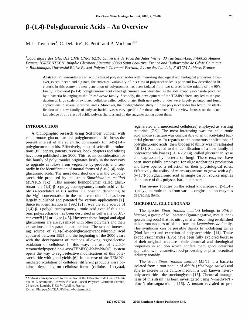
The native polysaccharide excreted by the M5N1CS strain was characterized. It was not soluble in 1 M H2SO4 and was resistant to acid hydrolysis even under drastic conditions (H2SO4 70 %) [2]. After reduction of all uronic acids, the polysaccharide could be hydrolysed and only glucose has been detected [16]. The reduced EPS has then been methy-lated and hydrolyzed with trifluoroacetic acid. After acetyla-tion, GC analysis has revealed the presence of a single peak corresponding to a 1,4,5-tri-O-acetyl-2,3,6-tri-O-methyl-glucitol, which proved that the monomers were 1 4 linked. 1H NMR analyses were monitored on polysaccharide. Sig-nals in the 2 ppm region, characteristic of O-acetyl groups were detected. The integration value ratio between the acetyl region (from 1.9 to 2.2 ppm) and the upfield and downfield regions (4.3 ppm to 5.1 ppm and 3.1 ppm to 4 ppm, respec-tively) has enabled to determine the acetylation degree (gen-erally about 0.5 acetate per glucuronic acid). It has been de-duced that the M5N1CS strain produced a homopolymer composed of -D-(1,4)-glucopyranosyluronic residues varia-bly acetylated at C3 and/or C2 position (Fig. 1). Conforma-tional and configurational features of deacetylated or acety-lated glucuronan have been investigated through glucuronan fibre X-ray diffraction analyses and molecular modeling [18, 19].
O
RO
OR
-OOC
OO
RO
OR
-OOC
O
n
Fig. (1). Glucuronan structure.
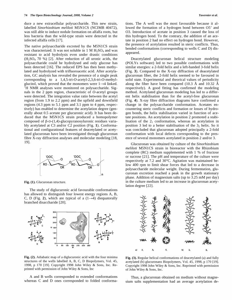
The study of diglucuronic acid favourable conformations has allowed to distinguish four lowest energy regions A, B, C, D (Fig. 2), which are typical of a (1 4) diequatorally branched disaccharide [20].
Fig. (2). Adiabatic map of a diglucuronic acid with the four minima structures of the wells labelled A, B, C, D Biopolymers, Vol. 45, 1998, p 170 [19]. Copyright 1998 John Wiley & Sons, Inc. Re-printed with permission of John Wiley & Sons, Inc
A and B wells corresponded to extended conformations whereas C and D ones corresponded to folded conforma-
tions. The A well was the most favourable because it al-lowed the formation of a hydrogen bond between O5’ and O3. Introduction of acetate in position 3 caused the loss of this hydrogen bond. To the contrary, the addition of an ace-tate in position 2 had no effect on hydrogen bond. However, the presence of acetylation resulted in steric conflicts. Thus, bended conformations (corresponding to wells C and D) dis-appeared.
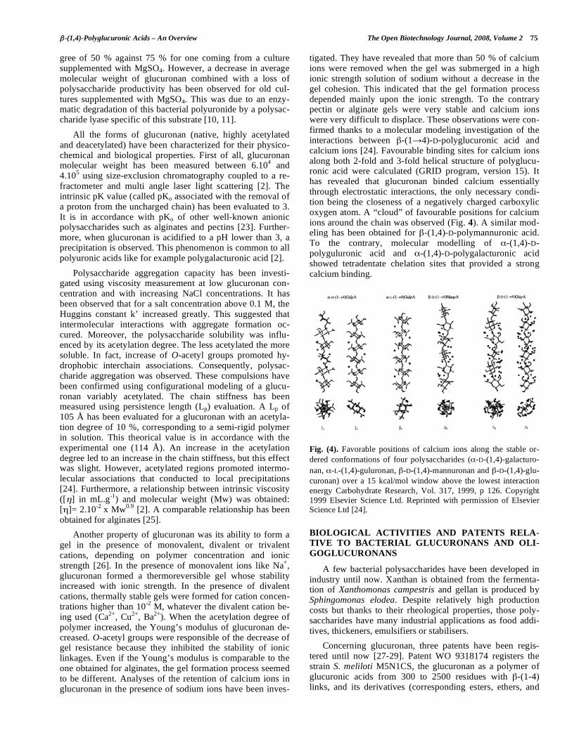
Deacetylated glucuronan helical structure modeling (POLYs software) led to two possible conformations with similar energies: a 2-fold helix and a left-handed 3-fold helix (Fig. 3). Compared to the X-ray diffraction of deacetylated glucuronan fiber, the 2-fold helix seemed to be favoured in solid state. Experimental and theorical values of periodicity along the fiber have been compared (10.3 Å and 10.32 Å respectively). A good fitting has confirmed the modeling method. Acetylated glucuronan modeling has led to a differ-ent helix stabilisation than for the acetyl-free glucuronan (Fig. 4). X-ray fibre diffraction diagrams have confirmed a change in the polysaccharide conformation. Acetates en-countering steric conflicts and formation or losses of hydro-gen bonds, the helix stabilisation varied in function of ace-tate positions. An acetylation in position 2 promoted a stabi-lisation of the 21 conformation, whereas an acetylation in position 3 led to a better stabilisation of the 32 helix. So it was concluded that glucuronan adopted principally a 2-fold conformation with local defects corresponding to the pres-ence of several monomers acetylated in position 2 and/or 3.
Glucuronan was obtained by culture of the Sinorhizobium meliloti M5N1CS strain in bioreactor with the Rhizobium complete (RC) medium supplemented with 1 % of fructose or sucrose [21]. The pH and temperature of the culture were respectively at 7.2 and 30°C. Agitation was maintained be-low 400 rpm to limit shear forces that led to a decrease in polysaccharide molecular weight. During fermentation, glu-curonan excretion reached a peak in the growth stationary phase. Addition of magnesium salts (up to 3.25 mM per day) in the culture medium led to an increase in glucuronan acety-lation degree [22].
Fig. (3). Regular helical conformations of deacetylated (a) and fully acetylated (b) glucuronans Biopolymers, Vol. 45, 1998, p 170 [19]. Copyright 1998 John Wiley & Sons, Inc. Reprinted with permission of John Wiley & Sons, Inc.
Thus, a glucuronan obtained on medium without magne-sium salts supplementation had an average acetylation de-
-(1,4)-Polyglucuronic Acids – An Overview The Open Biotechnology Journal, 2008, Volume 2 75
gree of 50 % against 75 % for one coming from a culture supplemented with MgSO4. However, a decrease in average molecular weight of glucuronan combined with a loss of polysaccharide productivity has been observed for old cul-tures supplemented with MgSO4. This was due to an enzy-matic degradation of this bacterial polyuronide by a polysac-charide lyase specific of this substrate [10, 11].
All the forms of glucuronan (native, highly acetylated and deacetylated) have been characterized for their physico-chemical and biological properties. First of all, glucuronan molecular weight has been measured between 6.104 and 4.105 using size-exclusion chromatography coupled to a re-fractometer and multi angle laser light scattering [2]. The intrinsic pK value (called pKo associated with the removal of a proton from the uncharged chain) has been evaluated to 3. It is in accordance with pKo of other well-known anionic polysaccharides such as alginates and pectins [23]. Further-more, when glucuronan is acidified to a pH lower than 3, a precipitation is observed. This phenomenon is common to all polyuronic acids like for example polygalacturonic acid [2].
Polysaccharide aggregation capacity has been investi-gated using viscosity measurement at low glucuronan con-centration and with increasing NaCl concentrations. It has been observed that for a salt concentration above 0.1 M, the Huggins constant k’ increased greatly. This suggested that intermolecular interactions with aggregate formation oc-cured. Moreover, the polysaccharide solubility was influ-enced by its acetylation degree. The less acetylated the more soluble. In fact, increase of O-acetyl groups promoted hy-drophobic interchain associations. Consequently, polysac-charide aggregation was observed. These compulsions have been confirmed using configurational modeling of a glucu-ronan variably acetylated. The chain stiffness has been measured using persistence length (Lp) evaluation. A Lp of 105 Å has been evaluated for a glucuronan with an acetyla-tion degree of 10 %, corresponding to a semi-rigid polymer in solution. This theorical value is in accordance with the experimental one (114 Å). An increase in the acetylation degree led to an increase in the chain stiffness, but this effect was slight. However, acetylated regions promoted intermo-lecular associations that conducted to local precipitations [24]. Furthermore, a relationship between intrinsic viscosity ([ ] in mL.g-1) and molecular weight (Mw) was obtained: [ ]= 2.10-2 x Mw0.9 [2]. A comparable relationship has been obtained for alginates [25].
Another property of glucuronan was its ability to form a gel in the presence of monovalent, divalent or trivalent cations, depending on polymer concentration and ionic strength [26]. In the presence of monovalent ions like Na+, glucuronan formed a thermoreversible gel whose stability increased with ionic strength. In the presence of divalent cations, thermally stable gels were formed for cation concen-trations higher than 10-2 M, whatever the divalent cation be-ing used (Ca2+, Cu2+, Ba2+). When the acetylation degree of polymer increased, the Young’s modulus of glucuronan de-creased. O-acetyl groups were responsible of the decrease of gel resistance because they inhibited the stability of ionic linkages. Even if the Young’s modulus is comparable to the one obtained for alginates, the gel formation process seemed to be different. Analyses of the retention of calcium ions in glucuronan in the presence of sodium ions have been inves-
tigated. They have revealed that more than 50 % of calcium ions were removed when the gel was submerged in a high ionic strength solution of sodium without a decrease in the gel cohesion. This indicated that the gel formation process depended mainly upon the ionic strength. To the contrary pectin or alginate gels were very stable and calcium ions were very difficult to displace. These observations were con-firmed thanks to a molecular modeling investigation of the interactions between -(1 4)-D-polyglucuronic acid and calcium ions [24]. Favourable binding sites for calcium ions along both 2-fold and 3-fold helical structure of polyglucu-ronic acid were calculated (GRID program, version 15). It has revealed that glucuronan binded calcium essentially through electrostatic interactions, the only necessary condi-tion being the closeness of a negatively charged carboxylic oxygen atom. A “cloud” of favourable positions for calcium ions around the chain was observed (Fig. 4). A similar mod-eling has been obtained for -(1,4)-D-polymannuronic acid. To the contrary, molecular modelling of -(1,4)-D-polyguluronic acid and -(1,4)-D-polygalacturonic acid showed tetradentate chelation sites that provided a strong calcium binding.
Fig. (4). Favorable positions of calcium ions along the stable or-dered conformations of four polysaccharides ( -D-(1,4)-galacturo-
nan, -L-(1,4)-guluronan, -D-(1,4)-mannuronan and -D-(1,4)-glu- curonan) over a 15 kcal/mol window above the lowest interaction energy Carbohydrate Research, Vol. 317, 1999, p 126. Copyright 1999 Elsevier Science Ltd. Reprinted with permission of Elsevier Science Ltd [24].
BIOLOGICAL ACTIVITIES AND PATENTS RELA-TIVE TO BACTERIAL GLUCURONANS AND OLI-
GOGLUCURONANS
A few bacterial polysaccharides have been developed in industry until now. Xanthan is obtained from the fermenta-tion of Xanthomonas campestris and gellan is produced by Sphingomonas elodea. Despite relatively high production costs but thanks to their rheological properties, those poly-saccharides have many industrial applications as food addi-tives, thickeners, emulsifiers or stabilisers.
Concerning glucuronan, three patents have been regis-tered until now [27-29]. Patent WO 9318174 registers the strain S. meliloti M5N1CS, the glucuronan as a polymer of glucuronic acids from 300 to 2500 residues with -(1-4) links, and its derivatives (corresponding esters, ethers, and
76 The Open Biotechnology Journal, 2008, Volume 2 Tavernier et al.
mixes of all derivatives). This patent also protects glucu-ronan for its use in food products, pharmaceutics, cosmetics or water purification, particularly as a gelifying, thickening, hydrating, stabilizing, chelating or floculating agent. A sec-ond patent (FR2781673) registers the immunostimulating properties of glucuronan and oligoglucuronans. Actually, glucuronans have shown immunostimulating activities on human blood monocytes. In comparison with alginate, glu-curonan induced a better production of Il-6 and TNF- cyto-kines and an equivalent production of Il-1 cytokine. The third patent (WO 9913855) reports a composition for cos-metics and dermopharmaceutical use containing glucuronan combined with an algae extract from Haematococcus pluvi-alis [29]. The product obtained has showed action of skin nourishment, care and regeneration.
The activity of partially O-sulfated and O-acetylated glu-curonans was assayed in tissue regeneration tests. Muscle regeneration is a complex phenomenon where specific agents of the extracellular matrix such as growth factors play a key role. In vivo, growth factor efficiency is countered by prote-olytic activities coming from the multiple proteases which are released at the site of the injury. By interaction with growth factors, heparan sulfate may enhance the bioavail-ability of these factors. Thus, sulfation procedures were ap-plied on glucuronan (dac = 0.7) and partially deacetylated glucuronan (dac = 0.2) in order to obtain heparan sulfate like. Sulfation of glucuronans by the SO3-pyridin complex was performed on tetrabutylammonium polymer salts which provided solubility in organic medium. The degree of sul-fation (dsS) estimated by conductimetric analyses were 1.4 and 1.6 for native and partially deacetylated glucuronan, respectively. Unfortunately, macroscopic and histological analyses of injured muscles treated by the sulfated polysac-charides showed global muscle degeneration because sam-ples still contained pyridin. So, another sulfation reagent was applied on the polymer samples in order to obtain pure sul-fated glucuronan. The SO3-dimethylformamide complex led to a higher dsS (1.7 and 2.6 for native and partially deacety-lated glucuronan, respectively). Muscular lesions were car-ried out on male rats Wistar. Under transitory anaesthesia, the Extensor Digitorum Longus muscles of the legs back were mechanically injured. A weak regenerating activity was observed with native, sulfated, or partially deacetylated glu-curonans. Authors postulated that the yield of acetate groups may modulate the specific activity. However the native and sulfated glucuronan showed a good regenerating activity [30].
Concerning oligoglucuronans, the earlier patent WO 9318174 described the potential efficiency of oligoglucu-ronans in various domains [31]. It claimed their use in farm-ing particularly for in vitro applications or for eliciting of plant natural defence mechanisms or also as additive for the coating of seeds. However, no example of these purposes were demonstrated until the patent FR 2795289 [31] where an application of plant natural defence elicitation by oligo-glucuronans has been described. The model used was com-posed of protoplasts from Rubus fruticosus L. treated by a pool of oligoglucurans with dp <10 at 400 g/L. These oli-gomers amplified a marker of defense reaction ( (1,3)-D-glucanase activity) by a factor of 1.5. The same claims were found in the FR 2885911 patent which revendicated also the use of the glucuronan lyase from Trichoderma sp. GL2 as a phytosanitary agent [32].
Another application concerned the immunostimulating properties on animal cells. It was reported the cytokine pro- duction (TNF- , Il-1 and Il-6) by human blood monocytes. The LMW glucuronans induced a bigger production of IL-6 and TNF- in the same conditions than those obtained by stimulating the cells with lipopolysaccharides or alginate molecules. Similar results were acquired for LMW and algi- nate molecules for the IL-1 cytokine [28].
CHEMICAL OXIDATION OF CELLULOSE
These last two years, lots of studies have been investi-gated to find inexhaustible ways to produce efficiently syn-thetic polyglucuronic acid by chemical oxidation of natural and abundant glucans such as cellulose and chitin. Cellulose is the world’s most abundant natural polysaccharide. This renewable resource, composed of -(1,4)-linked glucopy- ranosyl units, has been widely studied for the purpose of developing new biotechnologies for food, chemistry, phar-maceutical and fuels. Nevertheless, due to its intra- and in-termolecular hydrogen bonds, cellulose is basically insoluble in most solvents. Then it has appeared necessary to modify the crystalline structure of cellulose by chemical substitution of the hydroxyl groups, allowing cellulose to be less crystal-line and more water-soluble. Water soluble cellulose deriva-tives as many of other water-soluble polysaccharides play significant roles in many food, technical, industrial and medi- cal applications [33] such as colloidal stabilizer or emulsi-fier. These properties and the non-toxic nature of polysac-charides have allowed to include them within regulating sys-tems in pharmaceutical formulations to control the release rates of active substances [33]. In this context, cellulose chemical modifications such as esterification, have resulted in the development of biodegradable plastics used as food product packaging [34]. As already reported by Yalpani [35], polysaccharide chemical modifications represent promising ways to produce new polysaccharide structures possessing original properties.
Oxidized cellulose presents the advantage of being pro-duced from a renewable material and being less expensive than carboxymethylcellulose (CMC), even if both provide comparable properties. However, cellulose oxidation is dis-advantaged by the non-solubility of cellulose, making it inadequately accessible by oxidizing agents. Many medical applications attributed to polyglucuronic structures have been described for anionic cellulose such as sutures, hemo-stats, adhesion prevention devices, promotion of bone regen-eration, promotion of antibacterial activity, and use in perio-dontal therapy… [36]. Moreover, because polyglucuronic acids have abundant hydroxyl groups, they can form intra and intermolecular hydrogen bonds to constitute edible films with high gas-barrier properties [37]. Considering the num-ber of putative applications of these oxidized carbohydrates and their low representation in natural media, the selective oxidation of polysaccharide primary alcohol groups has been studied for more than half a century.
In the 40’s, the polyglucuronic acid production by the selective oxidation of cellulose using nitrogen oxides was reported [38, 39]. This reaction has been improved by per-forming the experimentation in phosphoric acid and sodium nitrite [40-42]. Unfortunately, a large depolymerization of cellulose could not be prevented due to uncontrolled reac-
-(1,4)-Polyglucuronic Acids – An Overview The Open Biotechnology Journal, 2008, Volume 2 77
tions such as oxidative scission of 1,2-diols or formation of dicarboxylic by-products. In 2006, a successful method us-ing (NO2/N2O4) in supercritic phase was proposed for the quantitative production of polyglucuronic acids [43]. How-ever the main drawback limiting this method is its dangerous and high-technological process.
If sodium nitrite is replaced by nitrate [44], oxidation yield is increased and polysaccharide depolymerization is lower. Consequently “greener chemical reagents” have been looked for. The stable nitroxyl radical 2,2,6,6-tetramethylpy- peridine-1-oxyl (TEMPO) in presence of NaOCl and NaBr has then been described as a specific catalyst for the regiose-lective oxidation of neutral polysaccharide primary hydroxyl groups [44, 45].
TEMPO/NaBr/NaOCl SYSTEM MEDIATED OXIDA-TION OF POLYSACCHARIDES
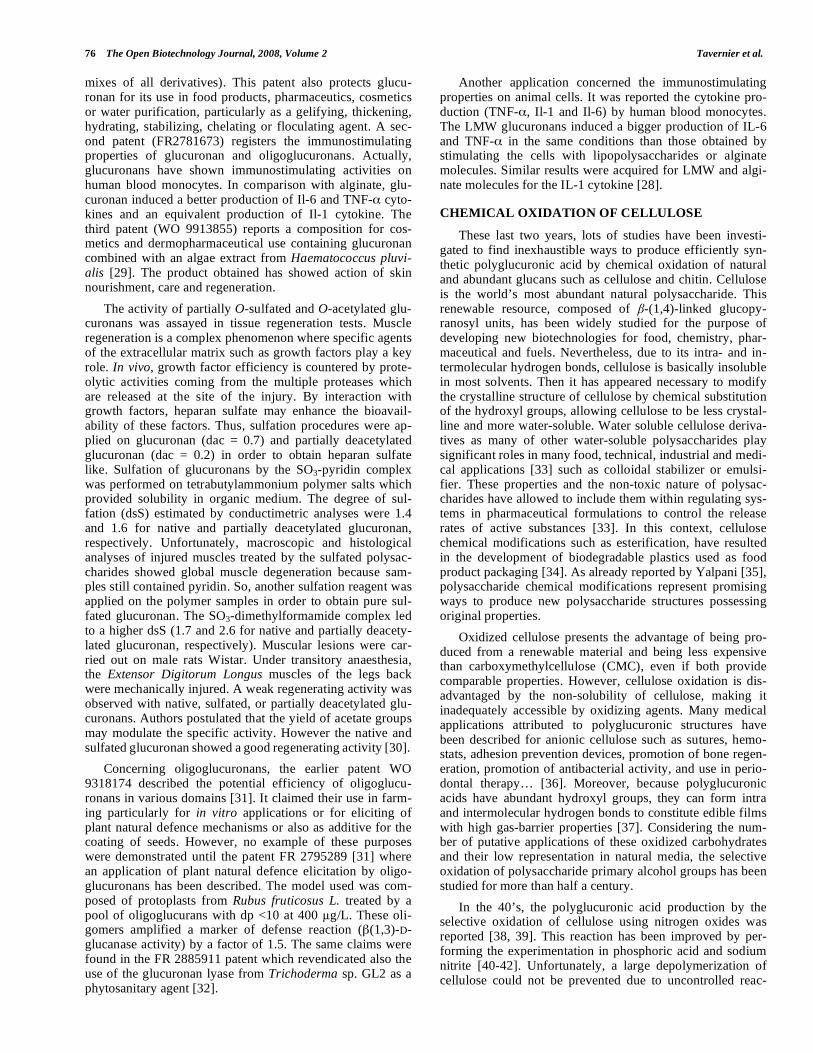
De Nooy et al. [44] first reported the efficiency of TEMPO/NaBr/NaOCl system for the yielded production of homogeneous polyuronic acid structures (Fig. 1). Conse-quently, TEMPO reagent has become the exclusive catalyst for the regioselective oxidation of high molecular weight polysaccharides. The advantages of this chemical process are its high reaction level, its high production yield, and the suit-able selectivity of primary alcohol groups (especially in car-bohydrates). Until now, lots of studies and patents have de-scribed procedures for TEMPO mediated oxidations of wa-ter-soluble natural polysaccharides and their derivatives, as well as water-insoluble polysaccharides such as chitin [46-48] and cellulose [8, 45, 49-52]. The water-soluble cellulose and chitin derivatives synthesized by the way of this reaction present a great potential with respect to their numerous rheological properties, such as gelling or thickening.
As shown in Fig. (5), production of polyglucuronic acid by primary alcohol oxidations is successful because it needs less than 1 mol % of TEMPO, aqueous sodium hypochlorite as inexpensive co-oxidant and sodium hydroxyde as mild base. Additives such as NaBr or KBr can be used to increase the rate of oxidation reaction. In fact, the oxidation reaction
is hampered by the high crystalline state of cellulosic and chitinic materials. It extremely decreases primary alcohol accessibility due to the hydrogen bond interactions. Even for a long oxidation experimentation (24 h) in alkaline medium (pH 10), the native polysaccharides keep being partially in-soluble [8].
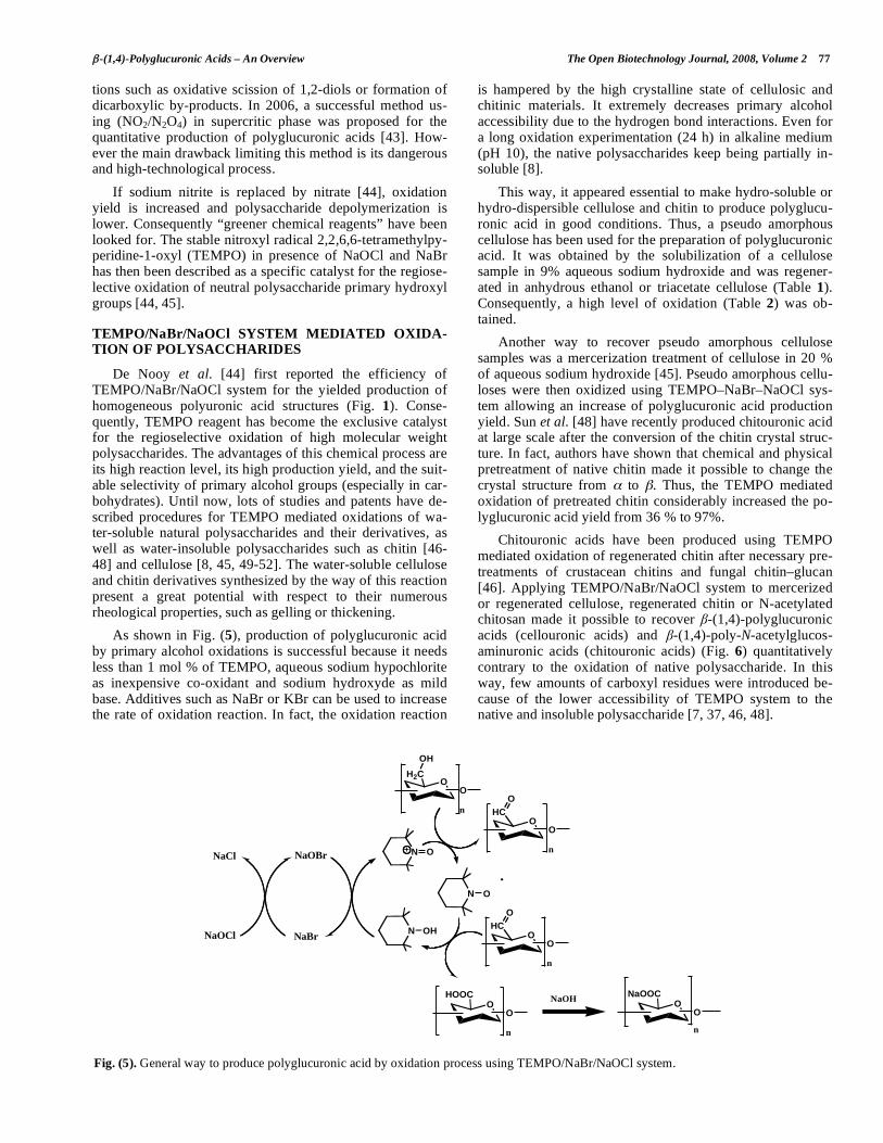
This way, it appeared essential to make hydro-soluble or hydro-dispersible cellulose and chitin to produce polyglucu-ronic acid in good conditions. Thus, a pseudo amorphous cellulose has been used for the preparation of polyglucuronic acid. It was obtained by the solubilization of a cellulose sample in 9% aqueous sodium hydroxide and was regener-ated in anhydrous ethanol or triacetate cellulose (Table 1). Consequently, a high level of oxidation (Table 2) was ob-tained.
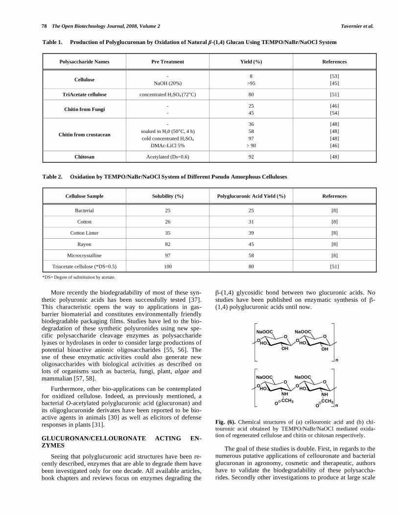
Another way to recover pseudo amorphous cellulose samples was a mercerization treatment of cellulose in 20 % of aqueous sodium hydroxide [45]. Pseudo amorphous cellu-loses were then oxidized using TEMPO–NaBr–NaOCl sys-tem allowing an increase of polyglucuronic acid production yield. Sun et al. [48] have recently produced chitouronic acid at large scale after the conversion of the chitin crystal struc-ture. In fact, authors have shown that chemical and physical pretreatment of native chitin made it possible to change the crystal structure from to . Thus, the TEMPO mediated oxidation of pretreated chitin considerably increased the po-lyglucuronic acid yield from 36 % to 97%.
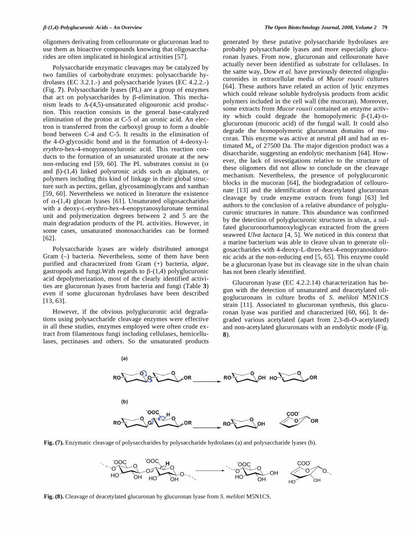
Chitouronic acids have been produced using TEMPO mediated oxidation of regenerated chitin after necessary pre-treatments of crustacean chitins and fungal chitin–glucan [46]. Applying TEMPO/NaBr/NaOCl system to mercerized or regenerated cellulose, regenerated chitin or N-acetylated chitosan made it possible to recover -(1,4)-polyglucuronic acids (cellouronic acids) and -(1,4)-poly-N-acetylglucos- aminuronic acids (chitouronic acids) (Fig. 6) quantitatively contrary to the oxidation of native polysaccharide. In this way, few amounts of carboxyl residues were introduced be-cause of the lower accessibility of TEMPO system to the native and insoluble polysaccharide [7, 37, 46, 48].
Fig. (5). General way to produce polyglucuronic acid by oxidation process using TEMPO/NaBr/NaOCl system.
N O
.
N O
N OH
OH2C
OH
O
n
O
HC
O
O
n
O
HOOC
O
n
NaOH
NaBr
NaOBr
NaOCl
NaCl
O
HC
O
O
n
O
NaOOC
O
n
78 The Open Biotechnology Journal, 2008, Volume 2 Tavernier et al.
More recently the biodegradability of most of these syn-thetic polyuronic acids has been successfully tested [37]. This characteristic opens the way to applications in gas-barrier biomaterial and constitutes environmentally friendly biodegradable packaging films. Studies have led to the bio-degradation of these synthetic polyuronides using new spe-cific polysaccharide cleavage enzymes as polysaccharide lyases or hydrolases in order to consider large productions of potential bioactive anionic oligosaccharides [55, 56]. The use of these enzymatic activities could also generate new oligosaccharides with biological activities as described on lots of organisms such as bacteria, fungi, plant, algae and mammalian [57, 58].
Furthermore, other bio-applications can be contemplated for oxidized cellulose. Indeed, as previously mentioned, a bacterial O-acetylated polyglucuronic acid (glucuronan) and its oligoglucuronide derivates have been reported to be bio-active agents in animals [30] as well as elicitors of defense responses in plants [31].
GLUCURONAN/CELLOURONATE ACTING EN-ZYMES
Seeing that polyglucuronic acid structures have been re-cently described, enzymes that are able to degrade them have been investigated only for one decade. All available articles, book chapters and reviews focus on enzymes degrading the
-(1,4) glycosidic bond between two glucuronic acids. No studies have been published on enzymatic synthesis of -(1,4) polyglucuronic acids until now.
Fig. (6). Chemical structures of (a) cellouronic acid and (b) chi-touronic acid obtained by TEMPO/NaBr/NaOCl mediated oxida-tion of regenerated cellulose and chitin or chitosan respectively.
The goal of these studies is double. First, in regards to the numerous putative applications of cellouronate and bacterial glucuronan in agronomy, cosmetic and therapeutic, authors have to validate the biodegradability of these polysaccha-rides. Secondly other investigations to produce at large scale
Table 1. Production of Polyglucuronan by Oxidation of Natural -(1,4) Glucan Using TEMPO/NaBr/NaOCl System
Polysaccharide Names Pre Treatment Yield (%) References
Cellulose -
NaOH (20%)
8
>95
[53]
[45]
TriAcetate cellulose concentrated H2SO4 (72°C) 80 [51]
Chitin from Fungi -
-
25
45
[46]
[54]
Chitin from crustacean
-
soaked in H20 (50°C, 4 h)
cold concentrated H2SO4
DMAc-LiCl 5%
36
58
97
> 90
[48]
[48]
[48]
[46]
Chitosan Acetylated (Ds=0.6) 92 [48]
Table 2. Oxidation by TEMPO/NaBr/NaOCl System of Different Pseudo Amorphous Celluloses
Cellulose Sample Solubility (%) Polyglucuronic Acid Yield (%) References
Bacterial 25 25 [8]
Cotton 26 31 [8]
Cotton Linter 35 39 [8]
Rayon 82 45 [8]
Microcrystalline 97 58 [8]
Triacetate cellulose (*DS=0.5) 100 80 [51]
*DS= Degree of substitution by acetate.
OO
HO
OH
NaOOC
OO
HO
OH
NaOOC
n
OO
HO
NH
NaOOC
OO
HO
NH
NaOOC
n
CCH3 CCH3
OO
-(1,4)-Polyglucuronic Acids – An Overview The Open Biotechnology Journal, 2008, Volume 2 79
oligomers derivating from cellouronate or glucuronan lead to use them as bioactive compounds knowing that oligosaccha-rides are often implicated in biological activities [57].
Polysaccharide enzymatic cleavages may be catalyzed by two families of carbohydrate enzymes: polysaccharide hy-drolases (EC 3.2.1.-) and polysaccharide lyases (EC 4.2.2.-) (Fig. 7). Polysaccharide lyases (PL) are a group of enzymes that act on polysaccharides by -elimination. This mecha-nism leads to -(4,5)-unsaturated oligouronic acid produc-tion. This reaction consists in the general base-catalyzed elimination of the proton at C-5 of an uronic acid. An elec-tron is transferred from the carboxyl group to form a double bond between C-4 and C-5. It results in the elimination of the 4-O-glycosidic bond and in the formation of 4-deoxy-l-erythro-hex-4-enopyranosyluronic acid. This reaction con-ducts to the formation of an unsaturated uronate at the new non-reducing end [59, 60]. The PL substrates consist in ( and )-(1,4) linked polyuronic acids such as alginates, or polymers including this kind of linkage in their global struc-ture such as pectins, gellan, glycosaminoglycans and xanthan [59, 60]. Nevertheless we noticed in literature the existence of -(1,4) glucan lyases [61]. Unsaturated oligosaccharides with a deoxy-L-erythro-hex-4-enopyranosyluronate terminal unit and polymerization degrees between 2 and 5 are the main degradation products of the PL activities. However, in some cases, unsaturated monosaccharides can be formed [62].
Polysaccharide lyases are widely distributed amongst Gram (–) bacteria. Nevertheless, some of them have been purified and characterized from Gram (+) bacteria, algae, gastropods and fungi.With regards to -(1,4) polyglucuronic acid depolymerization, most of the clearly identified activi-ties are glucuronan lyases from bacteria and fungi (Table 3) even if some glucuronan hydrolases have been described [13, 63].
However, if the obvious polyglucuronic acid degrada-tions using polysaccharide cleavage enzymes were effective in all these studies, enzymes employed were often crude ex-tract from filamentous fungi including cellulases, hemicellu-lases, pectinases and others. So the unsaturated products
generated by these putative polysaccharide hydrolases are probably polysaccharide lyases and more especially glucu-ronan lyases. From now, glucuronan and cellouronate have actually never been identified as substrate for cellulases. In the same way, Dow et al. have previously detected oligoglu-curonides in extracellular media of Mucor rouxii cultures [64]. These authors have related an action of lytic enzymes which could release soluble hydrolysis products from acidic polymers included in the cell wall (the mucoran). Moreover, some extracts from Mucor rouxii contained an enzyme activ-ity which could degrade the homopolymeric -(1,4)-D-glucuronan (mucoric acid) of the fungal wall. It could also degrade the homopolymeric glucuronan domains of mu-coran. This enzyme was active at neutral pH and had an es-timated Mw of 27500 Da. The major digestion product was a disaccharide, suggesting an endolytic mechanism [64]. How-ever, the lack of investigations relative to the structure of these oligomers did not allow to conclude on the cleavage mechanism. Nevertheless, the presence of polyglucuronic blocks in the mucoran [64], the biodegradation of cellouro-nate [13] and the identification of deacetylated glucuronan cleavage by crude enzyme extracts from fungi [63] led authors to the conclusion of a relative abundance of polyglu-curonic structures in nature. This abundance was confirmed by the detection of polyglucuronic structures in ulvan, a sul-fated glucuronorhamnoxyloglycan extracted from the green seaweed Ulva lactuca [4, 5]. We noticed in this context that a marine bacterium was able to cleave ulvan to generate oli-gosaccharides with 4-deoxy-L-threo-hex-4-enopyranosiduro- nic acids at the non-reducing end [5, 65]. This enzyme could be a glucuronan lyase but its cleavage site in the ulvan chain has not been clearly identified.
Glucuronan lyase (EC 4.2.2.14) characterization has be-gun with the detection of unsaturated and deacetylated oli-goglucuronans in culture broths of S. meliloti M5N1CS strain [11]. Associated to glucuronan synthesis, this glucu-ronan lyase was purified and characterized [60, 66]. It de-graded various acetylated (apart from 2,3-di-O-acetylated) and non-acetylated glucuronans with an endolytic mode (Fig. 8).
Fig. (7). Enzymatic cleavage of polysaccharides by polysaccharide hydrolases (a) and polysaccharide lyases (b).
O
COO-
OO
O
HO OH
-OOC
OO
HO OH
-OOC
O
H
OO
HOOH
-OOC
OH
HO OH
Fig. (8). Cleavage of deacetylated glucuronan by glucuronan lyase from S. meliloti M5N1CS.
80 The Open Biotechnology Journal, 2008, Volume 2 Tavernier et al.
Authors tried with poor success to employ this endopoly- glucuronic acid lyase in order to produce unsaturated oligo- glucuronans with putative biological activities [66]. In fact, this activity was highly specific of deacetylated glucuronan. No significant activity was observed when highly acetylated glucuronan was used as substrate. 2,3-di-O-acetyled residues had the most inhibitory effect.
These results agreed with those obtained with alginate lyases inhibited by O-acetylated mannurosyl residues [67, 68]. This high level of inhibition by acetate, associated to a low specific activity expressed by the bacterial strain synthe-sizing the polysaccharidic substrate suggested that this lyase was used by the micro-organism to control the molecular weight of its exopolysaccharide. This hypothesis was cor-roborated by literature data since polysaccharide lyase bio-synthesis is often associated to polysaccharide substrate pro-duction. Indeed, molecular biological studies have shown a genetic association between polysaccharide biosynthesis encoding genes and PL encoding genes. This association could be implicated in the regulation of medium viscosity for metabolite diffusions or for polysaccharide release [60, 69, 70].
With the same aim to generate anionic oligosaccharides at large scale, Delattre et al. [71] have investigated other sources of glucuronan lyases. Knowing the potential of fila-mentous fungi to produce polysaccharide cleavage enzyme and the previous characterisation of glucuronan pattern in fungi cell wall [3], a strain of Trichoderma sp. called Tricho-derma sp. GL2 was isolated from compost using bacterial glucuronan as single carbon source. This new fungal glucu-ronan lyase degraded all glucuronans (acetylated or not), ulvan extracted from the green seaweed Ulva lactuca and also cellouronate. [71, 55]. This endolytic enzyme (GL2) with a molecular weight of 27 kDa had a higher specific ac-tivity than the one from the S. meliloti M5N1CS strain.
The GL2 activity led to the formation of unsaturated oli-gomers with various degrees of polymerization depending of the substrate acetylation degree. The smallest substrate cleaved by the enzyme was an unsaturated trisaccharide. When deacetylated glucuronan was used, reaction products were a pool of oligomers from dp 1 to dp 3. The spontaneous transformation of the unsaturated monomer in -keto-glucuronic acid has been considered thanks to literature data [72]. When the glucuronan lyase from Trichoderma sp GL2 was applied to nascent glucuronan, product was an unsatu-rated oligomer with a dp 3 and not a mix of acetylated oli-gomers as noticed with the purified polymer [58]. The bio-logical function of this glucuronan lyase is unknown from now on. Nonetheless, a number of Trichoderma strains are well-known for their mycoparasitic action against phytopa-thogenic fungi. They can produce fungal cell wall cleavage enzymes when they are cultivated on specific substrates (like chitin or laminarin) or directly on fungal cell wall as carbon sources [73]. Moreover, glucuronan patterns or uronic acids are included in the structure of some fungal cell wall [3, 74]. These two knowledges suggested that this new glucuronan lyase could be implicated in the lysis of fungal cell wall in association with other polysaccharide cleavage enzymes [75]. At the same time of these investigations, other authors have screened from a sample of soil micro-organisms which were able to produce cellouronate lyase [56]. Surprisingly, even if cellouronate is not recognized as an abundant natural polymer, four different strains grew on media with cellouro-nate as sole carbone source (Table 3). Production of cel-louronate lyase activity was confirmed for all the strains. The one expressed by the strain Brevundimonas sp. SH203 has been purified, characterized and labelled CUL-I. Once more, this enzyme has revealed to be an endolyase with a high specificity to cellouronate. Residual activities were noticed with other polyuronates like alginate and amylouronate. Af-ter cellouronate degradation with CUL-I, the main products
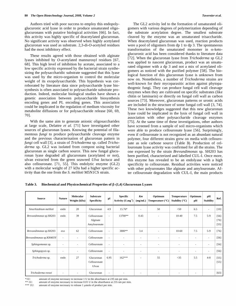
Table 3. Biochemical and Physicochemical Properties of -(1,4) Glucuronan Lyases
Source Nature Molecular
Weight (kDa)
Substrate
Specificity pI
Specific
Activity (U.mg-1
)
Km
(mg/mL)
Optimum
Temperature (°C)
Temperature
Stability (°C)
Optimum
pH
pH
Stability Ref.
Sinorhizobium meliloti endo 20 Glucuronan 4.9 15.76* - 50 <50 6.5 - [10]
Brevundimonas sp.SH203 endo 39 Cellouronate
Alginate
Amylouronate
- 13700** 0.43 - 10-40 7.5 5-9 [56]
[76]
[55]
Brevundimonas sp.SH203 exo 62 Cellouronate - 3800** 3.4 - 10-60 7.5 5-9 [76]
Brevundimonas sp.KH403Y - - Cellouronate - - - - - - - [56]
Sphingomonas sp. - - Cellouronate - - - - - - - [56]
Sphingopyxis sp. - - Cellouronate - - - - - - - [56]
Trichoderma sp. endo 27 Glucuronan
Cellouronate
Ulvan
6.95 162*** - 55 <35 5.5 4-8 [55]
[55]
Trichoderma reesei - - Glucuronan - - - - - - [63]
*1U: amount of enzyme necessary to increase 1 U in the absorbance at 235 nm per min. ** 1U: amount of enzyme necessary to increase 0.01 U in the absorbance at 235 nm per min. *** 1U: amount of enzyme necessary to release 1 mole of product per min.
-(1,4)-Polyglucuronic Acids – An Overview The Open Biotechnology Journal, 2008, Volume 2 81
were similar to those obtained after a degradation of deacety-lated glucuronan with the glucuronan lyase from Tricho-derma sp. GL2.
To resume, glucuronan and/or cellouronate lyases are until now the only enzymes working on -(1,4)-D-polyglucu- ronic acids. All the identified enzymes are endolyases. As these enzymes were discovered in various micro-organisms, we can deduce their abundance and diversity in nature. The isolation of an exolyase working specifically on oligoglucu-ronans and/or oligocellouronates could improve the compre-hension of polyglucuronic acid assimilation. Indeed, Konno et al. have recently identified an exocellouronate lyase (CUL-II) from the CUL-I producing strain (Brevundimonas sp. SH203). Until now, this is the first exolytic enzyme de-scribed for the degradation of -D-(1,4)-polyglucuronic acid. CUL-II can degrade saturated or unsaturated dimers in monomers more rapidly than the cellouronate polymer. Thus depolymerization of cellouronate is more efficient when CUL-I and CUL-II are both present in the mixture [76].
In future, to increase the number of proteins in this en-zyme family (EC 4.2.2.14), the clonage of glucuronan/cell- ouronate lyase genes seems to be unavoidable.
PURIFICATION OF POLY- AND OLIGOGLUCU-RONANS
Oligoglucuronans are negatively charged oligosaccha-rides composed of glucuronic acid residues, which can be partially acetylated on C2 and/or C3 position. They are ob-tained from enzymatic -elimination of glucuronan or cel-louronate with glucuronan/cellouronate lyases as described above. The oligoglucuronan structure can be analyzed in term of acetylation degree (dac) and polymerization degree after purification.
The most common treatment for the purification of -(1,4)-polyglucuronic acids is the precipitation (with addition of 3 volumes of alcohol or in acidic media) since the poly-saccharide has a high molecular weight [1, 27, 77, 78]. An-other useful and common method for purification is also the tangential ultrafiltration (UFT) on membranes with a mo-lecular weight cut off of 100 kDa [1, 10, 30, 58, 66, 78].
These techniques can be easily adapted to oligomer puri- fication. Alcohol precipitation with addition up to 7 volumes of alcohol allows to isolate a mixture of oligomers. In our team, alcohol precipitation is the common protocol to re-cover oligosaccharides obtained from enzymatic hydrolysis of glucuronan with Trichoderma sp. GL2 crude enzyme ex-tract. Another way is UFT because this process can be easily applied to all types of synthetized oligoglucuronans with the available membranes cut-off [79-81]. Delattre et al. [12, 57, 71, 78] investigated the production of oligoglucuronan by cleavage of highly (dac = 0.96) and normal O-acetylated glucuronan (dac = 0.7) during a fermentation process. A common S. meliloti M5N1CS culture was implemented in bioreactors. Fungal glucuronan lyase crude extract was added to broths before or after glucuronan synthesis. The fermentation media were centrifuged (34 000 g for 30 min) to remove bacteria. The supernatants containing poly- and oligoglucuronic fractions were treated by UFT through 100, 30, 10 and 5 kDa membranes successively. In these condi-tions, depolymerization of nascent mainly 3-O-acetylated glucuronan produced a large amount of a deacetylated oligo-
glucuronan of dp 3. With 2,3-di-O-acetylated glucuronan, the fungal glucuronan lyase crude extract made it possible to generate acetylated glucuronan with molecular weight below 100 kDa but did not allow to recover oligosaccharides. In 1999, Harscoat et al. [82] described the treatment of fermen-tation broths using dynamic cross-flow microfiltration (with a 0.2 m nylon membrane on a 16 cm rotating disc holder) to obtain a bacteria free medium. Improvements were no-ticed thanks to addition of valves on the holder which pro-duced an increase of the permeate flux. Moreover, the use of a rotating disc made it possible to recover the polysaccharide by UFT through a 50 kDa membrane [83]. The same tech-nique was employed using other cut-off corresponding to ultra- or nanofiltrations to separate various families of glucu-ronans and oligoglucuronans. The filtration module was composed of a 15 cm diameter aluminium disc equipped with 8 radial valves 6 mm high, rotating at adjustable speeds. The 190 cm2 membrane was fastened on the holder. The best result was obtained with a cascade of 3 ultrafiltrations at 50, 20 and 10 kDa followed by 2 nanofiltrations at 1 kDa and 700 Da. The first permeate at 50 kDa contained oligomers of dp < 25. The 20 kDa permeate contained oligomers of dp < 10. In the 10 kDa permeate oligomers of dp < 5 were recov-ered and finally oligoglucuronans with dp between 3 and 5 were collected in the 700 Da retentate [84].
All techniques described above allowed to separate fami-lies of different molecular weights but were not sufficiently accurate to obtain pure oligosaccharides with define dp and dac. So other methods such as liquid chromatography had to be used in order to purify oligosaccharides.
Unsaturated oligomers have been separated with various LC modes such as size exclusion, anion-exchange and pseu-dobioaffinity chromatography (Table 4). The most common detection method was UV absorption at around 230 nm at-tributed to -(4,5)-unsaturated uronic acids at the oligoglu-curonan non-reducing end, with routinely detectable amounts in the nanogram range. Other detection methods such as re-fractive index (RI) or evaporative light scattering detection (ELSD) could be easily used for sugar detection. The detec-tion mode was largely dependent on the kind of chromatog-raphy used.
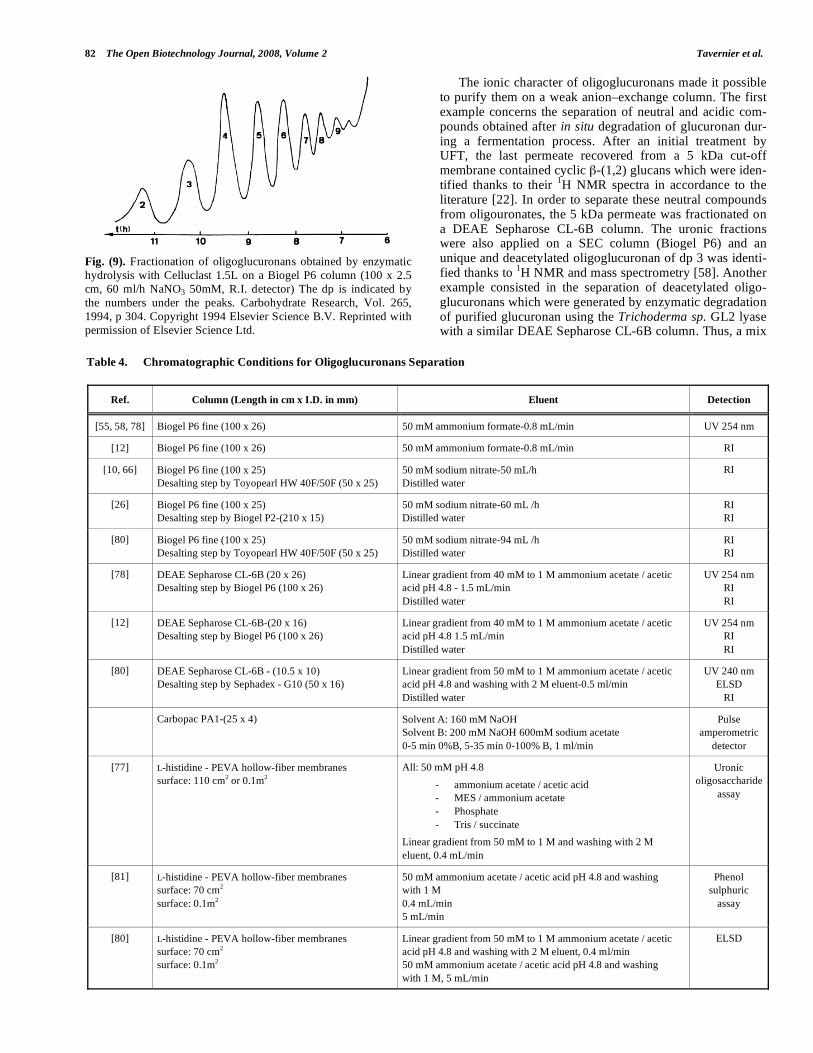
Dantas et al. [63], in the study of oligoglucuronate pre-pared by Celluclast 1.5L (Novo) hydrolysis, implemented a conventional size exclusion chromatography to separate eas-ily deacetylated oligomers up to dp 9 on a Biogel P6 column (Fig. 9). Many other authors [10, 12, 55, 58, 66, 78, 80] used this technique in order to purify oligoglucuronans. Nonethe-less, this kind of separation was disturbed by the polyanionic character of oligomers. In order to suppress this effect, an ionic strength had to be added in the eluent. Consequently a desalting step was necessary to recover pure oligosaccha-rides, unless a volatile eluent such as ammonium formate was employed. Analytical SEC-HPLC, used for example on Shodex columns (SB-802.5 coupled with SB-802), offered identical separation and could be used to monitor enzymatic degradation. However, same limitations than previously de-scribed have been observed, because of the polyanionic character. Good resolution for deacetylated oligoglucuronans were obtained but this technique revealed to be not enough efficient to separate accurately a mixture composed of acety-lated or deacetylated oligomers.
82 The Open Biotechnology Journal, 2008, Volume 2 Tavernier et al.
Fig. (9). Fractionation of oligoglucuronans obtained by enzymatic hydrolysis with Celluclast 1.5L on a Biogel P6 column (100 x 2.5 cm, 60 ml/h NaNO3 50mM, R.I. detector) The dp is indicated by the numbers under the peaks. Carbohydrate Research, Vol. 265, 1994, p 304. Copyright 1994 Elsevier Science B.V. Reprinted with permission of Elsevier Science Ltd.
The ionic character of oligoglucuronans made it possible to purify them on a weak anion–exchange column. The first example concerns the separation of neutral and acidic com-pounds obtained after in situ degradation of glucuronan dur-ing a fermentation process. After an initial treatment by UFT, the last permeate recovered from a 5 kDa cut-off membrane contained cyclic -(1,2) glucans which were iden-tified thanks to their 1H NMR spectra in accordance to the literature [22]. In order to separate these neutral compounds from oligouronates, the 5 kDa permeate was fractionated on a DEAE Sepharose CL-6B column. The uronic fractions were also applied on a SEC column (Biogel P6) and an unique and deacetylated oligoglucuronan of dp 3 was identi-fied thanks to 1H NMR and mass spectrometry [58]. Another example consisted in the separation of deacetylated oligo-glucuronans which were generated by enzymatic degradation of purified glucuronan using the Trichoderma sp. GL2 lyase with a similar DEAE Sepharose CL-6B column. Thus, a mix
Table 4. Chromatographic Conditions for Oligoglucuronans Separation
Ref. Column (Length in cm x I.D. in mm) Eluent Detection
[55, 58, 78] Biogel P6 fine (100 x 26) 50 mM ammonium formate-0.8 mL/min UV 254 nm
[12] Biogel P6 fine (100 x 26) 50 mM ammonium formate-0.8 mL/min RI
[10, 66] Biogel P6 fine (100 x 25) Desalting step by Toyopearl HW 40F/50F (50 x 25)
50 mM sodium nitrate-50 mL/h Distilled water
RI
[26] Biogel P6 fine (100 x 25) Desalting step by Biogel P2-(210 x 15)
50 mM sodium nitrate-60 mL /h Distilled water
RI RI
[80] Biogel P6 fine (100 x 25) Desalting step by Toyopearl HW 40F/50F (50 x 25)
50 mM sodium nitrate-94 mL /h Distilled water
RI RI
[78] DEAE Sepharose CL-6B (20 x 26) Desalting step by Biogel P6 (100 x 26)
Linear gradient from 40 mM to 1 M ammonium acetate / acetic acid pH 4.8 - 1.5 mL/min Distilled water
UV 254 nm RI RI
[12] DEAE Sepharose CL-6B-(20 x 16) Desalting step by Biogel P6 (100 x 26)
Linear gradient from 40 mM to 1 M ammonium acetate / acetic acid pH 4.8 1.5 mL/min Distilled water
UV 254 nm RI RI
[80] DEAE Sepharose CL-6B - (10.5 x 10) Desalting step by Sephadex - G10 (50 x 16)
Linear gradient from 50 mM to 1 M ammonium acetate / acetic acid pH 4.8 and washing with 2 M eluent-0.5 ml/min Distilled water
UV 240 nm ELSD
RI
Carbopac PA1-(25 x 4) Solvent A: 160 mM NaOH Solvent B: 200 mM NaOH 600mM sodium acetate 0-5 min 0%B, 5-35 min 0-100% B, 1 ml/min
Pulse amperometric
detector
[77] L-histidine - PEVA hollow-fiber membranes surface: 110 cm2 or 0.1m2
All: 50 mM pH 4.8
- ammonium acetate / acetic acid - MES / ammonium acetate - Phosphate - Tris / succinate
Linear gradient from 50 mM to 1 M and washing with 2 M eluent, 0.4 mL/min
Uronic oligosaccharide
assay
[81] L-histidine - PEVA hollow-fiber membranes surface: 70 cm2 surface: 0.1m2
50 mM ammonium acetate / acetic acid pH 4.8 and washing with 1 M 0.4 mL/min 5 mL/min
Phenol sulphuric
assay
[80] L-histidine - PEVA hollow-fiber membranes surface: 70 cm2 surface: 0.1m2
Linear gradient from 50 mM to 1 M ammonium acetate / acetic acid pH 4.8 and washing with 2 M eluent, 0.4 ml/min 50 mM ammonium acetate / acetic acid pH 4.8 and washing with 1 M, 5 mL/min
ELSD
-(1,4)-Polyglucuronic Acids – An Overview The Open Biotechnology Journal, 2008, Volume 2 83
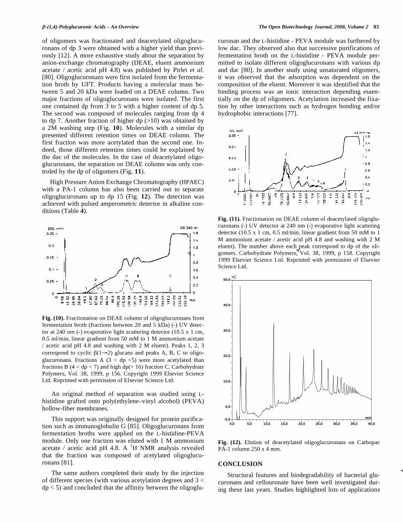
of oligomers was fractionated and deacetylated oligoglucu-ronans of dp 3 were obtained with a higher yield than previ-ously [12]. A more exhaustive study about the separation by anion-exchange chromatography (DEAE, eluent ammonium acetate / acetic acid pH 4.8) was published by Pirlet et al. [80]. Oligoglucuronans were first isolated from the fermenta-tion broth by UFT. Products having a molecular mass be-tween 5 and 20 kDa were loaded on a DEAE column. Two major fractions of oligoglucuronans were isolated. The first one contained dp from 3 to 5 with a higher content of dp 5. The second was composed of molecules ranging from dp 4 to dp 7. Another fraction of higher dp (>10) was obtained by a 2M washing step (Fig. 10). Molecules with a similar dp presented different retention times on DEAE column. The first fraction was more acetylated than the second one. In-deed, those different retention times could be explained by the dac of the molecules. In the case of deacetylated oligo-glucuronans, the separation on DEAE column was only con-troled by the dp of oligomers (Fig. 11).
High Pressure Anion Exchange Chromatography (HPAEC) with a PA-1 column has also been carried out to separate oligoglucuronans up to dp 15 (Fig. 12). The detection was achieved with pulsed amperometric detector in alkaline con-ditions (Table 4).
Fig. (10). Fractionation on DEAE column of oligoglucuronans from fermentation broth (fractions between 20 and 5 kDa) (-) UV detec-tor at 240 nm (-) evaporative light scattering detector (10.5 x 1 cm, 0.5 ml/min, linear gradient from 50 mM to 1 M ammonium acetate / acetic acid pH 4.8 and washing with 2 M eluent). Peaks 1, 2, 3 correspond to cyclic (1 2) glucans and peaks A, B, C to oligo-glucuronans. Fractions A (3 < dp <5) were more acetylated than fractions B (4 < dp < 7) and high dp(> 16) fraction C. Carbohydrate Polymers, Vol. 38, 1999, p 156. Copyright 1999 Elsevier Science Ltd. Reprinted with permission of Elsevier Science Ltd.
An original method of separation was studied using L-histidine grafted onto poly(ethylene–vinyl alcohol) (PEVA) hollow-fiber membranes.
This support was originally designed for protein purifica-tion such as immunoglobulin G [85]. Oligoglucuronans from fermentation broths were applied on the L-histidine-PEVA module. Only one fraction was eluted with 1 M ammonium acetate / acetic acid pH 4.8. A 1H NMR analysis revealed that the fraction was composed of acetylated oligoglucu-ronans [81].
The same authors completed their study by the injection of different species (with various acetylation degrees and 3 < dp < 5) and concluded that the affinity between the oligoglu-
curonan and the L-histidine - PEVA module was furthered by low dac. They observed also that successive purifications of fermentation broth on the L-histidine - PEVA module per-mitted to isolate different oligoglucuronans with various dp and dac [80]. In another study using unsaturated oligomers, it was observed that the adsorption was dependent on the composition of the eluent. Moreover it was identified that the binding process was an ionic interaction depending essen-tially on the dp of oligomers. Acetylation increased the fixa-tion by other interactions such as hydrogen bonding and/or hydrophobic interactions [77].
Fig. (11). Fractionation on DEAE column of deacetylated oligoglu-curonans (-) UV detector at 240 nm (-) evaporative light scattering detector (10.5 x 1 cm, 0.5 ml/min, linear gradient from 50 mM to 1 M ammonium acetate / acetic acid pH 4.8 and washing with 2 M eluent). The number above each peak correspond to dp of the oli-gomers. Carbohydrate Polymers, Vol. 38, 1999, p 158. Copyright 1999 Elsevier Science Ltd. Reprinted with permission of Elsevier Science Ltd.
Fig. (12). Elution of deacetylated oligoglucuronans on Carbopac PA-1 column 250 x 4 mm.
CONCLUSION
Structural features and biodegradability of bacterial glu-curonans and cellouronate have been well investigated dur-ing these last years. Studies highlighted lots of applications
_
min_
min
_
min
_
min_
min
_
min
_
nC
min
_
nC
_
min
_
min
_
nC
min
_
nC
_
min
_
min
0,0 5,0 10,0 15,0 20,0 25,0 30,0 35,0 40,0
-5,0
0,0
10,0
20,0
30,0
40,0
50,027 octobre # Oligo 10g/ IntAmp_
nC
min
nC
0,0 5,0 10,0 15,0 20,0 25,0 30,0 35,0 40,0
-5,0
0,0
10,0
20,0
30,0
40,0
50,027 octobre # Oligo 10g/ IntAmp_
nC
min
nC
84 The Open Biotechnology Journal, 2008, Volume 2 Tavernier et al.
in various industrial areas but patents were not always suc-cessfully associated with industrial developments. In this context the most promising way for valorisation could be the characterization of biological activities of oligomers. This implies to obtain them at large scale and as mixes well char-acterized (dp and dac). Thus degradation of macromolecules by glucuronan lyases associated to ultra and/or nanofiltration processes could open the way to new enzymatic reactors. Moreover, developments of immobilized glucuronan lyases would allow a better control of dp. However, significant im-provements of polyglucuronic acids degradation seem to be dependent on molecular biology engineering. The avaibility of bacterial and fungal genes coding for these enzymes could make achievable the enzyme directed mutagenesis by PCR strategies. So the biodegradation of acetylated glucuronans in oligomers, often described as bioactivators in animals and vegetables could be achieved. To conclude, the role of the glucuronan or cellouronate lyases in the physiology of mi-cro-organisms and their detection in other micro-organisms will be a challenge to identify other sources of natural glucu-ronans.
REFERENCES
[1] Courtois J, Courtois B. In-Biopolymers, Polysaccharides II, S De Baets, EJ Vandamme, A Steinbüchel (Eds), A Steinbuchel, Bel-gium 2002; 213-235.
[2] Heyraud A, Courtois J, Dantas L, Colin-Morel P, Courtois B. Structural characterization and rheological properties of an ex-tracellular glucuronan produced by a Rhizobium meliloti M5N1 mutant strain. Carbohydr Res 1993; 240(24): 71-78.
[3] De Ruiter GA, Josso SL, Colquhoun IJ, Voragen AGJ, Rombouts FM. Isolation and characterization of (1–4)-D-glucuronans from extracellular polysaccharides of moulds belonging to Mucorales. Carbohydr Polym 1992; 18:(1): 1-7.
[4] Ray B, Lahaye M. Cell-wall polysaccharides from the marine green algae Ulva "rigida" (Ulvales,Chlorophyta). Extraction and chemi-cal composition. Carbohydr Res 1995; 274: 251-261.
[5] Lahaye M, Brunel M, Bonnin E. Fine chemical structure analysis of oligosaccharides produced by an ulvan-lyase degradation of the water-soluble cell-wall polysaccharides from Ulva sp. (Ulvales, Chlorophyta). Carbohydr Res 1997; 304: 325-333.
[6] De Nooy AEJ, Besemer AC, van Bekkum H. Highly selective nitroxyl radical-mediated oxidation of primary alcohol groups in water-soluble glucans. Carbohydr Res 1995; 269: 89-98.
[7] Isogai A, Kato Y. Preparation of polyglucuronic acid from cellu-lose by TEMPO-mediated oxidation. Cellulose 1998; 5: 153-164.
[8] Tahiri C, Vignon MR. TEMPO-oxidation of cellulose: synthesis and characterizations of polyglucuronans. Cellulose 2000; 7: 177-188.
[9] Saito T, Yanagisawa M, Isogai A. TEMPO-mediated oxidation of native cellulose: SEC-MALLS analysis of water-soluble and in-soluble fractions in the oxidized products. Cellulose 2005; 12: 305-315.
[10] Da Costa A, Michaud P, Petit E, Courtois B, Courtois J. Purifica-tion and properties of a glucuronan lyase from sinorhizobium meliloti M5N1CS (NCIMB 40472). Appl Environ Microb 2001; 67(11): 5197-5203.
[11] Michaud P, Courtois J, Courtois B, et al. Physicochemical proper-ties of extracellular (1 4)- -D-glucuronan produced by the Rhizo-bium meliloti M5N1CS strain during fermentation: evidence of degradation by an exoenzyme activated by Mg2+. Int J Biol Mac-romol 1994; 16: 301-305.
[12] Delattre C, Michaud P, Lion JM, Courtois J, Courtois B. Produc-tion of glucuronan oligosaccharides using a new glucuronan lyase activity from a Trichoderma sp strain. J Biotechnol 2005; 118: 448-457.
[13] Kato Y, Habu N, Yamaguchi J, et al. Biodegradation of -1,4-linked polyglucuronic acid (cellouronic acid). Cellulose 2002; 9: 75-81.
[14] Gage DJ. Infection and invasion of roots by symbiotic, nitrogen-fixing rhizobia during nodulation of temperate legumes. Microbiol Mol Biol R 2004; 68: 280-300.
[15] Heyraud A, Rinaudi M, Courtois B. Comparative studies of extracellular polysaccharide elaborated by Rhizobium meliloti
strain M5N1 in defined medium and in non-growing cell suspensions. Int J Biol Macromol 1986; 8: 85-88.
[16] Courtois J, Seguin JP, Duclosmesnil S, et al. A (1 4)- -D-glucuronan excreted by a mutant of the Rhizobium meliloti M5N1 strain. J Carbohydr Chem 1993; 12: 441-448.
[17] Gonzales ML, Courtois J, Heyraud A, et al. Selection of a succino-glycan-deficient Rhizobium meliloti mutant producing a partially acetylated (1 4)- -D-glucuronan, symbiotic properties analysis. Ann NY Acad Sci 1996; 1: 53-60.
[18] Heyraud A, Dantas L, Courtois J, Courtois B, Hellbert W, Chanzy H. Crystallographic data on bacterial (1 4)- -D-glucuronan. Car-bohydr Res 1994; 258: 275-279.
[19] Braccini I, Heyraud A, Perez S. Three dimensional features of bacterial polysaccharide (1 4)- -D-glucuronan: a molecular mod-eling study. Biopolymers 1998; 45: 165-175.
[20] French AD, Dowd MK. Exploration of disaccharide conformations by molecular mechanics. J Mol Struct 1993; 286: 183-201.
[21] Courtois J, Seguin JP, Roblot C, et al. Exoplysaccharide production by the Rhizobium meliloti M5N1 CS strain. Location and quantita-tion of the sites of O-acetylation. Carbohydr Polym 1994; 25: 7-12.
[22] Michaud P, Roblot C, Courtois J, et al. Effect of Mg2+ on produc-tion and O-acetylation of glucuronan excreted by the Rhizobium
meliloti M5N1CS strain during fermentation. Lett Appl Microbiol 1995; 20: 110-112.
[23] Rinaudo M, Milas M. Interaction of monovalent and divalent coun-terions with some carboxylic polysaccharides. J Polym Sci 1974; 12: 2073-2081.
[24] Braccini I, Grasso RP, Perez S. Conformational and configurational features of acidic polysaccharides and their interactions with cal-cium ions : a molecular modeling investigation. Carbohydr Res 1999; 317: 119-130.
[25] Wedlock DJ, Fasihuddin BA, Phillips GO. Comparison of molecu-lar weight determination of sodium alginate by sedimentation-diffusion and light scattering, Int J Biol Macromol 1986; 8: 57-61.
[26] Dantas L, Heyraud A, Courtois J, Courtois B, Milas M. Physico-chemical properties of "exogel" exocellular (1 4)-D-glucuronan from Rhizobium meliloti strain M5N1CS (NCIMB 40472). Carbo-hydr Polym 1994; 24: 185-191.
[27] Courtois-Sambourg J, Courtois B, Heyraud A, Colin-Morel P, Rinaudo-Duhem M. Polymer compounds of the glycuronic acid, method of preparation and utilization particularly as gelifying, thickenning, hydrating, stabilizing, chelating or floculating means. WO 9318174. 1993.
[28] Courtois-Sambourg J, Courtois B. Use of glucuronan as an immu-nostimulating agent and process for its preparation. FR 2781673. 2000.
[29] Lintner K. Composition for cosmetic or dermopharmaceutical use containing a combination of algae extract and exoplysaccharides. WO9913855. 1999.
[30] Petit E, Papy-Garcia D, Muller G, Courtois B, Caruelle JP, Cour-tois J. Controlled Sulfatation of Natural Anionic Bacterial Polysac-charides Can Yield Agents with Specific Regenerating Activity in
Vivo. Biomacromolecules 2004; 5(2): 445-452. [31] Lienart Y, Heyraud A, Sevenou O. Utilisation des polyméres 1,4
beta-D-glycuronanes et d’oligosaccharides glycuroniques dérives en tant que phytosanitaires et/ou fertilisants. FR 2795289. 1999.
[32] Michaud P, Delattre C, Courtois B, Sambourg-Courtois J. Glucuro-nane lyase pour le clivage de polysaccharides constitues par ou contenant des glucuronanes, procédé de préparation et utilisation notamment dans le domaine phytosanitaire. FR 2885911. 2005.
[33] Florence AT, Attwood D. Physicochemical Principles of Pharmacy. 2nd ed. Macmillan London. 1988.
[34] Gourson C, Benhaddou R, Granet R, Krausz P, Saulnier L, Thi-bault JF. Preparation of biodegradable plastic in microwave oven and solvent-free conditions. C R Acad Sci Paris 1999; 2: 75-78.
[35] Yalpani M. A survey of recent advances in selective chemical and enzymic polysaccharide modifications. Tetrahedron 1985; 41: 2957-3020.
[36] Kumar V, Dong Y. Biodegradable oxidized cellulose esters. US patent. 2002086990. 2002.
-(1,4)-Polyglucuronic Acids – An Overview The Open Biotechnology Journal, 2008, Volume 2 85
[37] Kato Y, Kaminaga JI, Matsuo R, Isogai A. Oxygen Permeability and Biodegradability of Polyuronic Acids Prepared from Polysac-charides by TEMPO-Mediated Oxidation. J Polym Environ 2005; 13(3): 261-266.
[38] Yackel EC, Kenyon WO. Oxidation of cellulose by nitrogen diox-ide. J Am Chem Soc 1942; 64: 121-127.
[39] Maurer K, Reiff G. Oxidation of cellulose with NO2. J Macromol Chem 1943; 1: 27–34.
[40] Painter TJ. Preparation and periodate oxidation of C-6-oxycellulose: conformational interpretation of hemiacetal stability. Carbohydr Res 1977; 55: 95-103.
[41] Cesaro A, Delben F, Painter TJ, Paoletti S. New polyuronates from natural glucans. In Crescenzi V, Dea ICM, Stivala SS. (Eds.), New developments in industrial polysaccharides. New York: Gordon and Breach. 1985.
[42] Painter TJ, Cesaro A, Delben F, Paoletti S. New glucuronoglucans obtained by oxidation of amylose at position 6. Carbohydr Res 1985; 140: 61-68.
[43] Vignon M, Montanari S, Samain D, Condoret JS. Procédés d’oxydation contrôlée des polysaccharides. FR 2873700. 2006.
[44] DeNooy AEJ, Besemer AC, van Bekkum H. Highly selective ni-troxyl radical-mediated oxidation of primary alcohol groups in wa-ter-soluble glucans. Carbohydr Res 1985; 269: 89–98.
[45] Isogai A, Kato Y. Preparation of polyuronic acid from cellulose by TEMPO-mediated oxidation. Cellulose 1998; 5(3): 153-164.
[46] Muzzarelli RAA, Muzzarelli C, Cosani A, Tjerbojevich M. 6-Oxychitins, novel hyaluronan-like regiospecifically carboxylated chitins. Carbohydr Polym 1999; 39: 361-367.
[47] Kato Y, Kaminaga J, Matsuo R, Isogai A. TEMPO-mediated oxi-dation of chitin, regenerated chitin and N-acetylated chitosan. Car-bohydr Polym 2004; 58: 421-426.
[48] Sun L, Du Y, Yang J, et al. Conversion of crystal structure of the chitin to facilitate preparation of a 6-carboxychitin. Carbohydr Po-lym 2006; 66(2): 168-175.
[49] Chang PS, Robyt JF. Oxidation of primary alcohol groups of natu-rally occurring polysaccharides with 2,2,6,6-tetramethyl-1-piperidine oxoammonium ion. J Carbohydr Chem 1996; 15: 819-830.
[50] Fleury E, Vignon MR, Gomez Bujedo S. Procédé de préparation d’acide poly- ou copolyglucuronique. WO 03035699. 2002.
[51] Gomez-Bujedo S, Fleury E, Vignon MR. Preparation of cellouronic acids and partially acetylated cellouronic acids by tempo/naclo oxi-dation of water-soluble cellulose acetate. Biomacromolecules 2004; 5(2): 565-571.
[52] Saito T, Isogai A. TEMPO-mediated oxidation of native cellulose. The effect of oxidation conditions on chemical and crystal struc-tures of the water-insoluble fractions. Biomacromolecules 2004; 5(5): 1983-1989.
[53] Kitaoka T, Isogai A. Rosin sizing of pulps modified by TEMPO-mediated oxidation. Nord Pulp Paper Res J 2000; 15: 177-182.
[54] Muzzarelli RAA, Miliani M, Cartolari M, Tarsi R, Tosi G, Muzza-relli C. Polyuronans obtained by regiospecific oxidation of poly-saccharides from Aspergillus niger, Trichoderma reesei and Sapro-
legnia sp. Carbohydr Polym 2000, 43: 55-61. [55] Delattre C, Michaud P, Elboutachfaiti R, Courtois B, Courtois J.
Production of oligocellouronates by biodegradation of oxidized cel-lulose. Cellulose 2006; 13: 63-71.
[56] Konno N, Habu N, Maeda I, Azuma N, Isogai A. Cellouronate ( -1,4-linked polyglucuronate) lyase from Brevundimonas sp. SH203: Purification and characterization. Carbohydr Polym 2006; 64(4): 589-596.
[57] Delattre C, Michaud P, Courtois B, Courtois J. Oligosaccharides engineering from plants and algae - Applications in Biotechnology and therapeutic. Minerva Biotecnol 2005; 17: 107-117.
[58] Delattre C, Michaud P, Keller C, Courtois B, Courtois J. Production of oligoglucuronans by enzymatic depolymerization of nascent glucuronan. Biotechnol Progr 2005; 21(6): 1775-1781.
[59] Sutherland IW. Polysaccharide lyases. FEMS Microbiol Rev 1995; 16: 323-347.
[60] Michaud P, Da Costa A, Courtois B, Courtois J. Polysaccharide lyases - Recent development as biotechnological tools. Crit Rev Biotechnol 2003; 23(4): 233-266.
[61] Yu S, Refdahl C, Lundt I. Enzymatic description of the anhydro- fructose pathway of glycogen degradation: I. Identification and pu-rification of anhydrofructose dehydratase, ascopyrone tautom-
erase and -1,4-glucan lyase in the fungus Anthracobia melaloma. BBA - General Subjects 2004; 1672(2,3): 120-129.
[62] Shevchik VE, Condemine G, Robert-Baudouy J, Hugouvieux-Cotte-Pattat N. The exopolygalacturonate lyase PelW and the oli-gogalacturonate lyase OgL, two cytoplasmic enzymes of pectin ca-tabolism in Erwinia chrysanthemi 3937. J Bacteriol 1999; 181: 3912-3919.
[63] Dantas L, Courtois J, Courtois B, Seguin JP, Gey C, Heyraud A. NMR Spectroscopic investigation of oligoglucuronates prepared by enzymatic hydrolysis of (1 4)- -D-glucuronan. Carbohydr Res 1994; 265: 303-310.
[64] Dow JM, Villa VD. Oligoglucuronide production in Mucor rouxii: evidence for a role for endohydrolases in Hyphal extension. J Bac-teriol 1980; 142(3): 939-944.
[65] Lahaye M. NMR spectroscopic characterisation of oligosaccharides from two Ulva rigida ulvan samples (Ulvales, Chlorophyta) de-graded by a lyase. Carbohydr Res 1998; 314: 1-12.
[66] Da Costa A, Michaud P, Heyraud A, Colin-Morel P, Courtois B, Courtois J. Acetyl Substitution of Glucuronan Influences Glucu-ronan Cleavage by Glya from Sinorhizobium meliloti M5N1CS (NCIMB 40472). Carbohydr Polym 2003; 51(2): 223-228.
[67] Kennedy L, McDowell K, Sutherland IW. Alginases from Azotobacter species. J Gen Microbiol 1992; 138: 3007-3013.
[68] Skjak-Braek G, Espevik T. Application of alginate gels in biotech-nology and biomedicine. Carbohydr Eur 1996; 14: 19-25.
[69] Hatch RA, Schiller NL. Alginate lyase promotes diffusion of ami-noglycosides through the extracellular polysaccharide of mucoid Pseudomonas aeruginosa. Antimicrob Agents Chemother 1998; 42: 974-977.
[70] Boyd A, Ghosh M, May TB, Shinabarger D, Keogh R, Chakrabarty AM. Sequence of the algL gene of Pseudomonas aeruginosa and purification of its alginate lyase product. Gene 1993; 131: 1-8.
[71] Delattre C, Michaud P, Keller C, et al. Purification, characteriza-tion and biological properties of a novel polysaccharide lyase from Trichoderma sp. GL2. Appl Microb Biotechnol 2006; 70: 437-443.
[72] Preiss J, Ashwell G. Alginic acid metabolism in bacteria J Biol Chem 1962; 237: 309-316.
[73] De la Cruz J, Pintor-Toro JA, Benitez T, Llobell A, Romero LC. A novel endo- -1,3-glucanase, BGN13.1 involved in the mycopara-sitism of Trichoderma harzanium. J Bacteriol 1995; 177(23): 6937-6945.
[74] Schoffelmeer EAM, Klis FM, Sietsma JH, Cornelissen BJC. The cell wall of Fusarium oxysporum. Fungal Genet Biol 1999; (27): 275-282.
[75] Hjeljord L, Tronsmo A. 1998. Trichoderma and Gliocladium in biocontrol: an overview. In: Kubicek CP, Harman GE (eds) Tricho-derma and Gliocladium. Taylor & Francis, London, pp. 135-151.
[76] Konno N, Habu N, Maeda I, Azuma N, Isogai A. Purification and characterization of a novel cellouronate lyase from Brevundimonas sp. SH203. Carbohydr Res 2007; in press.
[77] Delattre C, Michaud P, Harnze K, Courtois B, Courtois J, Vijaya-lakshmi MA. Purification of oligouronides using hollow-fiber membrane functionalised with L-histidine. J Chromatogr A 2005; 1099(1-2): 121-126.
[78] Delattre C, Michaud P, Courtois J, Courtois B. Production of 0-acetylated oligouronides by depolymerization of a natural highly acetylated anionic bacterial polysaccharide. Enzyme Microb Tech 2007; 41(3): 250-257.
[79] Pau-Roblot C, Petit E, Sarazin C, et al. Studies of low molecular weight samples of glucuronans with various acetylation degree. Biopolymers 2002; 64(1): 34-43.
[80] Pirlet AS, Guentas L, Heyraud A, et al. Influence of acetyl sub-stituent on oligoglucuronans séparation by anion exchange chroma-tography. Carbohydr Polym 1999; 38(2): 155-160.
[81] Pirlet AS, Pitiot O, Guentas L, et al. Separation of low-rnolecular-mass acetylated glucuronans on L-histidine immobilized onto poly(ethylene-vinyl alcohol) hollow-fiber membranes. J Chroma-togr A 1998; 826(2): 157-166.
[82] Harscoat C, Jaffrin MY, Bouzerar R, Courtois J. Influence of fermentation conditions and microfiltration processes on membrane fouling during recovery of glucuronane polysaccharides from fer-mentation broths. Biotechnol Bioeng 1999; 65(5): 500-11.
[83] Brou A, Jaffrin MY, Ding LH, Courtois J. Microfiltration and ultrafiltration of polysaccharides produced by fermentation using a
86 The Open Biotechnology Journal, 2008, Volume 2 Tavernier et al.
rotating disk dynamic filtration system. Biotechnol Bioeng 2003; 82(4): 429-37.
[84] Mellal M, Ding LH, Jaffrin M, Delattre C, Michaud P, Courtois J. Extraction of oligoglucuronans of low degrees of polymerisation from a fermentation broth by cascade filtration using a rotating disk module. Desalination 2006; 199(1): 207-209.
[85] Bueno SMA, Haupt K, Vijayalakshmi MA. Separation of immuno-globulin G from human serum by pseudobioaffinity chromatogra-phy using immobilized L-histidine in hollow fibre membranes. J Chromatogr B Biomed Appl 1995; 667(1): 57-67.
Received: September 17, 2007 Revised: February 26, 2008 Accepted: February 26, 2008