THE JOURNAL OF CHEMISTRY Vol. No. 3, Issue pp. 1993 by … · THE JOURNAL OF BIOLOGICAL CHEMISTRY 0...
8
THE JOURNAL OF BIOLOGICAL CHEMISTRY 0 1993 by The American Society for Biochemistry and Molecular Biology, Inc Vol. 268, No. 3, Issue of January 25, pp. 2092-2099,1933 Printed in U. S. A. Novel 8-Base Pair Sequence (Drosophila DNA Replication-related Element) and Specific Binding Factor Involved in the Expression of Drosophila Genes for DNA Polymerase a! and Proliferating Cell Nuclear Antigen* (Received for publication, March 16, 1992) Fumiko Hirose$, Masamitsu YamaguchiS, Hiroshi HandaQ, Yukio Inomatall,and Akio MatsukageSII From the $Laboratory of Cell Biology, Aichi Cancer Center Research Institute, Chikusa-ku, Nagoya 464, the $Faculty of Bioscience and Biotechnology, Tokyo Institute of Technology, 4259 Nagatsuda-cho, Midori-ku, Yokohama 227, and the TDepartment of Applied Chemistry, Faculty of Science and Technology, Keio University, 3-14-1 Hiyoshi-cho, Kouhoku-ku, Yokohama 223, Japan Upstream regions containing a novel common 8-base pair (bp) palindromic sequence, 5”TATCGATA (Dro- sophila DNA replication-related element (DRE)), are required for thehigh expression of Drosophila genes for DNA polymerase a! and the proliferating cell nu- clear antigen (PCNA) (an auxiliary protein for DNA polymerase 6). Three DREs and one DRE are present in the DNA polymerase (Y gene (nucleotides -217, -83, and -30 with respect to the transcription initiation site) and in the PCNA gene (nucleotide -loo), respec- tively. Deletions or 2-bp insertional mutations of DRE sequences led to an extensive reduction of promoter activities of both genes. Chemically synthesized oligo- nucleotides containing DRE sequences greatly stimu- lated the activityof the heterologous promoter of the Drosophila metallothionein gene, in addition to the promoter of the PCNA gene, when they were placed upstream from these promoters in a normal or a re- verseorientation.Thestimulatoryeffectincreased synergistically and depended on the number of DREs. DRE activated the promoter when placed within 1.4 kilobases upstream from the promoter, but was much less active when placed 2.5 kilobases or more apart from the promoter. Using a gel mobility shift assay method, we obtained evidence for a proteinfactor (DREF) in the nuclear extract of cultured Drosophila cells (Kc cells), and this factor specifically binds to DREs of both genes. DNase I footprinting analysis indicated that DREF binds to the 24-bp DRE region of the DNA polymerase a! gene in which 8-bp palindromic sequences are centered. A UV cross-linking experi- ment revealed that a polypeptide of -90 kDa in the nuclear extract interacts directly with the DRE se- quence. Using DRE-conjugated latex particles, DREF was affinity-purified from the Kc cell nuclear extract. By comparing results obtained by SDS-polyacrylamide gel electrophoresis and gel mobility shift experiments, we concluded that DREF is associated with the 86-kDa polypeptide. On gel filtration chromatography, a single peak of DREF activity was recovered in fractions cor- *This work was supported in part by a grant-in-aid from the Ministry of Education, Science, and Culture of Japan. The costs of publication of this article were defrayed in part by the payment of page charges. This article must therefore be hereby marked “adoer- tisement” in accordance with 18 U.S.C. Section 1734 solely to indicate this fact. The nucleotide sequence(s) reported in thispaper has been submitted to the GenBankTM/EMBL Data Bank with accession numberfs) D90310 and M33950. 11 To whom correspondence should be addressed. Tel.: 81-52-762- 6111 (ext. 8818); Fax: 81-52-763-5233. responding to a molecular mass of 170 kDa, and the 86-kDa polypeptide was detected only in the corre- sponding fractions; thus, active DREF is probably a homodimeric form of the 86-kDa polypeptide. DREF may play important roles in coordinating expressions of Drosophila DNA replication-related genes. Onset of eukaryotic DNA replication requires activities of two levels of regulatory systems: one is the induction of coordinate expressions of many replication-related genes, and the other is the organization of protein complexes consisting of enyzymes and regulatory factors for initiation at replication origins and elongation of DNA strands. Biochemical studies revealed that cell cycle-dependent ac- tivations of DNA replication enzymes are regulated by phys- iological rather than genetic systems. Using cell-free replica- tion systems such as that for simian virus 40 DNA or extracts of Xenopus oocytes, it has been demonstrated that cell cycle- dependent organization and degradation of the DNA replica- tion enzyme complex may be mainly regulated through sys- tems such as protein phosphorylation by the cdc2-protein kinase (Nasheuer et at., 1991; D’Urso et al., 1990; Blow and Nurse, 1990). The abundance of mRNAs and activities of human DNA polymerase a, PCNA,’ murine thymidylate synthetase, and primase were seen to increase only marginally at the G1/S boundary in proliferating cells (Wahl et al., 1988; Chang et al., 1990; Ottabio et al., 1990; Lieberman et al., 1988; Tseng et at., 1989). A dramatic increase in expressions of animal cell DNA replication related-genes was observed when cells en- teredtheproliferatingstate.Expression of these genes is much reduced in quiescent cells reaching confluency or in associationwith cellular differentiation(Matsukageet al., 1987; Yamaguchi et al., 1990). Therefore, molecular mecha- nisms involved in the induction and repression of DNA rep- lication-related genes have to be elucidated. Little is known of cis-elements or trans-acting factors re- quired for the appropriate expression of animal cell DNA replication-related genes as only a few of these genes have been isolated. It has been suggested that many enzymes form enzyme complexes to carry out chromosomal DNA replication (Fry and Loeb, 1986). Therefore, a common molecular mech- anism responsible for the coordinate induction and repression The abbreviations used are: PCNA, proliferating cell nuclear antigen; bp, base pair(s); kb, kilobase(s); DRE, Drosophila DNA replication-related element; DREF, DRE-binding factor; CAT, chlor- amphenicol acetyltransferase. 2092
Transcript of THE JOURNAL OF CHEMISTRY Vol. No. 3, Issue pp. 1993 by … · THE JOURNAL OF BIOLOGICAL CHEMISTRY 0...
THE JOURNAL OF BIOLOGICAL CHEMISTRY 0 1993 by The American Society for Biochemistry and Molecular Biology, Inc Vol. 268, No. 3, Issue of January 25, pp. 2092-2099,1933
Printed in U. S. A.
Novel 8-Base Pair Sequence (Drosophila DNA Replication-related Element) and Specific Binding Factor Involved in the Expression of Drosophila Genes for DNA Polymerase a! and Proliferating Cell Nuclear Antigen*
(Received for publication, March 16, 1992)
Fumiko Hirose$, Masamitsu YamaguchiS, Hiroshi HandaQ, Yukio Inomatall, and Akio MatsukageSII From the $Laboratory of Cell Biology, Aichi Cancer Center Research Institute, Chikusa-ku, Nagoya 464, the $Faculty of Bioscience and Biotechnology, Tokyo Institute of Technology, 4259 Nagatsuda-cho, Midori-ku, Yokohama 227, and the TDepartment of Applied Chemistry, Faculty of Science and Technology, Keio University, 3-14-1 Hiyoshi-cho, Kouhoku-ku, Yokohama 223, Japan
Upstream regions containing a novel common 8-base pair (bp) palindromic sequence, 5”TATCGATA (Dro- sophila DNA replication-related element (DRE)), are required for the high expression of Drosophila genes for DNA polymerase a! and the proliferating cell nu- clear antigen (PCNA) (an auxiliary protein for DNA polymerase 6). Three DREs and one DRE are present in the DNA polymerase (Y gene (nucleotides -217, -83, and -30 with respect to the transcription initiation site) and in the PCNA gene (nucleotide -loo), respec- tively. Deletions or 2-bp insertional mutations of DRE sequences led to an extensive reduction of promoter activities of both genes. Chemically synthesized oligo- nucleotides containing DRE sequences greatly stimu- lated the activity of the heterologous promoter of the Drosophila metallothionein gene, in addition to the promoter of the PCNA gene, when they were placed upstream from these promoters in a normal or a re- verse orientation. The stimulatory effect increased synergistically and depended on the number of DREs. DRE activated the promoter when placed within 1.4 kilobases upstream from the promoter, but was much less active when placed 2.5 kilobases or more apart from the promoter. Using a gel mobility shift assay method, we obtained evidence for a protein factor (DREF) in the nuclear extract of cultured Drosophila cells (Kc cells), and this factor specifically binds to DREs of both genes. DNase I footprinting analysis indicated that DREF binds to the 24-bp DRE region of the DNA polymerase a! gene in which 8-bp palindromic sequences are centered. A UV cross-linking experi- ment revealed that a polypeptide of -90 kDa in the nuclear extract interacts directly with the DRE se- quence. Using DRE-conjugated latex particles, DREF was affinity-purified from the Kc cell nuclear extract. By comparing results obtained by SDS-polyacrylamide gel electrophoresis and gel mobility shift experiments, we concluded that DREF is associated with the 86-kDa polypeptide. On gel filtration chromatography, a single peak of DREF activity was recovered in fractions cor-
*This work was supported in part by a grant-in-aid from the Ministry of Education, Science, and Culture of Japan. The costs of publication of this article were defrayed in part by the payment of page charges. This article must therefore be hereby marked “adoer- tisement” in accordance with 18 U.S.C. Section 1734 solely to indicate this fact.
The nucleotide sequence(s) reported in thispaper has been submitted to the GenBankTM/EMBL Data Bank with accession numberfs) D90310 and M33950.
11 To whom correspondence should be addressed. Tel.: 81-52-762- 6111 (ext. 8818); Fax: 81-52-763-5233.
responding to a molecular mass of 170 kDa, and the 86-kDa polypeptide was detected only in the corre- sponding fractions; thus, active DREF is probably a homodimeric form of the 86-kDa polypeptide. DREF may play important roles in coordinating expressions of Drosophila DNA replication-related genes.
Onset of eukaryotic DNA replication requires activities of two levels of regulatory systems: one is the induction of coordinate expressions of many replication-related genes, and the other is the organization of protein complexes consisting of enyzymes and regulatory factors for initiation at replication origins and elongation of DNA strands.
Biochemical studies revealed that cell cycle-dependent ac- tivations of DNA replication enzymes are regulated by phys- iological rather than genetic systems. Using cell-free replica- tion systems such as that for simian virus 40 DNA or extracts of Xenopus oocytes, it has been demonstrated that cell cycle- dependent organization and degradation of the DNA replica- tion enzyme complex may be mainly regulated through sys- tems such as protein phosphorylation by the cdc2-protein kinase (Nasheuer et at., 1991; D’Urso et al., 1990; Blow and Nurse, 1990).
The abundance of mRNAs and activities of human DNA polymerase a, PCNA,’ murine thymidylate synthetase, and primase were seen to increase only marginally at the G1/S boundary in proliferating cells (Wahl et al., 1988; Chang et al., 1990; Ottabio et al., 1990; Lieberman et al., 1988; Tseng et at., 1989). A dramatic increase in expressions of animal cell DNA replication related-genes was observed when cells en- tered the proliferating state. Expression of these genes is much reduced in quiescent cells reaching confluency or in association with cellular differentiation (Matsukage et al., 1987; Yamaguchi et al., 1990). Therefore, molecular mecha- nisms involved in the induction and repression of DNA rep- lication-related genes have to be elucidated.
Little is known of cis-elements or trans-acting factors re- quired for the appropriate expression of animal cell DNA replication-related genes as only a few of these genes have been isolated. It has been suggested that many enzymes form enzyme complexes to carry out chromosomal DNA replication (Fry and Loeb, 1986). Therefore, a common molecular mech- anism responsible for the coordinate induction and repression
The abbreviations used are: PCNA, proliferating cell nuclear antigen; bp, base pair(s); kb, kilobase(s); DRE, Drosophila DNA replication-related element; DREF, DRE-binding factor; CAT, chlor- amphenicol acetyltransferase.
2092
Regulation of Drosophila D N A Replication Genes 2093
of animal cell DNA replication genes may exist. In budding yeast, there is a common 6-base pair (bp) sequence (MluI cell cycle box) and a specific binding factor (MBF MluI cell cycle box-binding factor) required for cell cycle-dependent expres- sion of a number of genes involved in DNA synthesis (Lown- ders et al., 1991; Verma et al., 1991).
In attempts to clarify the genetic basis for regulation of Drosophila DNA replication genes, we isolated genes for PCNA (Yamaguchi et al., 1990) and DNA polymerase (Y
(Hirose et al., 1991). In this report, we describe results indi- cating that regulatory elements containing a common 8-bp palindromic sequence (Drosophila DNA replication-related element (DRE)) and a specific binding factor (DREF) are required for the high expression of both PCNA and DNA polymerase CY genes. DREF was purified from cultured Dro- sophila cells and characterized, and the possible roles of DRE/ DREF in regulating DNA replication genes are discussed.
EXPERIMENTAL PROCEDURES Cell Culture-Kc cells derived from Drosophila early embryos were
grown at 25 "C in M(3)BF medium (Cross and Sang, 1978) supple- mented with 2% fetal calf serum in the presence of 5% COB and 95% air.
Oligonucleotides-Double-stranded 30-bp oligonucleotides contain- ing 24-bp DRE sequences and the 6-bp linker sequence recognizable by BglII and BamHI were chemically synthesized using an Applied Biosystems DNA synthesizer. DRE-P, DRE-aII, DRE-aIII, and DRE-PM oligonucleotides contain Sequences 1-4, respectively,
5'-gatccCTGCCTGCTATCGATAGATTCAGGa 3'-gGACGGACGATAGCTATCTAAGTCCtctag
5'-gatccAACGACACTATCGATATTACATTGa 3"gTTGCTGTGATAGCTATAATGTAACtctag
5'-gatccAGCTCTGTTATCGATATGTTCCCGa 3'-gTCGAGACAATAGCTATACAAGGGCtctag
5'-gatccCTGCCTGCTTACGATAGATTCAGGa 3'-gGACGGACGAATGCTATCTAAGTCCtctag
SEQUENCES 1-4
where lower-case letters indicate the linker sequence. A control double-stranded oligonucleotide referred to as 0 was also
synthesized to contain the 27-bp silencer sequence (positions -605 to -579) of the mouse DNA polymerase 0 gene (Yamaguchi et al., 1991a).
Plasmid Constructions-A 1152-bp DNA fragment from positions -1107 to +45 of the DNA polymerase a gene was isolated from the genomic clone (XDGPAO9) (Hirose et al., 1991) by digestion with BamHI and PstI, blunt-ended by T4 DNA polymerase, and then inserted into the blunt-ended SphI site of plasmid pOLCAT (Yama- guchi et al., 1988) to obtain -1107DPOLACAT. A set of 5'-end deletion derivatives of plasmid -1107DPOLACAT was constructed by digestion with Escherichia coli exonuclease 111 and S1 nuclease, as described (Henikoff, 1984). Deletion break points of these derivatives were determined by nucleotide sequencing.
To prepare 2-bp insertional mutations, the parental plasmid, -233DPOLACAT (a 5"deletion derivative from -1107DPOLAC- AT), was partially digested with ClaI. DNA fragments with a single cut at DRE-aI, DRE-aII, or DRE-a111 were isolated by agarose gel electrophoresis. Two-nucleotide overhangs of both strands were filled with T4 DNA polymerase, and the blunt-ended DNA was then self- ligated using T4 DNA ligase. Since ClaI digests between T and C of the 5"ATCGAT sequence present a t the center of DRE, the resulting constructs have an additional 2-bp GC sequence (ATCEGAT, in- serted nucleotides underlined). -67aIII(-) and -168P(-) were gen- erated using the same method, except that the parental DNAs were completely digested by ClaI. The resultant mutants were confirmed by nucleotide sequencing.
The CAT plasmids used for the experiments illustrated in Fig. 4 were made as follows. The chemically synthesized 24-bp DRE oligo- nucleotides were ligated in a head-to-tail manner by the method of Rosenfelt and Kelley (1986), with minor modifications. The phos- phorylated double-stranded oligonucleotide (4 pg) was dissolved in 200 p1 of reaction solution containing 70 mM Tris-HCl (pH 7.6), 10 mM MgCI,, 50 mM NaCI, 5 mM dithiothreitol, and 1 mM ATP. The
direct head-to-tail ligation was carried out by incubating the oligo- nucleotide in reaction solution containing T4 DNA ligase (2 units) at 16 "C for 12 b and then with BamHI (5 units) and BglII (5 units) at 37 "C for 2 h. After repeating three cycles of ligase/restriction enzyme reactions while adding the ligase and restriction endonucleases for each cycle, additional BglII (50 units) and BamHI (50 units) were added, and incubation was continued for an additional 3 h at 37 'c. The ligated oligonucleotides were phenol-extracted and separated by 2% agarose gel electrophoresis. Fragments ranging from 30 to 300 bp were recovered from the gel, blunt-ended using T4 DNA polymerase, and inserted into either the blunt-ended SalI or BamHI site of -86DPCNACAT (Yamaguchi et al., 1991b) or into either the blunt- ended SalI or ApaI sites of TATA-CAT. TATA-CAT was constructed by removing fragment k' from 5k"TATA-CAT (Han et al., 1989) with SalI digestion and self-ligation.
To investigate the position effect of DRE, three tandemly aligned DRE-P oligonucleotides were cloned in the BamHI site of pGEM3, excised by cutting with SmaI and HincII, and then inserted into the blunt-ended HaeII, PuuI,ApaI, or BamHI site of the -86DPCNACAT plasmid (see Table I). The number and orientation of DRE in these constructs were confirmed by nucleotide sequencing.
Plasmid pHP3, used as a probe for DNase I footprinting analysis and the UV cross-linking experiment, contains the HincII-PstI region (positions -292 to +45) of the Drosophila DNA polymerase a gene in the polylinker site of plasmid pBluescript SK(-).
DNA Transfection and CAT Assay-Kc cells (2 X 10⁶/dish) were grown in 60-mm plastic dishes for 24 h and transfected with 10 pg of the plasmid DNA by a calcium phosphate coprecipitation method, as described (Di Nocera and Dawid, 1983). Cells were harvested at 48 h after DNA transfection. Cell extracts for determination of CAT activities were prepared as described (Yamaguchi et al., 1988). To quantify CAT activities, spots corresponding to acetylated ['4C]chlor- amphenicols were taken from the thin-layer plates, and radioactivities were counted in a toluene-based scintillator. CAT activities were normalized to protein amounts, which were determined using a Bio- Rad protein assay kit (Bradford, 1976).
Preparation of Nuclear Extract-Kc cell nuclear extracts were prepared essentially as described by Topol et al. (1985). Kc cells were harvested and lysed by 20 strokes with a Dounce B-type glass ho- mogenizer in buffer A (10 mM Hepes (pH 7.6), 15 mM KCI, 2 mM MgCI2, 0.1 mM EDTA, 1 mM dithiothreitol, 0.25 mM phenylmethyl- sulfonyl fluoride, and 10 pg/ml leupeptin). After centrifugation at 1500 X g for 10 min, the nuclear pellet was resuspended in buffer B (15 mM Hepes (pH 7.6), 110 mM KCI, 3 mM MgCI2, 0.1 mM EDTA, and 1 mM dithiothreitol), the volume of which was four times (in milliliters) that of the original Kc cell weight (in grams). Ammonium sulfate (4 M, pH 7.6) was added dropwise to a final concentration of 0.36 M, and lysis of nuclei was carried out by gentle mixing for 30 min at 4 "C. The extract was centrifuged at 50,000 rpm for 30 min at 4 "C using a Beckman TLA100.3 rotor. The protein fraction contain- ing DREF activity was precipitated from the supernatant by adding 0.25 g of solid ammonium sulfate/ml. The protein was collected by centrifugation at 10,000 X g for 15 min at 4 "C and redissolved in buffer c (25 mM Hepes (pH 7.6), 40 mM KCI, 0.1 mM EDTA, 1 mM dithiothreitol, 10% glycerol, and 10 pg/ml leupeptin) with half the volume of the initial nuclear pellet. The resulting preparation was dialyzed against buffer C without leupeptin for 2 h at 4 "C. After centrifugation at 10,000 X g for 10 min at 4 "C, the supernatant was divided into aliquots and stored at -80 "C. The protein concentrations of nuclear extracts were 10-20 mg/ml, as determined by the Bio-Rad protein assay kit, using bovine serum albumin as a standard.
Gel Mobility Shift Analysis-Gel mobility shift analysis was per- formed as described by Hui and Suzuki (1990), with minor modifi- cations. Nuclear extract containing up to 5 pg of protein was incu- bated in 15 pl of reaction mixture containing 15 mM Hepes (pH 7.6), 60 mM KCI, 0.1 mM EDTA, 1 mM dithiothreitol, 12% glycerol, 1 pg/ ml poly(d1-dC), and 1 pg of sonicated calf thymus DNA (average size of 0.2 kb) on ice for 10 min. When necessary, unlabeled oligonucleo- tides as competitors (25 ng) were added at this step. Then, double- stranded 32P-end-labeled synthetic oligonucleotide (50 pg, 10,000 cpm) was added, and the mixture was incubated for 10 min on ice. DNA-protein complexes were electrophoretically resolved on a 6% polyacrylamide gel in 50 mM Trisporate (pH 8.3), 1 mM EDTA containing 2.5% glycerol at 25 "C. The gel was then dried and auto- radiographed.
DNase I Footprinting Analysis-DNase I footprinting analysis was performed essentially as described (Tora et al., 1988). The DNA fragment obtained from pHP3 (-292 to +45 region of the DNA polymerase a gene) was labeled at the 5'-end of the upper or lower


Rpgulation of Drosophila DNA Rqdication Gcncs ?ow,
-r I
FIG. 2. Mapping of regulatory region of Drosophila DNA polymerase a gene. 5"Deletion derivatives (2 pg each) of CAT plasmid DNA carrving upstream sequences of the DNA polymerase ( Y gene were transfected into Kc cells, and the CAT activity was determined. Results ohtained from four independent experiments were averaged and are expressed as values relative to that with -1107DI'OI,ACAT. CAT expression values are plotted against 5 ' - end point positions.
a pol.o gene m.0, m 11 .a) CAI ll8rtIl 1%1
R*lm
M l c u u l - E I00
211 011 1 - 4 - 18
4 m p a r u l ~ 7
211"l I 16
711om 1 I 18
PCNA gene oll ~
W * W . l ~ < < 0 2
*I -24 -1 A loo
- W - I 7 - C 8 .-ll-c K) dlbouD(Iu C O B
b ~ .,.l?,
!I I
:I8
.. .. . f: I ,I I n ..
1 m 1 : ,, I r I'f;r('\u' ~ r x l ' l ~ : w r . . & I I R I II +vx; .v .u ~ : ~ r ~ ~ ; , ~ ~ ~ ~ ~ . ~ , ~ . ~ ~ - ~ ~ ;
11111~ II 111 : \ l ~ l ~ ' ~ f ' ' ~ f ~ l T , ~ ~ l ~ ~ ~ ~ l ~ \ ~ l ~ ~ ~ l ' ~ ' ~ ~ ~ ' f ;
1)1(1..~ I' 1''""'1"1'1:~ ' l ' ~ ~ ' ~ ~ ~ ~ ~ ' ~ \ ' : , ~ l - l ' f ' & ~ ; l ;
1)It l ' 1-1 ~ . . l , f . [ . ~ . ~ (.(. ,. . I I , 5 ' , ? I(;A'l7I'CN~G
.. I O H . , ..
- FIG. 3. &-Regulatory elements required for expression of
DNA polymerase a and PCNA genes. n, schematic features of t h e reporter ( 'AT plasmids are diagrammatically illustrated. The regula- tory regions containing 8-hp palindromic sequences in common (DRE-cvI, DRE-nll , and DRI<-trllI of the DNA polymerase n gene rmd DRK-I' o f the PCNA gene) are indicated by closrd hoxrs. DREs with 2-hp (GC) insertional mutations are indicated hv carrts. 'The CAT activities ohtained using these constructs are expressed on the right as percentages of the wild-t.ype values (-23:IDPOLACAT and -IfXI)P('NACA'I'). Experiments were performed at least twice. h, nurleot ide sequences of DREs. Numhers on the left represent starting points, and the common 8-t~p palindromic sequences ( 5 ' - 'I'ATCGATA) containing the Clnl recognition sequence (indicated hy t h e hrnckrt) are boxrd. DRE-PM is a mutant oligonucleotide of DRE- I' with hase pair suhstitutions (undrrlinrd) used in the experimenh illustrated in Figs. 4-7.
designated DRE-nI, DRE-tvII, and DRE-nIII in distal to proximal order. Their locations correlated well with the re- gions in which deletions caused reduction of promoter activi- ties (Fig. 2) (Yamamchi e t al., 1991h).
To investigate the importance of DREs, 2-hp insertional mutations were introduced into the centers of palindromic sequences (Fig. 3a) . Mutation of any of t,he DNA polymerase ( Y gene DREs led to a reduction of CAT expression, although the mutation in DRE-nII was less effective than mutations in the other DREs. Insertional mutations in DRE-P (-168P(-)) and DRE-nIII, after removing the two distal elements (DRE-tu1 and DRE-nII) in the case of the DNA polymerase CY gene (-67nIII(-)), and also a deletion mutant lacking the Fi'-half of DRE-P (-97DPCNACAT) were made. Since reduction of CAT expression was extensive in all of these DHE-disrupted mutant.s, we concluded that the DREs are positive cis-regulatory elements.
For further confirmation of the stimulatory effects of DREs
on promoter activities, 30-hp DNA fragments consisting of 6- hp linkers and 24-hp DRE sequences identical to those of the PCNA and DNA polymerase n genes were chemically synthe- sized (Fig. 3 h ) . When the synthesized DHE-T' was ligated to t.he PCNA gene promoter (positions -86 to +24) in which the original DRE-P had been deleted, CAT expression was in- creased, depending on the numher of D R E - P copies ligated (Fig. 4, constructs 2-4) . Ligation of the DRE oligonucleotides in reverse orientation, alheit less effective than in the normal orientation, also stimulated CAT expression (constructs 5 and 6). DRE-P and DRE-nIlI were also stimulatory for the het- erologous TATA promoter isolated from the 1)rosophila me- tallothionein gene (constructs 9, IO, and 12-14) (Hunch rt al . . 1988). DRE-trlI or DRE-PM with 2-hp suhstitutions in the 8-hp palindromic sequence (see Fig. 3 h ) (Fig. 4, construct 16) did not stimulate CAT expression, even though multiple copies were introduced.
The finding that DRE-trII, although carrying the 8-hp palindromic sequence, is less stimulative than other DREs indicates the involvement of additional sequences for f u l l activity. I t should he pointed out that 2 thymitlvlate residues (indicated by dots ahove sequences in Fig. 3 h ) are commonly present 2 hp downstrenm from the palindromic sequences of DREs other than DRE-nII and may he important for the promoter stimulnting activities of DREs. As descrihed helow. DNase I footprinting analvsis revealed that these thymidylate
y CAT activity relative
(1.0)
11.2
118.4
156.7
0 4.9
@ @ 51.6 0 ' @ @
1.1
t
\ \ \
I . .
I1.0)
' 8.9
I 38.1
. 3.1
8.9
, 1 51.5
12.0
0.7
@ 1.5
t
0
FIG. 4. Effects of chemically synthesized DRRP an hamolo- gous and heterologous promoters. The srhrmntI(. lrnttrrr.; 1 1 1 tht. rrporter C A T plasmids arr diagrammatic all^ illtlstrntrtl on thr M r . Mnnomers or tandemlv 1ipatc.d oligomera o f 1)ItE oli~~lnlrclf~ntitlr.; were placed upstream ofthr IY'SA or mrtallothinnein grnr prcmntrr. as indicated. 'The orientations o f the insrrtrd I ) I t R ~IIIconurlroti(lr~* are also shown. b, normal orientation: 4, revrrw or i~*nt ;~t i r~n. 'l'hr CAT activities in Kc cells transfectrtl I I ~ plnsmitl.; wlth thwr cnn- structs are expressed on the right as valurs rrlntivr t o rhr~.;r with thr constructs without I ) H E ( c r m s f r w f s I and H 1. A?; l o r cur1.sfrucf.s 7 and 11, the distances hetween the 1)RF:s and prnmoter.; were indicated In kilohases. I 'M and Ac indicate chlornmphrnicnl and it.; aretvlated delivat ives, respectively.

residues are included in regions interacting with the specific binding factor(s) of I>REs.
Insertion of IIREs into regions 2.5 kb or more upstream from the transcription initiation sites did not significantlv stimulate the promoter act ivitv (Fig. 4, constructs 7 and I I ). T h e position effect of the DRE was further analvzed (Table I ) . Three tandemly aligned DREs were inserted into the indicated sites of -86DI'CNACAT. T h e DREs at positions up to 1.4 kb upstream from the t.ranscription initiation site stimulated the promoter activity at least 40-fold, whereas DREs at positions 2.5 or 2.8 kh upstream did so onlv margin-
1)rtcction Of I)HE,'l:"The nuclear extract was prepared from Kc cells, and a gel mobility shift assay was carried out to search for a protein factor(s) that can specifically recognize I.)JIE. Specific DNA-protein complexes with identical mobil- itv were detected using chemicallv synthesized DRE-P and I I I tE- t r I I I oligonucleotides as prohes (Fig. ,5a). No equivalent retarded hand was evident with DRE-nII or DRE-PM with a mutation in the 8-hp palindromic sequence (see Fig. 3h). T h e band shifted with :"l'-laheled DRE-nIII was diminished bv adding an excess amount of unlabeled DRE-P as a competitor; hence, a common factor can bind to DREs of both PCNA and DNA polymerase ( Y genes (Fig. 5h, lane 3 ) . We designated this h c t o r IIIIE-hinding factor. LIRE-nII was onlv slightlv com-
TARIX I I ' o s i t i o n c f f w t I)/<E: on thr nctitvif? of the PCNA grnr prornotrr
ally.
_" l ) I U < c loning s i te I,ocntion" Relntive CAT activity
2 S.D.h
-HC,I)IY'NAC'AT no DRE 1 S d l -9. l-lnr~l I
41.1 h 4.5 "-1
I'rwI '42.3 f .'%.2
- 1 :199 Apn I
42.1 f 2.1
h n l H I -.50.5 1.7 h 0.5
- I I O 1.2 f 0.2 -.,--. " 1,ocations o f D1iE are indicated as nucleotide positions upstream
I'rom thr transcription start site ( + I ) of the I'C'SA gene. ( A T ;lctivities (nvrrages h S.D.) are represented as values relative
t o that 0 1 -MI)I'CSAC'AT, which carries no 1)RE sequence. Values were ol)tainrtl trom triplicate independent transfections.
It .
a b D R E W * u l P 01 PU WDbe a l
ComWlllar - -a" P " " e,l,.Et - + + - + . - + - +"a p 01 8
"-77
c- - 0 c- - -
petitive with '"P-laheled DRE-trIII when a 500-fold excess amount was added; and in fact, the weak hinding capacity of DREF for DRE-tu11 was confirmed as described later. An unrelated sequence of similar size was not competitive for complex formation (lanc 5 1.
Determination of DREF-hinding Sites h). I1)Nn.w I Foot- printin,q Analysis-DDase I footprinting analysis demon- strated that DREF specifically recognized a11 three DRE sequences present upstream from the DNA polymerase ( r
gene. The upper and lower strand of the DNA region extended from positions -292 t o +45, was "P-labeled, and was used in the experiments illustrated in Fig. 6. Kc cell nuclear extract protected three regions of -24 hp at positinns -227 to -202. -89 to -67, and -37 to -14 of the upper strand (Fig. 60, lnnc I ). Three regions of the lower strand prohe almost correspond- ing to those of the upper strand were also protected (Fig. 6h. lane I ) . T h e regions protected from DNase I cleavage are represented in Fig. 1. The 8-bp palindromic sequence ( 5 ' - TATCGATA) was centered in all protected regions.
Specific interactions between DHE and DRJ<F were further confirmed by DNase I footprinting analysis using several kinds of DRE oligonucleotides as competitors (Fig. 6h. lnncs 2-5). Excess amounts of DRE oligonucleotides (DRE-trlIl and DRE-P, hut not DRE-PM) restored cleavage of the .'''I'- labeled prohe bv DNase I. The DRE-trlI oligonucleotide par- tiallv restored the cleavage (Innr 4 ) ; hence, the hinding of D R E F to DRE-WII is weaker than that to DRE-trl, DRE-trIII. and DRE-P. These results are consistent with the result shown in Fig. 5 .
Identification of DREF Polypcptidc hy ('1' Cross-linkins Method-The pol.ypeptide(s) that binds directly to DRE was identified bv LJV cross-linking experiments using as a probe
DNase I foolprlntlng a. upper strand b. lower strand
C o n p j l ~ a r - E c: -ro;;2 - i c E " c ~ l m -
.'$I i- ; n
I
d
1
a
1 ?
7 .. , * FIG. 6. Competitive DNase I footprinting analysis of DREF
binding to 5"upstream region of DNA polymernse n gene. DNase I lnotprinting analysis was prrfor tnrd on the, ~ ) ' - c . n t l - l ; t l ) c . l c ~ t i upper (0 ) or lower ( h ) strand of the S / ~ r , l - / ~ o m H I fragment Iron) pHI':% that contained n :%:K-hp fragment (positions -2 !V t o +.1.5,) n t the DNA polymerase (r gene. A s indicarerl. some reactions containrtl DRE oligonucleoticles as competitors, and all react ions enntainetl 20 ppof protein from Kc cell nuclear extract. A - and I(; + Al-secllwncinr reactions were put in the adjacent lanes. Competitor nliwnucleotide~ added were as follows. n. lonr / , no competitor: l n r w 2. l ) l < K . ~ t l l l . b. lonr I , no competitor; lnnc 2. 1)KE-nlIl: I n n r . 7 . 1)IiE-1': Inrw 4 . I)liE- c r l l : lnnr 5, l)RE-I'Ll. The regions protected from I)S;w I d igr l~ t~on are indicated 1,. hmchrts . T h r h>persensitive site is ind~catr t l hy an arrow. A control experiment without protein was not incl~rtled Irrrarw sensitivity of the prohe t o I)Sase I in the al)srner o f prntrin tva5 111-
I'old more or higher than that with t he nuclear ext met.
"
1 -" "
'1
1 -

UV crosslinking
competitor - - P al PU uv - + + + + + ld*
90
DREF 4
-90 kDa
17.4
M
I7
?e
1 2 3 4 3 0
probe: pol.0 gene -292 +45
FI~: . 7. UV cross-linking analysis of DRE-binding polypep- tide(s). Nuclearextract from Kccells (IiOpgofprotein) wnsincuhatrd in 1 3 p1 of solution containing 1.5 mM Hepes (pH 7 . 6 ) , 60 mM K('1. 0 . 1 mM EII'I'A, 1 mM tlithiothreitol, 12"; glvcerol, 1 pg of poIy(d1- dr). and 1 p g ofsonirated calf thymus DNA on ice for 10 min. Then. 1 pl ( 0 . 2 ng) of the "T-laheled and dHrUrd-srlhstitutetl prohe (posi- tions -292 to +45 of the DNA polymerase (r gene) was added t o the reaction solutions simultaneously with 1 pl (250 ng) of 1)liF: oligo- nucleotides as competitors. After an additional 10 min of incuhation o n ice, the reaction mixtures, except for that in l n n ~ I , were irradiated with IJV light on ice for 20 min, followed hy digestion with DNase I . Finnllv, the reaction products were electrophoresed on lop; SIIS- polvacrylamide gels. After staininR with Coomassie Brilliant Hlue, gels were dried and autoradiographed. The competitor oligonucleo- tides used were as follows: lnnc7.s I and 2, no competitor; l n n ~ .'{, DHE- 1 1 1 1 1 ; lnnc 4 , DRE-P; Innc 5 , DRE-mlI; lnnr h'; I)lIE-PM.
:I . 86 LDa C
I
m -
FIG. 8. Purification of DREF using DRE oligonucleotide- immobilized affinity latex particles. a and h, nuclear extract from Kc cells ( 3 mg 01' protein) was preincuhated with 2 pg of sonicated calf thvmus DNA and then mixed with 2.5 mg of DRF,-(rlII-conju- gated latex particles ( n ) or DliE-I'M-conjugated latex partirles ( h ) . The proteins that hound to DRE-conjugated latex particles were clluted stepwise with 100 pl each of elution huffers containing increas- ingconcentrationsof KCI. as indicated. Aliquots ( lOpl ) ofeach eluate w w e analvzerl hv electrophoresis on 10'; SDS-polvacrvlamide gels nnd stained with Coomassie Brilliant Hlne. c, with one aliquot ( 2 pl ) o f each eluate, the gel mohility shift assay using the D H E - t r l l l oligonrlrleot ide as a prohe was carried out as descrihed under "Exper- imental I'rocedures." I'rohes in the complex with DRRF and in free I'orm are indicated hv (' and F, respectivelv. d, the amount of the 86- k1)a polypeptide eluted from DRF:-~rlll-conjugated (A) and DRE- I'M-conjugated (0) latex particles was estimated hv densitometric scanning o f the SIX-polvacrylamide gels shown in n and h using bovine serum alhumin as a protein standard. The DREF activities c,luted from I)I~IS-crlll-conjugated (A) antl I)RE-I'M-conjugatetl (*) latex pclrt iclw are expressed as relat ive values lor Kc ext rart I>RI.:F wtivi tv Iwfnre fractionation (c, lane designated NI.'). NH, the fraction that was not retained hv DRE-conjugated latex particles; NI.., nuclear c'xt ract: M, size markers.
2098 Regulation of Drosophila D N A Replication Genes
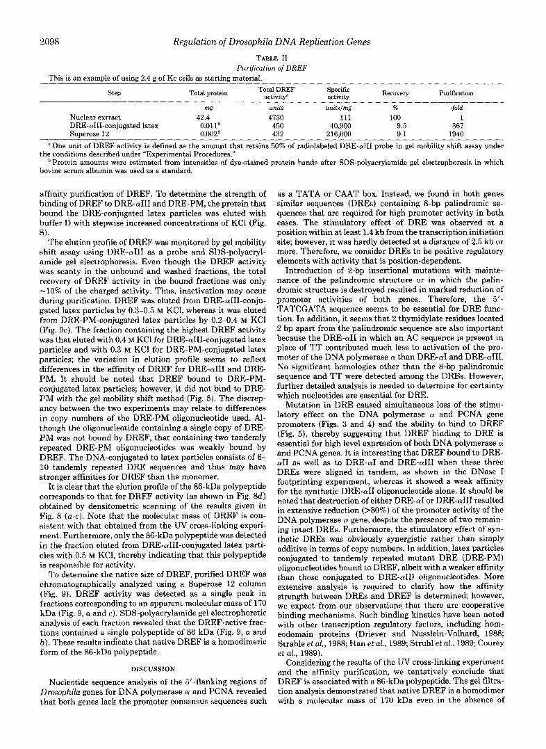
TABLE I1 Purification ojDREF
This is an example of using 2.4 g of Kc cells as starting material.
Step Total protein Recovery Purification Total DREF activity“
Specific activity
mg units unitslrng % -fold Nuclear extract 42.4 4730 111 100 1 DRE-aIII-conjugated latex O.O1lh 450 40,900 9.5 367 Superose 12 0.002h 432 216,000 9.1 1940
a One unit of DREF activity is defined as the amount that retains 50% of radiolabeled DRE-a111 probe in gel mobility shift assay under
* Protein amounts were estimated from intensities of dye-stained protein bands after SDS-polyacrylamide gel electrophoresis in which the conditions described under “Experimental Procedures.”
bovine serum albumin was used as a standard.
affinity purification of DREF. To determine the strength of binding of DREF to DRE-a111 and DRE-PM, the protein that bound the DRE-conjugated latex particles was eluted with buffer D with stepwise increased concentrations of KC1 (Fig. 8).
The elution profile of DREF was monitored by gel mobility shift assay using DRE-a111 as a probe and SDS-polyacryl- amide gel electrophoresis. Even though the DREF activity was scanty in the unbound and washed fractions, the total recovery of DREF activity in the bound fractions was only -10% of the charged activity. Thus, inactivation may occur during purification. DREF was eluted from DRE-aIII-conju- gated latex particles by 0.3-0.5 M KC1, whereas it was eluted from DRE-PM-conjugated latex particles by 0.2-0.4 M KC1 (Fig. 9c). The fraction containing the highest DREF activity was that eluted with 0.4 M KC1 for DRE-aIII-conjugated latex particles and with 0.3 M KC1 for DRE-PM-conjugated latex particles; the variation in elution profile seems to reflect differences in the affinity of DREF for DRE-a111 and DRE- PM. It should be noted that DREF bound to DRE-PM- conjugated latex particles; however, it did not bind to DRE- P M with the gel mobility shift method (Fig. 5) . The discrep- ancy between the two experiments may relate to differences in copy numbers of the DRE-PM oligonucleotide used. Al- though the oligonucleotide containing a single copy of DRE- PM was not bound by DREF, that containing two tandemly repeated DRE-PM oligonucleotides was weakly bound by DREF. The DNA-conjugated to latex particles consists of 6- 10 tandemly repeated DRE sequences and thus may have stronger affinities for DREF than the monomer.
It is clear that the elution profile of the 86-kDa polypeptide corresponds to that for DREF activity (as shown in Fig. 8 4 obtained by densitometric scanning of the results given in Fig. 8 (a-c). Note that the molecular mass of DREF is con- sistent with that obtained from the UV cross-linking experi- ment. Furthermore, only the 86-kDa polypeptide was detected in the fraction eluted from DRE-aIII-conjugated latex parti- cles with 0.5 M KC1, thereby indicating that this polypeptide is responsible for activity.
To determine the native size of DREF, purified DREF was chromatographically analyzed using a Superose 12 column (Fig. 9). DREF activity was detected as a single peak in fractions corresponding to an apparent molecular mass of 170 kDa (Fig. 9, a and c). SDS-polyacrylamide gel electrophoretic analysis of each fraction revealed that the DREF-active frac- tions contained a single polypeptide of 86 kDa (Fig. 9, a and b). These results indicate that native DREF is a homodimeric form of the 86-kDa polypeptide.
DISCUSSION
Nucleotide sequence analysis of the 5’-flanking regions of Drosophila genes for DNA polymerase a and PCNA revealed that both genes lack the promoter consensus sequences such
as a TATA or CAAT box. Instead, we found in both genes similar sequences (DREs) containing 8-bp palindromic se- quences that are required for high promoter activity in both cases. The stimulatory effect of DRE was observed at a position within at least 1.4 kb from the transcription initiation site; however, it was hardly detected at a distance of 2.5 kb or more. Therefore, we consider DREs to be positive regulatory elements with activity that is position-dependent.
Introduction of 2-bp insertional mutations with mainte- nance of the palindromic structure or in which the palin- dromic structure is destroyed resulted in marked reduction of promoter activities of both genes. Therefore, the 5 ’ - TATCGATA sequence seems to be essential for DRE func- tion. In addition, it seems that 2 thymidylate residues located 2 bp apart from the palindromic sequence are also important because the DRE-a11 in which an AC sequence is present in place of TT contributed much less to activation of the pro- moter of the DNA polymerase a than DRE-a1 and DRE-aIII. No significant homologies other than the 8-bp palindromic sequence and TT were detected among the DREs. However, further detailed analysis is needed to determine for certainty which nucleotides are essential for DRE.
Mutation in DRE caused simultaneous loss of the stimu- latory effect on the DNA polymerase a and PCNA gene promoters (Figs. 3 and 4) and the ability to bind to DREF (Fig. 5 ) , thereby suggesting that DREF binding to DRE is essential for high level expression of both DNA polymerase a and PCNA genes. It is interesting that DREF bound to DRE- a11 as well as to DRE-a1 and DRE-a111 when these three DREs were aligned in tandem, as shown in the DNase I footprinting experiment, whereas it showed a weak affinity for the synthetic DRE-a11 oligonucleotide alone. It should be noted that destruction of either DRE-a1 or DRE-a111 resulted in extensive reduction (>SO%) of the promoter activity of the DNA polymerase a gene, despite the presence of two remain- ing intact DREs. Furthermore, the stimulatory effect of syn- thetic DREs was obviously synergistic rather than simply additive in terms of copy numbers. In addition, latex particles conjugated to tandemly repeated mutant DRE (DRE-PM) oligonucleotides bound to DREF, albeit with a weaker affinity than those conjugated to DRE-a111 oligonucleotides. More extensive analysis is required to clarify how the affinity strength between DREs and DREF is determined; however, we expect from our observations that there are cooperative binding mechanisms. Such binding kinetics have been noted with other transcription regulatory factors, including hom- eodomain proteins (Driever and Nusslein-Volhard, 1988; Strahle et al., 1988; Han et al., 1989; Struhl et al., 1989; Courey et al., 1989).
Considering the results of the UV cross-linking experiment and the affinity purification, we tentatively conclude that DREF is associated with a 86-kDa polypeptide. The gel filtra- tion analysis demonstrated that native DREF is a homodimer with a molecular mass of 170 kDa even in the absence of

Regulation of Drosophila DNA Replication Genes 2099
DNA. If this evidence is combined with the finding that DREs contain a palindromic sequence, DREF can most likely bind t o DRE in a homodimeric form, as is the case with other transcription regulatory factors.
A typical example of the DREF preparation is summarized in Table 11. The DREF activity appeared to be labile because 90% was lost during single-step rapid purification using DRE- conjugated latex particles. On the other hand, the yield ex- ceeded 95% in the case of gel filtration. The DRE binding activity of the final preparation was almost completely lost during overnight storage at 4 "C. Therefore, the purification of DREF by conventional procedures would be difficult. In such circumstances, it should be emphasized that the rapid purification procedures using DNA-immobilized affinity latex particles (2 h) and the gel filtration with fast protein liquid chromatography (2 h) are useful.
In budding yeast, a hexamer sequence (5'-ACGCGT, des- ignated the MluI cell cycle box) and a trans-acting factor (MBF: MluI cell cycle box-binding factor) are required for the cell cycle-dependent expression of a number of genes involved in DNA synthesis (Lownders et al., 1991). Whereas it has been reported that the human genes for DNA polymerase a and PCNA have various cis-regulatory elements such as serum-responsive element, Sp-1, AP-1, AP-2 and E2F, it remains equivocal as to which of these elements is critical for induction or repression of gene expressions (Pearson et al., 1991). Therefore, it is of particular interest to determine whether Drosophila DNA replication-related genes other than the two genes described in this paper are also regulated by the DRE/DREF system. Cloning of more genes involved in DNA replication should facilitate elucidation of this problem.
Another interesting aspect of expression of Drosophila DNA polymerase N and PCNA is that both genes are repressed by the zerkunullt (zen) protein, one of the homeodomain proteins (Yamaguchi et al., 1991b and Footnote 2). The target sequence responsible for this repression by zen is localized between positions -119 and -86 of the 5"flanking region of the PCNA gene (Yamaguchi et al., 1991b), in which DRE-P is present. We obtained results suggesting that DRE is a target of the repression by Zen protein, although it does not involve direct binding to DRE (Yamaguchi et al., 1991b). During cellulari- zation and gastrulation of Drosophila embryos, expression of zen is restricted to the dorsal-most cells of the middle body region (Rushlow et al., 1987a, 1987b), an area where the presumptive optic lobe and amnioserosa are located, according to the embryo fate map (Campos-Ortega and Hartenstein, 1985; Foe, 1989). Cells in this region do not divide after the 14th cleavage cycle, which may be the result of the repression of DNA replication by zen. In embryos with a homozygous Zen- genotype (Lewis et al., 1980), we observed ectopic expres- sion of PCNA in the abnormally expanded dorsal region (Yamaguchi et al., 1991b). Thus, the lack of zen may have allowed the expression of DNA replication proteins and there- fore cell proliferation. Since repression of cell proliferation is evident during differentiation in various tissues, homeobox genes other than Zen may also be involved. We propose that both the induction and repression of DNA replication-related genes occur through the physiological and/or genetic altera- tions of DRE/DREF. From this point of view, the major interest of this study is that the expression of two DNA
F. Hirose, M. Yamaguchi, H. Handa, Y. Inomata, and A. Matsu- kage, unpublished data.
replication-related genes is controlled by regulatory elements with identical sequences and a common trans-acting factor. Further studies on the function of DRE/DREF and the reg- ulatory mechanisms for the gene coding for DREF should reveal the molecular mechanisms of cell proliferation and/or cell differentiation.
We are currently purifying DREF to allow for amino acid sequencing and gene cloning, and Southern/Western screen- ing of X phage expression libraries using DRE oligonucleotides as probes is underway.
Han, and James Manley for providing plasmid 5k"TATA-CAT; Mi- Acknowledgments-We are grateful to Michael Levine, Kyuhyung
Ae Yo0 for plasmid pAcGEM3; N. Kondo for technical assistance; and M. Ohara for helpful comments.
REFERENCES Blow, J. J., and Nurse, P. (1990) Cell 62,855-862 Bradford, M. M. (1976) Anal. Biochern. 72,248-254 Bunch, T. A,, Grinblat, Y., and Goldstein, L. S. B. (1988) Nucleic Acids Res.
Campos-Ortega, J. A,, and Hartenstein, V. (1985) The Embryonic Deoelopment
Chang, C.-D., Ottabio, L., Travari, S., Lipson, K. E., and Baserga, R. (1990)
Courey, A., Holtzman, D. A., Jackson, S. P., and Tjian, R. (1989) Cell 59,827-
Cross, D. P., and Sang, J. H, (1978) J. Embryol. Exp. Morphol. 4 5 , 161-172 Date, T., Yamaguch!, M., Hirose, F., Nlshmoto, Y., Tanlhara, K., and Matsu-
Di Nocera, P. P., and Dawid, I. B. (1983) Proc. Natl, Acad. Sci. U. S. A. 80,
Driever, W., and Nusslein-Volhard, C. (1988) Cell 54,887-898 D'Urso, G., Marraccino, R. L., Marshak, D. R., and Roberts, J. M. (1990)
Foe, V. (1989) Development (Carnb.) 107 , 1-22 FIT. M.. and Loeb. L. A. (1986) Animal Cell DNA Polymerases, CRC Press,
16, 1043-1061
of Drosophila rnelanogaster, Springer-Verlag, Berlin
Mol. Cell. Biol. 10, 3289-3296
836
kage, A. (1988) Baochenistry 27,2983-2990
7095-7098
Science 2 5 0 , 786-791
inc., Boca Raton, FL Han. K.. Levine. M.. and Manlev. J. L. (1989) Cell 56.573-583 Henikoff, S. (1984) Gene (Amst.j'28,351-359 Hirose, F., Yamaguchi, M., Nishida, Y., Masutani, M., Miyazawa, H., Hanaoka,
Hui, C.-c., and Suzuki, Y. (1990) Deu. Growth &Differ. 3 2 , 263-273 F., and Matsukage, A. (1991) Nucleic Acids Res. 19 , 4991-4998
Kawaguchi, H.,,Asai, A., Ohtsuka, Y., Watanabe, H., Wada, T., and Handa, H.
Lewis. R. A,. Wakimoto. B. T.. Denell. R. E.. and Kaufman. T. C. (1980) (1989) Nuckrc AcLds Res. 17,6231-6240
Genetics 95,383-397 '
Bid. 8,5280-5291
. .
Lieberman, H. B., Lin, P.-F., Yeh, D.-B., and Ruddle, F. H. (1988) Mol. Cell.
Lownders, N. F., Johnson, A. L., and Johnston, L. H. (1991) Nature 350,247- 9 m
Matsukage, A., Kitani, H., Yamaguchi, M., Kusakabe, M., Morita, T., and
Nasheuer, H.-P., Moore, A,, Wahl, A. F., and Wang, T. S.-F. (1991) J. Biol.
-""
Koshida, Y. (1987) Deu. B~ol. 117 , 226-232
Ottabio, L., Chang, C.-D., Travari, S., Calahretta, B., and Baserga, R. (1990)
Pearson, B. E., Nasheuer, H.-P., and Wang, T. S.-F. (1991) Mol. Cell. Biol. 11,
Chem. 266,7893-7903
Mol. Cell. Biol. 10 , 303-309
2081-2095 Rosenfelt, P. J., and Kelly, T. J. (1986) J. Biol. Chem. 2 6 1 , 1398-1408 Rushlow, C., Doyle, H., Hoey, T., and Levine, M. (1987a) Genes & Deo. 1 ,
Rushlow, C., Frasch, M., Doyle, H., and Levine, M. (1987b) Nature 330 , 583- 1268-1279
Samhrook, J., Fritsch, E. F., and Maniatis, T. (1989) Molecular Cloning: A 586
Laboratory Manual, Cold Spring Harbor Laboratory, Cold Spring Harbor, NY
Struhl, G., Struhl, K., and Macdonald, P. M. (1989 Stiahle, U., Schmid, W., and Schutz, G. (1988) EMBO J. 7.3389-3395
Topol, L., Ruden, D., Tora, L., Gauh, M. P
T:
and Parker, C. S. (1985) Cell 42,527-537 '., Mader, S., Dierich, A,, Bellard, M., and Chambon, P.
seng, B. Y., Prussak, C. E., and Almazan, M. T. (1989) Mol. Cell. Biol. 9 , (1988) EMBO J. 7,3771-3778
1940-1945 Verma, R., Pata outian, A., Gordon, C. B., and Campbell, J. (1991) Proc. Natl.
Acad. Sei. U .8 A. 88, 7155-7159 Wa_hLA.- F.,-Ge?s,,A, y., Spair, B. H., Wong, B. H., Korn, D., and Wang, T.
r, B., Dawid, I., Paisley, T., Zimarino, V., and Ueda,
) Cell 5 7 , 1259-1273
- id . 8,5016-5025
138. 1247-12.53 Wu, C., Wilson, S., Walke
s.-F. (1~88) MOL. Leu. fl
Yamaguchl, M., Hays H. (1987,) Science 2
8773-8787 156, Y., and Matsukage, A. (1988) Nucleic Acids Res. 16 ,
, ~~- ""
Yamaguchi, M., Nishida, Y., Moriuchi, T., Hirose, F., Hui, C.-c., Suzuki, Y.,
Yamaguchi, M., Hayashi, Y., Furukawa, K., Nishimoto, Y., and Matsukage, A.
Yamaguchi, M., Hirose, F., Nishida, Y., and Matsukage, A. (1991b) Mol. Cell.
and Matsukage, A. (1990) Mol. Cell. B~ol. 10,872-879
(1991a) Jpn. J. Cancer Res. (Gann) 8 2 , 72-81
Biol. 11,4909-4917