The Interaction between Arbuscular Mycorrhizal Fungi and ...
15
ORIGINAL RESEARCH published: 19 July 2016 doi: 10.3389/fmicb.2016.01089 Frontiers in Microbiology | www.frontiersin.org 1 July 2016 | Volume 7 | Article 1089 Edited by: Kumar Krishnamurthy, Tamil Nadu Agricultural University, India Reviewed by: Biswapriya Biswavas Misra, University of Florida, USA Joseph Davis Bagyaraj, Indian National Science Academy, India *Correspondence: Elsayed F. Abd_Allah [email protected] Specialty section: This article was submitted to Plant Biotic Interactions, a section of the journal Frontiers in Microbiology Received: 17 February 2016 Accepted: 29 June 2016 Published: 19 July 2016 Citation: Hashem A, Abd_Allah EF, Alqarawi AA, Al-Huqail AA, Wirth S and Egamberdieva D (2016) The Interaction between Arbuscular Mycorrhizal Fungi and Endophytic Bacteria Enhances Plant Growth of Acacia gerrardii under Salt Stress. Front. Microbiol. 7:1089. doi: 10.3389/fmicb.2016.01089 The Interaction between Arbuscular Mycorrhizal Fungi and Endophytic Bacteria Enhances Plant Growth of Acacia gerrardii under Salt Stress Abeer Hashem 1, 2 , Elsayed F. Abd_Allah 3 *, Abdulaziz A. Alqarawi 3 , Asma A. Al-Huqail 1 , Stephan Wirth 4 and Dilfuza Egamberdieva 4 1 Department of Botany and Microbiology, Faculty of Science, King Saud University, Riyadh, Saudi Arabia, 2 Department of Mycology and Plant Disease Survey, Agriculture Research Center, Plant Pathology Research Institute, Giza, Egypt, 3 Department of Plant Production, Faculty of Food and Agricultural Sciences, King Saud University, Riyadh, Saudi Arabia, 4 Leibniz Centre for Agricultural Landscape Research, Institute of Landscape Biogeochemistry, Müncheberg, Germany Microbes living symbiotically in plant tissues mutually cooperate with each other by providing nutrients for proliferation of the partner organism and have a beneficial effect on plant growth. However, few studies thus far have examined the interactive effect of endophytic bacteria and arbuscular mycorrhizal fungi (AMF) in hostile conditions and their potential to improve plant stress tolerance. In this study, we investigated how the synergistic interactions of endophytic bacteria and AMF affect plant growth, nodulation, nutrient acquisition and stress tolerance of Acacia gerrardii under salt stress. Plant growth varied between the treatments with both single inoculants and was higher in plants inoculated with the endophytic B. subtilis strain than with AMF. Co-inoculated A. gerrardii had a significantly greater shoot and root dry weight, nodule number, and leghemoglobin content than those inoculated with AMF or B. subtilis alone under salt stress. The endophytic B. subtilis could alleviate the adverse effect of salt on AMF colonization. The differences in nitrate and nitrite reductase and nitrogenase activities between uninoculated plants and those inoculated with AMF and B. subtilis together under stress were significant. Both inoculation treatments, either B. subtilis alone or combined with AMF, enhanced the N, P, K, Mg, and Ca contents and phosphatase activities in salt-stressed A. gerrardii tissues and reduced Na and Cl concentration, thereby protecting salt-stressed plants from ionic and osmotic stress-induced changes. In conclusion, our results indicate that endophytic bacteria and AMF contribute to a tripartite mutualistic symbiosis in A. gerrardii and are coordinately involved in the plant adaptation to salt stress tolerance. Keywords: AMF, endophyte, Acacia gerrardii, salinity, nutrition INTRODUCTION Salinity is a devastating environmental stress factor that severely affects plant growth and development (Barnawal et al., 2014). At the global level, particularly in arid and semiarid regions, salinity is considered an important constraint, and approximately 7% of global land has a high salt concentration, making this area unavailable for agriculture (Sheng et al., 2008; Ruiz-Lozano et al., 2012). Salinity reduces plant growth through osmotic as well as ionic constraints of major
Transcript of The Interaction between Arbuscular Mycorrhizal Fungi and ...
ORIGINAL RESEARCHpublished: 19 July 2016
doi: 10.3389/fmicb.2016.01089
Frontiers in Microbiology | www.frontiersin.org 1 July 2016 | Volume 7 | Article 1089
Edited by:
Kumar Krishnamurthy,
Tamil Nadu Agricultural University,
India
Reviewed by:
Biswapriya Biswavas Misra,
University of Florida, USA
Joseph Davis Bagyaraj,
Indian National Science Academy,
India
*Correspondence:
Elsayed F. Abd_Allah
Specialty section:
This article was submitted to
Plant Biotic Interactions,
a section of the journal
Frontiers in Microbiology
Received: 17 February 2016
Accepted: 29 June 2016
Published: 19 July 2016
Citation:
Hashem A, Abd_Allah EF,
Alqarawi AA, Al-Huqail AA, Wirth S
and Egamberdieva D (2016) The
Interaction between Arbuscular
Mycorrhizal Fungi and Endophytic
Bacteria Enhances Plant Growth of
Acacia gerrardii under Salt Stress.
Front. Microbiol. 7:1089.
doi: 10.3389/fmicb.2016.01089
The Interaction between ArbuscularMycorrhizal Fungi and EndophyticBacteria Enhances Plant Growth ofAcacia gerrardii under Salt Stress
Abeer Hashem 1, 2, Elsayed F. Abd_Allah 3*, Abdulaziz A. Alqarawi 3, Asma A. Al-Huqail 1,
Stephan Wirth 4 and Dilfuza Egamberdieva 4
1Department of Botany and Microbiology, Faculty of Science, King Saud University, Riyadh, Saudi Arabia, 2Department of
Mycology and Plant Disease Survey, Agriculture Research Center, Plant Pathology Research Institute, Giza, Egypt,3Department of Plant Production, Faculty of Food and Agricultural Sciences, King Saud University, Riyadh, Saudi Arabia,4 Leibniz Centre for Agricultural Landscape Research, Institute of Landscape Biogeochemistry, Müncheberg, Germany
Microbes living symbiotically in plant tissues mutually cooperate with each other by
providing nutrients for proliferation of the partner organism and have a beneficial effect
on plant growth. However, few studies thus far have examined the interactive effect of
endophytic bacteria and arbuscular mycorrhizal fungi (AMF) in hostile conditions and
their potential to improve plant stress tolerance. In this study, we investigated how the
synergistic interactions of endophytic bacteria and AMF affect plant growth, nodulation,
nutrient acquisition and stress tolerance of Acacia gerrardii under salt stress. Plant
growth varied between the treatments with both single inoculants and was higher in
plants inoculated with the endophytic B. subtilis strain than with AMF. Co-inoculated
A. gerrardii had a significantly greater shoot and root dry weight, nodule number, and
leghemoglobin content than those inoculated with AMF or B. subtilis alone under salt
stress. The endophytic B. subtilis could alleviate the adverse effect of salt on AMF
colonization. The differences in nitrate and nitrite reductase and nitrogenase activities
between uninoculated plants and those inoculated with AMF and B. subtilis together
under stress were significant. Both inoculation treatments, either B. subtilis alone or
combined with AMF, enhanced the N, P, K, Mg, and Ca contents and phosphatase
activities in salt-stressed A. gerrardii tissues and reduced Na and Cl concentration,
thereby protecting salt-stressed plants from ionic and osmotic stress-induced changes.
In conclusion, our results indicate that endophytic bacteria and AMF contribute to a
tripartite mutualistic symbiosis in A. gerrardii and are coordinately involved in the plant
adaptation to salt stress tolerance.
Keywords: AMF, endophyte, Acacia gerrardii, salinity, nutrition
INTRODUCTION
Salinity is a devastating environmental stress factor that severely affects plant growth anddevelopment (Barnawal et al., 2014). At the global level, particularly in arid and semiarid regions,salinity is considered an important constraint, and approximately 7% of global land has a highsalt concentration, making this area unavailable for agriculture (Sheng et al., 2008; Ruiz-Lozanoet al., 2012). Salinity reduces plant growth through osmotic as well as ionic constraints of major
Hashem et al. Alleviation of Salt Stress of A. gerrardii by Microbes
physiological and biochemical processes (Ahmad, 2010; Porcelet al., 2012; Abd_Allah et al., 2015a). This may in turn alterthe availability of nutrients for plant growth and affect theassociation with microbes living within the plant vicinity. Plantsare colonized bymicrobes, including endophytes, nitrogen-fixingbacteria and mycorrhizal fungi, which closely cooperate witheach other and can mediate important physiological processes,especially nutrient acquisition and plant tolerance to abioticstresses (Egamberdieva, 2012; Egamberdieva et al., 2013, 2015;Berg et al., 2013; Ahanger et al., 2014a; Abd_Allah et al.,2015a). Arbuscular mycorrhiza fungi (AMF) form beneficialsymbiotic associations with most plants and play a vital role inplant growth under various conditions by modifying the rootsystem and enhancing mobilization and the uptake of severalessential elements. They have also been reported to stimulateplant stress tolerance by enhancing enzymatic as well as non-enzymatic antioxidant defense systems (Wu et al., 2014; Ahmadet al., 2015), lipid peroxidation (Abd_Allah et al., 2015c), andphytohormone synthesis (Navarro et al., 2013). Endophyticbacteria colonize the internal tissues of their host plants andcan promote growth, stress tolerance, and nutrient uptake andprotect plants from soil-borne pathogens (Malfanova et al.,2011; Sessitsch et al., 2012). Microbes living symbiotically inplant tissues, such as mycorrhizal fungi, and nitrogen-fixingbacteria also mutually cooperate with each other by synthesizingbiologically active compounds and providing nutrients for thesurvival and proliferation of their partner organism (Marschneret al., 2001; Egamberdieva et al., 2013). These synergies amongendophytes are known to have beneficial effects on plants byimproving the availability of nutrients to plants and inducingplant defense against various stresses, including drought andsalinity.
The symbiotic relationship between legumes and nitrogen-fixing rhizobia is susceptible to abiotic factors, such as nutrientdeficiency, salinity, drought, acidity, and soil temperature, whichinduce failure of the infection and nodulation processes (Slatteryet al., 2001; Bouhmouch et al., 2005; Egamberdieva et al., 2013).An improvement in legume-rhizobia symbiotic performance byAMF has been reported for faba bean (Vicia faba) (Yinsuo et al.,2004), lucerne (Medicago sativa) (Ardakani et al., 2009), lentil(Lens culinaris) (Xavier and Germida, 2002), and common bean(Phaseolus vulgaris) (Tajini et al., 2012). The positive effects onplant growth and stimulation of stress tolerance by synergisticinteractions of root-colonizing, plant growth-promoting bacteria(PGPR) and AMF under hostile environments have beenextensively reviewed by Nadeem et al. (2014). These microbes arebelieved to act as essential bio-ameliorators of stress by regulatingthe nutritional and hormonal balance (Abd_Allah et al., 2015a,b;Egamberdieva et al., 2015, 2016) and inducing systemic toleranceto stress (Ruiz-Lozano et al., 2012).
Despite these beneficial associations of microbes, studiesexamining the interactions of endophytic bacteria and AMF inhostile environmental conditions are limited, especially wherecompetition for nutrient and niches in the rhizosphere is high.This knowledge is important for our understanding of therelationship between AMF and endophytic bacteria and theirpotential effect on plant stress tolerance and for the development
of crop management practices under hostile environmentalconditions.
Acacia gerrardii Benth. (Talh tree) is a small leguminous shrubthat is resistant to drought and salinity, forms nodules and canimprove the fertility of salt-affected arid soils. This tree is widelyused in Saudi Arabia for fuel, forage, medicine, food productionand also for agroforestry. In this study, we hypothesize thatthe improved salt tolerance and growth of A. gerrardii aremediated by (i) the effect of endophytic bacteria on mycorrhizaldevelopment and colonization in the roots of A. gerrardii and(ii) nodulation, nutrient acquisition by a synergistic interactionbetween endophytic bacteria and AMF.
MATERIALS AND METHODS
Plants and MicroorganismsIsolation of Endophytic BacteriaRoots of Talh trees were collected from the top 20 cm of soilin a natural meadow at Khuraim in Riyadh, Saudi Arabia.The samples were wrapped in a plastic bag and broughtto the laboratory, where they were incubated at 4◦C untilfurther processing. One gram of roots was surface-sterilized byimmersion in 70% ethanol, followed by 5% sodium hypochloritefor 5 min, and then rinsed in sterile distilled water four tosix times to eliminate the chlorine. The sterilized roots weremacerated, and the extracts were placed in a tube containing 9ml of sterile phosphate-buffered saline and then serially diluted.A 100 µl aliquot from the appropriate dilutions was platedon tryptic soy agar (TSA, Difco Laboratories, Detroit, USA)supplemented with 4% NaCl. The plates were incubated at 28◦Cfor 3 days, and all colonies that displayed differentiable colonymorphologies were selected from the plates and were re-streakedto purify the strains. To select strains with increased stresstolerance, the purified isolates were cultured in TSA mediumsupplemented with 3, 4, or 5% (w/v) NaCl.
Identification of the Selected StrainA highly salt-tolerant strain, which grew well in TSA mediumcontaining 4% NaCl, was identified. DNA was isolated by amodified version of the Töpper et al. (2010) protocol. Thefilters were re-suspended in 250 µl of lysozyme solution (1mgml−1 TE buffer, pH 7.4). Then, 250 µl of preheated (55◦C)lysis buffer (20 µg proteinase K ml−1 0.5% SDS) was addedto the solution. After incubation for 30 min at 55◦C, 80 µlof 5 M NaCl and 100 µl of preheated (55◦C) CTAB [10%(w/v) hexadecyltrimethylammonium bromide in 0.7% NaCl]were added. The solution was incubated for a further 10min at 65◦C followed by the addition of 500 µl chloroform:isoamylalcohol (24:1, v/v). The solution was centrifuged (16,000× g, 5 min) to separate the DNA from the remaining celldebris. Then, the top phase was transferred to a fresh tube,and the DNA was precipitated with isopropanol and later re-suspended in TE buffer (pH 7.4). Next, 16S rDNA was amplifiedby polymerase chain reaction (PCR) using universal forward16SF (5′-GAGTTTGATCCTGGCTCAG-3′) and reverse 16SR(5′-GAAAGGAGGTGATCCAGCC-3′) primers (Mohanta et al.,2015). The PCR reactions were 25 µl and contained 5 µl of
Frontiers in Microbiology | www.frontiersin.org 2 July 2016 | Volume 7 | Article 1089
Hashem et al. Alleviation of Salt Stress of A. gerrardii by Microbes
5 × buffer (TaKaRa Bio Inc.), 0.5 µl of dNTP mixture (10 mMof each dNTP, TaKaRa Bio Inc.), 1 µl of 2% BSA (Promega), 0.5µl of forward primer (10 µM), 0.5 µl of reverse primer (10 µM),0.125 µl of One Taq DNA Polymerase (New England Biolabs),15.375 MQ and 2 µl of template DNA. The PCR program (Bio-Rad DNA Engine) started with an initial denaturation step for30 s at 94◦C followed by 30 cycles of 15 s at 94◦C, 30 s at 55◦Cand 1.5 min at 68◦C. Before cooling to 4◦C, an extension periodof 20 min at 68◦C was incorporated into the program. ThePCR products were verified by gel electrophoresis on a 1.5%agarose gel stained with TAE (Tris-acetate-EDTA). Denaturinggradient gel electrophoresis (DGGE) was performed using theDCode system (Bio-Rad). Equal amounts of PCR products (6µl) were loaded onto 8% acrylamide gels with a denaturinggradient of 30–55% [where 100% denaturing is defined as 7M urea and 40% (v/v) formamide (Muyzer et al., 1995)] foroptimal separation of the PCR products. DGGE gels were runfor 19 h at 60 V and at 60◦C in 0.59 TAE buffer and stainedfor 30 min with SYBR Gold (Invitrogen) diluted 10,000-fold in19 × TAE buffer. Gels were visualized and digitized using theFujifilm Imaging System. The PCR product was purified, andnucleotide sequences were determined using automatic LI-CORDNA Sequencer 4000 L (Lincoln, USA). The sequences wereidentified using the basic local alignment search tool (BLAST)and comparisons with the GenBank nucleotide data bank fromthe National Center for Biotechnology Information (NCBI)(http://www.ncbi.nlm.nih.gov/).
Plant Growth-Promoting TraitsThe cellulose-degrading ability of bacterial isolates was analyzedby streaking inocula on cellulose Congo Red agar media, asdescribed by Gupta et al. (2012). Zones of clearance aroundand beneath the colony were detected, indicating enzymaticdegradation of cellulose.
The production of indole 3-acetic acid (IAA) was determinedas described by Bano and Musarrat (2003). Briefly, bacterialstrains were grown in TSB medium. After 3 days, 1 ml ofeach culture was pelleted by centrifugation, and the supernatantwas discarded. Cell pellets were washed with 1 ml of PBSand re-suspended in PBS. One milliliter of cell suspension(corresponding to a cell density of 107 cells/ml) was addedto 10 ml of TSB supplemented with tryptophan (100 µg/ml).After 3 days of cultivation, 2 ml aliquots of bacterial cultureswere centrifuged at 13,000 × g for 10 min. One milliliter ofsupernatant was transferred to a fresh tube to which 100 µg/mlof 10 mM orthophosphoric acid and 2 ml of reagent (1 ml of0.5 M FeCl3 in 50 ml of 35% HClO4) were added. After 25 min,the absorbance of the sample was measured at 530 nm. The IAAconcentration in cultures was calculated using a calibration curveof pure IAA as the standard.
The phosphate-solubilizing activity of the bacterial strains wasdetermined on Pikovskaya agar (Pikovskaya, 1948) containingprecipitated tricalcium phosphate. The bacterial culture grown inTSAmedium for 2 days was streaked on the surface of Pikovskayaagar plates and incubated for 3 days. The presence of a clear zonearound bacterial colonies was considered to be an indicator ofpositive P solubilization.
Arbuscular Mycorrhizal FungiAMF were isolated from the soil surrounding the roots ofA. gerrardii. AMF were extracted by wet sieving, decantingand sucrose density gradient centrifugation as described byDaniels and Skipper (1982) and modified by Utobo et al.(2011). Briefly, 100 g of soil was placed in a 10 L bucket,and 5 L of tap water was added to the soil and mixed wellto produce a soil-water suspension. The suspension was leftfor 5 min to allow insoluble and heavy particles to settle,and the suspension was sequentially sieved through ASTM-500, ASTM-250, ASTM-150, and ASTM-50 sieves to extract thespores using a wet sieving and decanting method (Gerdemannand Nicolson, 1963). The sieved residues were filtered throughWhatman filter paper No. 1. After water filtration, the filterpaper was examined under a stereo-binocular microscope at 25× magnification. Morphologically similar spores were selectedfor identification. AMF species were identified based on thedescription of subcellular structures (spore color, shape, surfaceornamentation, spore contents, and wall structures) of asexualspores provided by the International Culture Collection ofVesicular and Arbuscular Mycorrhizal Fungi (INVAM 2012)1
and other descriptive protocols (Bethlenfalvay and Yoder, 1981;Schüßler and Walker, 2010; Redecker et al., 2013).
Propagation of AMF in Trap CulturesThe trap culture protocol described by Stutz and Morton (1996)was used in the current study to propagate the most efficientmycorrhizal isolates. Sterilized sand (121◦C for 3days) wasinoculated with single spores from each mycorrhizal isolate.Surface-sterilized seeds [0.5% (v/v) NaOCl used] of Sorghumsudanense were sown (20 seeds/pot) at a depth of 2 cm in eachpot (5 kg capacity) of trap cultures. The pots were incubated ina plant growth chamber at 25 ± 2◦C with 18 h photoperiod,750 µmol m−2 s−1 photosynthetic photon flux density, and70–75% relative humidity for 3months. Half-strengthHoagland’ssolution was used to irrigate the pots. The trap culture was usedas the mycorrhizal inoculum and was added to the experimentalsoil as 25 g of trap soil culture (approx. 100 spores/g trap soil)/pot.Soil not inoculated with mycorrhiza served as the control.
Determination of Arbuscular MycorrhizalColonizationAt the end of the pot experiment (12 weeks), fine roots werecollected from the lateral root system and fixed in formalin/aceticacid/alcohol (v/v/v) (FAA) solution until further processing.Roots were stained with trypan blue in lactophenol (Phillips andHayman, 1970) and assessed formycorrhizal infection. Roots thatwere pigmented after clearing were bleached in alkaline hydrogenperoxide (0.5% NH4OH and 0.5% H2O2 v/v in water) to removeany phenolic compounds (Kormanik and McGraw, 1982) beforeacidification (0.05 M HCl). To assess mycorrhizal colonization,stained root segments (one cm in length) were mounted onglass slides with lactophenol and were observed under a digitalcomputerized microscope (model DP-72, Olympus) at 20 ×
1INVAM - International Culture Collection of Arbuscular & Vesicular-Arbuscular
Mycorrhizal Fungi. Available online at: www.invam.caf.wvu.edu/index.html
(Accessed February 6, 2012).
Frontiers in Microbiology | www.frontiersin.org 3 July 2016 | Volume 7 | Article 1089
Hashem et al. Alleviation of Salt Stress of A. gerrardii by Microbes
magnification. A minimum of 50 segments for each replicatesample were observed to assess structural colonization of AMFassociated with roots. Twenty or more segments were mountedon each slide and examined under the microscope. The presenceof mycelia, vesicles and arbuscules was recorded and analyzed toassess structural colonization.
Germination of SeedsAcacia gerrardii seeds were surface-sterilized by immersion for5 min in concentrated sulfuric acid followed by 3 min in 70%ethanol and were rinsed five times with sterile, distilled water.
Germination tests were carried out in Petri dishes (Ø85 × 15 mm) containing 1% water agar. Salinity conditionswere established by adding 50, 100, 150, 200, 250, and 300 mMNaCl. Twenty healthy and surface-sterilized seeds were placedon each Petri dish and were arranged in a randomized completeblock design with three replications. Eventually, the Petri disheswere covered with a polyethylene sheet to avoid the loss ofthe moisture through evaporation and kept in the plant growthchamber at 28◦C. The seeds were observed daily, and the percentgermination was recorded after 10 days of incubation. Seeds wereconsidered to have germinated when the emerging radicles weregreater than 0.5 cm long.
Plant Growth ConditionThis experiment was carried out in the growth chamber of thePlant Production Department, Faculty of Food & AgriculturalSciences, King Saud University, Riyadh, Saudi Arabia. Talh treeseeds were provided by a personal nursery in Alghat, Riyadhthat produces tree seedlings. The soil used was loamy sand soilwith the following properties (%): sand (87.6), clay (7.2), silt(5.2), organic carbon (0.12), total nitrogen (0.005), pH 7.5. Thesand was washed with 1.0 N H2SO4 for 1 h, followed by 1.0 NCa carbonate, and was then washed using distilled water. Thesurface-sterilized seeds were sown in acid-washed sterile sandand kept in a plant growth chamber under the same conditionsdescribed for trap cultures for 1 month.
The bacterial strain was grown in TSB medium for 2 days, and1 ml of culture suspension was pelleted by centrifugation. Thesupernatant was discarded, and the cell pellets were washed with1 ml phosphate-buffered saline and diluted to a cell density of 108
cells/ml. Roots of 1-month-old seedlings were immersed in thebacterial suspension for 30 min and sown in pots (1 seedling perpot) filled with 2 kg of sandy loam soil mixed with mycorrhizalinoculum [i.e., 25 g of trap soil culture or approx. 100 spores/gtrap soil (M = 80%)/pot]. The experiment was a completelyrandomized design with five replicates for each treatment: (i)Control without microbes, (ii) Bacillus subtilis (BS), (iii) AMF,and (iv) B. subtilis combined with AMF (BS+AMF). Plants weregrown in a greenhouse for 12 weeks with average day/nighttemperatures of 25◦C/18◦C and were supplemented with tapwater as required. Salinity was established by adding NaCl tothe irrigation solution to obtain a constant concentration of 250mM. At harvest, plants were harvested carefully, and shoots wereseparated from roots and dried to constant weight at 100◦C.Fresh nodules were used to estimate leghemoglobin (LB) levelsas well as the activities of nitrite reductase, nitrate reductase, and
nitrogenase. Fresh leaf samples were used to assess the content ofphotosynthetic pigments.
Nodulation, Leghemoglobin, and CrudeProtein ContentsThe nodule fresh weight (FW) and the number of nodules perplant root were determined. The leghemoglobin concentrationof root nodules was estimated using the method of Keilin andWang (1945). Fresh nodules (2.0 g) were ground to a fine powderin liquid N2 and transferred to 50 mM KPO4 (pH 7.4) buffercontaining 1 mM EDTA. The mixture was stirred until it thawedinto a homogenate at a final temperature of 2◦C, transferredto centrifuge tubes and centrifuged at 4◦C and 10,000 × gfor 10 min. The leghemoglobin-containing supernatant wascollected and maintained at a known volume with 50 mMKPO4 (pH 7.4) buffer as described above. The color intensitythat developed was recorded spectrophotometrically at 710 nmagainst a blank, which contained 50 mM KPO4 (pH 7.4) bufferwith 1 mM EDTA. Leghemoglobin concentration was expressedas mg/g nodule (FW). The micro-Kjeldahl method described byAllen (1953) was used to estimate the total nitrogen content ofoven-dried (110◦C for two successive weights) nodules. Crudeprotein concentration (%, w/w) was determined mathematicallyby multiplying total nitrogen content by the general factor forcereal protein determination, 6.25, as described in the AOACOfficial Method 992.23 (AOAC Association of Official AnalyticalChemists, 1995).
Nitrate and Nitrite Reductase andNitrogenase ActivityNitrate and nitrite reductase activities in the nodules wereassayed using the methods of Hageman and Hucklesby (1971)and Finka et al. (1977), respectively. Phosphate buffer (pH7.5) containing 0.1 M potassium nitrate and 5% n-propanolwas used to extract the reductases. The optical density of thesamples was measured spectrophotometrically at 550 nm. Astandard curve of potassium nitrite was used as a reference.The activity of nitrate and nitrite reductases was expressed asµmNO2 released/h/g FW and µmNO2 disappeared/h/g freshwt, respectively. Nitrogenase (EC 1.7.99.2) activity (ARA) wasdetermined by acetylene reduction with the known weight ofthe nodules collected from all nodulated root portions of theplants, following the method of Herdina and Silsbury (1990).Gas samples were analyzed for ethylene produced in the reactionusing a Shimadzu GC-14B gas chromatograph equipped with aPorapak R column (Ligero et al., 1986).
Determination of Photosynthetic PigmentsPhotosynthetic pigments were extracted from leaf samples in 80%acetone as described by Arnon (1949). Fresh leaf samples (0.5g) were extracted in 80% acetone (v/v). The extracted materialwas centrifuged at 10, 000 × g for 10 min. The optical densityof the supernatants was recorded at 480, 645, and 663 nm usinga UV-visible spectrophotometer (T80 UV/VIS spectrometer, PGInstruments Ltd., USA). A blank with 80% acetone served as thecontrol.
Frontiers in Microbiology | www.frontiersin.org 4 July 2016 | Volume 7 | Article 1089
Hashem et al. Alleviation of Salt Stress of A. gerrardii by Microbes
Estimation of Acid and AlkalinePhosphatase ActivityFresh root samples were extracted with 0.1 M acetate buffer (pH5.0) and 0.1 M Tris HCI buffer (pH 8.2) for acid phosphatase(AP) (EC 3.1.3.2) and alkaline phosphatase (ALP) (EC 3.1.3.1),respectively, as described by Gianinazzi-Pearson and Gianinazzi(1976). AP and ALP were assayed following the methods ofIkawa et al. (1964) and Torriani (1967), respectively. In thesemethods, the reaction mixture of AP contained 0.2 ml ofenzyme extract and 1.0 ml of 5.5 mM p-nitrophenol phosphatein 5.5mM citrate buffer (pH 4.8). For the ALP assays, 0.05M Tris-citrate (pH 8.5) was used instead of citrate buffer(pH 4.8). The reaction mixtures were incubated at 37◦C for30 min, and the reactions were stopped by adding 10 ml of200 mM NaOH. Absorbance was recorded at 410 nm, andactivity was expressed as µmol p-nitrophenol released min/mgprotein.
Determination of Mineral ContentsTo determine the mineral contents, oven-dried leaves werepowdered, and the powder was digested with 98% H2SO4
and 30% H2O2. The total nitrogen (N) content in leaftissues was determined following the semi-micro Kjedahlprocedure using a nitrogen analyzer (Kjedahl 2300; FOSS,Hoganas, Sweden). The phosphorus (P) content was estimatedusing the vanadomolybdophosphoric colorimetric method(Jackson, 1962). A standard curve (10–100 µg/ml) of potassiumdihydrogen phosphate (KH2PO4) was used as the reference.The contents of Na+, K+, Mg2+, and Ca2+ in plant leaves wereestimated as described by Wolf (1982) using a flame photometer(Jenway Flame Photometer, Bibby Scientific Ltd., Stone-Staffs,UK). The chloride (Cl−) concentration was directly estimatedin the digested extracts using a chloride analyzer (Model 926,Sherwood Scientific Ltd., Cambridge, UK), as described byAbd_Allah et al. (2015b).
Statistical AnalysesDuncan’s multiple range test was performed using one-wayanalysis of variance (ANOVA) for a completely randomizeddesign by SPSS-21 software, and significant differences in meanswere determined by the least significant differences (LSD) (p =
0.05) test. In addition, the correlation coefficients among theparameters studied were calculated and are presented inTables 3,5, Table S3.
RESULTS
Identification of MicroorganismsUsing nucleotide homology and phylogenetic analysis of the 16SrRNA gene sequences, we grouped the microbe that showed thehighest salt tolerance in a cluster containing B. subtilis (GenBankAccession Number: JX188065.1) with 99% sequence similarity.The genome sequence of B. subtilis BERA71 was depositedin GenBank with the accession number KX090253. The strainproduced IAA in nutrient broth containing 1.5% (260 mM)NaCl and cellulose and also solubilized mineral phosphorus fromtricalcium phosphate used in solid medium.
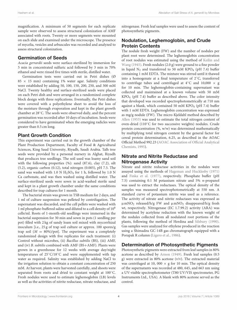
Photomicrographs in (Figures 1A–F) showing sporemorphology of AMF (Claroideoglomus etunicatum, Rhizophagusintraradices, Funneliformis mosseae) used in the currentstudy. Figures 1A,B Crushed spore of C. etunicatum showingdepressions on the surface and two wall layers: L1, an outerpermanent rigid layer with some plasticity and an unevenouter surface. L2, A layer consisting of laminae that increase inthickness (∼8.0 µm thick) is rigid, exhibiting some swelling andspreading when broken. R. intraradices: Figures 1C,D Crushedyellow-brown spore, globose or subglobose in shape, consists ofthree layers. L1, Outer layer has a sloughing spore wall (SSW),hyaline, mucilaginous spores that stain pale pink. L2: A rigidhyaline layer attached firmly to the underlying laminae [L3] asa sub-laminae layer of the spore wall (SLW). L3: Inner layeras laminae of the spore wall (LSW), which is continuous withthe innermost layer of the subtending hypha. The subtendinghypha (SH) is cylindrical to slightly flared with three layers thatare continuous with the three layers of the spore wall. The plugpartitions the spore from the hyphal contents (septum, S). Aseptum (S) occludes the hyphal attachment of a thin-walledspore of the pale morph close to the spore base. F. mosseae:Figures 1E,F Crushed spore showing the wall structure asthree layers. L1, Outer wall, hyaline, mucilaginous, forming asloughing granular layer (SSW). L2, Hyaline, generally rigid,consisting of a thin adherent sublayer attached firmly to theunderlying laminae (L3). L3, Inner layer consisting of laminaewith minute depressions covering the surface and separatingL1 and L2. The sporocarp (SC) is subglobose, light brown, andsurrounded by a peridium (P). Developed intact spores (IS)were observed. The subtending hypha is funnel-shaped with awidth ranging between 16 and 32 µm (Figure 1F). As shownin Figure 1E, the subtending hypha consists of two layers (L1,L3), with the sum of thickness ranging between 2.4 and 4.8 µm.A germ tube emerges from the lumen (funnel-shaped) of thesubtending hypha, originating from the occlusion-recoveredseptum. Sporocarps dramatically produce numerous infectivehyphae (Figure 1E). The spores in the current isolates from soiladhering to the roots of A. gerrardii were identified as F. mosseae(syn.Glomus mosseae), R. intraradices (syn.Glomus intraradices),and C. etunicatum (syn. Glomus etunicatum).
Plant Growth ParametersThe germination of A. gerrardii seeds was tested at NaClconcentrations of 50–300 mM. The results showed that increasesin salt concentration decreased the germination rate of the seedscompared to that of the control seeds (water only) (from 87 to3%). Because germination of the seeds was strongly impaired by300 mM NaCl, we used 250 mM NaCl in all further salt stressexperiments.
The growth of A. gerrardii was strongly impaired by salinitywhen plants were not inoculated with AMF or endophyticbacteria. Salinity reduced shoot height and shoot dry weight by61 and 62%, respectively, and root length and dry weight werereduced by 35 and 38%, respectively (Table 1), compared withnon-stressed plants. Salt-stressed shoots were 35% shorter androot depth 61% less in plants treated with 250 mM NaCl thannon-stressed plant shoots and roots.
Frontiers in Microbiology | www.frontiersin.org 5 July 2016 | Volume 7 | Article 1089
Hashem et al. Alleviation of Salt Stress of A. gerrardii by Microbes
FIGURE 1 | (A–F) (40X). AMF spores were isolated from soil samples of A. gerrardii. (A,B) The microscopic investigation indicated that the crushed spores of C.
etunicatum were globose, sub-globose in shape, orange to red-brown in color and consisted of two layers (L1 and L2),. (C,D) Describes the spore morphology of R.
intraradices as globose to subglobose in shape and whose crushed spores consisted of three layers (L1, L2, and L3) and prominent subtending hyphae. (E,F) The
microscopic investigation revealed that the spores of F. mosseae are clustered together within a compact peridium. The shape of the spores was globose to
subglobose, and the spore wall consisted of three layers (L1, L2, and L3).
TABLE 1 | Shoot and root growth of Acacia gerrardii under salt stress after inoculation with B. subtilis and AMF alone and in combination.
Treatments SH (cm) SDW (g) RD (cm) RDW (g) SH/RD SDW/RDW
0 mM NaCl Control 53.4± 1.7 1.94±0.2 70.9± 2.1 2.78±0.3 0.75± 0.04 0.70± 0.03
BS 78.4± 1.9 2.95±0.3 87.0± 2.4 3.66±0.4 0.90± 0.05 0.80± 0.04
AMF 61.4± 1.5 2.51±0.2 75.7± 2.3 3.24±0.5 0.81± 0.06 0.77± 0.03
BS + AMF 80.2± 2.1 3.55±0.4 92.1± 3.1 3.87±0.4 0.87± 0.06 0.91± 0.05
250 mM NaCl Control 20.8± 1.0 0.74±0.1 46.0± 1.2 1.72±0.1 0.45± 0.02 0.43± 0.01
BS 41.6± 1.4 1.61±0.2 63.6± 1.5 2.42±0.2 0.65± 0.03 0.67± 0.03
AMF 29.1± 1.2 1.13±0.1 56.8± 1.3 2.09±0.2 0.51± 0.03 0.54± 0.03
BS + AMF 48.1± 1.3 1.82±0.2 66.4± 1.4 2.63±0.1 0.72± 0.04 0.69± 0.04
LSD p < 0.05: 2.80 0.08 1.31 0.12 0.04 0.04
SH, shoot height; SDW, shoot dry weight; RD, root depth; RDW, root dry weight; ±, standard deviation.
Frontiers in Microbiology | www.frontiersin.org 6 July 2016 | Volume 7 | Article 1089
Hashem et al. Alleviation of Salt Stress of A. gerrardii by Microbes
Plant growth depended strongly on the presence or absence ofboth AMF and the endophytic bacterium, BS. The inoculationof plants with B. subtilis alone or with AMF enhanced theroot and shoot growth of non-stressed A. gerrardii, and thedifference between the uninoculated and co-inoculated plantswas significant (Table 1). Roots inoculated with the endophyticBS alone were significantly longer (22%) than uninoculated rootsand were longer (14%) than roots inoculated with AMF alone(Table 1). In general, plant growth responded positively to the BSinoculation compared to AMF alone.
Salt-stressedA. gerrardii inoculated with endophytic B. subtilisalone grew better than salt-treated uninoculated plants. Rootdry weight increased by 40% and shoot dry weight by 118%in the presence of 250 mM NaCl (Table 1). Shoot height androot length also responded positively to a single inoculation ofBS. Root and shoot growth varied between treatments with thesingle inoculants and were higher in plants inoculated with theendophytic BS strain than with AMF. However, the interactionof AMF and rhizobia affected plant productivity positivelycompared to a single inoculation. Co-inoculated A. gerrardiihad significantly higher shoot and root weight than plantsinoculated with AMF or BS alone under NaCl stress (Table 1,Figure S1).
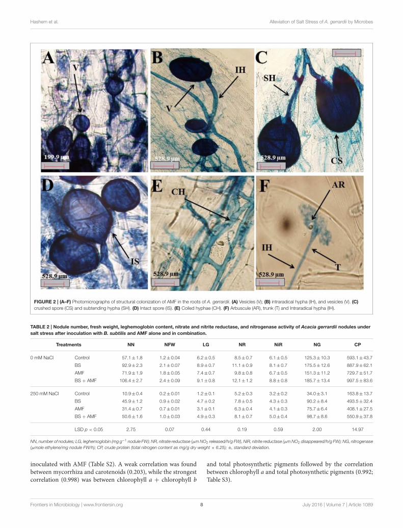
Colonization of AMF in Plant RootsThe colonization of fine A. gerrardii roots by AMF is shownin Figures 2A–F. The roots were colonized with different AMFmorphological structures, such as vesicles (Figure 2A), spores(Figure 2C), mycelium hyphae, intraradical hyphae (Figure 2B),subtending hyphae (Figure 2C), coiled hyphae (Figure 2E), andarbuscules (Figure 2F).
The percentage of 67.6, 55.8, and 13.8 of AMF that colonizedthe roots of A. gerrardii were in the form of mycelia, vesiclesand arbuscules, respectively, with a total spore density of 707.8spores/g of experimental soil (Table S1). NaCl stress decreasedspore density, the presence of mycelia, vesicles and arbusculesby 24.8, 63.6, 20.7, and 60.4%, respectively, compared with thecontrol treatment. Endophytic BS alleviated the adverse impactsof salt on spore density and mycorrhizal fungal colonization,and total spore count, mycelium, vesicles and arbuscules wereincreased by 27, 96, 14, and 23%, respectively, comparedwith those in the salt stress treatment group. In the absenceof salt stress, the endophytic BS significantly increased bothspore intensity and mycelium by 78.6 and 29.5%, respectively.However, both vesicles and arbuscules decreased by 48.38and 44.92%, respectively, compared to those of the controltreatment.
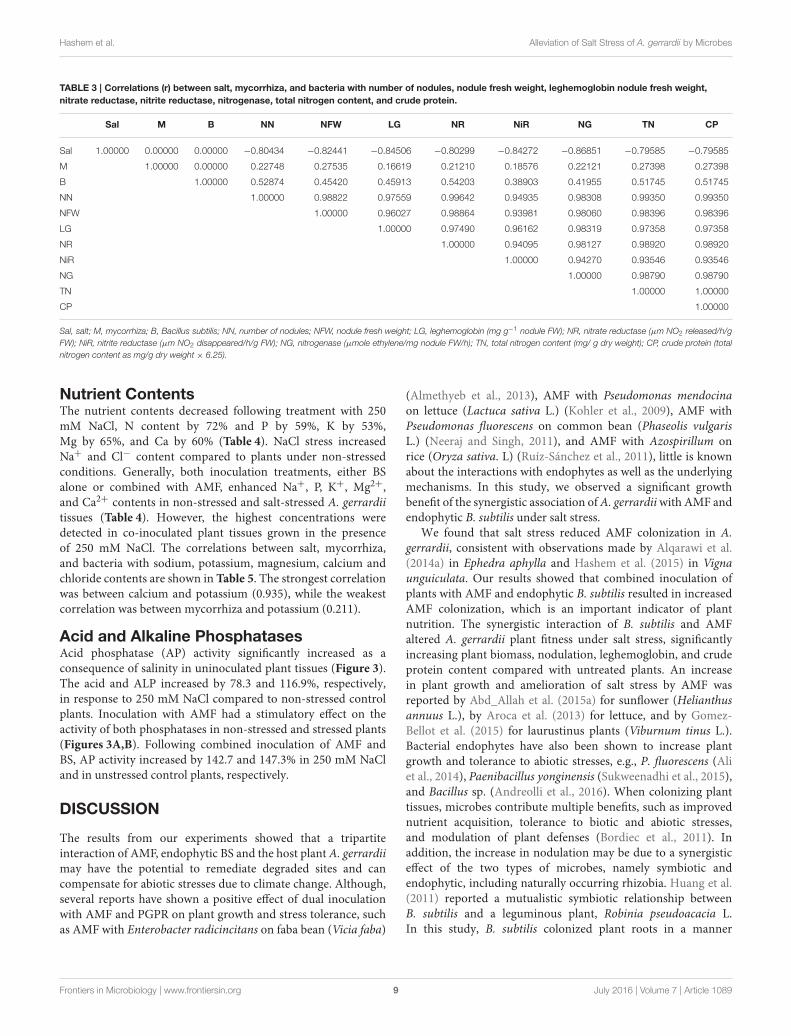
Number of Nodules, Nodule Fresh Weight,and Leghemoglobin ContentThe number of nodules, nodule FW and leghemoglobin contentwere reduced by 80.8, 80.04, and 80.6%, respectively, in salt-stressed plants relative to control, unstressed plants (Table 2). A.gerrardii grown in AMF-infected soil showed a higher numberof nodules, nodule FW and leghemoglobin content (25.9, 51.8,and 18.02%, respectively) than plants grown in control soil inboth non-saline and saline conditions. The root nodulation of
A. gerrardii depended strongly on the presence or absence ofAMF and/or the endophytic bacterium, B. subtilis (Table 2). BSand AMF alone improved the symbiotic performance of salt-stressed A. gerrardii. However, plants co-inoculated with AMFand B. subtilis produced three times more nodules, nodule FW,and leghemoglobin content in 250 mM NaCl than those inuninoculated plants.
Nitrate Reductase, Nitrite Reductase,Nitrogenase, and Crude ProteinBoth AMF and endophytic BS applied alone to non-stressed andstressed plants increased the nitrate (NR) and nitrite reductase(NIR) as well as nitrogenase activity. Inoculation of AMFincreased nitrate reductase, nitrite reductase and nitrogenaseactivity by 15.4, 10.1, and 20.7%, respectively, while BS enhancedthe activity of these enzymes by 31.5, 32.2, and 40.06%,respectively (Table 2).
The combined inoculation of AMF and BS increased nitratereductase, nitrite reductase, and nitrogenase activity in thenodules of A. gerrardii by 42.5, 43.7, and 48.2%, respectively(Table 2), while salt-stressed plants showed a 38.5, 46.4, and72.8% decline in nitrate reductase, nitrite reductase, andnitrogenase activity, respectively. The differences in enzymeactivities between uninoculated plants and those co-inoculatedwith AMF and BS under salt stress were significant, andnitrate reductase activity increased by 56%, nitrite reductaseactivity by 53% and nitrogenase activity by 189% (Table 2). Thecrude protein content increased in response to all microbialinoculation treatments regardless of whether salt was present.The combination of BS and AMF produced even better resultsbecause co-inoculated salt-stressed plant nodules containedsignificantly more protein than uninoculated salt-stressed ones(Table 2).
Table 3 show the correlations between salt, mycorrhiza, andbacteria with number of nodules, nodule FW, leghemoglobinnodule FW, nitrate reductase, nitrite reductase, nitrogenase, totalnitrogen content, and crude protein. The results indicated a slightcorrelation between mycorrhiza and nitrite reductase (0.185).However, there was a strong correlation between the number ofnodules and nitrate reductase (0.996).
Photosynthetic PigmentsThe photosynthetic pigments [chlorophyll a (ChlA), chlorophyllb (ChlB)], carotenoids and total chlorophyll (TChl) content inA. gerrardii were lower in plants without microbial inoculantsor with only one inoculant. When plants were grown in thepresence of AMF or BS, photosynthetic pigments increased,ChlA by 19.4 or 30.3%, ChlB by 12.6 or 28.3%, carotenoidsby 41.8 or 62.5% and total chlorophyll content by 15.4 or32.5%, respectively, compared to uninoculated plants (Table S2).Salinity reduced the content of ChlA, ChlB, carotenoids andTChl in A. gerrardii by 32.9, 40.5, 93.01, and 41.1%, respectively.Plants inoculated with endophytic BS and grown in soil infestedwith AMF showed an increase in the content of photosyntheticpigments. The content of ChlA, ChlB, carotenoids and TChlvaried between the single inoculant treatments and was higherin plants inoculated with the endophytic BS strain than in those
Frontiers in Microbiology | www.frontiersin.org 7 July 2016 | Volume 7 | Article 1089
Hashem et al. Alleviation of Salt Stress of A. gerrardii by Microbes
FIGURE 2 | (A–F) Photomicrographs of structural colonization of AMF in the roots of A. gerrardii. (A) Vesicles (V); (B) intraradical hypha (IH), and vesicles (V). (C)
crushed spore (CS) and subtending hypha (SH). (D) Intact spore (IS). (E) Coiled hyphae (CH). (F) Arbuscule (AR), trunk (T) and Intraradical hypha (IH).
TABLE 2 | Nodule number, fresh weight, leghemoglobin content, nitrate and nitrite reductase, and nitrogenase activity of Acacia gerrardii nodules under
salt stress after inoculation with B. subtilis and AMF alone and in combination.
Treatments NN NFW LG NR NiR NG CP
0 mM NaCl Control 57.1± 1.8 1.2± 0.04 6.2± 0.5 8.5± 0.7 6.1±0.5 125.3± 10.3 593.1± 43.7
BS 92.9± 2.3 2.1± 0.07 8.9± 0.7 11.1± 0.9 8.1±0.7 175.5± 12.6 887.9± 62.1
AMF 71.9± 1.9 1.8± 0.05 7.4± 0.7 9.8± 0.8 6.7±0.5 151.3± 11.2 729.7± 51.7
BS + AMF 106.4± 2.7 2.4± 0.09 9.1± 0.8 12.1± 1.2 8.8±0.8 185.7± 13.4 997.5± 83.6
250 mM NaCl Control 10.9± 0.4 0.2± 0.01 1.2± 0.1 5.2± 0.3 3.2±0.2 34.0± 3.1 163.8± 13.7
BS 45.9± 1.2 0.9± 0.02 4.7± 0.2 7.8± 0.5 4.3±0.3 90.2± 8.4 493.5± 32.4
AMF 31.4± 0.7 0.7± 0.01 3.1± 0.1 6.3± 0.4 4.1±0.3 75.7± 6.4 408.1± 27.5
BS + AMF 50.6± 1.6 1.0± 0.03 4.9± 0.3 8.1± 0.7 5.0±0.4 98.7± 8.6 550.9± 37.8
LSD p < 0.05 2.75 0.07 0.44 0.19 0.59 2.00 14.97
NN, number of nodules; LG, leghemoglobin (mg g−1 nodule FW); NR, nitrate reductase (µmNO2 released/h/g FW), NiR, nitrite reductase (µmNO2 disappeared/h/g FW); NG, nitrogenase
(µmole ethylene/mg nodule FW/h); CP, crude protein (total nitrogen content as mg/g dry weight × 6.25); ±, standard deviation.
inoculated with AMF (Table S2). A weak correlation was foundbetween mycorrhiza and carotenoids (0.203), while the strongestcorrelation (0.998) was between chlorophyll a + chlorophyll b
and total photosynthetic pigments followed by the correlationbetween chlorophyll a and total photosynthetic pigments (0.992;Table S3).
Frontiers in Microbiology | www.frontiersin.org 8 July 2016 | Volume 7 | Article 1089
Hashem et al. Alleviation of Salt Stress of A. gerrardii by Microbes
TABLE 3 | Correlations (r) between salt, mycorrhiza, and bacteria with number of nodules, nodule fresh weight, leghemoglobin nodule fresh weight,
nitrate reductase, nitrite reductase, nitrogenase, total nitrogen content, and crude protein.
Sal M B NN NFW LG NR NiR NG TN CP
Sal 1.00000 0.00000 0.00000 −0.80434 −0.82441 −0.84506 −0.80299 −0.84272 −0.86851 −0.79585 −0.79585
M 1.00000 0.00000 0.22748 0.27535 0.16619 0.21210 0.18576 0.22121 0.27398 0.27398
B 1.00000 0.52874 0.45420 0.45913 0.54203 0.38903 0.41955 0.51745 0.51745
NN 1.00000 0.98822 0.97559 0.99642 0.94935 0.98308 0.99350 0.99350
NFW 1.00000 0.96027 0.98864 0.93981 0.98060 0.98396 0.98396
LG 1.00000 0.97490 0.96162 0.98319 0.97358 0.97358
NR 1.00000 0.94095 0.98127 0.98920 0.98920
NiR 1.00000 0.94270 0.93546 0.93546
NG 1.00000 0.98790 0.98790
TN 1.00000 1.00000
CP 1.00000
Sal, salt; M, mycorrhiza; B, Bacillus subtilis; NN, number of nodules; NFW, nodule fresh weight; LG, leghemoglobin (mg g−1 nodule FW); NR, nitrate reductase (µm NO2 released/h/g
FW); NiR, nitrite reductase (µm NO2 disappeared/h/g FW); NG, nitrogenase (µmole ethylene/mg nodule FW/h); TN, total nitrogen content (mg/ g dry weight); CP, crude protein (total
nitrogen content as mg/g dry weight × 6.25).
Nutrient ContentsThe nutrient contents decreased following treatment with 250mM NaCl, N content by 72% and P by 59%, K by 53%,Mg by 65%, and Ca by 60% (Table 4). NaCl stress increasedNa+ and Cl− content compared to plants under non-stressedconditions. Generally, both inoculation treatments, either BSalone or combined with AMF, enhanced Na+, P, K+, Mg2+,and Ca2+ contents in non-stressed and salt-stressed A. gerrardiitissues (Table 4). However, the highest concentrations weredetected in co-inoculated plant tissues grown in the presenceof 250 mM NaCl. The correlations between salt, mycorrhiza,and bacteria with sodium, potassium, magnesium, calcium andchloride contents are shown in Table 5. The strongest correlationwas between calcium and potassium (0.935), while the weakestcorrelation was between mycorrhiza and potassium (0.211).
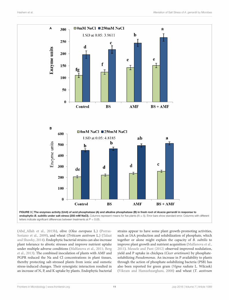
Acid and Alkaline PhosphatasesAcid phosphatase (AP) activity significantly increased as aconsequence of salinity in uninoculated plant tissues (Figure 3).The acid and ALP increased by 78.3 and 116.9%, respectively,in response to 250 mM NaCl compared to non-stressed controlplants. Inoculation with AMF had a stimulatory effect on theactivity of both phosphatases in non-stressed and stressed plants(Figures 3A,B). Following combined inoculation of AMF andBS, AP activity increased by 142.7 and 147.3% in 250 mM NaCland in unstressed control plants, respectively.
DISCUSSION
The results from our experiments showed that a tripartiteinteraction of AMF, endophytic BS and the host plant A. gerrardiimay have the potential to remediate degraded sites and cancompensate for abiotic stresses due to climate change. Although,several reports have shown a positive effect of dual inoculationwith AMF and PGPR on plant growth and stress tolerance, suchas AMF with Enterobacter radicincitans on faba bean (Vicia faba)
(Almethyeb et al., 2013), AMF with Pseudomonas mendocinaon lettuce (Lactuca sativa L.) (Kohler et al., 2009), AMF withPseudomonas fluorescens on common bean (Phaseolis vulgarisL.) (Neeraj and Singh, 2011), and AMF with Azospirillum onrice (Oryza sativa. L) (Ruíz-Sánchez et al., 2011), little is knownabout the interactions with endophytes as well as the underlyingmechanisms. In this study, we observed a significant growthbenefit of the synergistic association ofA. gerrardiiwith AMF andendophytic B. subtilis under salt stress.
We found that salt stress reduced AMF colonization in A.gerrardii, consistent with observations made by Alqarawi et al.(2014a) in Ephedra aphylla and Hashem et al. (2015) in Vignaunguiculata. Our results showed that combined inoculation ofplants with AMF and endophytic B. subtilis resulted in increasedAMF colonization, which is an important indicator of plantnutrition. The synergistic interaction of B. subtilis and AMFaltered A. gerrardii plant fitness under salt stress, significantlyincreasing plant biomass, nodulation, leghemoglobin, and crudeprotein content compared with untreated plants. An increasein plant growth and amelioration of salt stress by AMF wasreported by Abd_Allah et al. (2015a) for sunflower (Helianthusannuus L.), by Aroca et al. (2013) for lettuce, and by Gomez-Bellot et al. (2015) for laurustinus plants (Viburnum tinus L.).Bacterial endophytes have also been shown to increase plantgrowth and tolerance to abiotic stresses, e.g., P. fluorescens (Aliet al., 2014), Paenibacillus yonginensis (Sukweenadhi et al., 2015),and Bacillus sp. (Andreolli et al., 2016). When colonizing planttissues, microbes contribute multiple benefits, such as improvednutrient acquisition, tolerance to biotic and abiotic stresses,and modulation of plant defenses (Bordiec et al., 2011). Inaddition, the increase in nodulation may be due to a synergisticeffect of the two types of microbes, namely symbiotic andendophytic, including naturally occurring rhizobia. Huang et al.(2011) reported a mutualistic symbiotic relationship betweenB. subtilis and a leguminous plant, Robinia pseudoacacia L.In this study, B. subtilis colonized plant roots in a manner
Frontiers in Microbiology | www.frontiersin.org 9 July 2016 | Volume 7 | Article 1089
Hashem et al. Alleviation of Salt Stress of A. gerrardii by Microbes
TABLE 4 | Mineral contents of Acacia gerrardii under salt stress after inoculation with B. subtilis and AMF alone and in combination.
Treatments Accumulation of elements (mg/g dry weight)
Na K Mg Ca Cl N P
0 mM NaCl Control 11.3± 0.9 28.3±2.5 2.1± 0.2 2.8±0.2 8.1± 0.6 94.8± 8.2 1.6± 0.1
BS 15.7± 1.2 32.9±3.1 3.5± 0.3 4.0±0.3 5.2± 0.3 142.0± 12.4 1.8± 0.1
AMF 12.4± 1.1 29.6±2.7 2.2± 0.2 3.6±0.3 7.0± 0.5 116.7± 10.3 2.9± 0.2
BS + AMF 17.0± 1.4 35.1±3.2 2.8± 0.2 4.6±0.4 4.6± 0.2 159.6± 13.5 3.2± 0.2
250 mM NaCl Control 37.3± 3.1 13.3±1.1 0.7± 0.04 1.1±0.06 27.2± 1.9 26.2± 2.3 0.6± 0.04
BS 26.2± 2.4 23.5±2.1 1.6± 0.1 2.2±0.2 13.7± 1.1 78.9± 6.4 1.0± 0.07
AMF 28.8± 2.5 18.3±1.6 1.0± 0.08 2.0±0.2 19.4± 1.7 65.3± 6.1 1.2± 0.1
BS + AMF 25.0± 2.3 26.6±2.4 1.8± 0.1 2.4±0.2 11.0± 0.8 88.1± 7.6 1.4± 0.1
LSD p<0.05 1.51 0.54 0.11 0.25 0.28 2.14 0.16
Na, sodium; K, potassium; Mg, magnesium; Ca, calcium; N, nitrogen; P, phosphorous; ±, standard deviation.
TABLE 5 | Correlations (r) between salt, mycorrhiza, and bacteria with sodium, potassium, magnesium, calcium, and chloride.
Sal M B Na K Mg Ca Cl
Sal 1.00000 0.00000 0.00000 0.88260 −0.80382 −0.79170 −0.81818 0.79031
M 1.00000 0.00000 −0.10702 0.21197 −0.02390 0.28581 −0.20716
B 1.00000 −0.08547 0.51986 0.54310 0.42853 −0.46092
Na 1.00000 −0.85107 −0.75276 −0.77220 0.90348
K 1.00000 0.91692 0.93540 −0.97884
Mg 1.00000 0.90114 −0.88184
Ca 1.00000 −0.88885
Cl 1.00000
Sal, salt; M, mycorrhiza; B, Bacillus subtilis; Na, sodium; K, potassium; Mg, magnesium; Ca, calcium; Cl, chloride.
similar to the infection of root hairs by rhizobia and formedbacteroids inside plant cortical cells. Inoculation of plants withcellulose-producing B. subtilis resulted in more nodules andhigher nitrogenase activity than the uninoculated control andAMF-inoculated plants. Rhizobial symbionts penetrate deeperplant tissues by producing cellulase, which can completely erodethe root-hair wall at the site of infection (Sindhu and Dadarwal,2001). The synthesis of cell wall-degrading enzymes by B. subtiliscould help explain themechanism underlying rhizobial entry intotarget root hair cells to form nodules.
Increased nitrogenase activity following treatments with B.subtilis or B. subtilis combined with AMF resulted from theirpositive impact on the activity of enzymes such as nitratereductase and nitrite reductase. Nitrate and nitrite reductasecontrol the conversion of nitrate into ammonia and result inthe formation of amino acids (Iqbal et al., 2015). In Trifoliumalexandrinum L. and Trifolium resupinatum L. (Zarea et al.,2011), as well as in Vicia faba (Hashem et al., 2014), inoculationof AMF enhanced plant growth by improving nitrogenase activityand nodule formation.
High salt concentrations induce alterations in the synthesisof chlorophyll-related proteins and components of the oxygen-evolving complex, resulting in reduced photosynthetic efficiency(Alqarawi et al., 2014a,b). Altered de novo synthesis of proteinsand the associated pigment-related components due to salinity
has negative effects on the synthesis of photoassimilates andhence reduces the growth rate of plants. The combinedinoculation of AMF and B. subtilis increase photosyntheticpigments, which may be a collective result of many positivechanges induced by AMF and PGPR. The enhanced chlorophyllcontent due to AMF inoculation under normal as well as NaCl-stressed conditions corroborates the reports of Aroca et al.(2013) in lettuce, Alqarawi et al. (2014a) in E. aphylla andAbd_Allah et al. (2015a) in Sesbania sesban. Recently, in salt-stressed Brassica juncea, Ahmad et al. (2015) demonstratedthe positive impact of Trichoderma harzianum inoculation ongrowth via improved chlorophyll synthesis. InOcimum basilicumgrown under water stress, inoculation of PGPR (Pseudomonassp. and Bacillus lentus) increased chlorophyll synthesis as wellphotosynthetic electron transport and also mitigated the negativeimpact of water stress (Heidari and Golpayegani, 2012).
Salt stress inhibits the uptake of essential mineral elements,such as K, Mg, Ca, N, and P, because of the antagonisticrelationship of sodium. By reducing the uptake of magnesium,salt stress affects plant photosynthetic efficiency by altering thesynthesis of chlorophyll molecules. Reduced uptake of nitrogendirectly affects the nitrogen metabolic potential as well as aminoacid synthesis in plants (Näsholm et al., 2009). Improved plantnutrient uptake under salt stress conditions by AMFwas reportedin many studies e.g., for common bean (Phaseolus vulgaris)
Frontiers in Microbiology | www.frontiersin.org 10 July 2016 | Volume 7 | Article 1089
Hashem et al. Alleviation of Salt Stress of A. gerrardii by Microbes
FIGURE 3 | The enzymes activity (Unit) of acid phosphatase (A) and alkaline phosphatase (B) in fresh root of Acacia gerrardii in response to
endophytic B. subtilis under salt stress (250 mM NaCl). Columns represent means for five plants (N = 5). Error bars show standard error. Columns with different
letters indicate significant differences between treatments at P < 0.05.
(Abd_Allah et al., 2015b), olive (Olea europaea L.) (Porras-Soriano et al., 2009), and wheat (Triticum aestivum L.) (Talaatand Shawky, 2014). Endophytic bacterial strains can also increaseplant tolerance to abiotic stresses and improve nutrient uptakeunder multiple adverse conditions (Malfanova et al., 2011; Berget al., 2013). The combined inoculation of plants with AMF andPGPR reduced the Na and Cl concentrations in plant tissues,thereby protecting salt-stressed plants from ionic and osmoticstress-induced changes. Their synergistic interaction resulted inan increase of N, P, and K uptake by plants. Endophytic bacterial
strains appear to have some plant growth-promoting activities,such as IAA production and solubilization of phosphate, whichtogether or alone might explain the capacity of B. subtilis toimprove plant growth and nutrient acquisition (Malfanova et al.,2011). Messele and Pant (2012) observed improved nodulation,yield and P uptake in chickpea (Cicer arietinum) by phosphate-solubilizing Pseudomonas. An increase in P availability to plantsthrough the action of phosphate-solubilizing bacteria (PSB) hasalso been reported for green gram (Vigna radiata L. Wilczek)(Vikram and Hamzehzarghani, 2008) and wheat (T. aestivum
Frontiers in Microbiology | www.frontiersin.org 11 July 2016 | Volume 7 | Article 1089
Hashem et al. Alleviation of Salt Stress of A. gerrardii by Microbes
L.) (Panhwar et al., 2011). In earlier studies, it was reportedthat salt and drought stresses inhibit the production of plantgrowth regulators in plant tissues (Debez et al., 2001). Theadditional supply of hormones by endophytes in plant tissue canstimulate the root system, thereby facilitating the absorption ofmore nutrients from the soil, especially under stress conditions(Malfanova et al., 2011; Berg et al., 2013). Studies by Kavinoet al. (2010), Heidari et al. (2011), and Lavakush et al. (2014)also support the positive impact of PGPR on the mineralnutrient status of plants under normal and stressed conditionsin determining the physiological strength of a plant. ImprovedMg content affects chlorophyll, while P and N contribute tothe energy budget of a cell, and Ca serves as an importantcellular messenger for downstream signaling (Ahanger et al.,2014b). Improved K uptake is associated with reduced Nauptake in AMF- and PGPR-inoculated plants and results in anenhanced K/Na ratio, an important aspect for the maintenanceof physiological cellular functioning (Abd_Allah et al., 2015a,b;Ahanger et al., 2015).
Phosphatases are responsible for the hydrolysis of a rangeof organic P compounds and provide mineral phosphate tothe plant (Tazisong et al., 2015). In our study, increased acidand ALP was observed in A. gerrardii grown under salt stress.Similarly, enhanced AP activity under salt stress was alsoobserved in Medicago sativa (Ehsanpour and Amini, 2003)and may be due to the fact that salt stress suppresses plantgrowth, P uptake, transport and utilization (Dracup et al., 1984).An increase in the activities of phosphatases (alkaline andacidic phosphatase) following the combined inoculation of A.gerrardii plants with AMF and B. subtilis support the findingsof Amaya-Carpio et al. (2009) in Ipomoea carnea sp. fistulosaand Kebrabadi et al. (2014) in Fraxinus rotundifolia. DuringP deficiency or impaired P uptake, plants release or activateacid phosphates and exude carboxylates and phosphatases toenhance P solubilization and uptake (Veneklaas et al., 2003).An increase in the activity of phosphatases in inoculated plants,either with AMF or B. subtilis, alone or in combination,can contribute to the release of bound P to maximize its
uptake and transport. Maize plants inoculated with B. subtilisshowed an increase in phosphatase activity compared touninoculated controls (Hussain et al., 2013). Salinity-stressedplants showed lower P accumulation in the above-groundplant parts, which may be due to the toxicity of high saltconcentrations.
CONCLUSION
Our observations in this study indicate that endophytic bacteriaand AMF that live within the plant tissues of A. gerrardii arecoordinately involved in the plant’s adaptation to stress tolerance.Inoculation of plants with AMF and endophytic B. subtilisincreased plant growth and nutrient acquisition and improvedsymbiotic performance of A. gerrardii. In addition, endophytic B.subtilis increased AMF germination and root colonization of A.gerrardii under salt stress.
AUTHOR CONTRIBUTIONS
AAH, Provided the seeds and seedlings. AH, Provided AMF andmycological analysis. EA, All biochemical analysis, writing andgraphing. AAA, Statistical analysis. SW, Revision and editing.DE, writing, revision and editing.
ACKNOWLEDGMENTS
The authors would like to extend their sincere appreciation tothe Deanship of Scientific Research at King Saud University forits funding of this research through the Research Group Projectno RGP-271.
SUPPLEMENTARY MATERIAL
The Supplementary Material for this article can be foundonline at: http://journal.frontiersin.org/article/10.3389/fmicb.2016.01089
REFERENCES
Abd_Allah, E. F., Hashem, A., Alqarawi, A. A., and Alwhibi, M. S. (2015b).
Alleviation of adverse impact of salt in Phaseolus vulgaris l. by arbuscular
mycorrhizal fungi. Pak. J. Bot. 47, 1167–1176.
Abd_Allah, E. F., Hashem, A., Alqarawi, A. A., Bahkali, A. H., and Alwhibi, M. S.
(2015a). Enhancing growth performance and systemic acquired resistance of
medicinal plant Sesbania sesban (L.) Merr using arbuscular mycorrhizal fungi
under salt stress. Saudi J. Bio. Sci. 22, 274–283.
Abd_Allah, E. F., Hashem, A., Alqarawi, A. A., and Hend, A. (2015c). Alleviation
of adverse impact of cadmium stress in sunflower (Helianthus annuus l.) by
arbuscular mycorrhizal fungi. Pak. J. Bot. 47, 785–795.
Ahanger, M. A., Agarwal, R. M., Tomar, N. S., and Shrivastava,. M. (2015).
Potassium induces positive changes in nitrogen metabolism and antioxidant
system of oat (Avena sativa L cultivar Kent). J. Plant Int. 10, 211–223. doi:
10.1080/17429145.2015.1056260
Ahanger,M. A., Hashem, A., Abd_Allah, E. F., and Ahmad, P. (2014a). “Arbuscular
mycorrhiza in crop improvement under environmental stress,” in Emerging
Technologies and Management of Crop Stress Tolerance, Vol. 2, eds P. Ahmad
and S. Rasool (San Diego, CA: Academic Press; Elsevier), 69–95.
Ahanger, M. A., Tyagi, S. R., Wani, M. R., and Ahmad, P. (2014b). “Drought
tolerance: role of organic osmolytes, growth regulators, and mineral nutrients,”
in Physiological Mechanisms and Adaptation Strategies in Plants Under
Changing Environment, eds P. Ahmad and M. R. Wani (New York, NY:
Springer; Science+Business Media), 25–55.
Ahmad, P. (2010). Growth and antioxidant responses in mustard (Brassica juncea
L.) plants subjected to combined effect of gibberellic acid and salinity. Arch.
Agro. Soil Sci. 56, 575–588. doi: 10.1080/03650340903164231
Ahmad, P., Hashem, A., Abd-Allah, E. F., Alqarawi, A. A., John, R., Egamberdieva,
D., et al. (2015). Role of Trichoderma harzianum in mitigating NaCl stress
in Indian mustard (Brassica juncea L) through antioxidative defense system.
Front. Plant Sci. 6:868. doi:10.3389/fpls.2015.00868
Ali, S., Charles, T. C., and Glick, B. R. (2014). Amelioration of high
salinity stress damage by plant growth-promoting bacterial endophytes
that contain ACC deaminase. Plant Physiol. Biochem. 80, 160–167. doi:
10.1016/j.plaphy.2014.04.003
Allen,M. B. (1953). Experiments in Soil Bacteriology, 1st Edn.Minneapolis: Burgess
Pub. Co.
Almethyeb, M., Ruppel, S., Paulsen, H. M., Vassilev, N., and Eichler-Löbermann, B.
(2013). Single and combined applications of arbuscular mycorrhizal fungi and
Frontiers in Microbiology | www.frontiersin.org 12 July 2016 | Volume 7 | Article 1089
Hashem et al. Alleviation of Salt Stress of A. gerrardii by Microbes
Enterobacter radicincitans affect nutrient uptake of faba bean and soil biological
characteristics. Agric. Fores. Res. 3, 229–234. doi: 10.3220/LBF_2013_229-234
Alqarawi, A. A., Abd_Allah, E. F., and Hashem, A. (2014a). Alleviation of salt-
induced adverse impact via mycorrhizal fungi in Ephedra aphylla Forssk. J.
Plant Inter. 9, 802–810. doi: 10.1080/17429145.2014.949886
Alqarawi, A. A., Abd_Allah, E. F., Hashem, A., Al-Huqail, A. A., and Al-Sahli, A.
A. (2014b). Impact of abiotic salt stress on some metabolic activities of Ephedra
alata Decne. J. Food Agr. Environ. 12, 620–625.
Amaya-Carpio, L., Davies, F. T., Fox, T., and He, C. (2009). Arbuscular
mycorrhizal fungi and organic fertilizer influence photosynthesis, root
phosphatase activity, nutrition, and growth of Ipomoea carnea ssp. fistulosa.
Photosynthetica 47, 1–10. doi: 10.1007/s11099-009-0003-x
Andreolli, M., Lampis, S., Zapparoli, G., Angelini, E., and Vallini, G. (2016).
Diversity of bacterial endophytes in 3 and 15 year-old grapevines
of Vitis vinifera cv. Corvina and their potential for plant growth
promotion and phytopathogen control. Microb. Res. 183, 42–52. doi:
10.1016/j.micres.2015.11.009
AOAC (Association of Official Analytical Chemists) (1995). Official Methods of
Analysis of AOAC International, 16th Edn., Vol. 2. Arlington, VA: AOAC
International.
Ardakani, M. R., Pietsch, G., Moghaddam, A., Raza, A., and Friedel, J. K. (2009).
Response of root properties to tripartite symbiosis between lucerne (Medicago
sativa L.), rhizobia and mycorrhiza under dry organic farming conditions. Am.
J. Agric. Biol. Sci. 4, 266–277. doi: 10.3844/ajabssp.2009.266.277
Arnon, D. I. (1949). Copper enzymes in isolated chloroplasts. polyphenoloxidase
in Beta vulgaris. Plant Physiol. 24, 1–15. doi: 10.1104/pp.24.1.1
Aroca, R., Ruiz-Lozano, J. M., Zamarreno, A., Paz, J. A., Garcia-Mina, J. M., Pozo,
M. J., et al. (2013). Arbuscular mycorrhizal symbiosis influences strigolactone
production under salinity and alleviates salt stress in lettuce plants. J. Plant
Physiol. 170, 47–55. doi: 10.1016/j.jplph.2012.08.020
Bano, N., and Musarrat, J. (2003). Characterization of a new Pseudomonas
aeruginosa strain NJ-15 as a potential biocontrol agent. Curr. Microb. 46,
324–328. doi: 10.1007/s00284-002-3857-8
Barnawal, D., Bharti, N., Maji, D., Chanotiya, C. S., and Kalra, A. (2014).
ACC deaminase-containing Arthrobacter protophormiae induces NaCl stress
tolerance through reduced ACC oxidase activity and ethylene production
resulting in improved nodulation and mycorrhization in Pisum sativum. J.
Plant Physiol. 171, 884–894. doi: 10.1016/j.jplph.2014.03.007
Berg, G., Alavi, M., Schmidt, C. S., Zachow, C., Egamberdieva, D., Kamilova,
F., et al. (2013). “Biocontrol and osmoprotection for plants under saline
conditions,” inMolecularMicrobial Ecology of the Rhizosphere, ed F. J. De Bruijn
(Hoboken, NJ: Wiley-Blackwell).
Bethlenfalvay, G. J., and Yoder, J. F. (1981). The Glycine-Glomus-Rhizobium
symbiosis. I. Phosphorus effect on nitrogen fixation and mycorrhizal infection.
Physiol. Plant. 52, 141–145. doi: 10.1111/j.1399-3054.1981.tb06047.x
Bordiec, S., Paquis, S., Lacroix, H., Dhondt, S., Ait Barka, E., Kauffmann, S., et al.
(2011). Comparative analysis of defence responses induced by the endophytic
plant growth-promoting rhizobacterium Burkholderia phytofirmans strain
PsJN and the non-host bacterium Pseudomonas syringae pv. pisi in grapevine
cell suspensions. J. Exp. Bot. 62, 595–603. doi: 10.1093/jxb/erq291
Bouhmouch, I., Souad-Mouhsine, B., Brhada, F., and Aurag, J. (2005). Influence of
host cultivars and Rhizobium species on the growth and symbiotic performance
of Phaseolus vulgaris under salt stress. J. Plant Physiol. 162, 1103–1113. doi:
10.1016/j.jplph.2004.12.003
Daniels, B. A., and Skipper, H. D. (1982). “Methods for the recovery and
quantitative estimation of propagules from soil,” in Methods and Principles
of Mycorrhizal Research, ed N. C. Schenck (St. Paul Minnesota: American
Phytopathological Society), 29–36.
Debez, A., Chaibi, W., and Bouzid, S. (2001). Effect du NaCl et de regulatoeurs de
croissance sur la germination d’Atriplex halimus L. Cahiers Agric. 10, 135–138.
Dracup, M. N. H., Barrett-Lennard, E. G., Greenway, H., and Robson, A. D.
(1984). Effect of phosphorus deficiency on phosphatase activity of cell walls
from roots of subterranean clover. J. Exp. Bot. 35, 466–480. doi: 10.1093/jxb/35.
4.466
Egamberdieva, D. (2012). Pseudomonas chlororaphis: a salt tolerant bacterial
inoculant for plant growth stimulation under saline soil conditions. Acta
Physiol. Plant. 34, 751–756. doi: 10.1007/s11738-011-0875-9
Egamberdieva, D., Berg, G., Lindström, K., and Räsänen, L. A. (2013). Alleviation
of salt stress of symbiotic Galega officinalis L. (goat’s rue) by co-inoculation
of rhizobium with root colonising Pseudomonas. Plant Soil 369, 453–465. doi:
10.1007/s11104-013-1586-3
Egamberdieva, D., Jabborova, D., and Berg, G. (2015). Synergistic interactions
between Bradyrhizobium japonicum and the endophyte Stenotrophomonas
rhizophila and their effects on growth, nodulation and nutrition of soybean
under salt stress. Plant Soil 1–11.
Egamberdieva, D., Li, L., Lindström, K., and Räsänen, L. (2016). A synergistic
interaction between salt tolerant Pseudomonas and Mezorhizobium strains
improves growth and symbiotic performance of liquorice (Glycyrrhiza uralensis
Fish.) under salt stress. Appl. Microb. Biotechnol. 100, 2829–2841. doi:
10.1007/s00253-015-7147-3
Ehsanpour, A. A., and Amini, F. (2003). Effect of salt and drought stress on acid
phosphatase activites in alfalfa (Medicago sativa L.) explants under in vitro
culture. Afr. J. Biotech. 2, 133–135. doi: 10.5897/AJB2003.000-1026
Finka, R. L., Warner, R. L., and Muzit, T. J. (1977). Effect of herbicides on in vivo
nitrate and nitrite reduction.Weed Sci. 25, 18–22.
Gerdemann, J. W., and Nicolson, T. H. (1963). Spores of mycorrhizal Endogone
extracted from soil by wet sieving and decanting. Trans. Brit. Mycol. Soc. 46,
235–244. doi: 10.1016/S0007-1536(63)80079-0
Gianinazzi-Pearson, V., and Gianinazzi, S. (1976). Enzymatic studies on the
metabolism of vesicular-arbuscular mycorrhiza. I. Effect of mycorrhiza
formation and phosphorus nutrition on soluble phosphatase activities in onion
roots. Physiol. Veg. 14, 833–841.
Gomez-Bellot, M. J., Nortes, P. A., Ortuno, M. F., Romero, C., Fernandez-García,
C., and Sanchez-Blanco, M. J. (2015). Influence of arbuscular mycorrhizal
fungi and treated wastewater onwater relations and leaf structure alterations
of Viburnum tinus L. plants during both saline and recovery periods. J. Plant
Physiol. 188, 96–105. doi: 10.1016/j.jplph.2015.09.007
Gupta, P., Samant, K., and Sahu, A. (2012). Isolation of cellulose-degrading
bacteria and determination of their cellulolytic potential. Int. J. Microbiol.
2012:578925. doi: 10.1155/2012/578925
Hageman, R. H., and Hucklesby, D. P. (1971). “Nitrate reductase from higher
plants,” in Methods in Enzymology, ed A. S. Pietro (New York, NY: Academic
Press), 491–503.
Hashem, A., Abd_Allah, E. F., Alqarawi, A. A., Al-Didamony, G., Al-Whibi, M.,
Egamberdieva, D., et al. (2014). Alleviation of adverse impact of salinity on
faba bean (Vicia faba L.) by arbuscular mycorrhizal fungi. Pak. J. Bot. 46,
2003–2013.
Hashem, A., Abd_Allah, E. F., Alqarawi, A. A., and Egamberdieva, D.
(2015). Induction of salt stress tolerance in cowpea (Vigna unguiculata
L. Walp) by arbuscular mycorrhizal fungi. Legume Res. 38, 579–588. doi:
10.18805/lr.v38i5.5933
Heidari, M., and Golpayegani, A. (2012). Effects of water stress and inoculation
with plant growth promoting rhizobacteria (PGPR) on antioxidant status and
photosynthetic pigments in basil (Ocimum basilicum L.). J. Saudi Society Agri.
Sci. 11, 57–61. doi: 10.1016/j.jssas.2011.09.001
Heidari, M., Mousavinik, S. M., and Golpayegani, A. (2011). Plant growth
promoting rhizobacteria (PGPR) effect on physiological parameters and
mineral uptake in basil (Ociumum basilicum L.) under water stress. J. Agri. Bio.
Sci. 6, 6–11.
Huang, B., Lv, C., Zhuang, P., Zhang, H., and Fan, L. (2011). Endophytic
colonisation of Bacillus subtilis in the roots of Robinia pseudoacacia L. Plant
Biol. 13, 925–931. doi: 10.1111/j.1438-8677.2011.00456.x
Hussain, M. I., Asghar, H. N., Akhtar, M. J., and Arshad, M. (2013). Impact of
phosphate solubilizing bacteria on growth and yield of maize. Soil Environ. 32,
71–78.
Ikawa, I. J., Nisizawa, K., and Mira, N. K. (1964). Specifications of several acid
phosphatases from plant sources. Nature 203, 939–940. doi: 10.1038/203939a0
Iqbal, N., Umar, S., and Khan, N. A. (2015). Nitrogen availability regulates
proline and ethylene production and alleviates salinity stress in mustard
(Brassica juncea). J. Plant Physiol. 178, 84–91. doi: 10.1016/j.jplph.2015.0
2.006
Jackson, M. L. (1962). Soil Chemical Analysis. New York, NY: Prentice Hall.
Kavino, M., Harish, S., Kumar, N., Saravanakumar, D., and Samiyappan, R. (2010).
Effect of chitinolytic PGPR on growth, yield and physiological attributes of
Frontiers in Microbiology | www.frontiersin.org 13 July 2016 | Volume 7 | Article 1089

Hashem et al. Alleviation of Salt Stress of A. gerrardii by Microbes
banana (Musa spp.) under field conditions. Appl. Soil Ecol. 45, 71–77. doi:
10.1016/j.apsoil.2010.02.003
Kebrabadi, B. Z., Matinizadeh, M., Daryayi, M. G., and Salehi, A. (2014). Changes
in acid and alkaline phosphatase enzyme activity in rhizosphere ash Fraxinus
rotundifolia and its correlation with soil and plant phosphorus. J. Biod. Env.
Sci. 4, 233–238.
Keilin, D., andWang, Y. L. (1945). Hemoglobin in the root nodules of leguminous
plants. Nature 155, 227–229. doi: 10.1038/155227a0
Kohler, J., Hernandez, J. A., Caravaca, F., and Roldan, A. (2009). Induction
of antioxidant enzymes is involved in the greater effectiveness of a PGPR
versus AM fungi with respect to increasing the tolerance of lettuce to severe
salt stress. Environ. Exp. Bot. 65, 245–252. doi: 10.1016/j.envexpbot.2008.
09.008
Kormanik, P. P., and McGraw, A. C. (1982). “Quantification of vesicular-
arbuscularmycorrhizal in plant roots,” inMethods and Principles ofMycorrhizal
Research, ed N. C. Schenck (St. Paul, MN: American Phytopatholgy Society),
37–46.
Lavakush, Y. J., Verma, J. P., Jaiswal, D. K., and Kumar, A. (2014). Evaluation
of PGPR and different concentration of phosphorus level on plant growth,
yield and nutrient content of rice (Oryza sativa). Ecol. Engin. 62, 123–128. doi:
10.1016/j.ecoleng.2013.10.013
Ligero, F., Lluch, C., and Olivares, J. (1986). Evolution of ethylene from roots of
Medicago sativa plants inoculated with Rhizobiummeliloti. J. Plant Physiol. 125,
361–365. doi: 10.1016/S0176-1617(86)80158-4
Malfanova, N., Kamilova, F., Validov, S., Shcherbakov, A., Chebotar, V.,
Tikhonovich, I., et al. (2011). Characterization of Bacillus subtilis HC8, a novel
plant-beneficial endophytic strain from giant hogweed. Microb. Biotech. 4,
523–532. doi: 10.1111/j.1751-7915.2011.00253.x
Marschner, P., Yang, C. H., Lieberei, R., and Crowley, D. E. (2001). Soil and plant
specific effects on bacterial community composition in the rhizosphere. Soil
Biol. Biochem. 33, 1437–1445. doi: 10.1016/S0038-0717(01)00052-9
Messele, B., and Pant, L. M. (2012). Effects of Inoculation of Sinorhizobium ciceri
and phosphate solubilizing bacteria on nodulation, yield and nitrogen and
phosphorus uptake of chickpea (Cicer arietinum L.) in Shoa Robit Area. J.
Biofertil. Biopest. 3:129. doi: 10.4172/2155-6202.10000129
Mohanta, M. K., Saha, A. K., Saleh, D. K. M. A., Islam, M. S., Mannan, K. S. B., and
Fakruddin, M. (2015). Characterization of Klebsiella granulomatis pathogenic
to silkworm, Bombyx mori L. 3 Biotech. 5, 577–583. doi: 10.1007/s13205-014-
0255-4
Muyzer, G., Teske, A., Wirsen, C. O., and Jannasch, H. W. (1995). Phylogenetic
relationships of Thiomicrospira species and their identification in deep-sea
hydrothermal vent samples by denaturing gradient gel electrophoresis of 16S
rDNA fragments. Arch. Microb. 164, 165–171. doi: 10.1007/BF02529967
Nadeem, S. M., Ahmadm, M., Zahir, Z. A., Javaid, A., and Ashraf, M. (2014).
The role of mycorrhizae and plant growth promoting rhizobacteria (PGPR) in
improving crop productivity under stressful environments. Biotech. Adv. 32,
429–448. doi: 10.1016/j.biotechadv.2013.12.005
Näsholm, T., Kielland, K., and Ganeteg, U. (2009). Uptake of organic nitrogen by
plants. New Phytol. 182, 31–48. doi: 10.1111/j.1469-8137.2008.02751.x
Navarro, J. M., Perez-Tornero, O., and Morte, A. (2013). Alleviation of salt
stress in citrus seedlings inoculated with arbuscular mycorrhizal fungi
depends on the rootstock salt tolerance. J. Plant Physiol. 171, 76–85. doi:
10.1016/j.jplph.2013.06.006
Neeraj and Singh, K. (2011). Organic amendments to soil inoculated arbuscular
mycorrhizal fungi and Pseudomonas fluorescens treatments reduce the
development of root-rot disease and enhance the yield of Phaseolus vulgaris
L. Eur. J. Soil Biol. 47, 288–295. doi: 10.1016/j.ejsobi.2011.07.002
Panhwar, Q. A., Radziah, O., Khanif, Y. M., and Naher, U. A. (2011). Application of
boron and zinc in the tropical soils and its effect on maize (Zea mays L.) growth
and soil microbial environment. Aust. J. Crop Sci. 5, 1649–1654.
Phillips, J. M., and Hayman, D. S. (1970). Improved procedures for clearing
and staining parasitic and vesicular-arbuacular mycorrhizal fungi for rapid
assessment of infection. Trans. Br. Mycol. Soc. 55, 158–161. doi: 10.1016/S0007-
1536(70)80110-3
Pikovskaya, R. I. (1948). Mobilization of phosphorus in soil connection with the
vital activity of some microbial species.Microbiologiya 17, 362–370.
Porcel, R., Aroca, R., and Ruiz-Lozano, J. M. (2012). Salinity stress alleviation using
arbuscular mycorrhizal fungi. A review. Agron. Sust. Dev. 32, 181–200. doi:
10.1007/s13593-011-0029-x
Porras-Soriano, A., Sorano-Marintin, M. L., Porras-Piedra, A., and Azcon, P.
(2009). Arbuscular mycorrhizal fungi increased growth, nutrient uptake and
tolerance to salinity in olive trees under nursery conditions. J. Plant Physiol.
166, 1350–1359. doi: 10.1016/j.jplph.2009.02.010
Redecker, D., Schüßler, A., Stockinger, H., Stürmer, S. L., Morton, J. B., and
Walker, C. (2013). An evidence-based consensus for the classification of
arbuscular mycorrhizal fungi (Glomeromycota). Mycorrhiza 23, 515–531. doi:
10.1007/s00572-013-0486-y
Ruiz-Lozano, J. M., Porcel, R., Azcon, R., and Aroca, R. (2012). Regulation by
arbuscular mycorrhizae of the integrated physiological response to salinity in
plants. New challenges in physiological and molecular studies. J. Exp. Bot. 63,
4033–4044. doi: 10.1093/jxb/ers126
Ruíz-Sánchez, M., Armada, E., Munoz, Y., de Salamone, I. E. G., Aroca, R.,
Ruíz-Lozano, J. M., et al. (2011). Azospirillum and arbuscular mycorrhizal
colonization enhance rice growth and physiological traits under well-
watered and drought conditions. J. Plant Physiol. 168, 1031–1037. doi:
10.1016/j.jplph.2010.12.019
Schüßler, A., and Walker, C. (2010). The Glomeromycota: A Species List with New
Families and Genera. Gloucester.
Sessitsch, A., Hardoim, P., Döring, J., Weilharter, A., Krause, A., Woyke, T., et al.
(2012). Functional characteristics of an endophyte community colonizing rice
roots as revealed by metagenomic analysis. Mol. Plant Microbe Interact. 25,
28–36. doi: 10.1094/MPMI-08-11-0204
Sheng, M., Tang, M., Chan, H., Yang, B., Zhang, F., and Huang, Y. (2008).
Influence of arbuscular mycorrhizae on photosynthesis and water status of
maize plants under salt stress. Mycorrhiza 18, 287–296. doi: 10.1007/s00572-
008-0180-7
Silsbury, J. H. H. (1990). Growth nitrogen accumulation and partitioning, and
N2 fixation in faba bean (Vicia faba cv. Fiord) and pea (Pisum sativum
cv. Early Dun). Field Crops Res. 24, 173–188. doi: 10.1016/0378-4290(90)
90036-B
Sindhu, S. S., and Dadarwal, K. R. (2001). Chitinolytic and cellulolytic
Pseudomonas sp. antagonistic to fungal pathogens enhances nodulation by
Mesorhizobium sp. cicer in chickpea. Microbiol. Res. 156, 353–358. doi:
10.1078/0944-5013-00120
Slattery, J. F., Conventry, D. R., and Slattery, W. J. (2001). Rhizobial ecology
as affected by the soil environment. Aust. J. Exp. Agric. 41, 289–298. doi:
10.1071/EA99159
Stutz, J. C., and Morton, J. B. (1996). Successive pot cultures reveal high species
richness of arbuscular mycorrhizal fungi in arid ecosystems. Can. J. Bot. 74,
1883–1889. doi: 10.1139/b96-225
Sukweenadhi, J., Kima, Y. J., Choi, E., Koh, S. C., Lee, S. W., Kim, Y. J., et al. (2015).
Paenibacillus yonginensis DCY84T induces changes in Arabidopsis thaliana
gene expression against aluminum, drought, and salt stress. Microb. Res. 172,
7–15. doi: 10.1016/j.micres.2015.01.007
Tajini, F., Trabelsi, M., and Drevona, J. J. (2012). Combined inoculation with
Glomus intraradices and Rhizobium tropici CIAT899 increases phosphorus
use efficiency for symbiotic nitrogen fixation in common bean (Phaseolus
vulgaris L.). Saudi J. Biol. Sci. 19, 157–163. doi: 10.1016/j.sjbs.2011.
11.003
Talaat, N. B., and Shawky, B. T. (2014). Protective effects of arbuscular mycorrhizal
fungi on wheat (Triticum aestivum L.) plants exposed to salinity. Environ. Exp.
Bot. 98, 20–31. doi: 10.1016/j.envexpbot.2013.10.005
Tazisong, I. A., Senwo, Z. N., and He, Z. (2015). Phosphatase hydrolysis of organic
phosphorus compounds. Adv. Enz. Res. 3, 39–51. doi: 10.4236/aer.2015.32005
Töpper, B., Larsen, A., Thingstad, T. F., Thyrhaug, R., and Sandaa, R.-A. (2010).
Bacterial community composition in an Arctic phytoplankton mesocosm
bloom: the impact of silicate and glucose. Polar Biol. 33, 1557–1565. doi:
10.1007/s00300-010-0846-4
Torriani, A. M. (1967). “Alikaline phosphatase from Escherichia coli,” in Procedures
in Nucleic Acid Research, eds G. L. Cantoni and D. L. Davies (New York, NY:
Harper and Row Publishers, Inc.), 224–235.
Utobo, E. B., Ogbodo, E. N., and Nwogbaga, A. C. (2011). Techniques for
extraction and quantification of arbuscular rmycorrhizal fungi. Libyan Agric.
Res. Center. Int. 2, 68–78.
Veneklaas, E. J., Stevens, J., Cawthray, G. R., Turner, S., Grigg, A. M., and Lambers,
H. (2003). Chickpea and white lupin rhizosphere carboxylates vary with soil
properties and enhance phosphorus uptake. Plant Soil 248, 187–197. doi:
10.1023/A:1022367312851
Frontiers in Microbiology | www.frontiersin.org 14 July 2016 | Volume 7 | Article 1089
Hashem et al. Alleviation of Salt Stress of A. gerrardii by Microbes
Vikram, A., and Hamzehzarghani, H. (2008). Effect of phosphate solubilizing
bacteria on nodulation and growth parameters of greengram (Vigna radiate L.
Wilczek). Res. J. Microbial. 3, 62–72. doi: 10.3923/jm.2008.62.72
Wolf, B. (1982). A comprehensive system of leaf analysis and its use for
diagnosing crop nutrient status. Comm. Soil Sci. Plant Anal. 13, 1035–1059.
doi: 10.1080/00103628209367332
Wu, Q. S., Ying-Ning, Z., and Abd-Allah, E. F. (2014). “Mycorrhizal Association
and ROS in Plants,” in Oxidative Damage to Plants, ed P. Ahmad (New York,
NY: Academic Press; Elsevier), 453–475.
Xavier, L. J. C., and Germida, J. J. (2002). Response of lentil under controlled
conditions to coinoculation with arbuscular mycorrhizal fungi and rhizobia
varying in efficacy. Soil Biol Biochem. 34, 181–188. doi: 10.1016/S0038-
0717(01)00165-1
Yinsuo, J., Vincent, M. G., and Colin, J. S. (2004). The Influence of Rhizobium
and arbuscular mycorrhizal fungi on nitrogen and phosphorus accumulation
by Vicia faba. Ann. Bot. 94, 251–258. doi: 10.1093/aob/mch135
Zarea, M. J., Karimi, N., Goltapeh, E. M., and Ghalavand, A. (2011). Effect
of cropping systems and arbuscular mycorrhizal fungi on soil microbial
activity and root nodule nitrogenase. J. Saudi Soc. Agri. Sci. 10, 109–120. doi:
10.1016/j.jssas.2011.04.003
Conflict of Interest Statement: The authors declare that the research was
conducted in the absence of any commercial or financial relationships that could
be construed as a potential conflict of interest.
Copyright © 2016 Hashem, Abd_Allah, Alqarawi, Al-Huqail, Wirth and
Egamberdieva. This is an open-access article distributed under the terms of
the Creative Commons Attribution License (CC BY). The use, distribution or
reproduction in other forums is permitted, provided the original author(s) or licensor
are credited and that the original publication in this journal is cited, in accordance
with accepted academic practice. No use, distribution or reproduction is permitted
which does not comply with these terms.
Frontiers in Microbiology | www.frontiersin.org 15 July 2016 | Volume 7 | Article 1089