Role of arbuscular mycorrhizal fungi and bioamendments in ...
Upload
datura498762Category
view
220download
0
8/12/2019 Arbuscular Mycorrhizal Fungi Promote the Growth
http://slidepdf.com/reader/full/arbuscular-mycorrhizal-fungi-promote-the-growth 1/8
Arbuscular Mycorrhizal Fungi Promote the Growth of Ceratocarpus arenarius (Chenopodiaceae) with NoEnhancement of Phosphorus Nutrition
Tao Zhang1,2., Ning Shi1,2., Dengsha Bai3, Yinglong Chen4, Gu Feng1,2*
1 College of Resources and Environmental Sciences, China Agricultural University, Beijing, China, 2 Centre for Resource, Environment and Food Security, China AgriculturalUniversity, Beijing, China, 3 Institute of Nuclear Technology and Biotechnology, Xinjiang Academy of Agricultural Science, Urumqi, Xinjiang, China, 4 School of Earth and
Environment, The University of Western Australia, Crawley, Perth, Australia
Abstract
The mycorrhizal status of plants in the Chenopodiaceae is not well studied with a few controversial reports. This studyexamined arbuscular mycorrhizal (AM) colonization and growth response of Ceratocarpus arenarius in the field and agreenhouse inoculation trial. The colonization rate of AM fungi in C. arenarius in in-growth field cores was low (around 15%).Vesicles and intraradical hyphae were present during all growth stages, but no arbuscules were observed. Sequencinganalysis of the large ribosomal rDNA subunit detected four culturable Glomus species, G. intraradices, G. mosseae, G.etunicatum and G. microaggregatum together with eight unculturable species belong to the Glomeromycota in the rootsystem of C. arenarius collected from the field. These results establish the mycotrophic status of C. arenarius. Both in the fieldand in the greenhouse inoculation trial, the growth of C. arenarius was stimulated by the indigenous AM fungal communityand the inoculated AM fungal isolates, respectively, but the P uptake and concentration of the mycorrhizal plants did not
increase significantly over the controls in both experiments. Furthermore, the AM fungi significantly increased seedproduction. Our results suggest that an alternative reciprocal benefit to carbon-phosphorus trade-off between AM fungiand the chenopod plant might exist in the extremely arid environment.
Citation: Zhang T, Shi N, Bai D, Chen Y, Feng G (2012) Arbuscular Mycorrhizal Fungi Promote the Growth of Ceratocarpus arenarius (Chenopodiaceae) with NoEnhancement of Phosphorus Nutrition. PLoS ONE 7(9): e41151. doi:10.1371/journal.pone.0041151
Editor: Matthias Rillig, Freie Universitat Berlin, Germany
Received January 20, 2012; Accepted June 18, 2012; Published September 5, 2012
Copyright: 2012 Zhang et al. This is an open-access article distributed under the terms of the Creative Commons Attribution License, which permitsunrestricted use, distribution, and reproduction in any medium, provided the original author and source are credited.
Funding: The study was supported by the National Science Foundation of China (No. 30770341 and the Innovative Group Grant No. 31121062). The funders hadno role in study design, data collection and analysis, decision to publish, or preparation of the manuscript.
Competing Interests: The authors have declared that no competing interests exist.
* E-mail: [email protected]
. These authors contributed equally to this work.
Introduction
Plant species belonging to the Chenopodiaceae show high
drought and salinity resistance and tolerance to nutrient deficien-
cy, and often grow in psammophytic or halophytic plant
communities. They are pioneer plants in colonization and
settlement of harsh edaphic environments which are affected by
salt or drought, and therefore play crucial roles in erosion control
and rehabilitation of desert ecosystem.
Arbuscular mycorrhizas (AM) are characterized by the forma-
tion of unique structures such as arbuscules and vesicles by fungi of
the phylum Glomeromycota, which have traits that are distinct
from the other fungal groups [1,2]. AM fungi are significant
drivers of nutrient cycling [3], and they promote seedling
establishment in degraded ecosystems [4,5]. In some extreme
environments, some plants species are unable to survive without
AM fungi [6,7]. Chenopods are generally regarded as non- AM
plants for the arbuscules are very rarely observed in their roots [8].
However, increasing evidence, including microscopic character-
ization, demonstrates that many chenopods can be well colonized
by AM fungi both in the field and in pot cultures
[9,10,11,12,13,14]. For example, Sengupta and Chaudhuri
(1990) found high levels of colonization in two Chenopods,
Arthrocnemum indicum and Suaeda maritima , in salt marshes of the
Ganges delta [9]. Recently, Aleman and Tiver (2010) observed the
characteristically structures of the symbionts with both arbuscules
and vesicles present in some South Australian species of
Chenopodiaceae [14], though their frequency of occurrence was
relatively low.
Previous studies on mycorrhizal associations in the roots of
chenopods have often been descriptive concerned with the
colonization dynamics or characteristic structures of the symbionts
without molecular identification. To our knowledge, very few
studies have examined the ecological interactions between the AM
fungi and chenopods. Williams et al. [15] first reported thatinoculation with Glomus mosseae increased the growth of Atriplex
canescens grown in sterilized soil. However, the growth response of
chenopods to AM fungi under field conditions remains undocu-
mented.
Ceratocarpus arenarius is a dominant desert annual in the
Chenopodiaceae [16,17]. It plays an important role in sand dune
stabilization in the Gurbantunggut Desert, the second largest
desert in central Asia [18]. Our previous observations found
vesicles and hyphae but no arbuscule structures in roots of this
plant species. Accordingly, we classified it as a possible AM fungi-
colonized plant [12,19]. The objectives of the present study were
PLOS ONE | www.plosone.org 1 September 2012 | Volume 7 | Issue 9 | e41151
8/12/2019 Arbuscular Mycorrhizal Fungi Promote the Growth
http://slidepdf.com/reader/full/arbuscular-mycorrhizal-fungi-promote-the-growth 2/8
to verify, using molecular and microscopic methods, whether C.
arenarius is able to be colonized by AM fungi, and to examine the
growth response of C. arenarius to AM fungal inoculation under
greenhouse and field conditions.
Materials and Methods
Ethics statement All field work was approved by the Dzungaria Basin Nature
Reserve Committee and performed to conform with the Regula-tions of the People’s Republic of China on Nature Reserves. No
endangered or protected species were involved in this fieldinvestigation.
Study areaThe Gurbantunggut Desert is a fixed and semi-fixed desert. It is
located at the hinterland of the Dzungaria Basin (34u099 –49u089N,
73u259 –96u249E) in Xinjiang, northwestern China. Its annual
accumulative temperature varies from 3,000 to 3,500uC. Annual
precipitation is 70–150 mm with a mean of 143 mm, mainly falling in spring and autumn. During winter, the desert is usually covered
by snow to a depth of more than 20 cm. Annual evaporation in thisregion is more than 2,000 mm. The average soil moisture content in
the topsoil (top 30 cm) is 2 to 5% in April and only 0.8 to 1.2% in
May in inter-dune depressions and base of the dunes [20].
A series of experiments were conducted to verify whether or notC. arenarius can be colonized by AM fungi and the effects of AM
fungi on plant growth. Firstly, we assessed the level of colonization
by AM fungi in C. arenarius in the field by using microscopic
observation and a molecular probe method (Experiment 1).
Secondly, we determined the effects of indigenous AM fungi on
the growth of C. arenarius on-site in the Gurbantunggut Desert
(Experiment 2). Thirdly, the effects of inoculation with AM fungi
on the growth of C. arenarius were investigated under greenhouse
conditions (Experiment 3).
Experiment 1: Mycorrhizal colonization and AM fungispecies in the roots of field grown C. arenarius
Root samples were collected from the Gurbantunggut Desert
weekly over five weeks in spring (beginning 4 April 2009). Ten
seedlings were collected at each time. Entire plants were dug outusing a hand trowel, and the roots and shoots were separated in
the laboratory. Shoots were oven dried at 70uC and for 72 h and
the dry weights were recorded.
Fresh roots of the plants collected from the field were cut into 1-
cm-long segments and thoroughly mixed. A sub-sample (ca. 0.2 g)
was cleared with 10% (w/v) KOH at 90uC in a water bath for 20–
30 min and stained with 0.5% (w/v) Trypan blue according to the
method of Brundrett [21]. Frequency of mycorrhizas (F%), extent of
root cortex colonization (M%), and abundance of arbuscules (A%)
and vesicles (V%) in the root system were calculated according to
Trouvelot et al. [22] using the MYCOCALC (http://www.dijon.
inra.fr/mychintec/Mycocalc-prg/download.html) program. The re-
maining root system was cut into smallpieces, oven dried overnight at
70uC, and stored in air-tight containers for subsequent AM fungal
DNA processing as described by Dumbrell et al. (2011) [23].
The roots samples were ground to a homogenous powder using
a ball bearing grinder. The DNA was extracted from 50 mg of
mixed powder following the direction for use of the Plant Genomic
DNA Kit (Tiangen Biotech Beijing Co., Ltd.). Fungal isolates were
identified by the large subunit (LSU) region of ribosomal RNA-
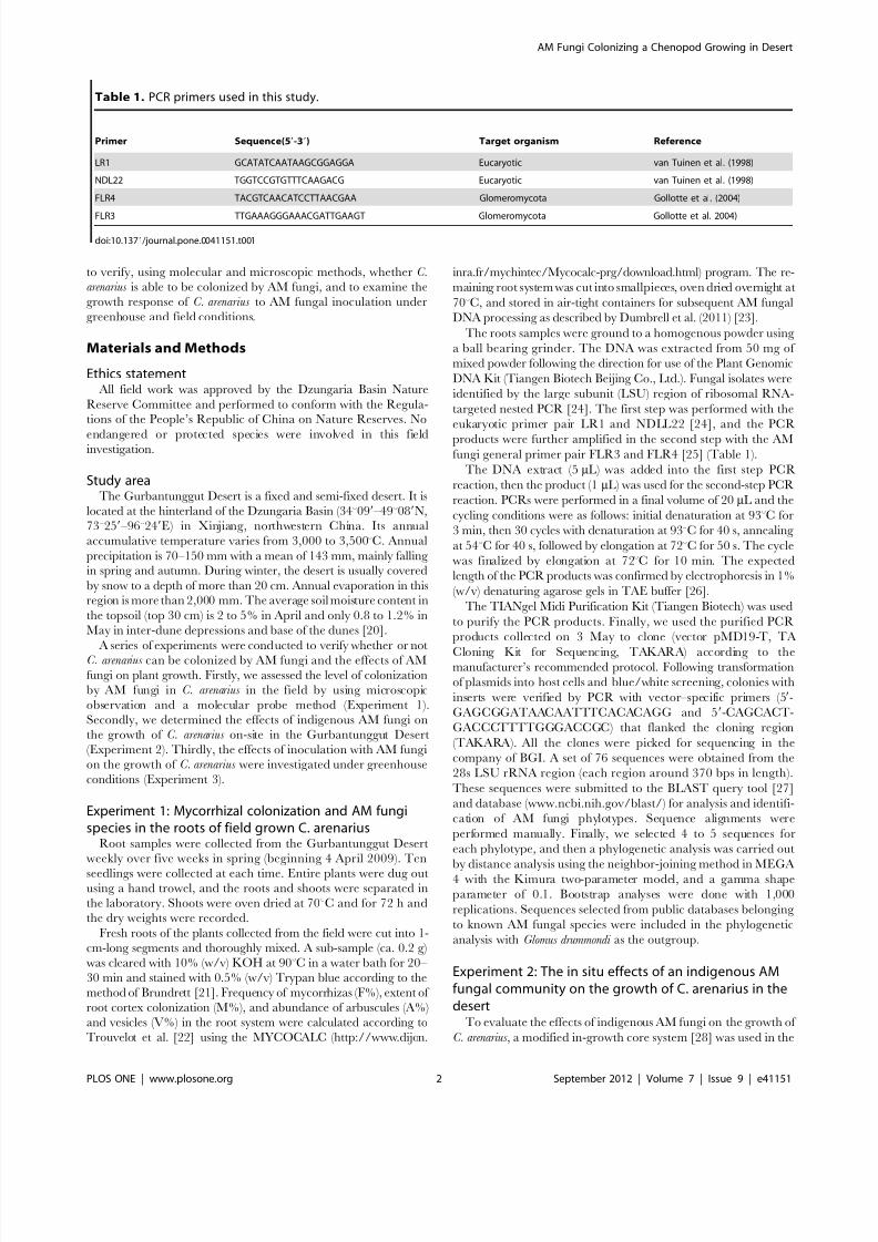
targeted nested PCR [24]. The first step was performed with the
eukaryotic primer pair LR1 and NDLL22 [24], and the PCR
products were further amplified in the second step with the AMfungi general primer pair FLR3 and FLR4 [25] (Table 1).
The DNA extract (5 mL) was added into the first step PCR
reaction, then the product (1 mL) was used for the second-step PCR
reaction. PCRs were performed in a final volume of 20 mL and the
cycling conditions were as follows: initial denaturation at 93uC for
3 min, then 30 cycles with denaturation at 93uC for 40 s, annealing
at 54uC for 40 s, followed by elongation at 72uC for 50 s. The cycle
was finalized by elongation at 72uC for 10 min. The expected
length of the PCR products was confirmed by electrophoresis in 1%
(w/v) denaturing agarose gels in TAE buffer [26].
The TIANgel Midi Purification Kit (Tiangen Biotech) was used
to purify the PCR products. Finally, we used the purified PCR
products collected on 3 May to clone (vector pMD19-T, TA
Cloning Kit for Sequencing, TAKARA) according to themanufacturer’s recommended protocol. Following transformation
of plasmids into host cells and blue/white screening, colonies with
inserts were verified by PCR with vector–specific primers (59-
GAGCGGATAACAATTTCACACAGG and 59-CAGCACT-
GACCCTTTTGGGACCGC) that flanked the cloning region
(TAKARA). All the clones were picked for sequencing in the
company of BGI. A set of 76 sequences were obtained from the
28s LSU rRNA region (each region around 370 bps in length).
These sequences were submitted to the BLAST query tool [27]
and database (www.ncbi.nih.gov/blast/) for analysis and identifi-
cation of AM fungi phylotypes. Sequence alignments were
performed manually. Finally, we selected 4 to 5 sequences for
each phylotype, and then a phylogenetic analysis was carried out
by distance analysis using the neighbor-joining method in MEGA4 with the Kimura two-parameter model, and a gamma shape
parameter of 0.1. Bootstrap analyses were done with 1,000
replications. Sequences selected from public databases belonging
to known AM fungal species were included in the phylogenetic
analysis with Glomus drummondi as the outgroup.
Experiment 2: The in situ effects of an indigenous AMfungal community on the growth of C. arenarius in thedesert
To evaluate the effects of indigenous AM fungi on the growth of
C. arenarius , a modified in-growth core system [28] was used in the
Table 1. PCR primers used in this study.
Primer Sequence(59-39) Target organism Reference
LR1 GCATATCAATAAGCGGAGGA Eucaryotic van Tuinen et al. (1998)
NDL22 TGGTCCGTGTTTCAAGACG Eucaryotic van Tuinen et al. (1998)
FLR4 TACGTCAACATCCTTAACGAA Glomeromycota Gollotte et al. (2004)
FLR3 TTGAAAGGGAAACGATTGAAGT Glomeromycota Gollotte et al. 2004)
doi:10.1371/journal.pone.0041151.t001
AM Fungi Colonizing a Chenopod Growing in Desert
PLOS ONE | www.plosone.org 2 September 2012 | Volume 7 | Issue 9 | e41151
8/12/2019 Arbuscular Mycorrhizal Fungi Promote the Growth
http://slidepdf.com/reader/full/arbuscular-mycorrhizal-fungi-promote-the-growth 3/8
desert in 2009. Cores were constructed using polyvinylchloride
(PVC) water pipe (3.2 cm inner diameter, 3.5 cm outer diameter
in 20-cm sections). Two rectangular windows (8 cm length, 2 cm
width) were cut symmetrically into the pipe, and the distance from
window to top was 7 cm and to bottom was 5 cm. A 30- mm nylon
mesh (allowing hyphal but not root penetration) was attached to
the core window. The base of each pipe was sealed using a rubber
plug to prevent entry by mycelia or roots.
Soil collected from the desert with the same properties as
described above was sterilized with 10 k Gy 60Co c-rays and then
air-dried. Two hundred grams of the sterilized soil were placedinto each pipe. Seeds of C. arenarius were collected from the desert
in June 2008 and stored at 220uC before use. The seeds were
surface-sterilized in 10% hydrogen peroxide for 10 min and rinsed
at least 5 times in deionized (DI) water. Seeds were pre-incubated
on moist filter paper for 48 h until the radicles appeared. Ten
uniform seeds were sown per pipe and the seedlings were thinned
to three respectively after emergence.
The experimental design was a random block with two
treatments, namely rotated (prevent AM fungal colonization of
the plants) and static (no restriction of AM fungi colonization of
the plants). Ten blocks were randomly set in a five 262 m2 plot
which was selected before the germination of the ephemeral plants
to avoid any interference from heterogenity in resources or
adjacent plant composition. Holes were drilled with a larger PVC
tube to install the experimental pipes, and the gap between the
sandy soil and the pipe wall was filled with the soil. The PVC pipes
were inserted with the top 3 cm above the soil surface. In the
rotated treatment the pipes were rotated approx. 45u around their vertical axes every two days in order to limit hyphal penetration of
the core [28]. In the static treatment, the cores remained static to
allow any indigenous AM fungal mycelium to colonize the plant
roots in the pipe. Five seeds were supplemented and seedlings were
thinned to one in each pipe. Plants were harvested 60 days after
sowing. Mycorrhizal colonization in the root system was deter-
mined as described above. Seed number was recorded, and shoot
biomass was determined after oven drying at 70uC for 48 h. Oven-
dried shoots were ground and ashed in a muffle furnace at 300uC
for 3 h and at 550uC for 5 h. The ash was dissolved in 2% (v/v)
HCl. Phosphorus concentration was determined by inductively
coupled plasma-atomic emission spectroscopy (ICP-AES; Perkin
Elmer Optima 3300DV).
Experiment 3: Effects of inoculation with indigenous AMfungi on the growth of C. arenarus under greenhouseconditions
The three dominant AM fungal species, Glomus mosseae , Glomus
etunicatum, and Glomus intradices [19], were previously isolated from
the desert soil and propagated in the soil with red clover and
sorghum for 4 months in a glasshouse. The mycorrhizal inoculum
consisted of root fragments, hyphae, spores (approx 200 spores in
5 g soil) and soil. In the mycorrhizal treatment each pot was
inoculated with 150 g inoculum mix (a mix of all three AM fungi
species at 1:1:1). The same amount of the inoculum mix treated
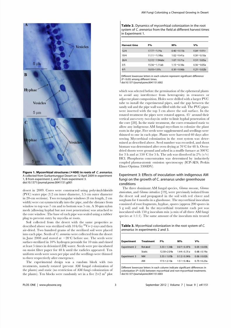
Figure 1. Mycorrhizal structures ( 400) in roots of C. arenarius .A collected from Gurbantunggut Desert on 12 April 2009 in experiment1, B from experiment 2, and C from experiment 3.doi:10.1371/journal.pone.0041151.g001
Table 2. Dynamics of mycorrhizal colonization in the rootsystem of C. arenarius from the field at different harvest timesin Experiment 1.
Harvest time F% M% V%
12/4 17.7763.19a 0.4060.11b 0.0460.01c
19/4 11.1163.18bc 1.0260.41a 0.5060.10a
26/4 12.2262.94abc 1.0760.21a 0.5360.05a
3/5 15.5661.11ab 1.1560.18a 0.5660.05a
9/5 10.00+1.92c 0.3060.06b 0.2560.02b
Different lowercase letters in each column represent significant difference(P ,0.05) among different times.doi:10.1371/journal.pone.0041151.t002
Table 3. Mycorrhizal colonization in the root system of C.
arenarius in experiments 2 and 3.
Experiment Treatment F% M% V%
Experiment 2 Rot ated 3.3361.3 6b 0 .0 760. 07 b 0. 0360.03b
Static 12.50+2.09a 1.4460. 31 a 0. 6860.19a
Experiment 3 NM 3.3361.0 5b 0 .1 260. 04 b 0. 0660.02b
AM 17.562.1 0a 1 .5 160. 46 a 0. 7360.23a
Different lowercase letters in each column indicate significant differences incolonization (P ,0.05) between mycorrhizal and non-mycorrhizal treatments.doi:10.1371/journal.pone.0041151.t003
AM Fungi Colonizing a Chenopod Growing in Desert
PLOS ONE | www.plosone.org 3 September 2012 | Volume 7 | Issue 9 | e41151

8/12/2019 Arbuscular Mycorrhizal Fungi Promote the Growth
http://slidepdf.com/reader/full/arbuscular-mycorrhizal-fungi-promote-the-growth 4/8
with 10 k Gy 60Co c-rays was used for the non-mycorrhizal
treatment.
The experiment was a randomized block factorial design with
two treatments, namely a mycorrhizal treatment (AM) and a non-
mycorrhizal treatment (NM), each replicated 10 times. To
minimize differences in the rhizosphere microbial communities
between mycorrhizal and non-mycorrhizal treatments, 10 mL of
filtrate free from mycorrhizal propagules from the inoculum
mixture was added to each pot of the non-mycorrhizal treatment,and 10 ml of deionized water to each inoculated pot.
At the start of the experiment, 2 kg sterile sand soil combined
with 150 g AM fungal inoculum was placed in the PVC pipe
(20 cm tall and 10 cm in diameter, and with a PVC cap on the
bottom). The seed germination rate of C. arenarius is relative low
under laboratory conditions and we therefore transplanted
seedlings directly from the field. Five-day-old seedlings with two
cotyledons and 2-cm in height were collected from the desert. The
seedlings were pre-selected according to height and cotyledon size.
The selected seedlings were immersed into a solution of 1.5 g l21
benomyl for 5 min to reduce any effect from indigenous AM fungi
on the root surface [29]. Ten seedlings were transplanted into each
pot and thinned to five per pot 7 days after transplanting. The pots
were watered three times each week. No nutrients were added
during the growth period. The plants were harvested 60 days after
transplanting when as they approached maturity. Mycorrhizal
colonization, P concentration, seed number and shoot biomass
were determined as described above.
Statistical analysisPercentage data of root length colonized by AM fungi
comprising colonization rate, abundance of arbuscules and
vesicles, were normalized by arcsine transformation prior to
statistical analysis. Data were subjected to one-way analysis of
variance (ANOVA) using SPSS software version 16.0 (SPSS Inc.,
Chicago, IL, USA). Critical differences between pairs of mean
values were compared by least significant difference (LSD) at the
5% level.
Results
Mycorrhizal colonizationVesicles and intraradical hyphae were observed in the root
systems of C. arenarius (Figure 1A) collected from the field
(Experiment 1) but no typical arbuscules were found. A relatively
low level of colonization rate with a mean of 15% was detected
from spring to summer. Significant differences were found in
colonization percentage among different sampling times ranging
from 10 to 20% ( P ,0.05) (Table 2). The highest colonization rate
appeared at the first harvest. The extents of root cortex
colonization and vesicle abundance were about 1% and 0.5%,
respectively (Table 2).
Vesicles and intraradical hyphae were also detected in the root
systems of C. arenarius in mycorrhizal treatments from the field
experiment (Experiment 2) (Figure 1B) and the pot inoculation
experiment (Experiment 3) (Figure 1C), and the presence of
mycorrhiza (F%) in the roots of C. arenarius from mycorrhizal
treatments were 3.75 and 5.25 times higher than in non-
mycorrhizal treatments, respectively. There were significantdifferences in mycorrhizal colonization (M%) and abundance of
vesicles (V%) between the mycorrhizal and non-mycorrhizal
treatments (Table 3).
AM fungal species in root systems of C. arenarius
growing in the field A PCR product of about 380 bp was amplified using the primer
pair FLR3-FLR4 especially for amplifying Glomeromycota LSU
rDNA sequences. In our experiment 1, the PCR detections in the
period from 19 April to 9 May were successful but on others dates
were less satisfactory (Figure 2). This indicates that these roots
were colonized by AM fungi.
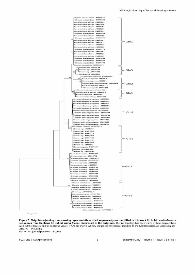
The phylogenetic analysis identified AM fungal species present
in the root system of C. arenarius under the field condition. All the
isolated AM fungi were clustered into five groups, and each
sequence was matched published sequences with .97% similarity.
Glo1 matched Glomus intraradices (GenBank Accession
no. FM865597), Glo2 matched Glomus microaggregatum
(AF389004), Glo3, Glomus mosseae (AM158954), Glo4, Glomus
etunicatum (AY541886), and Glo5, while clearly Glomus , was not
closely related to sequences from any named strains (Figure 3).
Growth response of C. arenarius to AM fungiInoculation with the AM fungi increased the growth of the
plants. In Experiment 2, the shoot biomass and seed number from
static cores increased by 180% and 100% (Table 4) respectively
when compared to that from rotated cores. Similarly, inExperiment 3, significant differences in shoot biomass and seed
number were observed between mycorrhizal and non-mycorrhizaltreatments with increments of 125% and 73.9% (Table 4)
respectively, comparing with non-mycorrhizal treatments. There
were no significant differences in root biomass or root/shoot ratio
between mycorrhizal and non-mycorrhizal treatments in experi-
ment 2 and 3.
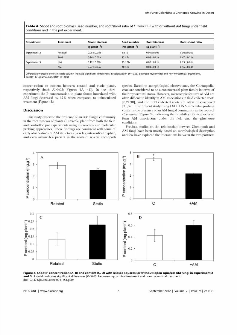
P acquisitionThe P content in shoot tissues of mycorrhizal plants was
generally higher than that of non-mycorrhizal ones, but there was
no significant difference in P acquisition in both experiments
(Figure 4). In Experiment 2, there were no differences in shoot P
Figure 2. PCR detection of Glomeromycota (FLR3-FLR4) in colonized roots from experiment 1 (three replicates per sampling date).(W) indicates water control, (M) indicates molecular weight marker 100 bp ladder (Tiangen Biotech).doi:10.1371/journal.pone.0041151.g002
AM Fungi Colonizing a Chenopod Growing in Desert
PLOS ONE | www.plosone.org 4 September 2012 | Volume 7 | Issue 9 | e41151
8/12/2019 Arbuscular Mycorrhizal Fungi Promote the Growth
http://slidepdf.com/reader/full/arbuscular-mycorrhizal-fungi-promote-the-growth 5/8
Figure 3. Neighbour-joining tree showing representatives of all sequence types identified in this work (in bold), and referencesequences from Genbank (in italics), using Glomus drummondi as the outgroup. The five topology has been tested by bootstrap analysiswith 1000 replicates, and all bootstrap values .70% are shown. All new sequences have been submitted to the GenBank database (Accession nosJN805771–JN805847).doi:10.1371/journal.pone.0041151.g003
AM Fungi Colonizing a Chenopod Growing in Desert
PLOS ONE | www.plosone.org 5 September 2012 | Volume 7 | Issue 9 | e41151
8/12/2019 Arbuscular Mycorrhizal Fungi Promote the Growth
http://slidepdf.com/reader/full/arbuscular-mycorrhizal-fungi-promote-the-growth 6/8
concentration or content between rotated and static plants,
respectively (both P .0.05; Figures 4A, 4C). In the third
experiment the P concentration in plant shoots inoculated with
AM fungi decreased by 37% when compared to uninoculated
treatment (Figure 4B).
Discussion
This study observed the presence of an AM fungal communityin the root systems of plants C. arenarius plant from both the field
and controlled pot experiments using microscopy and molecular
probing approaches. These findings are consistent with some of
early observations of AM structures (vesicles, intraradical hyphae,
and even arbuscules) present in the roots of several chenopods
species. Based on morphological observations, the Chenopodia-
ceae are considered to be a controversial plant family in terms of
their mycorrhizal status. However, microscopic features of AM are
often difficult to identify in AM associations in field-collected roots
[8,21,30], and the field collected roots are often misdiagnosed
[31,32]. Our present study using LSU rDNA molecular probing
confirms the presence of an AM fungal community in the roots of
C. arenarius (Figure 3), indicating the capability of this species to
form AM associations under the field and the glasshouseconditions.
Previous studies on the relationship between Chenopods and
AM fungi have been mostly based on morphological description
and few have explored the interactions between the two partners
Table 4. Shoot and root biomass, seed number, and root/shoot ratio of C. arenarius with or without AM fungi under fieldconditions and in the pot experiment.
Experiment Treatment Shoot biomass Seed number Root biomass Root/shoot ratio
(g plant21) (No plant21) (g plant21)
Experiment 2 Rotated 0.0560.01b 661b 0.0160.00a 0.3660.05a
Static 0.146
0.01a 126
2a 0.026
0.01a 0.476
0.11aExperiment 3 NM 0.1260.00b 2365b 0.0260.01a 0.1360.01a
AM 0.2760.03a 4066a 0.0460.01a 0.1660.04a
Different lowercase letters in each column indicate significant differences in colonization (P ,0.05) between mycorrhizal and non-mycorrhizal treatments.doi:10.1371/journal.pone.0041151.t004
Figure 4. Shoot P concentration (A, B) and content (C, D) with (closed squares) or without (open squares) AM fungi in experiment 2and 3. Asterisk indicates significant differences (P ,0.05) between mycorrhizal treatment and non-mycorrhizal treatment.doi:10.1371/journal.pone.0041151.g004
AM Fungi Colonizing a Chenopod Growing in Desert
PLOS ONE | www.plosone.org 6 September 2012 | Volume 7 | Issue 9 | e41151

8/12/2019 Arbuscular Mycorrhizal Fungi Promote the Growth
http://slidepdf.com/reader/full/arbuscular-mycorrhizal-fungi-promote-the-growth 7/8
such as the role of the indigenous AM fungal community in the
growth and nutrient uptake of Chenopods. Our present results,from both the on-site field trial and sterilized-soil pot culture,
demonstrated that the indigenous AM fungal community hadpositive effects on the growth of C. arenarius (Table 4). Our results
are compatible with early reports that inoculation with AM fungus
increased the growth of Atriplex canescens [15] or Atriplex nummularia
[33,34] growing in sterilized soil. Asghari et al. (2005) suggested
that plant age is an important factor influencing colonizationpercentage [33]. They found that inoculation with AM fungi
decreased P concentrations at harvest after 6 weeks harvest but
significantly increased P concentrations at 10 weeks, while
inoculation with AM fungi significantly increased plant dry weight
at 6 weeks, but not at 10 weeks.
AM symbionts generally increase plant P acquisition, and plant
growth resulting from AM colonization (i.e. the ‘mycorrhizal
growth response’, MGR) varies depending on the plant-fungus-
environment combinations, and this is usually explained by a
simple cost–benefit approach [35]. MGR may not necessarily be
related to changes in the phosphorus content of plants because the
mycorrhizal phosphorus uptake pathway can dominate the
phosphate supply to plants irrespective of growth responses [36].
It is known that trade-off in exchange of photosynthetic carbon for
soil-derived nutrients, particularly phosphorus, is a fundamentalprinciple of symbiotic mycorrhizal associations [35,37]. P flow at
the plant-fungal interface is of benefit to the plant and also a signal
to sustain the existence of the fungi in the roots. If a fungus is
unable to supply P to the plant, its growth will be terminated [38].
A recent study showed that plants can detect, discriminate, and
reward the best fungal partners with more carbohydrates. In turn,
the fungal partners enforce cooperation by increasing nutrient
transfer only to those roots providing more carbohydrates [39].
Therefore, it is clear in the present study the fungal partners
delivered phosphorus to C. arenarius because they colonized and
promoted the growth of host plant. Colonization by AM fungi did
not increase the P accumulation in plant tissues (Figure 4). One
possible explanation for the decrease in P concentration in the
present study might be in the involvement of other mechanisms
such as enhanced water uptake by the AM fungi [40]. Increased
water uptake may have made a greater contribution than
increased phosphorus acquisition on the growth of C. arenarius ,
with a subsequent phosphorus dilution effects in the shoots
(Figure 4). Further investigation is necessary to explore themechanisms by which the reciprocal process between AM fungi
and the chenopod in addition to any effects on plant P nutrion.Plant nutrient use efficiency is defined as biomass production
per unit nutrient element (such as phosphorus) and is one of the
most important functional traits for plant survival in Nature.
Mycorrhizal plants with lower phosphorus use efficiency than non-
mycorrhizal plants have been reported in agricultural crops
[41,42]. However, our present results indicate that phosphorus use
efficiency in mycorrhizal plant is higher than in non-mycorrhizal
controls in terms of lower phosphorus concentration and higherdry weight of shoots of plants colonized by AM fungi. In this
context it is important to note that such a functional trait may be
an important strategy for this desert plant to use a limited
phosphorus resource economically in the nutrient-poor habitat.
Arbuscules have been considered as the key site of interchange
of carbon and phosphorus between root cells and the hyphae of
fungi of the phylum Glomeromycota, which are defining
characteristics to distinguish AM fungi from other types of fungi
[1]. Our molecular results verify the presence of AM fungi in the
root systems of C. arenarius , but no arbuscules or hyphal coils were
observed microscopically. These results may suggest that intrar-
adical hyphae play a role in C/P exchange in chenopods.
However, various explanations have been proposed regarding thisissue. Aleman and Tiver (2010) suggested that AM fungi may not
contribute greatly to the nutrient exchange between the fungi and
plant roots of chenopod species in which very low presence of
arbuscules was observed [14]. Asghari et al. (2005) reported that
inoculation with Glomus mosseae lowered P concentration of Atriplex
nummularia at the sixth week but increased the P concentration at
the tenth week of growth [33]. Plenchettea and Duponnois (2005)
reported that inoculation with Glomus intraradices increased the
biomass and P concentration of Atriplex nummularia but no
arbuscules were observed [34]. Manjarrez et al. (2010) recently
showed that arbuscules are not an absolute requirement for
phosphorus transfer to plants [43]. All these evidences above are
implying that, beside arbuscules, an intraradical structure which is
responsible for carbon/nutrients exchange must exist in Cheno-
pods and AM fungal symbionts. More work is required to
determine what factors actually control the beneficial effects of
AM fungi on Chenopods.
Many studies have documented that in extremely adverse
environments, such as those affected by salinity or drought
habitats, Chenopods are often the dominant species in plant
community. These plants follow the ‘r ’ strategy for they have a
high fecundity (particularly higher seed productivity), short
generation time and an ability rapidly to exploit pulses of nutrient
availability [44,45]. Our current results show that AM fungi
greatly increased the seed production of C. arenarus , indicating that
the desert Chenopods take advantage of the local AM fungal
community to explore and efficiently utilize soil resources to
strengthen their ‘r ’ strategy. Such functional traits are crucial for
the species to survive in the desert habitat.In summary, our result show that C. arenarius , a member of the
Chenopodiaceae, was colonized by AM fungi with mycorrhizal
structures presented in the root systems. Although AM fungi did
not enhance the P concentration (the decline in Experiment 2 was
significant as shown in Figure 4B) or uptake by the shoots, shoot
biomass and plant seed production did increase significantly under
both greenhouse and field conditions. Our results suggest that an
alternative reciprocal process between AM fungi and the
chenopod may exist in addition to C-P trade-off in the extremely
arid desert environment.
Author Contributions
Conceived and designed the experiments: TZ GF. Performed the
experiments: TZ NS DB. Analyzed the data: TZ GF. Contributedreagents/materials/analysis tools: TZ NS GF. Wrote the paper: TZ GF
YC.
References
1. Brundrett MC (2002) Coevolution of roots and mycorrhizas of land plants. New
phytol 154: 275–304.
2. Javot H, Pumplin N, Harrison MJ (2007) Phosphate in the arbuscular
mycorrhizal symbiosis: transport properties and regulatory roles. Plant Cell
Environ 30: 310–322.
3. Helgason T, Fitter AH (2009) Natural selection and the evolutionary ecology of
the arbuscular mycorrhizal fungi (Phylum Glomeromycota). J Exp Bot 60: 2465–
2480.
4. Nie M, Wang Y, Yu J, Xiao M, Jiang L, et al. (2011) Understanding plant-
microbe interactions for phytoremediation of petroleum-polluted soil. Plos One
6(3): e17961. doi:10.1371/journal. pone.0017961.
AM Fungi Colonizing a Chenopod Growing in Desert
PLOS ONE | www.plosone.org 7 September 2012 | Volume 7 | Issue 9 | e41151
8/12/2019 Arbuscular Mycorrhizal Fungi Promote the Growth
http://slidepdf.com/reader/full/arbuscular-mycorrhizal-fungi-promote-the-growth 8/8
5. White JA, Tallaksen J, Charvat I (2008) The effects of arbuscular mycorrhizal
fungal inoculation at a roadside prairie restoration site. Mycologia 100: 6–11.
6. Pennisi E (2004) The secret life of fungi. Science 304: 1620–1622.
7. Rodriguez R, Redman R (2008) More than 400 million years of evolution and
some plants still can’t make it on their own: plant stress tolerance via fungal
symbiosis. J Exp Bot 59: 1109–1114.
8. Smith SE, Read DJ (2008) Mycorrhizal symbiosis 3rd ed: Boston: Academic
Press.
9. Sengupta A, Chaudhuri S (1990) Vesicular arbuscular mycorrhiza (VAM) in
pioneer salt-marsh plants of the Ganges River delta in West Bengal (India). Plant
Soil 122: 111–113.
10. Aguilera LE, Gutierrez JR, Moreno RJ (1998) Vesiculo arbuscular mycorrhizaeassociated with saltbushes Atriplex spp. (Chenopodiaceae) in the Chilean arid
zone. Rev Chil Hist Nat 71: 291–302.
11. O’Connor PJ, Smith SE, Smith FA (2001) Arbuscular mycorrhizal associations
in the southern Simpson Desert. Aust J Bot 49: 493–499.
12. Shi ZY, Feng G, Christie P, Li XL (2006) Arbuscular mycorrhizal status of
spring ephemerals in the desert ecosystem of Junggar Basin, China. Mycorrhiza
16: 269–275.
13. Wilde P, Manal A, Stodden M, Sieverding E, Hildebrandt U, et al. (2009)
Biodiversity of arbuscular mycorrhizal fungi in roots and soils of two salt
marshes. Environ microbiol 11: 1548–1561.
14. Aleman R, Tiver F (2010) Endomycorrhizal infection levels among chenopod
plant species at Port Wakefield, South Australia. T Roy Soc South Aust 134: 1–
4.
15. Williams SE, Wollum AG, Aldon EF (1974) Growth of Atriplex-Canescens (Pursh)
Nutt improved by formation of vesicular-arbuscular mycorrhizae. Soil Sci Soc of
Am J 38: 962–965.
16. Mao Z, Zhang D (1994) Conspectus of ephemeral flora in northern Xinjiang.
Arid Zone Res 11: 1–26 (in Chinese with English a bstract).
17. Gao R, Wei Y (2007) Amphicarpy of Ceratocarpus arenarius (Chenopodiaceae) in
Junggar Desert. Acta Bot Yunnan 29: 300–302 (in Chinese with English
abstract).
18. Huang PY (2002) Shrub vegetation and to restoration in arid areas. Science
Press (in Chinese).
19. Shi ZY, Zhang LY, Li XL, Feng G, Tian CY, et al. (2007) Diversity of
arbuscular mycorrhizal fungi associated with desert ephemerals in plant
communities of Junggar Basin, northwest China. Appl Soil Ecol 35: 10–20.
20. Wang XQ, Jiang J, Lei JQ, Zhao CJ (2004) Relationship between spring
ephemeral plants distribution and soil moisture on longitudinal dune surface in
Gurbantunggut desert. Chinese J Appl Ecol 15: 556–560 (in Chinese with
English abstract).
21. Brundrett MC (2009) Mycorrhizal associations and other means of nutrition of
vascular plants: understanding the global diversity of host plants by resolving
conflicting information and developing reliable means of diagnosis. Plant Soil
320: 37–77.
22. Trouvelot A, Kough JL, Gianinazzi-Pearson (1986) Mesure du taux de
mycorhization VA d’un systeme radiculaire. Recherche de methodes d’estima-
tion ayant une signification functionnelle. In: Physiological and Genetic Aspectsof Mycorrhizae. Gianinazzi-Pearson V. and S. Gianinazzi. eds. INRA Press,
Paris: 217–221.
23. Dumbrell AJ, Ashton PD, Aziz N, Feng G, Nelson M, et al. (2011) Distinct
seasonal assemblages of arbuscular mycorrhizal fungi revealed by massively
parallel pyrosequencing. New phytol 190: 794–804.
24. Van Tuinen D, Jacquot E, Zhao B, Gollotte A, Gianinazzi-Pearson V (1998)
Characterization of root colonization profiles by a microcosm community of
arbuscular mycorrhizal fungi using 25S rDNA-targeted nested PCR. Mol Ecol 7:
879–887.
25. Gollotte A, van Tuinen D, Atkinson D (2004) Diversity of arbuscularmycorrhizal fungi colonising roots of the grass species Agrostis capillaris andLolium perenne in a field experiment. Mycorrhiza 14: 111–117.
26. Armstrong KA (1983) Molecular-Cloning - a Laboratory Manual - Maniatis,T,Fritsch,Ef, Sambrook, J Q Rev of Biol 58: 234–234.
27. Altschul SF, Madden TL, Schaffer AA, Zhang JH, Zhang Z, et al. (1997)Gapped BLAST and PSI-BLAST: a new generation of protein database searchprograms. Nucleic Acids Res 25: 3389–3402.
28. Johnson D, Leake JR, Read DJ (2001) Novel in-growth core system enablesfunctional studies of grassland mycorrhizal mycelial networks. New Phytol 152:555–562.
29. O’Connor PJ, Smith SE, Smith EA (2002) Arbuscular mycorrhizas influenceplant diversity and community structure in a semiarid herbland. New Phytol154: 209–218.
30. Brundrett M (2004) Diversity and classification of mycorrhizal associations. BiolRev 79: 473–495.
31. Olsson PA (1999) Signature fatty acids provide tools for determination of thedistribution and interactions of mycorrhizal fungi in soil. FEMS Microbiol Ecol29: 303–310.
32. Dickie IA, FitzJohn RG (2007) Using terminal restriction fragment lengthpolymorphism (T-RFLP) to identify mycorrhizal fungi: a methods review.Mycorrhiza 17: 259–270.
33. Asghari HR, Marschner P, Smith SE, Smith FA (2005) Growth response of
Atriplex nummularia to inoculation with arbuscular mycorrhizal fungi at differentsalinity levels. Plant Soil 273: 245–256.
34. Plenchette C, Duponnois R (2005) Growth response of the saltbush Atriplex nummularia L. to inoculation with the arbuscular mycorrhizal fungus Glomus intraradices . J Arid Environ 61: 535–540.
35. Smith FA, Grace EJ, Smith SE (2009) More than a carbon economy: nutrienttrade and ecological sustainability in facultative arbuscular mycorrhizalsymbioses. New Phytol 182: 347–358.
36. Smith SE, Smith FA, Jakobsen I (2003) Mycorrhizal fungi can dominatephosphate supply to plants irrespective of growth responses. Plant Physiol 133:16–20.
37. Fitter AH (2006) What is the link between carbon and phosphorus fluxes inarbuscular mycorrhizas? A null hypothesis for symbiotic function. New Phytol172: 3–6.
38. Javot H, Penmetsa RV, Terzaghi N, Cook DR, Harrison MJ (2007) A Medicagotruncatula phosphate transporter indispensable for the arbuscular mycorrhizalsymbiosis. P Nat Acad Sci USA 104: 1720–1725.
39. Kiers ET, Duhamel M, Beesetty Y, Mensah JA, Franken O, et al. (2011)Reciprocal rewards stabilize cooperation in the mycorrhizal symbiosis. Science333: 880–882.
40. Allen MF (1983) Formation of Vesicular-Arbuscular Mycorrhizae in Atriplex gardneri (Chenopodiaceae) - Seasonal Response in a Cold Desert. Mycologia 75:773–776.
41. Zhu YG, Smith SE, Barritt AR, Smith FA (2001) Phosphorus (P) efficiencies andmycorrhizal responsiveness of old and modern wheat cultivars. Plant Soil 237:249–255.
42. Marschner P, Rengel Z (2010) The effects of plant breeding on soil microbes.Soil Microbiol Sustain Crop Product: 297–314.
43. Manjarrez M, Christophersen HM, Smith SE, Smith FA (2010) Corticalcolonisation is not an absolute requirement for phosphorus transfer to plants inarbuscular mycorrhizas formed by Scutellospora calospora in a tomato mutant:evidence from physiology and gene expression. Funct Plant Biol 37: 1132–1142.
44. Grime JP (1979) Competition and the struggle for existence. In Populationdynamics (Anderson RM, Turner BD and Taylor LR eds.). 20th The BritishEcological Society Symposium London, England, Blackwell Scientific Publica-tions: Oxford, P123–140.
45. Gutterman Y (2002) Survival strategies of annual desert plants: Springer Verlag.
AM Fungi Colonizing a Chenopod Growing in Desert
PLOS ONE | www.plosone.org 8 September 2012 | Volume 7 | Issue 9 | e41151