The Human Skeleton - Kenan Fellows Program is the study of the human skeleton, which includes all...
29
The Human Skeleton Bone and Bone Growth Bone is living tissue, and, as such, can grow and remodel during a person’s lifetime. The three types of bone cells are the osteoblasts, which are responsible for bone growth; the osteoclasts, which are active in bone resorption; and the osteocytes, which serve a regulatory function, adjusting the circulating levels of bone minerals. Because bone is malleable, it can be modified through exercise, disease, injury, and diet. Osteology Osteology is the study of the human skeleton, which includes all bones of the body. It is important to know the correct descriptive terminology when speaking of the various bones and regions, i.e., the bone of the upper leg is the femur, not the thigh-bone. Craniology is the study of the head and face. This portion of the body formerly received attention because it was thought to provide more detailed information than the rest of the body with respect to the evolutionary trends in human physical morphology. However, in recent years the postcranial body has been studied extensively for evidence of age at time of death, estimate of stature and bodily proportions, and presence of injury and disease. Although we will focus on the human skeleton, the same bones, with some modification in shape, are found in non-human primates and other mammals, for example, the dog. Learning the 206 bones of the skeleton sounds like a formidable task, but many bones are paired, such as the right and left femur (pl. femora), right and left parietals, and right and left ribs. If the non- paired bones are identified first, the paired bones are much easier to recognize. The skeleton may be separated into two parts, the cranial (skull) portion (usually also includes the hyoid) and the postcranial portion, that is, all bones below the skull. The axial region includes those bones of the trunk and thorax,
Transcript of The Human Skeleton - Kenan Fellows Program is the study of the human skeleton, which includes all...

The Human Skeleton
Bone and Bone Growth
Bone is living tissue, and, as such, can grow and remodel during a
person’s lifetime. The three types of bone cells are the osteoblasts, which are
responsible for bone growth; the osteoclasts, which are active in bone
resorption; and the osteocytes, which serve a regulatory function, adjusting the
circulating levels of bone minerals. Because bone is malleable, it can be
modified through exercise, disease, injury, and diet.
Osteology
Osteology is the study of the human skeleton, which includes all bones
of the body. It is important to know the correct descriptive terminology when
speaking of the various bones and regions, i.e., the bone of the upper leg is the
femur, not the thigh-bone. Craniology is the study of the head and face. This
portion of the body formerly received attention because it was thought to
provide more detailed information than the rest of the body with respect to the
evolutionary trends in human physical morphology. However, in recent years
the postcranial body has been studied extensively for evidence of age at time of
death, estimate of stature and bodily proportions, and presence of injury and
disease.
Although we will focus on the human skeleton, the same bones, with
some modification in shape, are found in non-human primates and other
mammals, for example, the dog. Learning the 206 bones of the skeleton
sounds like a formidable task, but many bones are paired, such as the right and
left femur (pl. femora), right and left parietals, and right and left ribs. If the non-
paired bones are identified first, the paired bones are much easier to recognize.
The skeleton may be separated into two parts, the cranial (skull) portion
(usually also includes the hyoid) and the postcranial portion, that is, all bones
below the skull. The axial region includes those bones of the trunk and thorax,

including the sacrum. The appendicular region includes the bones of the
upper and lower limbs, shoulder and pelvic regions, hands, and feet.
The next few pages will illustrate the bones of the cranial and the
postcranial skeleton as well as provide for you a list of general and directional
definitions. This list will introduce you to terminology that often is used in
human skeletal studies.
As you work with the diagrams and other materials, note the articulations
of the various bones. For example, observe that one nasal bone articulates
with (that is, meets or touches another bone by way of a suture, the juncture
edge of each bone) the other nasal bone, one of two maxilla bones, and the
unpaired frontal bone. Some bones, like the occipital bone, are easily
observed, but others, like the vomer and ethnoid bones, are difficult to identify
because they are part of the internal support structure of the nose and midfacial
region. These bones are illustrated in the diagram of a sagittal sectioning of the
skull. Pterion refers to the region where the frontal, parietal, temporal, and
sphenoid bones meet. Asterion refers to the region where the occipital,
parietal, and temporal bones meet. Note that there is a right and left pterion and
asterion region.
Turning to the postcranial skeleton, note which bones are part of the
pectoral (shoulder) girdle and the pelvic girdle. Use the articulated skeleton in
the laboratory as well as the diagrams provided to distinguish the bones of the
hands (carpals, metacarpals, and phalanges) and feet (tarsals, metatarsals,
and phalanges).
Planes of Orientation
In studying the human body and skeleton, it is convenient to use certain
properly defined planes of orientation for descriptive purposes. It can readily be
seen that an infinite number of possible planes can be thought to pass through
the body in any direction. In osteometry the following planes are particularly
important.
The Frankfurt plane is defined as the horizontal plane of the skull
determined by the landmark called the porion (left or right) and the lowest point

on the inferior border on the left orbit (orbitale). It is often called the eye-ear
plane. This position roughly corresponds to that of the head of an individual
standing at attention and looking straight ahead. Proper orientation of the skull
is important since some landmarks (i.e., opisthocranion), and the
measurements involved, will be in error without it.
Except for the internal organs, the bodies of all vertebrates are bilaterally
symetrical along a median plane. This plane, called the median sagittal plane,
passes from the sagittal suture of the skull downward, thus dividing the body
and skull into symmetrical left and right halves.

Directional Definitions
Dorsal the back or upper side (posterior in human anatomy)
Ventral the under side, stomach side (anterior in human anatomy)
Lateral to the side, right and left
Anterior, cephalic, or cranial nearer the front of the body (in bipeds this means ventral)
Posterior, caudal the tail end of the animal (inferior in anatomy)
Median mid-line of the body, also called sagittal
Central the part of a system nearest the middle of the animal
Peripheral the part nearest the surface
Proximal mass of the body, as the thigh
Distal away from the main mass of the body, as the toes
Superficial on or near the surface
Deep some distance below the surface
Superior above (anterior in animals)
Inferior below (posterior in animals)
Medial toward the mid-line of the body
Mid-Sagittal Plane the imaginary plane that transects the body along the mid-point into the mirrored left and right sides
Vick, Robbins, Smith; A Laboratory Manual of Methods and Techniques in Physical Anthropology

Vick, Robbins, Smith; A Laboratory Manual of Methods and Techniques in Physical Anthropology

General Descriptive Definitions
The skull consists of all of the bones that comprise the head. The cranium or
calvarium refers to the skull minus the mandible or lower jaw. The skull cap, or
the calvarium minus its base, is called a calva or calotte.
Aperture opening on surface of space within a bone, e.g., nasal aperture
Boss a rounded eminence or bulging of bone, e.g., parietal bosses
Canal long perforation in bone, e.g., occipital condyloid canal
Capitulum a small articular swelling, e.g., capitulum near head of rib
Caput rounded articular eminence generally with a neck, e.g., head of
radius
Condyle bony enlargement bearing an articulating surface, e.g., occipital condyle
Foramen short perforation through bone, e.g., mental foramen
Crest prominent border or margin, a distinct linear elevation or ridge, e.g., supramastoid crest
Fissure narrow slit through bone, e.g., superior orbital fissure on sphenoid bone
Fovea a shallow pit on the bone, e.g., fovea on head of femur
Lines roughened ridge on the bone, e.g., linea aspera on femur
Meatus outlet, opening in temporal bone, e.g., auditory meatus
Fossa deeper pit in single bone or formed by several bones, e.g., mandibular fossa of temporal bone
Process a marked projection or prominence on a bone, e.g., mastoid process
Sinus closed space within the bone, e.g., maxillary sinuses, frontal sinuses
Spine a slender narrow or pointed bony projection, e.g., the spines of thoracic vertebrae
Tubercle small bony tuber (projection), e.g., genial tubercle

Tuberosity broad thick rough eminence, e.g., ischial tuberosity
Vick, Robbins, Smith; A Laboratory Manual of Methods and Techniques in Physical Anthropology
Evidence Used in Determination of Sex (Sexual Dimorphism)
Humans vary in their degree of sexual dimorphism (difference in body
form between male and female). Humans vary approximately 10-12% between
male and female. Sexual dimorphism varies in form mostly by body size and
features such as robustness and muscularity. Sexual dimorphism can be used to
determine the sex of unknown individuals. In general, males are larger and more
robust, with heavier muscle markings and larger teeth. Generally, in humans, the
skull, long bones, and pelvis are all used to estimate sex. The best diagnostic
evidence is the pelvis, however.
PELVIS MALE FEMALE
Greater Sciatic Notch
Narrow angle relatively deep
Wide angle relatively shallow
Preauricular Sulcus Infrequent, sometimes absent Common, mostly present
Pubic Symphysis (less reliable) Deeper in males
Obturator Foramen
(less reliable) Relatively large and oval Relatively small and triangular
Subpubic Angle Under 90 degrees, narrow Generally 90 degrees or higher, wide
SKULL
Paired Sometimes absent, but if present , single and in the midline

Orbital Rim Sharpness Rounded and dull to the touch Distinct and sharp edged
Temporal Ridges Muscle attachments larger, more rugged Muscle attachments slight
Nuchal Ridges Muscle attachments larger, more rugged Muscles attachments slight
Supraorbital Ridges Larger, heavier Smaller or absent
Mastoid Process Medium to large (thumb size), usually projecting below the below of the skull
Small, (little finger size), usually does not project below the skull
Posterior Zygomatic Root
Continues into supramastoid crest above auditory meatus, heavier, greater length
Underdeveloped, lighter and more compressed
Mandible
Heavier jaw, more “square” chin that has two points connected by more or less a
straight line. The gonial angle is generally < 125 degrees
More narrow and “pointed”(single prominence), ramus is more gracile. The gonial angle is generally > 125
degrees
Skull Overall Rougher, larger, more rugged Smoother, smaller, more rounded, adolescent-like
OTHER
Postcranial General
Heavier; heavy muscle marking compared to female; larger head of
humerus; larger head of femur
Light muscle markings; smaller bones and joints in general
Vick, Robbins, Smith; A Laboratory Manual of Methods and Techniques in Physical Anthropology

Brothwell, Digging Up Bones
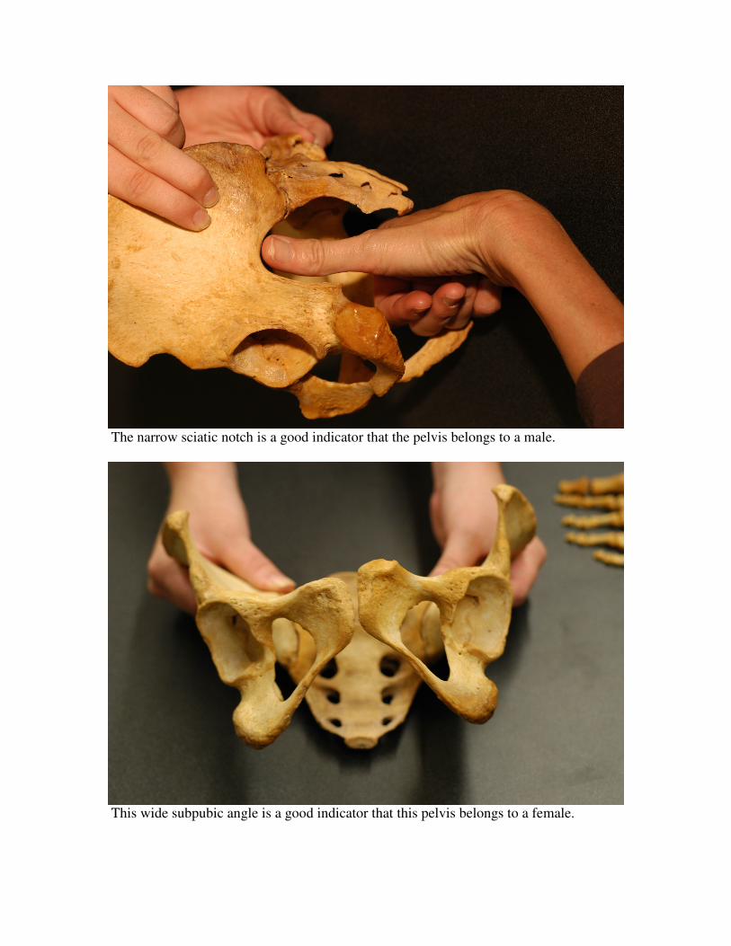
The narrow sciatic notch is a good indicator that the pelvis belongs to a male.
This wide subpubic angle is a good indicator that this pelvis belongs to a female.

Evidence Used in Determination of Age Skeletal Evidence
Lines of evidence used in the determination of age involve examination of epiphyseal union, closing of cranial sutures, and changes in the pubic symphysis. The epiphysis is a cartilaginous area of bone growth located near the ends of long bones. As individuals mature, these epiphyses gradually ossify and join the diaphysis, in a timed sequence. In flat bones, such as the skull, growth occurs from the center of the bone. Upon maturation, growth stops and the sutures gradually close. The pubic symphysial face also changes over time.
Times of epiphyseal union of long bones vary somewhat due to nutrition and individual variability and can therefore serve only as an approximate indicator of age up to about 25-30 years. The sutures of the skull begin closing at approximately 17 years of age; finally, in very old age, the sutures are completely fused. Suture fusing begins on the inside of the skull and proceeds to the outside. Average ages for suture closing have been determined. However, because of inter-individual variation, one should be very cautious in using suture closure data for age estimation. Stages in the aging of the pubic symphysis have also been determined. This measure is more useful than other measures since the changes extend into later life.
A. Fused by 40 years of age B. Fused by 65 years of age C. Fused by 72 years of age D. Fused by 80 years of age E. Fused by 40 years of age F. Fused by 50 years of age G. Fused by 80 years of age H. Fused by 65 years of age
A. Fuses between 18 and 25 years of age
Wolfe, 1983

Age Estimation of Immature Skeletons
In general, immature remains are those of individuals who are less than 20
years old. Complete fusion of all major epiphyses has occurred and all teeth
have erupted. Of course, the age of occurrence of these events varies depending
on sex, race, nutrition and other factors.
I. Appearance of Ossification Centers
A. Appearance of ossification centers occurs from birth to 15 years.
B. The centers themselves rarely survive in archeological/forensic
context because of their fragile nature.
II. Epiphyseal Union
A. Most commonly used in teenage years (10-20 years)
B. Standards available for humerus, clavicle, scapula, hip, elbow,
hand, wrist, foot, ankle and knee.
C. Epiphyseal union should be considered a process rather that a
event.
D. A range of four years is seen between fusion onset in early-
maturing and completion of fusion in late-maturing individuals.
E. Females are an average of two years in advance of males in
epiphyseal union.
F. Radiographs of the epiphysis provide earlier age estimates.
III. Bone Size
A. In individuals from the prenatal period to about 6-7 years, the length
of the long bone diaphyses can be used to estimate age.
B. Growth rates vary greatly among groups and sexes.

C. For prenatal to birth skeletons, use long bone length to get an
estimate of body length, then use body length to get estimate of
age.
D. For postnatal skeletal remains, the diaphyseal length can be used
directly to estimate age.
IV. Dental calcification and eruption
A. Most accurate age indicator in subadults.
B. Dental development largely controlled by genetic factors and is,
therefore, relatively less susceptible to environmental factors.
C. Most studies provide calcification and eruption data for specific
groups (e.g. white, black, Indian, male, female)
D. Age estimates are available for calcification of deciduous (milk) and
permanent teeth.

Epiphyseal Union
Male and Female age ranges in years for complete fusion of epiphyses
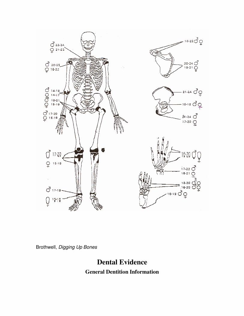
Brothwell, Digging Up Bones
Dental Evidence General Dentition Information

Teeth provide much useful information to the physical anthropologist. Due
to their hardness, they very often fossilize. Teeth can give us insights about the
life ways of the individuals to whom they belonged. For example, eruption and
wear patterns can provide information about age. Moreover, teeth can provide
data on�nutrition and diet.
Occlusal surface top of crown that comes in contact with teeth of the opposing jaw when the mouth is closed
Crown part of the tooth above the alveolus
Neck joins crown and root(at the gum line)
Root anchors the tooth in the bone
Buccal cheek side of tooth
Lingual tongue side of tooth
Mesial toward the midline of chin
Distal away from the midline of chin
Groove natural valleys between cusps or other tooth parts
Alveolus socket in the maxilla and mandible in which the root of The tooth is anchored Cusp a pronounced elevation on the crown surface of the
tooth Bicuspid a tooth with two cusps (premolars) Incisor a tooth with one cutting edge
DISTAL
MESIAL
L INGUAL
BUCCAL
ROOT
NECK
CROWN
Vick, Robbins, Smith; A Laboratory Manual of Methods and Techniques in Physical Anthropology

Human Dentition
Deciduous Maxillary Dentition
Permanent Maxillary Dentition
Vick, Robbins, Smith; A Laboratory Manual of Methods and Techniques in Physical Anthropology
Incisor Incisor Canine Molar Molar Incisor Incisor Canine Premolar Premolar Molar Molar Molar
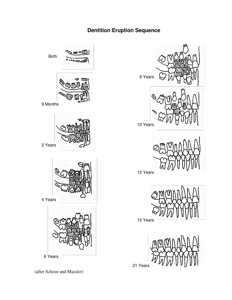
Dentition Eruption Sequence
Birth
9 Months
2 Years
4 Years
6 Years
10 Years
8 Years
12 Years
15 Years
21 Years (after Schour and Massler)

Evidence Used in Determination of Race
Race is much harder to determine, as there is much interindividual
variation within the various ethnic groups. The following characteristics may be
used to aid in racial identification.
Asian
Asian skulls have a flat, moon-like face. This is caused partly by
the fact that the cheek bone protrudes forward. Asians usually have what
is called an edge-to-edge bite; this occurs when teeth of the opposing jaw
touch each other when the mouth is closed. The incisors are generally
shovel-shaped, and the malars (or zygomatics) are robust and flaring.
There is usually no crowding of the teeth.

African
African skulls generally have rounded foreheads, and a wide nasal
opening that lacks the nasal sill seen in Caucasoids. Africans typically have what
is called an over-bite; this happens when the top teeth protrude farther than the
bottom teeth. The incisors are generally blade-form, and the malars are small
and retreating. There is usually no crowding of the teeth
European
The European skull comes to a point along the midline and cheek bones
do not extend forward. The nose in narrow and high bridged, with a nasal sill
that dams the nasal opening. Europeans have a “flat” face in the dental area,
which is opposite of the African face. The incisors are generally blade-form, and
the malars are small and retreating, just as the African dentition. There is
frequently crowding of the teeth, particularly the impacted third (3rd) molars.
Burns, Forensic Anthropology Training Manual

INDIVIDUAL BONES OF THE CRANIUM
(BASAL VIEW)
SAGITTAL SUTURE CORONAL SUTURE
PTERION REGION
TEMPORAL
ETHMOID
PARIETAL
FRONTAL
SPHENOID NASAL
INFERIOR NASAL CONCHA
MAXILLAE
MANDIBLE
VOMER
TEMPORAL LINE
LACRIMAL
ZYGOMATIC
INDIVIDUAL BONES OF THE CRANIUM (FRONTAL VIEW)
Vick, Robbins, Smith; A Laboratory Manual of Methods and Techniques in Physical Anthropology

MAXILLAE
ZYGOMATIC ARCH (MALAR)
MAXILLA
VOMER SPHENOID
PARIETAL
PALATINE
TEMPORAL
STYLOID PROCESS
OCCIPITAL CONDYLE
PARIETAL
OCCIPITAL
FORAMEN MAGNUM
INDIVIDUAL BONES OF THE CRANIUM (BASAL VIEW)
Vick, Robbins, Smith; A Laboratory Manual of Methods and Techniques in Physical Anthropology
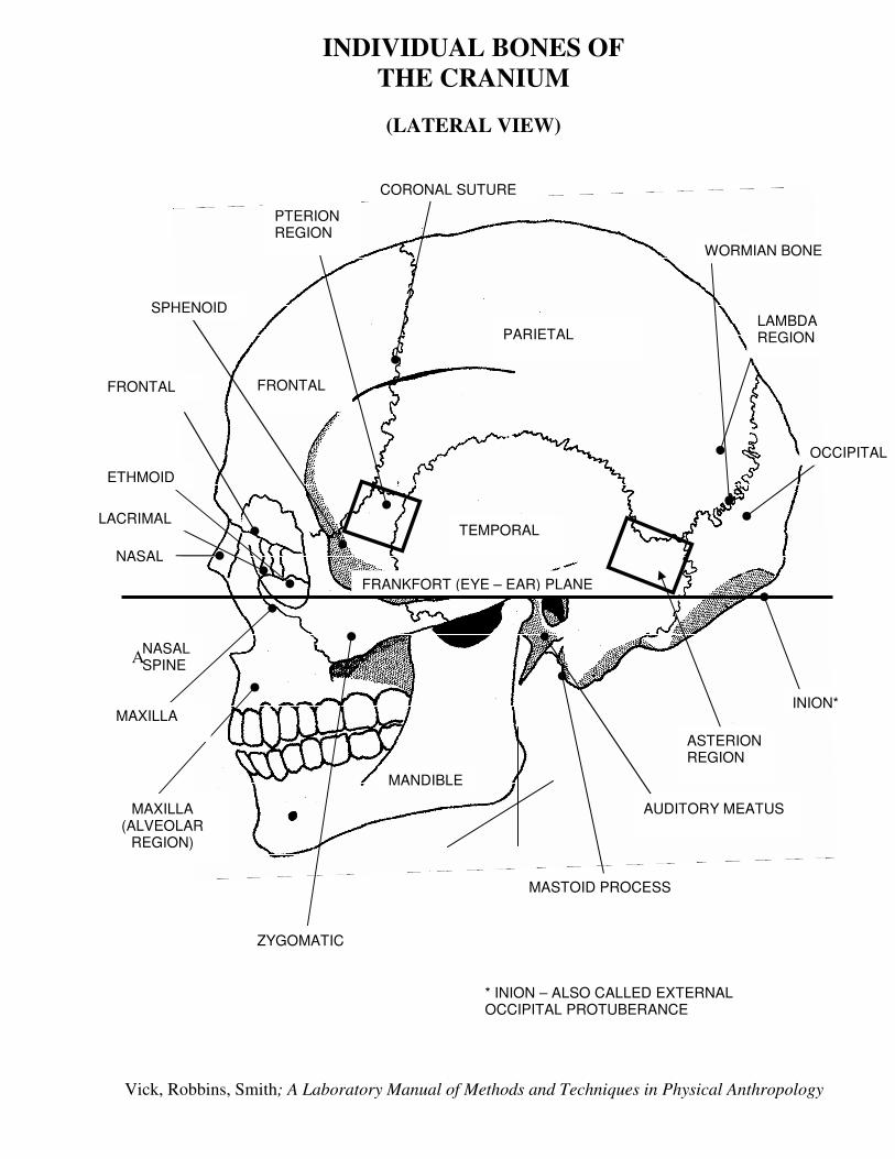
INDIVIDUAL BONES OF THE CRANIUM
(LATERAL VIEW)
CORONAL SUTURE
PARIETAL
FRONTAL
PTERION REGION
TEMPORAL
FRANKFORT (EYE – EAR) PLANE
OCCIPITAL
INION*
ASTERION REGION
WORMIAN BONE
LAMBDA REGION
MANDIBLE
SPHENOID
MASTOID PROCESS
FRONTAL
NASAL
NASAL SPINE
ZYGOMATIC
* INION – ALSO CALLED EXTERNAL OCCIPITAL PROTUBERANCE
LACRIMAL
ETHMOID
AUDITORY MEATUS MAXILLA (ALVEOLAR
REGION)
MAXILLA
A
Vick, Robbins, Smith; A Laboratory Manual of Methods and Techniques in Physical Anthropology

SAGITTAL SECTION OF THE SKULL
(ILLUSTRATING INTERNAL STRUCTURE OF THE SKULL)
SPHENOPALATINE FORAMEN
ETHMOID
OCCIPITAL
PTERYGOID PLATE OF SPHENOID BONE
PALATINE
NASAL
SUPERIOR NASAL CONCHA
MIDDLE NASAL CONCHA
LACRIMAL
INFERIOR NASAL CONCHA
MAXILLA
NASAL PROCESS OF FRONTAL
ETHMOID
CRISTA GALLI (ETHMOID)
SELLA TURCICA (SPHENOID)
VOMER
MAXILLA
INFERIOR NASAL CONCHA
PALATOMAXILLARY PROCESS
PALATINE
INTERNAL PTERYGOID PLATE (SPHENOID)
SAGITTAL SECTION LATERAL TO THE CENTER
(ILLUSTRATING INTERNAL STRUCTURE OF THE
SKULL)
Vick, Robbins, Smith; A Laboratory Manual of Methods and Techniques in Physical Anthropology

CRANIUM
CERVICAL VERTEBRAE
CLAVICLE
HUMERUS
THORACIC VERTEBRAE
LUMBAR VERTEBRAE
PELVIS (ILLIUM)
SACRUM
COCCYGEAL VERTEBRAE
CARPALS
PHALANGES
TIBIA
FIBULA
TARSALS
PHALANGES
SCAPULA
STERNUM
ULNA
RADIUS
PELVIS (PUBIS)
METACARPALS
FEMUR
PATELLA
METATARSALS
BONES OF THE SKELETON

Vick, Robbins, Smith; A Laboratory Manual of Methods and Techniques in Physical Anthropology
Bibliography
Bass, W. M. 1989. Human Osteology: A Laboratory and Field Manual of the
Human
Skeleton, 3rd ed. Columbia, MO: Missouri Archaeological Society.
Brothwell, Dr. 1992. Digging up Bones, 3rd ed. Ithaca, New York: Cornell
University Press.
Burns, Karen Ramey. 1999. Forensic Anthropology Training Manual. Upper
Saddle River, New Jersey: Prentice-Hall.
Byers, Steven N. 2002. Introduction to Forensic Anthropology: A Textbook. A
Pearson Education Company 75 Arlington Street Boston, MA: Boston,
MA.
Schour, l. and M. Massler
The development of the human dentition. Journal of the American Dental
Association 28:1153-1160.
Vick, Laura, L. Robbins, and L. Smith. 2003. A Laboratory Manual of Methods
and
Techniques in Physical Anthropology. Raleigh, NC: Department of
Anthropology, Peace College.

What and How Many?
When one is dealing with bony remains, one must first determine to what
species (one or more than one) the remains belong. In other words, is only one species represented? Or are you dealing with more than one species? Another question involves the minimum number of individuals (MNI) represented in the remains. Clues such as shape, size, side, and number are used to answer these question. In addition, color, degree of weathering, etc., can also offer valuable information.
One of the first steps in analysis involves sorting. Place bones of the same type together as, for example, all femurs versus all humeri. If homologous bones have very different shapes, they probably represent different species. Next look at size; two differently sized humeri will indicate at least two different individuals. With good understanding of the skeletal structure and relative size differences of the various bones, size can also be used as a clue even when one has non-homologous bones. Now check for side. An animal can only have one left femur or one left innominate bone (left half of the pelvis).
Color and weathering are less valuable clues since part of a skeleton may have been exposed to sunlight while another part has been covered by dirt or debris. However, these clues can sometimes help one in assigning remains to one or more than one individual.
Demonstration / Exercise
“Taphonomy Exercise” bags will be assigned to student groups. Each bag contains some rhesus monkey bones (very similar to human bones) as well as some other bones. You will use clues such as size, size, shape, number, color, and weathering to determine the answers to the questions below.

After your group is ready to “present” your conclusions, check these with the instructor or lab assistant.)
Examine carefully the bones found in your bag. Bag ID Number ______
How many species do you observe? at least