The Human Genome - Columbia
19
1 The Human Genome • 6 billion base pairs of DNA ~ 3 meters of DNA • Approximately 30,000 – 70,000 genes Approximately 80-100,000 proteins • These genes are spread across 24 different chromosomes • One chromosome each from each parent, for a total of 23 pairs (24 different chromosomes) or 46 chromosomes per somatic cell Chromatin Compaction Metaphase chromosome is compacted into a structure that is 50,000 times shorter than its extended length • Cell Cycle ~ 17 – 18 hrs • Mitosis 1-2 hrs Cell will be visible as Interphase Nucleus Majority of Time • DNA is replicated during S-phase in preparation for mitosis Meiosis 1 Meiosis 1 Normal Disjunction (Separation) Disappearance of nuclear membrane Cytoplasm divides Formation of meiotic spindle Bivalent centromeres separate Meiosis II Cells move directly from telophase I to metaphase II
Transcript of The Human Genome - Columbia
1
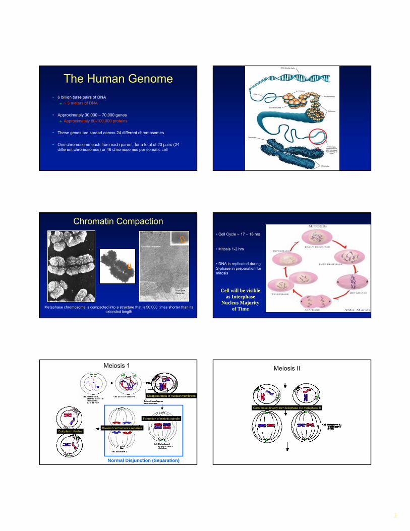
The Human Genome• 6 billion base pairs of DNA
~ 3 meters of DNA
• Approximately 30,000 – 70,000 genesApproximately 80-100,000 proteins
• These genes are spread across 24 different chromosomes
• One chromosome each from each parent, for a total of 23 pairs (24 different chromosomes) or 46 chromosomes per somatic cell
Chromatin Compaction
Metaphase chromosome is compacted into a structure that is 50,000 times shorter than its extended length
• Cell Cycle ~ 17 – 18 hrs
• Mitosis 1-2 hrs
Cell will be visible as Interphase
Nucleus Majority of Time
• DNA is replicated during S-phase in preparation for mitosis
Meiosis 1Meiosis 1
Normal Disjunction (Separation)
Disappearance of nuclear membrane
Cytoplasm divides
Formation of meiotic spindle
Bivalent centromeres separate
Meiosis II
Cells move directly from telophase I to metaphase II
2
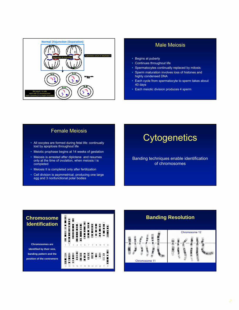
Normal Disjunction (Separation)
Separation of chromatidsCytokinesis occurs in telophase II
Net result = 4 cellseach with 23 chromosomes
each consisting of a single chromatid
Male Meiosis
• Begins at puberty• Continues throughout life• Spermatocytes continually replaced by mitosis• Sperm maturation involves loss of histones and
highly condensed DNA• Each cycle from spermatocyte to sperm takes about
40 days• Each meiotic division produces 4 sperm
Female Meiosis
• All oocytes are formed during fetal life: continually lost by apoptosis throughout life
• Meiotic prophase begins at 14 weeks of gestation
• Meiosis is arrested after diplotene and resumes only at the time of ovulation, when meiosis I is completed
• Meiosis II is completed only after fertilization
• Cell division is asymmetrical, producing one large egg and 3 nonfunctional polar bodies
Cytogenetics
Banding techniques enable identification of chromosomes
Chromosomes are
identified by their size,
banding pattern and the
position of the centromere
Chromosome Identification
Banding Resolution
Chromosome 11
Chromosome 12
3
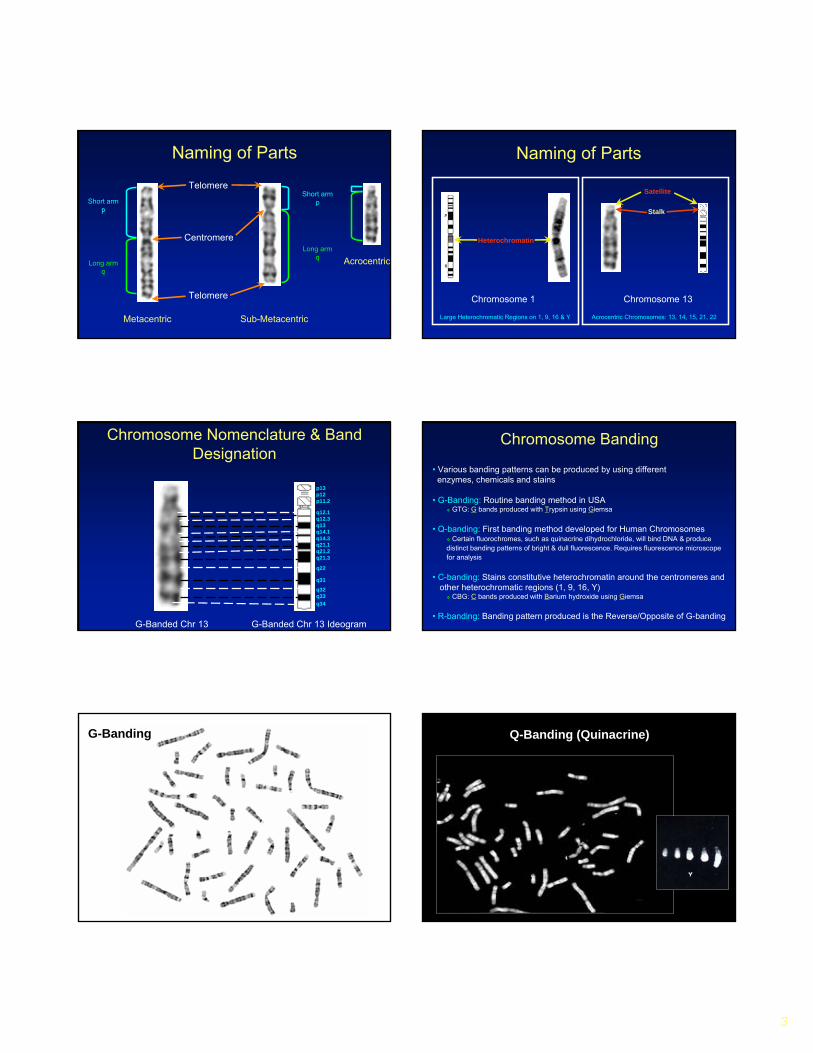
Naming of Parts
Telomere
Telomere
Centromere
Short armpShort arm
p
Long armq
Long armq
Metacentric Sub-Metacentric
Acrocentric
Heterochromatin
Stalk
Chromosome 1 Chromosome 13
Satellite
Naming of Parts
Large Heterochromatic Regions on 1, 9, 16 & Y Acrocentric Chromosomes: 13, 14, 15, 21, 22
Chromosome Nomenclature & Band Designation
G-Banded Chr 13 G-Banded Chr 13 Ideogram
q12.1q12.3q13q14.1q14.3q21.1q21.2q21.3
q22
q31
q32q33q34
p13p12p11.2
Chromosome Banding
• Various banding patterns can be produced by using differentenzymes, chemicals and stains
• G-Banding: Routine banding method in USAGTG: G bands produced with Trypsin using Giemsa
• Q-banding: First banding method developed for Human ChromosomesCertain fluorochromes, such as quinacrine dihydrochloride, will bind DNA & produce
distinct banding patterns of bright & dull fluorescence. Requires fluorescence microscope for analysis
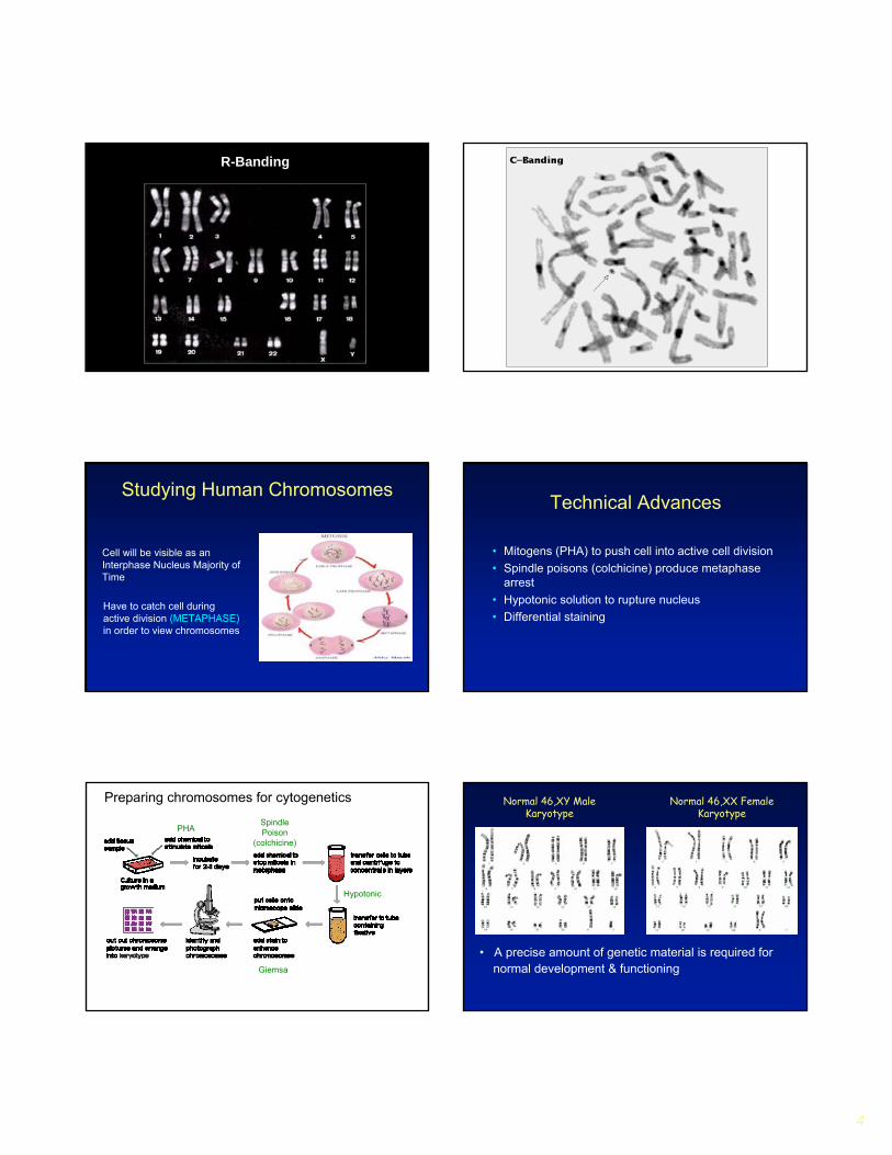
• C-banding: Stains constitutive heterochromatin around the centromeres andother heterochromatic regions (1, 9, 16, Y)
CBG: C bands produced with Barium hydroxide using Giemsa
• R-banding: Banding pattern produced is the Reverse/Opposite of G-banding
G-Banding Q-Banding (Quinacrine)
4
R-Banding
Cell will be visible as an Interphase Nucleus Majority of Time
Studying Human Chromosomes
Have to catch cell during active division (METAPHASE)in order to view chromosomes
Technical Advances
• Mitogens (PHA) to push cell into active cell division• Spindle poisons (colchicine) produce metaphase
arrest• Hypotonic solution to rupture nucleus• Differential staining
Preparing chromosomes for cytogenetics
PHASpindle Poison
(colchicine)
Hypotonic
Giemsa
Normal 46,XY Male Karyotype
Normal 46,XX Female Karyotype
• A precise amount of genetic material is required fornormal development & functioning

5
Genetic Imbalance• An incorrect amount of genetic material in a
conceptus may disturb/distort the normal growth pattern
Zygote Blastula Embryo Fetus
Adult Adolescent Newborn
Chromosomal Imbalance• An imbalance in the amount of chromosomal
material may involve 100’s to 1000’s of genes and generally has more catastrophic effects
Zygote Blastula Embryo Fetus
Adult Adolescent Newborn
Chromosomal Imbalance
• May involve the gain or loss of a whole chromosome (complete aneuploidy) or of part of a chromosome (partial aneuploidy)
• The abnormality may occur in the non-mosaic or mosaic state (Mosaicism = Various chromosome complements in different cells)
• Monosomy (one missing) is generally more devastating than trisomy (one extra)
Chromosomal Imbalance• Most (complete) autosomal trisomies & all
(complete) autosomal monosomies are so catastrophic that their presence in a conceptus is not compatible with survival
• Trisomies, monosomy X and polyploids are the most common abnormalities observed in spontaneous abortions
• ~ 66% of first trimester spontaneous abortions• ~20% of 2nd trimester spontaneous abortions
Frequency of Chromosome Abnormalities
%
25Infants with congenital heart disease
12-15 Children with mental retardation
0.3 Livebirths
7Late fetal deaths and stillbirths
60-70Early miscarriages
8-10Early recognized conceptions (>4 wks)
85“Poor” preimplantation embryos
30-40“Good” preimplantation embryos
20-30Oocytes
8Sperm
Source
Chromosomal Imbalance and Pregnancy*Loss
• 65% Trisomies• 11% Monosomies• 11% Triploidies• 7.5% Multiple Aneuploidies• 5.5% Tetraploid and structural
Trisomy 16• Most common trisomy observed in POC studies• Never seen in liveborn
Trisomy 21 & 22• Next most common (equally)
*Recognized pregnancies

6
Chromosomal
Tetraploidy (92,XXXX)
Triploidy (69,XXX)
Imbalance
Survivable Chromosomal Imbalance
• Only a few complete non-mosaic aneuploidies are observed in liveborns.
Down syndrome (Tri 21), Edward Syndrome (Tri 18), PatauSyndrome (Tri13), Turner Syndrome (Mono X)
• All other imbalances will contain much smaller chromosomal regions (partial aneuploidy) that would allow for the organisms to survive….Albeit with clinical abnormalities (in most cases)
Numerical abnormalities
• Ploidy: The category of chromosome changes which involve the addition or loss of complete sets of chromosomes.
• TriploidyThe possession of one complete extra set of chromosomes.Usually caused by polyspermy, the fertilisation of an egg by more than one sperm.Such embryos will usually spontaneously abort.
• TetraploidyUsually the result of a failure of the first zygotic division. It is also lethal to the embryo.Any other cell division may also fail to complete properly and in consequence a very small proportion of tetraploid cells can sometimes be found in normal individuals (mosaicism).
Autosomal Numerical abnormalities
• AneuploidyThe category of chromosome changes which do not involve whole sets. It is usually the consequence of a failure of a single chromosome (or bivalent) to complete division.
• Monosomies All autosomal monosomies are lethal in very early embryogenesis.Most abort too early even to be recognised as a conception.
• Down syndrome, trisomy 21The incidence of trisomy 21 rises sharply with increasing maternal age.
Age Related Risks for Trisomy at the Time of CVS and Amniocentesis
0123456789
10111213141516171819202122
16 17 18 19 20 21 22 23 24 25 26 27 28 29 30 31 32 33 34 35 36 37 38 39 40 41 42 43 44 45 46 47 48 49
Maternal Age
Freq
uenc
y (%
)
CVS
Amniocentesis
7
Clinical Phenotypes of Chromosomal Abnormalities
• Associated with Developmental Delay/MR• Alteration of facial morphogenesis to produce characteristic
facial features• Growth delay• Malformations of the internal organs - especially cardiac
Indication for chromosome analysis = MCA/MR
Survivable Chromosomal Imbalance
Trisomy 13 (47,XY,+13) Trisomy 18 (47,XY,+18) Trisomy 21 (47,XY,+21)
• Only a few full non-mosaic aneuploidies are observed in liveborns
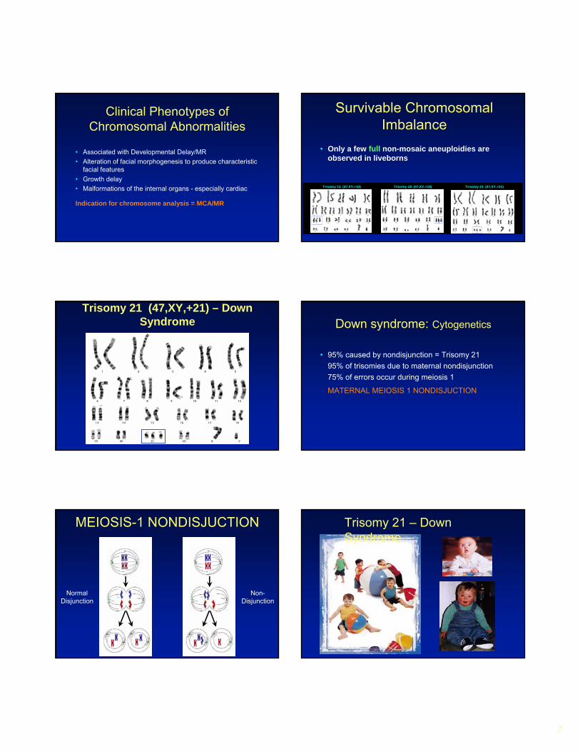
Trisomy 21 (47,XY,+21) – Down Syndrome Down syndrome: Cytogenetics
• 95% caused by nondisjunction = Trisomy 2195% of trisomies due to maternal nondisjunction75% of errors occur during meiosis 1
MATERNAL MEIOSIS 1 NONDISJUCTION
MEIOSIS-1 NONDISJUCTION
NormalDisjunction
Non-Disjunction
Trisomy 21 – Down Syndrome
8
Trisomy 21 Down Syndrome
• Hypotonia• Redundant neck fold/flat occiput• Low set ears with characteristic pinnae• Protruding/large tongue• Abnormal dermatoglyphics
Simian line and clinodactylyWide space between 1st & 2nd toes
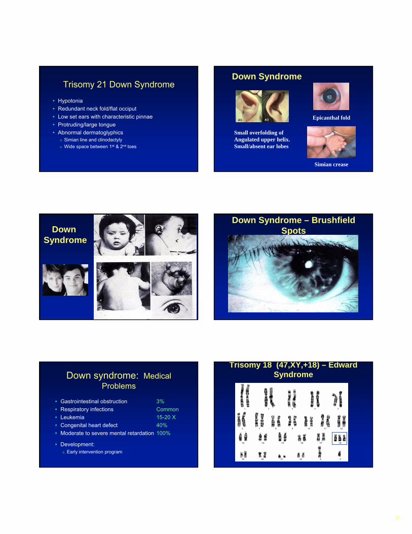
Epicanthal fold
Simian crease
Down Syndrome
Small overfolding ofAngulated upper helix.Small/absent ear lobes
17.jpgDown Syndrome
Down Syndrome – BrushfieldSpots
Down syndrome: Medical Problems
• Gastrointestinal obstruction 3%• Respiratory infections Common• Leukemia 15-20 X• Congenital heart defect 40%• Moderate to severe mental retardation 100%
• Development:Early intervention program
Trisomy 18 (47,XY,+18) – Edward Syndrome
9
Trisomy 18• Incidence 1:3333 live births• Most common abnormality in stillbirths with multiple
congenital abnormalities• Prenatal growth deficiency resulting in a small for gestational
age infant (SGA)• 90% congenital heart defect VSD• 10% alive at one year• Marked developmental disability
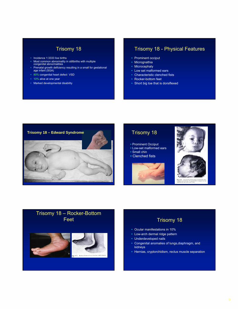
Trisomy 18 - Physical Features• Prominent occiput• Micrognathia• Microcephaly• Low set malformed ears• Characteristic clenched fists• Rocker-bottom feet• Short big toe that is dorsiflexed
Trisomy 18 – Edward Syndrome Trisomy 18
• Prominent Occiput• Low-set malformed ears• Small chin• Clenched fists
Trisomy 18 – Rocker-Bottom Feet Trisomy 18
• Ocular manifestations in 10%• Low-arch dermal ridge pattern• Underdeveloped nails• Congenital anomalies of lungs,diaphragm, and
kidneys• Hernias, cryptorchidism, rectus muscle separation

10
Trisomy 18 – High Morbidity & Mortality
Trisomy 18 - Medical Management
• Feeding difficulties GE reflux • Apnea• Seizures• Slow post natal growth• Developmental disability/ mental retardation• Scoliosis
Trisomy 13 (47,XY,+13) – PatauSyndrome Trisomy 13
• Incidence 1:5,000 births• Distinctive malformation pattern
(Craniofacial and Central Nervous System)
• 95% spontaneously aborted• Survival rate and development similar to Trisomy 18
Trisomy 13 Patau Syndrome
• Microcephaly with sloping forehead • Holoprosencephaly• Ophthalmologic abnormalities
microphthalmia or anophthalmiaColobomata of iris and ciliary body
• Cleft lip +/- palate• Low set ears with abnormal helices
Trisomy 13 Patau Syndrome
• Cardiac defects: ASD, PDA, VSD• Males: cryptorchidism ; Females: Bicornuate uterus• Polycystic kidneys• Aplasia cutis congenita• Polydactyly of hands +/- feet• Rockerbottom feet
11
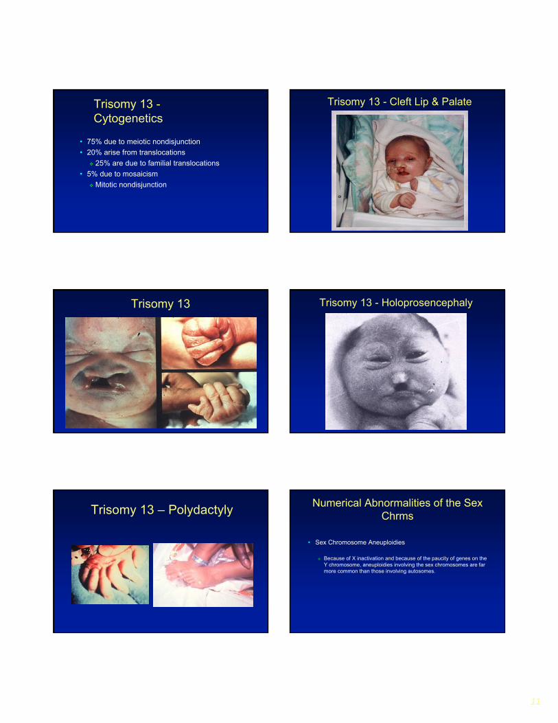
Trisomy 13 -Cytogenetics
• 75% due to meiotic nondisjunction• 20% arise from translocations
25% are due to familial translocations• 5% due to mosaicism
Mitotic nondisjunction
Trisomy 13 - Cleft Lip & Palate
Trisomy 13 Trisomy 13 - Holoprosencephaly
Trisomy 13 – Polydactyly Numerical Abnormalities of the Sex Chrms
• Sex Chromosome Aneuploidies
Because of X inactivation and because of the paucity of genes on the Y chromosome, aneuploidies involving the sex chromosomes are farmore common than those involving autosomes.
12
Sex Chromosome Abnormalities
• Turner syndrome• Klinefelter syndrome (XXY)• Triple X • XYY
Numerical Abnormalities of the Sex Chrms
• Turner syndrome 45,X
The incidence is about 1 in 5000 female births but this is only the tip of the iceberg because 99% of Turner syndrome embryos are spontaneously aborted.
Individuals are very short, they are usually infertile. Characteristic body shape changes include a broad chest with widely spaced nipples and may include a webbed neck.
IQ and lifespan are unaffected.
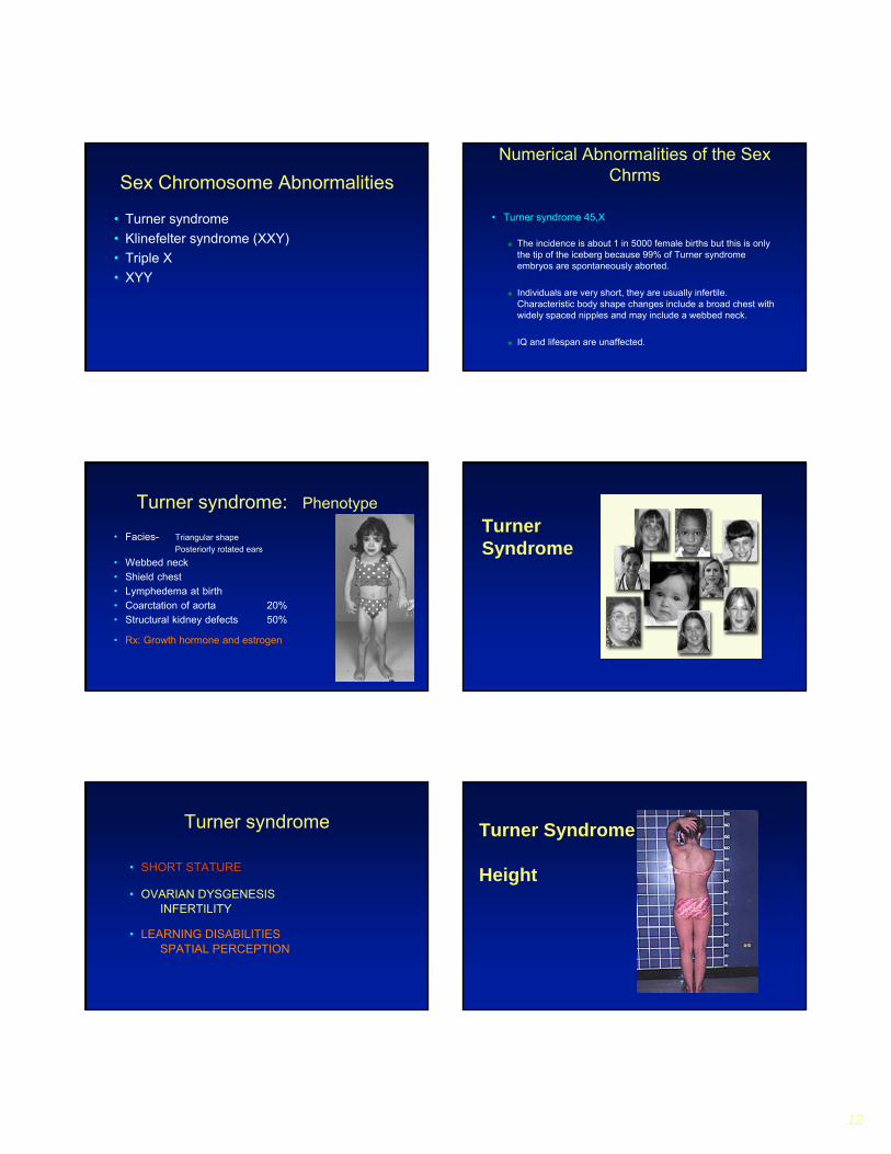
Turner syndrome: Phenotype
• Facies- Triangular shape Posteriorly rotated ears
• Webbed neck• Shield chest• Lymphedema at birth• Coarctation of aorta 20%• Structural kidney defects 50%
• Rx: Growth hormone and estrogen
Turner Syndrome
Turner syndrome
• SHORT STATURE
• OVARIAN DYSGENESISINFERTILITY
• LEARNING DISABILITIESSPATIAL PERCEPTION
Turner Syndrome
Height
13
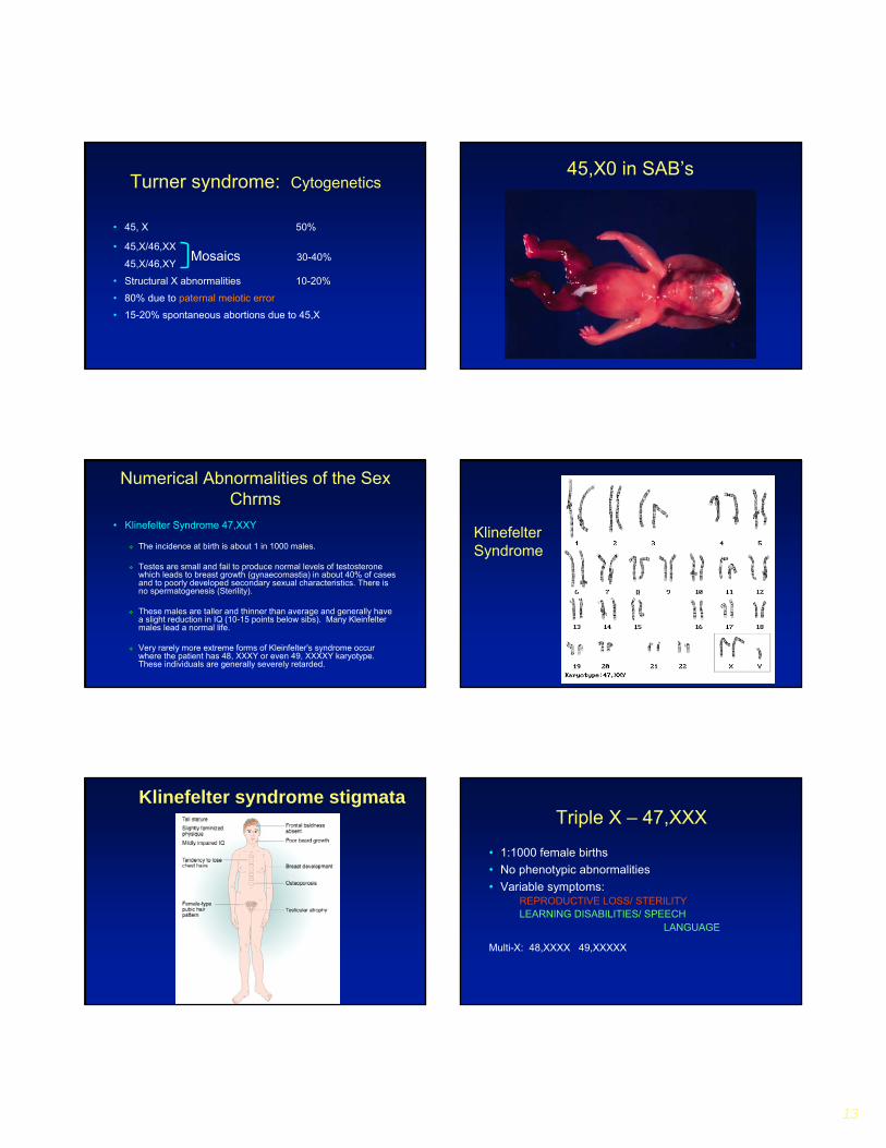
Turner syndrome: Cytogenetics
• 45, X 50%
• 45,X/46,XX
45,X/46,XY
• Structural X abnormalities 10-20%
• 80% due to paternal meiotic error
• 15-20% spontaneous abortions due to 45,X
Mosaics 30-40%
45,X0 in SAB’s
Numerical Abnormalities of the Sex Chrms
• Klinefelter Syndrome 47,XXY
The incidence at birth is about 1 in 1000 males.
Testes are small and fail to produce normal levels of testosterone which leads to breast growth (gynaecomastia) in about 40% of cases and to poorly developed secondary sexual characteristics. There is no spermatogenesis (Sterility).
These males are taller and thinner than average and generally have a slight reduction in IQ (10-15 points below sibs). Many Kleinfeltermales lead a normal life.
Very rarely more extreme forms of Kleinfelter's syndrome occur where the patient has 48, XXXY or even 49, XXXXY karyotype. These individuals are generally severely retarded.
KlinefelterSyndrome
Klinefelter syndrome stigmataTriple X – 47,XXX
• 1:1000 female births• No phenotypic abnormalities• Variable symptoms:
REPRODUCTIVE LOSS/ STERILITYLEARNING DISABILITIES/ SPEECH
LANGUAGE
Multi-X: 48,XXXX 49,XXXXX

14
Numerical Abnormalities of the Sex Chrms
• XXX femalesAbout one woman in 1000 has an extra X chromosome. It seems to do little harm, individuals are fertile and do not transmit the extra chromosome.
They do have a reduction in IQ comparable to that of Kleinfelter's males (10-15 point below sibs).
Variants− Multi-X: 48,XXXX 49,XXXXX− Mild to moderate MR− Variable dysmorphic features
Numerical Abnormalities of the Sex Chrms
• 47,XYY malesIncidence 1 in 1000 male births. May be without any symptoms.
Males are tall but normally proportioned.
10 - 15 points reduction in IQ compared to sibs? (IQ:93-109/109-147)
More common in high security institutions than chance would suggest? (Problems with impulse control?)
XYYSyndrome
Structural Rearrangements
• Translocations
• Inversions
Multiple options for Gametes only 2 of which are balanced
Structural Rearrangements –Translocations & Inversions
15
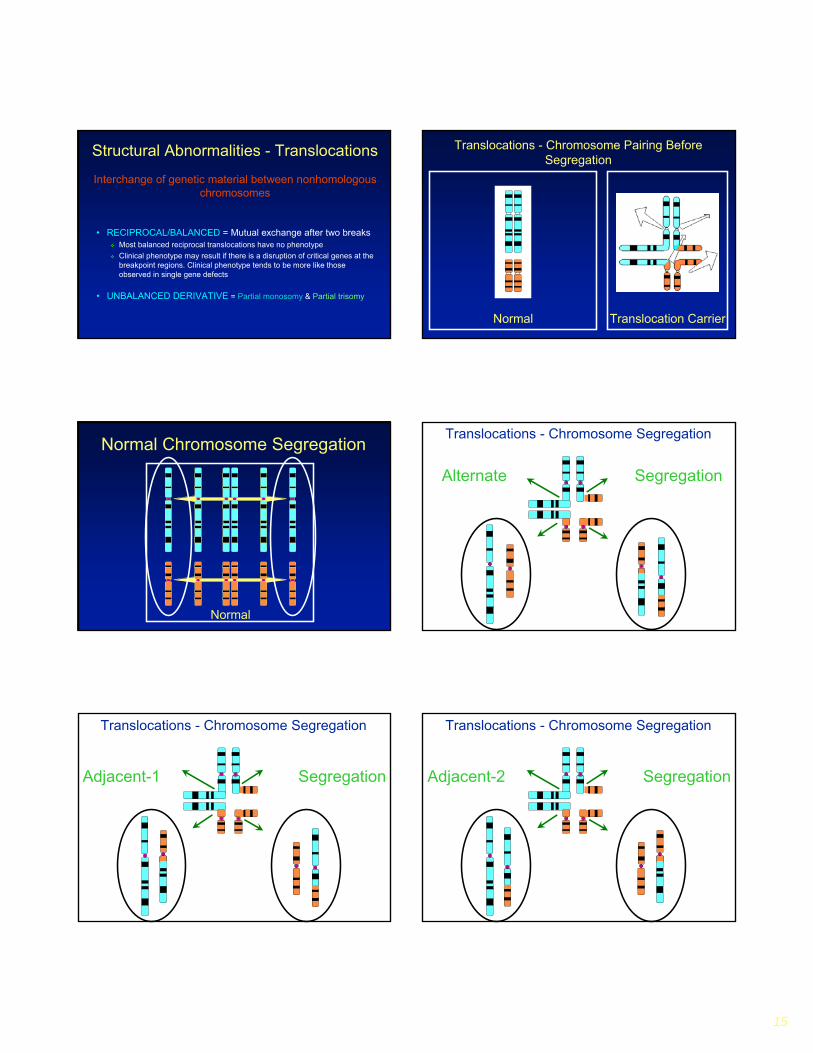
Structural Abnormalities - Translocations
• RECIPROCAL/BALANCED = Mutual exchange after two breaksMost balanced reciprocal translocations have no phenotypeClinical phenotype may result if there is a disruption of critical genes at the breakpoint regions. Clinical phenotype tends to be more like those observed in single gene defects
• UNBALANCED DERIVATIVE = Partial monosomy & Partial trisomy
Interchange of genetic material between nonhomologouschromosomes
Translocations - Chromosome Pairing Before Segregation
Normal Translocation Carrier
Normal Chromosome Segregation
Normal
Translocations - Chromosome Segregation
Alternate Segregation
Translocations - Chromosome Segregation
Adjacent-1 Segregation
Translocations - Chromosome Segregation
Adjacent-2 Segregation

16
Normal Normal Balanced Unbalanced Unbalanced
Unbalanced Unbalanced Unbalanced Unbalanced Unbalanced
Alternate Segregation Adjacent-1 Segregation Adjacent-2 Segregation
3:1 Segregation 3:1 Segregation 3:1 Segregation 3:1 Segregation 4:0 Segregation
Translocations
Robertsonian Translocation• Translocation between acrocentric chromosomes. Short
arms are lost and long arms fuse at centromere (5% of Down syndrome)
Two AcrocentricChromosomes
RobertsonianTranslocation
Short Arm is Lost
Short Arm Fusion Long Arm Fusion
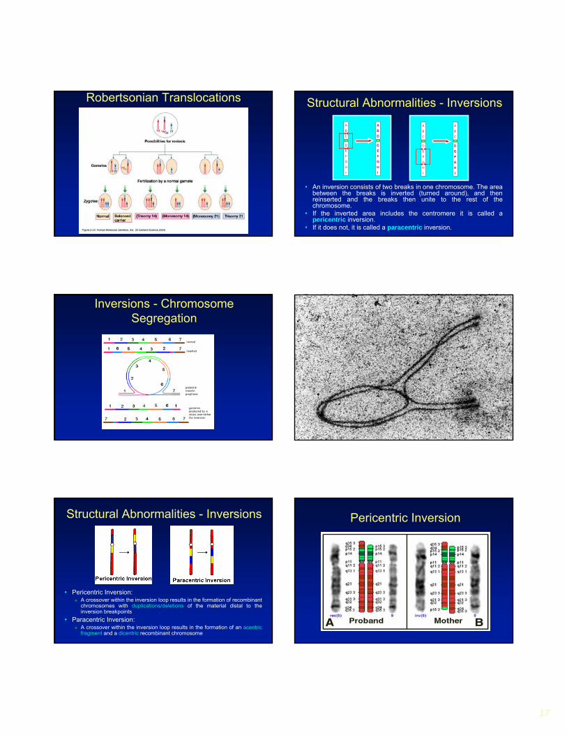
Robertsonian Translocations
Normal Normal Balanced UnbalancedAlternate Segregation Adjacent-Segregation
Unbalanced3:1 Segregation
Unbalanced3:1 Segregation
Unbalanced3:1 Segregation
Unbalanced4:0 Segregation
17
Robertsonian Translocations
• An inversion consists of two breaks in one chromosome. The area between the breaks is inverted (turned around), and then reinserted and the breaks then unite to the rest of the chromosome.
• If the inverted area includes the centromere it is called a pericentric inversion.
• If it does not, it is called a paracentric inversion.
Structural Abnormalities - InversionsA
B
C
A
B
E
F
G
H
I
A
B
F
G
H
I
C
D
E
D
E
F
G
H
I
D
C
A
B
C
A
B
F
G
H
I
C
D
E G
F
E
D
H
I
D
E
F
G
H
I
Inversions - Chromosome Segregation
• Pericentric Inversion:A crossover within the inversion loop results in the formation of recombinant chromosomes with duplications/deletions of the material distal to the inversion breakpoints
• Paracentric Inversion:A crossover within the inversion loop results in the formation of an acentric fragment and a dicentric recombinant chromosome
Structural Abnormalities - Inversions Pericentric Inversion

18
RingChromosomes
Translocations & Inversions
• BALANCED
Most balanced rearrangements have no phenotype
Clinical phenotype may result if there is a disruption of critical genes at the breakpoint regions. Clinical phenotype tends to be more like those observed in single gene defects
• UNBALANCED
Partial Monosomy/Trisomy
Deletions• 5p- Cri-du Chat
• 4p- Wolf-Hirschhorn
Deletion 5p - Cri Du Chat Syndrome Cri Du Chat Syndrome - Older childre

19
Cri du Chat Karyotype Deletion 4p - Wolf- Hirschhorn Syndrome
Deletion 4p Cytogenetics Wolf Hirschhorn Syndrome
• Deletion in the terminal band 4p16.3• 87% of cases due to de novo interstitial deletion of
paternal origin• 13% due to unbalanced product of a parental
reciprocal translocation
Deletion 4p Karyotype
• Unbalanced Translocations Partial Monosomy & Partial Trisomy
• Deletions Partial Monosomy
• Duplications Partial Trisomy
• Ring & marker chromosomes Partial Trisomy
• Recombinant Inversion derivatives Partial Monosomy & Partial Trisomy
• Isochromosomes Partial Trisomy/Tetrasomy or Partial Monosomy & Partial Trisomy
“Viable” Chromosome Imbalance
“Viable” Chromosome Imbalance
• 1 in 150 Livebirths• 10-15% Mentally retarded population
Higher percentage when cryptic rearrangements areincluded
• Most case reports involve partial aneuploidy