Treatment of complicated crown-root fracture in a single ...
The Effect of Crown Lengthon the Fracture Resistanceof Posterior Porcelain and
Glass-Ceramic Crowns
s.S. Scherrer, Dr Med Denf
W.G. de Rijk, MS, DDS, PhD"
School of Dentistry and University of Texas Health ScienceCenter
This in vitro study measured the changes in the fractureresistance of posterior crowns as a function of crown length.The crowns, 10 for each group, were made of a feldspafhicporcelain (Ceramco), a glass-ceramic material (Dicor), andan alumina-reinforced glass (In-Ceram). Three differentcrown lengths were tested on acrylic resin dies. Therestorations were fractured in a testing machine using a steelball, 12.7 mm in diameter, that contacted the occlusalsurface at three distinct points. Statistical analysis wasperformed using tbe Weibull distribution. The fractureresistance increased significantly with increasing crownlength for all crown materials. Int J Prosthodont 1992;5:550-557.
T he longevity of all-porcelain and glass-ceramiccrowns has been of concern to clinicians
because the restoration may fracture suddenly andcatastrophically, a phenomenon rarely seen inmetal ceramic restorations. A clinical evaluation ofall-ceramic posterior and anterior crowns usingCorestore, Dicor, and Hi-Ceram has been ongoingsince 1986 at the University of Geneva, Switzer-land, and has been previously reported by Scherreret al'^ and Pisa et al.^ In those clinical studies noneof the restorations were bonded to the dentin; allwere placed with a luting agent. After 3 years, a
'Visiting Assistant Professor, Department of Restorative Den-tistry, UThISC; from the School of Dental Medicine, 19, rueBarthelemy-Menn, 1205 Geneva, Switzerland.
"Associate Professor, Department of Restorative Dentistry,University of Texas Health Science Center Dental School atSan Antonio, 7703 Floyd Curl Drive, San Antonio, Texas78284-7890.
Reprint requests: W.C. de Rijk.A part of the experimental results presented in this paper havebeen presented before the International Association of DentalResearch 69th General Session in Acapuico, Mexico in April1991.This research was supported in part by a Young Scientist Award(Dr Scherrer) from the Swiss Nationai Foundation for ScientificResearch (FNSRS).
high rate of failure ranging from 15% to 22% wasfound (unpublished data), mostly with posteriorcrowns, a finding that is consistent with other clin-ical studies of all-ceramic restorations as reportedby Anusavice."" Several factors including porcelainthickness, crown configuration, cement layer, andthe supporting tooth structure are consideredmajor factors in determining the overall strength ofthe restoration. From these factors the crown con-figuration was selected as the area of interest, as itis believed that the longer the crown the greaterthe probability of failure because of the increasein circumferential stresses in the crown. This theoryis based on the fact that loading a crown seatedon a tapered preparation markedly increases cir-cumferential stresses. Finite-element analysis byHojjatie and Anusavice^ has shown that, indeed,the maximum stresses occur at or near the marginof a crown. That investigation also showed that thestresses decrease when the ratio of occlusal thick-ness to crown length is increased, making a shortercrown appear stronger. In a study by DeRand,^where both length and contour were changed, itwas claimed that crown length is of minor impor-tance.
The purpose of this study was to determine thechanges in the fracture resistance of three all-ceramic posterior crown restorations as a function
ournsi o I Pios( h odontic 550 Volume S. Number 6. 1992

Se h errer/de Riik Effect of Crown Length on Fracture Resistance
of crown length. To separate the effect of increas-ing crown length from the effect of material choice,three totally different ceramic systems wereselected; a feldspathic porcelain, a glass-ceramicmaterial, and an alumina-reinforced glass. The feld-spathic porcelain used here is usually marketed formetal ceramic applications and not for all-porcelaincrowns.
Materials and Methods
Die Preparation
For this study three crown lengths were selected:an occlusal cover (O¡, a crown half the length ofthe tooth (H], and a full-length crown (C) (Figs 1and 2]. This crown has been termed a full crownbecause it refers to the length of the crown,whereas the term complete crown is reserved toindicate full circumferential coverage. All crownlength preparations were sequentially created ona single Ivorine maxillary third molar (ColumbiaDentoform, New York, NY). The tooth prepara-tions were made manually using diamond burs.The occlusal reduction of 1.5 mm followed theocciusai morphology of the Ivurine tooth, keepingan angle of approximately 150° at the central fossabetween the lingual cusp and the two buccalcusps. Subsequent to the occlusal reduction a rep-lica of the tooth was made. The half-crown con-figuration was made on the same tooth by addingonly a 90° shoulder preparation, 1 mm wide at halfthe length of a full-crown preparation, withoutmodifying the original occlusal preparation. Theaxiai walls had a taper of approximately 6°. At thispoint, a replica was again made of the tooth prep-
Fig 1 Three different acrylic resin die configurafions. (Leftto right)7he occiusai cover, the half crown, and fhe full-crowndie.
aration. Finally, the full-crown configuration wascompleted as an extension in the length ofthe halfcrown, with a 90° shoulder preparation at the levelof the cementoenamel junction. Using thismethod, all dies had exactly the same occlusal con-figuration.
[fach crown configuration was reproduced as adie in a microwave-processed acrylic resin (Para-gon Denture Resin, Hygienic Co, Akron, Ohio).The dies were made from an elastomeric impres-sion material (Fxpress, 3M, Dental Products Div, StPaul, Minn). Wax was poured into the impressionand the resulting pattern was processed in a micro-waveable denture flask (FRP Flask, Justi Products,Oxnard, Calif), All dies were coated with two appli-cations of a die-spacer medium (PDQ, Whip-MixCo, Louisville, Ky) to provide space for the lutingagent. Three different porcelains were evaluatedfor each crown configuration. A castable glass-
Fig 2 A schematic representation ofthe three crown configurations, with the loading sphere shownin position on fhe full crown.
i to lume5, Number 6, 1992 551 The International lournal ol Prosthodontici

Efíecl oí Crown Length on Fracture Resista Scherrer/de Rijk
ceramic nnaterial (D [Dicor, Dentsply Int Inc, York,Pa]), a feldspathic porcelain (C [Ceramco Bodyporcelain A-2, provided to the investigators whenCeramco was still a division of |ohnson & Johnson,Fast Windsor, N)]¡, and an alumina-reinforced glass(I [In-Ceram, Vident, Baldwin Park, Calif]), Thedesired occlusal geometry is that of a simplifiedthird-molar crown with three cusps and an occlusalthickness of 1,6 mm (± 0,1 mm). The occlusalsurface contour must be such that a steel ball, 12,7mm in diameter, produces three well-defined con-tact points (Fig 3], The castable glass-ceramic per-mi t ted accurate dup l i ca t i on of a singleconfiguration by using identical duplicate wax pat-terns. To minimize variations in the thickness andshape of the surface, no colorant was applied tothe surface. For crowns made from the other twomaterials used in this study, the occlusal surfacehad to be reproduced manually. Care was takento reproduce occlusal contours as best as possible,by controlling groove angles with specific cuttinginstruments and by repeated measurement of theocclusal thickness and the position of the occlusalcontact points.
Variation in all measurements approximated 0,1mm. To preserve the accuracy of the occlusalgeometry, none of the crowns was glazed. The finalfinish of all restorations was obtained by polishingwith a polishing kit (Shofu Dental Products, Tokyo,lapan). The occlusal contact points were obtainedby coating the steel ball with red ink and placingit on the crown,
Castable Glass-Ceramic Crowns (Dicor)
Because the Dicor crowns were produced by thelost-wax technique, duplication of the occlusal sur-face was achieved by making a metal template that,when combined with the die, produced identicalwax patterns when impressions of the die-templatecombination were made and the impression wasfilled with melted wax. The Dicor crowns were castand cerammed according to the manufacturer'sinstructions. The occlusal surface of each Dicorcrown was finished as described previously,
Feldspathic-Porcelain Crowns (Ceramco)
The feldspathic crowns were hand fabricated onrefractory dies (Polyvest, Whip-Mix Co) obtainedby duplicating the Dentoform master die of eachtooth preparation. Two firings were necessary forthe occlusal cover; three to four firings were nec-essary for the half and full crowns. The refractorydie was removed using aluminum-oxide abrasion
(alumina 50 ßm at 0,5 MPa) and the crown wasseated on the acrylic resin die. The occlusal surfaceof each crown was then shaped and finished,
Alumina-Reiniorced Glass Crowns (In-Ceram)
The In-Ceram system consisted of an alumina-glass coping and a veneer of feldspathic porcelain(Vitadur-N, Vident, Baldwin Park, Calif), The cop-ings were provided by the manufacturer and fit theacrylic resin dies developed for this study. The por-celain veneer was applied in two firing cycles, withthe occlusal surface again being shaped and fin-ished. The copings had a thickness of 0,42 mm ( ±0,07 mm) with the overall thickness of the occlusalsurface still kept at 1,6 mm (± 0,1 mm) as withthe other units. The surface was again finished withthe same porcelain finishing kit.
Crown Luting
Prior to cementation, the Dicor and the Ceramcorestorations were acid etched and silanized usingthe Dicor Light Activation Kit (Dentsply Int), Thecrowns, a total of 90 units, were luted using DicorLight-Activated Cement (dual polymerization,opaque) on their respective acrylic resin dies usinga light exposure of 20 seconds per side of eachcrown (for a total of 100 seconds per crown). Dur-ing the luting procedure finger pressure wasapplied to retain the position of the crown, as rec-ommended by the manufacturer. Restorationswere stored in a dry room for 24 hours prior tofracture,
Fracture-Strength Determination
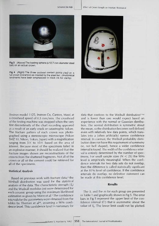
Fracture loads, recorded in Newtons, wereapplied to the occlusal surface with a steel ball,12.7 mm in diameter, that contacted the crown atthree distinctive points (a light petroleum jelly coat-ing was applied between the steel ball and theporcelain to reduce the effect of surface rough-ness). The effect of a lubricant placed on theindenter is still a subject of debate as to whetherit helps in avoiding discrepancies resulting fromsurface roughness and the elastic modulus differ-ence between indenter and tested surface. Basedon the discussion by Johnson et al,^ the use of alubricant on the indenter was included in the pro-tocol. The configuration of the crown under theball and the resultant occlusal contact points areshown in Figs 3 and 4. Compression loading wasperformed using a Universal testing machine
The InleTnational Journal of Pro^lhodontics 552 •5, Numbers, 1992
Scherrer/de Ri|k Effect of Crown LeiiBlfi on Fracture
Fig 3 (Above) The loading sphere (a 12,7-mm diameter steelball) on an actual crown.
Rg 4 (Right) The three occlusal contact points (red) on afull crown ¡Ceramco) as created by the steel ball, ¡Anatomicallandrtiarks have been emphasized in black ink for clarity)
(Instron model 1125, Instron Co, Canton, Mass) ata crosshead speed of 0,5 mm/min. The crossheadof the testing machine was stopped when the veryfirst discontinuity of the chart recording appearedas a result of an early crack or catastrophic failtjre.The fracture pattern of each crown was photo-graphed using a stereoscopic microscope (NikonSMZ 10, Nikon, Tokyo, Japan) with a magnificationranging from 3X to 10X based on the area ofinterest. Because most of the specimens failed inan explosive manner, it should be realized that thefracture images shown are reconstructions of thecrowns from the shattered fragments. Not all of thecrown or all of the cement could be retrieved forall of the specimens.
Statistical Analysis
Based on previous work with fracture data,*' theWeibull distribution was used for the statisticalanalysis of the data. The characteristic strength (So)and the Weibull modulus (in) were determined foreach ceramic group using the maximum likelihoodestimation as given by Mann et al,' The confidenceintervals for the parameters were obtained from thetables by Thoman et aV", assuming a 90% confi-dence level. This confidence level is customary for
data that conform to the Weibull distribution'^ '̂and is lower than one would expect based onexperience with the normal or Gaussian distribu-tion. The normal distribution is symmetric aboutthe mean, so the distribution becomes well definedeven with relatively few data points, which trans-lates into a (often artificially) narrow confidenceinterval. In contrast, the Weibull probability distri-bution does not have this requirement of symmetry(ie, not bell shaped), hence a wider confidenceintervalis found. The width of the confidence inter-val is entirely determined by the number of spec-imens. For small sample sizes (N < 25) the 90%level is empirically meaningful. When the confi-dence intervals for two data sets do not overlap,then the difference is called statistically significantat the 81 % level of confidence. If the confidenceintervals do overlap, no definitive statement canbe made at this confidence level.
Results
The S,¡ and the m for each group are presentedin Table 1 and graphically shown in Fig 5, The errorbars in Fig 5 represent the upper limit of the con-fidence interval (Cl) that is asymmetric about thevalue of So, The lower limit could not be plotted.
• 5, Number 6, 1992 5 5 3 ! The inlernalional Journal of Prosthodorlics

Effect of Crown Lengtii on Fracture Resislanc Scherrer/de Rijk
Table 1 Charaderistic Strength and Weibull Modulus, Including the ConfidenceIntervals at 90%*
Material
Ceramco
Dicor
In-Ceram
Configuration
Occlusal (CO)Half (CH)Full (CF)
Occlusal (DO)Half (DH)Full (DF)
Occlusal (10)Half (IH)Full (IF)
Weibullmodulus (m)
6.69.27.4
5.37.05.1
3,06,58,5
Cl(m)
3.6-8.95.1-13.54.1-10.0
2.9-7.2d.0-9.92.8-6.9
1.7-4.14.7-11.54.7-11.5
S„(N)
478667963
633700852
1130t6291965
CI(So)
432-529620-715880-1051
559-715637-767748-967
904-14011506-17561622-2120
•Ten specimens per group.
CO CH CF OO OH DF lO IH IFCERAMCO DiCOR IN-CERAM
Table 2 The ts lor the loglog Transformed Data
Material
Fig 5 The characteristic strengths for each group Of mate-riais ana crown iengths. CO, Ceramco occlusal cover; CH;Ceramco halt-length crown; CF, Ceramco fuli-length crown;DO, Dicor ooclusal cover; DH, Dicor half-length crown; DF,Dicor full-length crown; 10, In-Ceram occlusal cover; liH, In-Ceram haif-length crown; IF, In-Ceram full-iength crown.
Table 1 presents the actual Cl for each of theparameters. With the exception of the group lO,the differences found between the values for mwere not significant. The cumulative probability offailure F(5] as a function of the applied load is pre-sented for each of the materials in Figs 6 through8. The data presented in Fig 6 are shown in aloglog(1/(l — f) vs Iog(5) plot in Fig 9. The linearityof the curves in Fig 9 indicates that the Weibulldistribution is applicable {Table 2|.
The fracture patterns observed were very con-sistent between crown configurations. The longerthe crown, the more cracks seen in the occiusalsurface, and more of these cracks traversed theocclusal contact points. In Fig 10 the fracture pat-tern of a full crown is shown with multiple cracksthrough three contact points whereas for an occlu-
Con figuration
Ceramco
Dicor
In-Ceram
Occlusal (CO)Half (CH)Full (CF)Occiusal (DO)Half (DH)Full (DF)
Occlusal (10)Half (IH)Full (tF)
0.96030.95450.95760.98030.92780.9789
0.96930.98390.9528
Ten specimens per group.
Table 3 Number of Contact Points in the Path ofCrack vs Crown Configuration
Configuration
OcclusaiHalfFull
0
1301
Number ofcontact points
1 2
8 86 172 11
3
17
16
sal cover, as shown in Fig 11, only one continuouscrack is observed connecting two contact points.These findings are summarized in Table 3.
Discussion
In light of the literature^'*"'' indicating that thetensile stresses are the cause of fractures of all-ceramic restorations and that the tensile stressesincrease when a tapered restoration is displacedapically. it was surprising to find that for all of thematerials used in this study, an increase in crownlength produced an increase in the fracture resis-
The International lournai of Prosihodomi« 554 •5, Nurrber 6. 1992

Seh errer/de Rijk Effect ol Crûwn Length on Fracture Resistance
OCCUSAL D HALFS, = t130N So
+ FULLS„ - 1965N
500 1000 1500 2000 2500
APPLIED LOAD (N)
Fig S Cumuiative probability of failure for the three crownconfigurations ofthe In-Ceram/Vitadur-N crowns.
FAIL
UR
EP
RO
B O
FC
UM
UL
•
1.00 r
0.80
0.60 1
0.40 !-
0.20 1-
0.00 —0
OCCUSAL D HALF -f FULLSo = 633N S„ = 700N S„ = 852N
U
5 00 1000 1500 2000 2500
APPLIED LOAD (N)
Fig 7 Cumuiative probability of failure for the three crownconfigurations ot the Dicor crowns.
OCCUSAL D HALF -I- FULLS„ = 478N So = 667N S, = 965N
1000 1500 2000 2500
APPLIED LOAD (N)
Fig 8 Cumulative probability of failure for the three crownconfigurations of the feldspathic-porcelain crowns.
OCCUSAL D HALF -I- FULLm = 3.0 m = 8.5 m = 8.5N
5.80 6.60 7.40 8.20 9.00
LOG LCAD (N)
Fig 9 in-Ceram crown fracture vaiues as Weibull loglcgtransformea flata. (Note that ali logarithms are natural or basee logarithms)
Fig 10 (Left) A typical fracture patternof a full crown (feldspathic). This is thesame crown as seen in Fig 4.
Fig 11 (Right) A typical fracture pat-tern for an occiusai cover (Dicor).
Volumes. Number 6, 1992 555 The International louinal of Prosthodontics
Effecl of Crown Length on Frscnire Resistan.
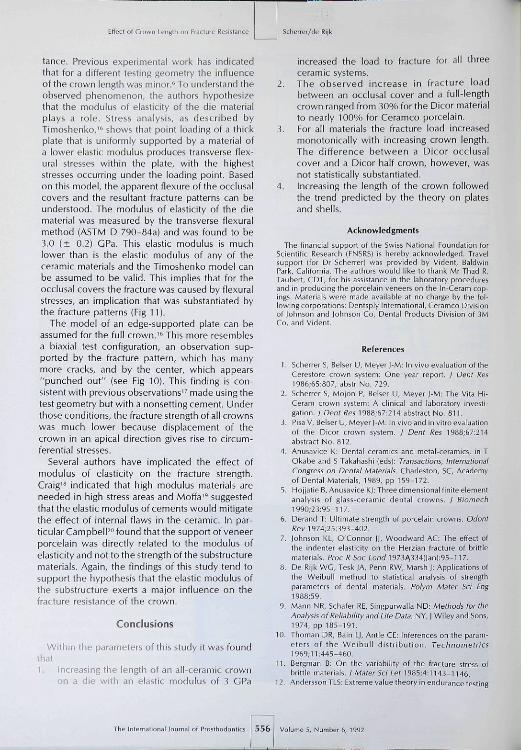
tance. Previous experimental work has indicatedthat for a different testing geometry the influenceof the crown length was minor.^ To understand theobserved phenomenon, the authors hypothesizethat the modulus of elasticity of the die materialplays a role. Stress analysis, as described byTimoshenko,'" shows that point loading of a thickplate that is uniformly supported by a material ofa lower eiastic modulus produces transverse flex-ural stresses within the plate, with the higheststresses occurring under the loading point. Basedon this model, the apparent flexure of the occlusalcovers and the resultant fracture patterns can beunderstood. The modulus of elasticity of the diematerial was measured by the transverse flexuralmethod (ASTM D 790-84a) and was found to be3.0 (± 0.2) GPa. This elastic modulus is muchlower than is the elastic modulus of any of theceramic materials and the Timoshenko model canbe assumed to be valid. This implies that for theocclusal covers the fracture was caused by flexuralstresses, an implication that was substantiated bythe fracture patterns (Fig 11).
The model of an edge-supported plate can beassumed for the full crown."' This more resemblesa biaxial test configuration, an observation sup-ported by the fracture pattern, which has manymore cracks, and by the center, which appears"punched out" (see Fig 10). This finding is con-sistent with previous observations" made using thetest geometry but with a nonsetting cement. Underthose conditions, the fracture strength of al! crownswas much lower because displacement of thecrown in an apical direction gives rise to circum-ferential stresses.
Several authors have implicated the effect ofmodulus of elasticity on the fracture strength.Craig'^ indicated that high modulus materials areneeded in high stress areas and Moffa''' suggestedthat the elastic modulus of cements would mitigatethe effect of internal flaws in the ceramic. In par-ticular Campbell" found that the support of veneerporcelain was directly related to the modulus ofelasticity and not to the strength ofthe substructurematerials. Again, the findings of this study tend tosupport the hypothesis that the elastic modulus ofthe substructure exerts a major influence on thefracture resistance of the crown.
Conclusions
Within the parameters of this study it was foundthat1. Increasing the length of an all-ceramic crown
on a die with an elastic modulus of 3 GPa
Scherier/rie
increased the load to fracture for all tbreeceramic systems.
2. The observed increase in fracture loadbetween an occlusal cover and a full-lengthcrown ranged from 30% for the Dicor materialto nearly 100% for Ceramco porcelain.
3. For all materials the fracture load increasedmonotonically with increasing crown length.The difference between a Dicor occlusalcover and a Dicor half crown, however, wasnot statistically substantiated,
4. Increasing the length of the crown followedthe trend predicted by the theory on platesand shells.
Acknowledgments
The financial support of the Swiss National Foundation forScientific Research (FNSRS) is hereby acknowledged. Travelsupport (for Dr Scherrer) was provided by Vident, BaldwinPark, California. The authors would like to thank Mr Thad R.Taubert, CDT, for his assistance in the laboratory proceduresand in producing the porcelain veneers on the In-Ceram cop-ings. Materials were made available at no charge by the fol-lowing corporations: Dentsply International, Ceramco Divisionof lohnson and lohnson Co, Dental Products Division of 3MCo, and Vident.
References
1. Scherrer S, Belser U, Meyer j-M: In vivo evaluation oftheCereitore crown system: One year report. / Dent Res1966;&5:ßO7, abstr No. 729.
2. Scherrer S, Mojon P, Belser U, Meyer J-M: The Vila hli-Ceram crown system: A clinical and laboratory investi-gation. I Dent Res 1938:67:214 abstract No. 811.
3. Pisa V, Belser U, Meyer J-M: In vivo and in vitro evaluationof the Dicor crown system. / Den! Res 19B8;67:214abstract No. 812.
4. Anusavice K: Dental ceramics and metal-ceramics, in TOkabe and S Takahashi |eds|: Transactions, InternationalCongress on Dental Materials. Charieston, SC, Academyof Dental Materials, 1989, pp 159-172.
5. i-lo|jatie B,AnusaviceKJ: Three dimensional finite elementanalysis of glass-ceiamic dental crowns. J Biomech1990:33:95-117.
6. Derand T: Ultimate strength of porcelain crowns. OdontRev 1974;25:393-402.
7. lohnson KL, O'Connor JI, Woodward AC: The effect ofthe indenter elasticity on the hierzian fracture of brittlematerials. Proc R Soc io/id 1973)a|334¡Jan¡:95-n7.
B. De Rijk WG, Tesk JA, Penn RW, Marsh J: Applications ofthe Weibuil method to statistical analysis of strengthparameters of dental materials. Polym Mater 5ci Eng1988:59.
9. Mann NR. Schäfer RE, Singpurwalla ND: Methods for theAnalysis of Reliability and Life Data. NV, | Wiley and Sons,1974, pp 185-191.
10. Thoman DR, Bain L|, Antle CE: Inferences on the param-eters of the Weibuil distribution. Technometrics1969:11:445-460.
11. Bergman B: On ihe variability of the fracture stress ofbrittle materials. J Mater Sei Let 1985;4:1143-1146.
1 2. Andersson TLS: Extreme value theory in endurance testing
of Prosthoduntil 5 5 6 Volume 5. Number 6. 1992

Se h errer/de Rijk
of bsll and roller bearings, m Eggwertz S, Lind NC (eds);Probabilistic Methods in the Mechanics of Solids andStructures. Berlin, Springer-Verlag, 1985, pp 41-S3,
13, Dehoff PH, Anusavice KJ: Tempering stresses in feld-spathic porcelains, / Dent Res 1989;69:1 34-138,
14, Derand T: Effect of variation of the shape of the core onstresses in a loaded model of a porcelain crown, OdontRev)'1974;25;n-26,
15, Slarling LB: Transfer molded "all ceramic crowns"; TheCerestore system, in O'Brien W], Craig RG (eds]: CeramicEngineering and Science Proceedings. American CeramicSociety, 1985, pp 41-56,
Effecl of Crown Length on Fracture Résistanc
16. Timoshenko S, Vi/oinowski-Krieger S: Theory of Plates andShells, ed 2, New York, McGraw-Hill Book Co Inc 1959pp 259-281,
17, ScherrerS, de Rijk WG: Factors in the fracture resisfaneeof posterior all-ceramic crowns, / Dent Res 199l;70;434,abstract No, 1342.
16, Craig RC, Peyton FA: Elastic and mechanical propertiesof human dentin. / DenI Res 195B;37;71Ü-713,
t9, Moffa IP; Porceiain materials, Adv Dent Res 19ee;2;3-6,20, Campbeil SD: A comparative strength study of metal
ceramic and all-ceramic esthetic materials: Modulus ofrupture. I Prosthet Denf 1989;62:476-479.
Literature Abstracts.
A Clinical Examination of Ceramic (Cerec) Inlays
Several new materials and teofiniques for making ceramic inlays have been introduced onto themarket, Tfie Cerec system (Siemens AG, Bensfieim, Germany) was introduced in 1988, The aim of thepresent study was to evaiuate the quality of Cerec inlays made by dentists in gênerai practice, A totaiOf 205 Cerec ceramic inlays placed by 8 dentists in 72 patients were examined independently by 3calibrated evaluators 12 to 34 months after insertion, using the criteria of the California DentalAssociation (CDA) and certain periodontal variables. Neither proximal plaque nor bleeding on probingwas seen more often in relationship to Cerec inlay surfaces than to homologous control surfaces.Excellent CDA ratings for ooior were obtained for 57% of the inlays. The corresponding exceilentratings were for surface, 26%; for anatomic form, 55%: and for margin integrity, 83%, Obviousdifferences were seen among the participating dentists with regard to the clinicai quality ot Cerecinlays. Even tfiough the present study covered only a limited time of 12 to 24 months, the authorsconcluded that the system may function vi;ell in the hands of a skilled dentist. However, the authorspoint out that the long-term performance of the Cerec technique, at present, cannot be predicted.
Sjögren G, Bergman M, Molin M. Bessing C, Acta Odontol Scani}^992•.50.•\^^-^^B. References. 17. Reprints: QöranSjögren, Department of Dental Materials and T80hno[ogy, Faculty of Odontology, University of Llniea, S-901 87 Umea,Sweaeri-íííc/ja/-íy f l . Seals, Jr, DDS. MEd, MS. Depariment ot ProsthoOontics, The University ot Texas HeaithScience Center at San Antonio, Texas
Keratinization of Palatal Mucosa Beneath Metal-BasedRemovable Partial Dentures
Tissue response to denture wearing, including altered epithelial structure and functicnai integrity, maybe associated with the development of mucosal inflammation and underlying bone résorption. Thisinvestigation addressed these issues by assessing the use of an adhesive tape, celi-sampiingtechnique, as opposed to the more common celi-smeaf technique, to sampie oral epitheiium and byusing the adhesive tape technique to examine heratinization of the palatai mucosa beneath tooth-supported removable partiai dentures. Results suggested that, as with complete dentures, tooth-supported, cobait-chromium-based removable partiai dentures cause a significant shift in thekeratinization pattern of the supporting epithelium from one dominated by orthokeratinizafion to one inwfiich half the surface cells are parakeratinized, Iti spite of the reduced tissue loading that resultafrom the elements of tooth support and retention inherent in metal-based removable partialdentures, the underlying tissue appears likely lo undergo structural changes that may affect theirfurtctional integrity, as well as the long-term success of the prosthesis.
HarJnasuta S, Howlett JA, J DenM992:20i152-15S. References: 19, Heprints; Dr J.A, Howlett, Department ofProsthetic Dentistry, Institute ot Dental Surgery, University of London, Gray's Inn Road, London WC1X 8LD, UnitedKingdom-Dawö ft Cagna. DMD, Depanment of Prosthodontics, The University of Texas Healttt Science Center atSan Antonio, San Antonio, Texas
Volume 5, Number 6, 1992 557 The Internstionai Journal of Prosthodontics