The Circulatory System The circulatory system moves nutrients, gases, and wastes to and from cells...
46
The Circulatory System • The circulatory system moves nutrients, gases, and wastes to and from cells • Single-celled organisms obtain oxygen and nutrients directly across the surface of the cell • Multi-cellular organisms require methods for transporting materials to and from cells which are far removed from the external environment
-
date post
20-Dec-2015 -
Category
Documents
-
view
219 -
download
1
Transcript of The Circulatory System The circulatory system moves nutrients, gases, and wastes to and from cells...

The Circulatory System
• The circulatory system moves nutrients, gases, and wastes to and from cells
• Single-celled organisms obtain oxygen and nutrients directly across the surface of the cell
• Multi-cellular organisms require methods for transporting materials to and from cells which are far removed from the external environment

Invertebrate Circulatory Systems
• Sponges and most Cnidarians use water from the environment as a circulatory fluid
• Pseudocoelomate invertebrates (e.g., roundworms) use the fluids of the body cavity for circulation (=gastrovascular cavity)
• Larger animals have tissues that are several cells thick, such that many cells are too far away from the body surface or digestive cavity to exchange materials with the environment


• In Cnidarians, respiration occurs via diffusion directly through their tissues
• A gastrovascular cavity is used for digestion and transport

Invertebrate Circulatory Systems

Invertebrate Circulatory Systems
• Open circulatory system – No distinction between blood and the interstitial fluid; hemolymph– Most Molluscs and Arthropods – A tubular muscle, or heart, pumps hemolymph
through a network of channels and body cavities, before draining back to the central cavity
– Hemolymph directly
bathes the internal
organs

The Circulatory System
• Closed circulatory system – The circulating fluid, or blood, is enclosed within blood vessels that transport it away from – and back to – the heart– All vertebrates, cephalopod molluscs, and
annelids– Consists of
heart, bloodvessels andblood

Vertebrate Circulatory Systems
• Surface area – as the physiological complexity of animals increased, so too did the need for more surface area to transport and exchange nutrients and oxygen (and remove CO2 and metabolic wastes)
• Adaptations have allowed the development of large body size and locomotion

Vertebrate Circulatory Systems
http://www.amnh.org/exhibitions/permanent/ocean/images/03_oceanlife/features/06_whales/whale.jpg
http://en.wikipedia.org/wiki/File:Bar-headed_Goose_-_St_James%27s_Park,_London_-_Nov_2006.jpg
Check out: http://www.youtube.com/watch?v=yd_w3biT3TU

Vertebrate Circulatory Systems
• Fish evolved a 2-chambered heart to increase efficiency of gas exchange in gills
First to contract
1.
2.
3. 4.

Vertebrate Circulatory Systems
• The evolution of lungs in amphibians involved a major change in the pattern of circulation – a second pumping circuit
• After blood is pumped from the heart through pulmonary arteries to the lungs, it is returned to the heart via pulmonary veins– Double circulation – gives boost to speed/pressure
at which blood is transported to the rest of the body– Pulmonary circulation moves blood between the
heart and lungs; Systemic circulation moves blood between the heart and the rest of the body

Heart
1. Deoxygenated blood from body is pumped through the heart and to the lungs
2. Oxygenated blood is returned to heart to be pumped to rest of the body
1b.
1a.
2a.
2b.

Vertebrate Circulatory Systems
• Amphibians and most reptiles have a 3-chambered heart– 2 atria and 1 ventricle– Some mixing of oxygenated and
deoxygenated blood• Right atrium receives deoxygenated blood from the
systemic circulation, and the left atrium receives oxygenated blood (pulmonary) from the lungs – no mixing in the atria
• Separation of pulmonary and systemic incomplete in ventricle


Amphibian and Reptilian Circulation
• Amphibians obtain additional oxygen via diffusion through their (moist) skin
• Reptiles have a septum that partially subdivides the ventricle– Separation is complete in Crocodilians
(septum divides ventricle into 2 separate ventricles; a 4-chambered heart)
– Further reduces mixing of blood in the heart– Atria receive blood returning to the heart– Ventricles pump blood out of the heart

Mammalian and Avian (and Crocodilian) Circulatory Systems
• Oxygenated and deoxygenated blood does not mix; completely separated– 4-chambered heart: 2 atria, 2 ventricles– Right atrium receives deoxygenated blood from
the body and delivers it to the right ventricle which pumps it to the lungs (pulmonary); the left atrium receives oxygenated blood from the lungs and delivers it to the left ventricle, which pumps it to the rest of the body (systemic)
RA RV LUNGS LA LV REST OF BODYpulmonary systemic

1a.
1b.2a.
2b.
1. Deoxygenated blood from body is pumped through the heart and to lungs2. Oxygenated blood is returned to heart to be pumped to rest of the body

Mammalian and Avian (and Crocodilian) Circulatory Systems
• The sinus venosus is present, but reduced, in amphibians and (further reduced) in reptiles
• In mammals and birds, the sinus venosus is present only as a remnant of tissue in the wall of the right atrium = sinoatrial (SA) node– Pacemaker, site where the impulses that
initiate the heartbeat originate

The Four-Chambered Heart
• The heart functions as a two-cycle pump– Both atria fill with blood and simultaneously
contract, emptying the blood into the ventricles (atrial contraction)
– Both ventricles also contract at the same time, pushing blood into the pulmonary and systemic circulations (ventral contraction)
RA RV LUNGS LA LV REST OF BODYpulmonary systemic

The Four-Chambered Heart
• The cardiac cycle includes the atrial and ventricular contraction, and the resting period between these two
• Atrioventricular (AV) valves maintain unidirectional flow between the atria and the ventricles: tricuspid (right) and bicuspid (left)
• Semilunar valves maintain unidirectional flow out of the ventricles to the arterial systems– Pulmonary valve located at exit of the right ventricle– Aortic valve located at the exit of the left ventricle– Valves open and close as the heart goes through its
cycle

The Four-Chambered Heart: Diastole (resting) phase
• Blood returns to the resting heart through veins that empty into the right and left atria
• As blood fills the atria and pressure rises, the AV valves open and blood flows into the ventricles
• The ventricles become ~80% full from this process; contraction of the atria fills the remaining 20%
• Ventricles are relaxed = diastole phase

The Four-Chambered Heart: Systole (ventricle contraction) phase
• Following a slight delay from the diastole phase, the ventricles contract = systole phase
• Contraction of the ventricles increases the pressure within each chamber, causing the AV valves to forcefully close; this forces the semilunar valves open and blood flows into the arterial systems
• As the ventricles relax, closing of the semilunar valves prevents backflow

Heart Contraction and Blood Flow
• http://www.nhlbi.nih.gov/health/dci/Diseases/hhw/hhw_pumping.html

The Four-Chambered Heart and the Blood Vessels
• The aorta (and all its branches) are systemic arteries, carrying oxygen-rich blood from the left ventricle to all parts of the body
• Right and left pulmonary arteries deliver oxygen-depleted blood from the right ventricle to the right and left lungs
• Pulmonary veins return oxygenated blood from the lungs to the left atrium of the heart

The Four-Chambered Heart and the Blood Vessels
• Arteries – carry blood away from the heart; branch into arterioles
• Capillaries – where materials (O2, CO2, nutrients, metabolic wastes) are exchanged
• Veins – carry deoxygenated blood back towards the heart

The Four-Chambered Heart and the Blood Vessels
• Coronary arteries are the first branches off the aorta and supply the heart muscle with oxygenated blood
• Blood from the body’s organs (now low in O2) returns to the heart via systemic veins, which empty into 2 major veins– Superior vena cava – drains the upper body– Inferior vena cava – drains the lower body– Both empty into the right atrium
Measuring Blood Pressure
• As the ventricles contract, they generate tremendous pressure, which is transferred through the arteries once the AV valve opens
• Arteries contain large amounts of elastin, an elastic protein which allows for dilation/stretching and rebound
• A pulse results from changes in pressure as arteries expand and contract with blood flow
Measuring Blood Pressure
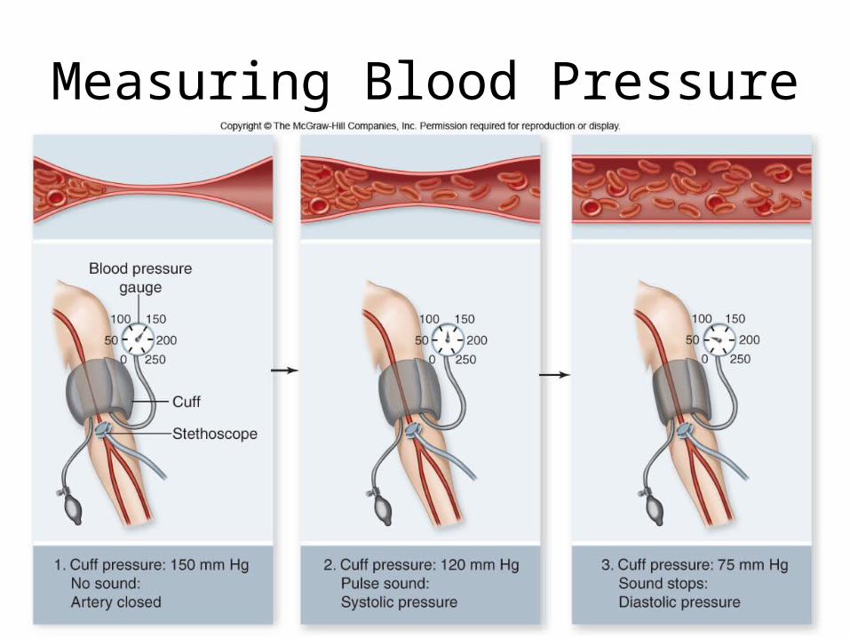
• Blood pressure is a general indicator of cardiovascular health
• Arterial blood pressure can be measured with a sphygmomanometer at the brachial artery on the inside part of the arm
• A tightened cuff stops the flow of blood to the lower part of the arm; As the cuff is loosened, blood begins pulsating through the artery = systolic pressure; ventricles are contracting
• As the cuff is loosened further, the vessel is no longer distorted and the pulsing stops = diastolic pressure; ventricles are relaxed
Measuring Blood Pressure

Measuring Blood Pressure
• Systolic pressure is the peak pressure at which ventricles are contracting
• Diastolic pressure is the minimum pressure between heartbeats at which the ventricles are relaxed
• Written as a ratio of systolic over diastolic
• Typical blood pressure is 120/75; >150 systolic or >90 diastolic = hypertension

Contraction of Cardiac Muscle
• Each cell in heart produces an action potential (electrical signal that is stimulus for cell to contract); fairly long in duration; 250 milliseconds from start to finish
• Can’t start another action potential until the other is completely finished
• The sinoatrial (SA) node located in the wall of the right atrium acts as a pacemaker by producing spontaneous action potentials

Contraction of Cardiac Muscle
• Action potentials are generated by a constant leakage of Na+ ions into the cell that depolarize the membrane
• When the threshold is reached, the action potential occurs
• Allows heart muscle to carry signal over distance; conducted rapidly over both ventricles by a network of fibers, including Purkinje fibers, which spread electrical activity to rest of heart

Contraction of Cardiac Muscle
• An electrocardiogram (ECG or EKG) records the electrical activity of the heart
• Illustrates via electrodes how the cells of the heart depolarize and repolarize during the cardiac cycle (action potentials)– Depolarization causes contraction of the heart– Repolarization causes relaxation– Depolarization of the atria produces first peak;
depolarization of ventricles produce second, larger peak
– Repolarization produces third peak

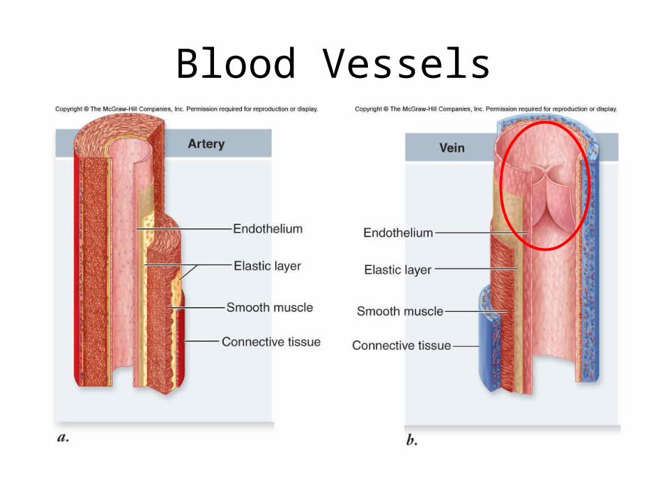
Blood Vessels
• The walls of arteries and veins have three layers:– Epithelium (innermost layer)– Smooth muscle with elastin fibers– Connective tissue (outermost layer)
• Arteries have thicker walls than veins; arterioles have less elastin than arteries
• Capillaries only have the inner epithelium layer (site of gas/nutrient/waste exchange)
Blood Vessels

Blood Vessels: Capillaries
• Blood flows slower through capillaries because of larger total cross-section– Enables materials to be exchanged– By the time blood reaches the end of the
capillary, it releases some of its oxygen and nutrients and picks up CO2 and waste products

Blood Vessels: Veins
• Less muscle is needed in veins because the pressure in veins is only ~1/10th of that in the arteries
• Venous pressure alone is not sufficient to return blood to the heart
• Thus, skeletal muscles surrounding the veins contract to move blood by squeezing the veins; the venous pump

Blood Vessels: Veins
• Internal valves in veins (venous valves) ensure that blood continues to heart; operate as one-way swinging doors
• Skeletal muscles on outside of vein contracts, pushes against vein, causing blood to flow towards heart; this opens one-way valves up, pressure is released and cannot return through valves (one-way transport to heart)

Blood Vessels: Veins

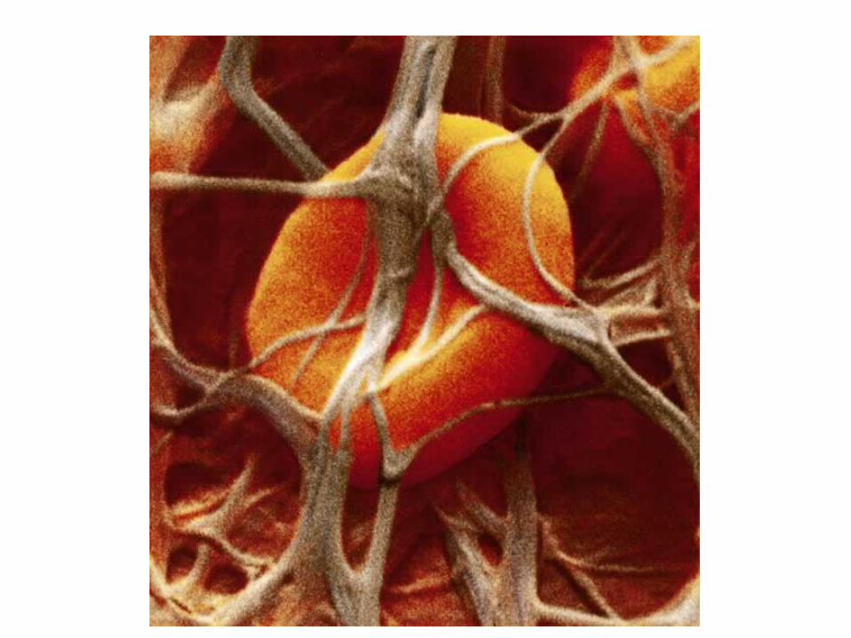
Components of Blood
• Blood serves to transport, regulate, and protect
• Blood is composed of a fluid-matrix known as plasma, within which reside different cells and other ‘elements’ – Blood cells (red & white) and platelets– Ions, proteins– Nutrients, wastes and hormones

Components of Blood
• Red blood cells, Erythrocytes– Most numerous: 5 million/mL
– Transport O2 and CO2 (hemoglobin in vertebrates)
– Mammalian erythrocytes lack nuclei

Components of Blood
• White blood cells; Leukocytes– Fewer in number; 1-2 for every 1000
erythrocytes– Larger in size, and have nuclei– Not confined to blood as erythrocytes are; can
migrate out of blood into surrounding interstitial fluid or into the lymphatic system – where your body fights infection
– Function in body’s defense

Components of Blood• Platelets; Thrombocytes
– Cell fragments that pinch off from larger cells in the bone marrow
– Following injury to a blood vessel, platelets release clotting factors (proteins) into the blood
– When platelets contact collagen, they stick to it ; results in release of several factors which activate other platelets
• Conversion of fibrogen (soluable) to fibrin (insoluable); fibrin threads cross-link, connecting platelets and trapping other cells in network = blood clot

4. Threads of fibrin trap erythrocytes and form a clot.
1. Vessel is damaged, exposing surrounding tissue to blood.
5. Once tissue damage is healed, the clot is dissolved.
3. Cascade of enzymatic reactions is triggered by platelets, plasma factors, and damaged tissue.
2. Platelets adhere and become sticky, forming a plug.
Prothrombin
Thrombin
Thrombin
Fibrinogen
Fibrin
Formation of Blood Clot

Blood Types
• Three alleles denote presence of specific glycoproteins on the surface of blood cells– Type A, B, O (and AB)– Each contains antibodies of other types
• Rh factor – presence or absence of Rh protein– Positive (have) or negative (do not have)– Negatives will form antibodies against Rh
blood upon exposure