The biology behind the counts: tooth development related ...
20
Stewart BE and Stewart REA (2014) The biology behind the counts: tooth development related to age estimation in beluga (Delphinapterus leucas). NAMMCO Scientific Publications. doi: http://dx.doi.org/10.7557/3.3195 Creative Commons License The biology behind the counts: tooth development related to age estimation in beluga (Delphinapterus leucas) Barbara E. Stewart 1 and Robert E.A. Stewart 2,* 1 )Sila Consultants, 1218 Marchand Road, Howden, Manitoba, R5A 1J6, Canada 2 )Department of Fisheries and Oceans, Freshwater Institute, 501 University Crescent, Winnipeg, Manitoba, R3T 2N6, Canada * Current Address: Sila Consultants, 1218 Marchand Road, Howden, Manitoba, R5A 1J6, Canada ABSTRACT The widely accepted method of determining ages of beluga is to count dentine growth layer groups (GLGs) in median, longitudinal sections of a tooth. It is essential to understand how these growth layers form and to consider developmental factors that can confound their enumeration to be able to provide meaningful age estimates. Here we provide information on, and illustrate, the developmental biology of beluga teeth as it relates to interpreting GLGs. Key factors are: evaluating the presence and occlusal wear of fetal dentine; interpreting early- formed diagnostic features such as the neonatal line; assessing the last-formed growth layer adjacent to the pulp cavity; identifying the presence of nodes at the dentine-cementum interface to assist in counting GLGs; and recognizing pulp stones and accessory lines in the dentine which may hinder the age estimate process. INTRODUCTION Age estimates are of fundamental importance to management plans and scientific studies (Johnston et al. 1987, Heide-Jørgensen et al. 1994). Belugas are toothed whales (Family Monodontidae) for which age estimates are derived by counting growth layer groups (GLGs, IWC 1980) in median, longitudinal sections of teeth (NAMMCO in review). The accuracy and precision of age estimates are enhanced by an understanding of the biological development of beluga teeth and the consistent interpretation of GLGs seen in optimally prepared thin sections (Stewart 2012, NAMMCO in review). Here we describe and illustrate GLG development in teeth of beluga and review factors that affect accuracy and precision in GLG counts. Important diagnostic features of teeth used in age estimation are presented. For interpretive purposes, it is considered that beluga deposit one GLG per year (Stewart et al. 2006); there is no a priori reason why divergence from the typical mammalian pattern should be expected (Sergeant 1981).
Transcript of The biology behind the counts: tooth development related ...
Stewart BE and Stewart REA (2014) The biology behind the counts: tooth development related to age estimation in beluga (Delphinapterus leucas). NAMMCO Scientific Publications. doi: http://dx.doi.org/10.7557/3.3195
Creative Commons License
The biology behind the counts: tooth
development related to age estimation in
beluga (Delphinapterus leucas) Barbara E. Stewart1 and Robert E.A. Stewart2,*
1)Sila Consultants, 1218 Marchand Road, Howden, Manitoba, R5A 1J6, Canada 2)Department of Fisheries and Oceans, Freshwater Institute, 501 University Crescent,
Winnipeg, Manitoba, R3T 2N6, Canada * Current Address: Sila Consultants, 1218 Marchand Road, Howden, Manitoba, R5A 1J6,
Canada
ABSTRACT
The widely accepted method of determining ages of beluga is to count dentine growth layer
groups (GLGs) in median, longitudinal sections of a tooth. It is essential to understand how
these growth layers form and to consider developmental factors that can confound their
enumeration to be able to provide meaningful age estimates. Here we provide information
on, and illustrate, the developmental biology of beluga teeth as it relates to interpreting GLGs.
Key factors are: evaluating the presence and occlusal wear of fetal dentine; interpreting early-
formed diagnostic features such as the neonatal line; assessing the last-formed growth layer
adjacent to the pulp cavity; identifying the presence of nodes at the dentine-cementum
interface to assist in counting GLGs; and recognizing pulp stones and accessory lines in the
dentine which may hinder the age estimate process.
INTRODUCTION
Age estimates are of fundamental importance to management plans and
scientific studies (Johnston et al. 1987, Heide-Jørgensen et al. 1994). Belugas
are toothed whales (Family Monodontidae) for which age estimates are
derived by counting growth layer groups (GLGs, IWC 1980) in median,
longitudinal sections of teeth (NAMMCO in review). The accuracy and
precision of age estimates are enhanced by an understanding of the biological
development of beluga teeth and the consistent interpretation of GLGs seen
in optimally prepared thin sections (Stewart 2012, NAMMCO in review).
Here we describe and illustrate GLG development in teeth of beluga and
review factors that affect accuracy and precision in GLG counts. Important
diagnostic features of teeth used in age estimation are presented. For
interpretive purposes, it is considered that beluga deposit one GLG per year
(Stewart et al. 2006); there is no a priori reason why divergence from the
typical mammalian pattern should be expected (Sergeant 1981).

Stewart and Stewart (2014)
NAMMCO Scientific Publications, Volume 10 2
MATERIALS AND METHODS
Beluga jaws and teeth used to inform this discussion comprise over 2500
animals sampled as part of ongoing research conducted by the Department of
Fisheries and Oceans (DFO), Winnipeg. The one fetal beluga mandible we
have been able to obtain (HI-08-06 fetus) was a male sampled at Hendrickson
Island, Mackenzie Delta on July 5, 2008. The whale was 160 cm long. The
mandibles were x-rayed, prior to tooth extraction, to examine unerupted teeth
using the Eureka digital x-ray program Image Pilot (Peak Kilovoltage: 50;
milliampere second: 2.5; measurement: 3 cm). The right mandible had been
dissected in the field and the first, second, and fifth teeth were missing when
it arrived in the lab. The left mandible remained intact and contained seven
teeth.
All teeth extracted from mandibles, including the fetal jaw, were processed
for age estimates following Stewart (2012). Briefly, mandibles were
designated as left and right (MNL and MNR) and the teeth numbered
sequentially from the mandibular symphysis. Teeth selected for age estimates
(MNR 2 and 5 usually) were mounted on wooden blocks using hot glue, and
then longitudinal thin sections (0.3 mm) were cut from the middle of the tooth,
stored in 70% ETOH, and kept wet while being viewed microscopically with
transmitted light. In these conditions one translucent (light) growth layer plus
one opaque (dark) growth layer comprise one GLG and one GLG is
interpreted as a one year cycle (Stewart et al. 2006). All photomicrographs
presented here are of wet thin sections, viewed with transmitted light, on a
Nikon SMZ800 dissecting microscope at 10-63 power. While photographs
are important illustrative tools, these images in particular and photographs in
general are not reliable for final age estimation (NAMMCO in review). The
best sections for age determination are high quality median sections (Stewart
2012, NAMMCO in review). Some sections that are less than optimal are
presented here to display specific characteristics although they would not be
used for age determination.
GENERAL DESCRIPTION
Gross morphology
Beluga teeth are single rooted, conical, and non-cusped. The homodont
dentition occurs in the maxilla and mandible. A maximum of 40 teeth is
present although the number of teeth in each jaw may vary (Doan and Douglas
1953, Kleinenberg et al. 1969). Beluga teeth have an indeterminate growth
form (IWC 1980, Stewart 2012); as GLGs are deposited, tooth diameter
increases and the tooth becomes longer, resulting in the eruption of the teeth
from the gingiva. Occlusal wear starts shortly thereafter (Ishiyama 1987).

Stewart and Stewart (2014)
NAMMCO Scientific Publications, Volume 10 3
The beluga has a unique status as a monophyodont (Uhen 2009) although
there is no a priori reason for this designation. To unequivocally establish the
presence/absence of deciduous dentition, x-ray analysis of fetal jaws is
required. The one fetal jaw we have been able to x-ray showed one set of
unerupted, permanent teeth in situ just prior to birth and no deciduous teeth
(Fig. 1). Stewart (2012) reported 3 of 2,707 jaws processed contained
resorbing, deciduous dentition (one tooth in each whale) suggesting
diphyodonty. The presence of deciduous teeth in some jaws established that
they do occur, but their postnatal incidence or persistence appears low. In
utero development and resorption of deciduous dentition is found in several
other marine mammals including walrus (Odobenus rosmarus divergens, Fay
1982), harp seal (Phoca groenlandica, Stewart and Stewart 1987), and ringed
seal (Phoca hispida, Stewart et al. 1998). Most beluga samples examined
came from Inuit subsistence harvests and rarely include winter samples when
it is likely fetuses are developing deciduous dentition (Stewart and Stewart
1989).
Fig. 1. X-ray of a beluga fetus mandible. HI-08-06 fetus. The
right mandible was dissected in the field; the first, second, and
fifth teeth (MNR-1, -2, and -5 respectively) are missing. The left
mandible is intact; 7 teeth are present in the tooth row. All the
teeth were embedded in the gingiva.

Stewart and Stewart (2014)
NAMMCO Scientific Publications, Volume 10 4
In terms of accurate age-estimation, it is important that the teeth used contain
a life-long record of GLG formation. Belugas are born with one full set of
unerupted dentition (Stewart 2012) and fetal dentine may be present in teeth
with a considerable number of GLGs e.g., over 60. It remains that no
systematic shedding of the teeth present at birth is evident. For the purpose of
age estimates, the teeth present at birth are considered to be the permanent
dentition.
Eruption of the teeth from the gingiva occurs postnatally (Brodie 1971), but
as the process is highly variable, it is not a good indicator of relative or
absolute age. For example, unerupted teeth were observed in beluga up to 6
years of age; partially erupted teeth were seen in 3 to 10 year olds; and fully
erupted teeth were evident in beluga as young as 3 years (Stewart 2012). It
has been reported that erupting teeth are tritubercular, with small auxiliary
cusps on each side of the anterior and posterior faces of the tooth (Douglas
1951, Kleinenberg et al. 1969). We have observed auxiliary cusps rarely in
unerupted teeth.
Teeth grow in diameter and length by the addition of cementum and dentine
derived from cementoblasts and odontoblasts respectively. Physiological
feedback is thought to influence the timing, structure, and thickness of
depositions leading to the formation of growth layers, but no definitive
mechanism has been identified (Klevezal 1996). At birth, the tooth is cone-
shaped and additional layers of dentine are added to the inside surface along
the pulp cavity (creating a “stack of cones”) while cementum layers are added
to the outer surface of the tooth that is still below the gingiva.
Opposing the natural tendency for an increase of tooth length and eruption
are occlusal forces. Occlusion occurs when opposing teeth from the maxilla
and mandible contact each other (Doan and Douglas 1953, Sergeant 1973)
and may result in significant erosion of the crown. The interplay between
tooth growth and occlusal wear of erupted teeth continues throughout a
beluga’s life. The angle of wear and extent of erosion is often highly variable
among teeth from an individual (Fig. 2). Also, occlusal wear is often, but not
always, faster in males than females and varies among beluga stocks (Heide-
Jørgensen et al. 1994).
Tooth size and shape vary with the position in the jaw (Fig. 2). Teeth near the
front of the jaw and in the middle of the tooth row tend to be straighter than
those at the rear. The teeth in the middle of the tooth row tend to be larger
than the others (Heide-Jørgensen et al. 1994).

Stewart and Stewart (2014)
NAMMCO Scientific Publications, Volume 10 5
Fig. 2. Variation in size and shape of beluga teeth from one jaw.
HI-09-01, MNR-1 through MNR-9 (left to right). Different
occlusal wear patterns in the distal surface are apparent also. The
extracted teeth show remnants of the periodontal ligament,
especially dark at the gingival line, indicating the portion of the
tooth contained within the alveolar socket.
Typically, there is no closure or obstruction of the pulp cavity at the root tip.
However, the root tips of very old beluga may have considerably reduced
diameters as the teeth taper deeply into the alveolar socket.
Internal structure
Enamel is present in unerupted teeth but is quickly eroded in erupted teeth
(Ishiyama 1987). The thin (7–10 μm) enamel is prismless, made of fine
crystal groups perpendicular to the surface and has incremental lines roughly
parallel to the surface (Ishiyama 1987). However, since enamel wears away
quickly, it plays no role in age estimation (Stewart 2012).
In beluga, GLGs are seen in both the dentine and cementum (Brodie 1969,
1971, Sergeant 1973, 1981) of thin-sectioned teeth. In median sections,
dentine growth layers appear as wide, chevron-shaped bands in the large, ˄-
shaped pulp cavity of young whales, and become thinner and flatter near the
root tips of older whales (Stewart 2012). Dentine is the preferred tissue for
age estimates as GLGs usually are better defined and the GLG counts equal
or exceed those of cementum, particularly in older beluga. Typically,
cementum deposits are asymmetric – the anterior face of the tooth has broader
cementum than the posterior face – and thinner than dentine growth layers.
Fetal dentine and fetal cementum develop in utero and can be seen in thin
sections of both near-term fetal (Fig. 3a) and newborns’ teeth (Fig. 3b). A
small enamel layer on the cusp tip was also present in the unsectioned teeth
(not seen in the photographs) but as it is not a diagnostic growth layer for age
estimates it will not be noted further. Fetal dentine may be recognized as a
distinctive homogeneous layer without inclusions at the cusp of the tooth.

Stewart and Stewart (2014)
NAMMCO Scientific Publications, Volume 10 6
Fig. 3. First-formed growth layers in a beluga tooth. (a) HI-08-06
fetus, MNR-3; (b) neonate AREP86-14, MNR-5. Both teeth have
fetal dentine (fd) and thin fetal cementum (fc). No postnatal
growth layers have been deposited in (b). The fetal cementum on
the anterior surface (left side of section) is slightly thicker and
higher on the cusp compared to the posterior face.
Fetal cementum has a characteristic graininess, is never as dense as
subsequent translucent growth layers, and extends to the level of the fetal
dentine terminus or slightly below. The fetal cementum is thickest distally
and tapers to the root tip. The presence of fetal material is the baseline for the
depositional record to follow.
The first dentine growth layer deposited after the fetal dentine is translucent,
extremely thin, and not always well defined. After this interface between the
fetal dentine and postnatal dentine, a thin neonatal line (IWC 1980), the first
opaque layer associated with birth, may be present. There is a time lag of
unknown duration between birth and the formation of the neonatal line in the
tooth. For teeth collected beyond this period, the neonatal line cannot always
be identified consistently and reliably. The neonatal line may be absent
a) b)

Stewart and Stewart (2014)
NAMMCO Scientific Publications, Volume 10 7
altogether or ambiguous (Fig. 4a,b), present as a short line extending from the
mid-line of the dentine before “fading” out (Fig. 4c), or appear as a longer
line that extends fully to the dentine-cementum interface (Fig. 4d). If the
neonatal line reaches the dentine-cementum interface, it does not extend the
length of the tooth significantly.
The variability of recognizing a neonatal line is not a significant problem with
respect to generating age estimates. Fetal dentine and fetal cementum can be
identified as uniquely textured tissues and, at their proximal ends, are close
to the neonatal line at the dentine-cementum interface. The presence of fetal
dentine and fetal cementum is sufficient to identify the prenatal portion of the
tooth in the absence of a distinct neonatal line. Conversely, if the prenatal
portion of the tooth is eroded, the neonatal line is likely to be lost too so a
systematic error of including the residual neonatal line as an age class growth
layer is unlikely.
A topographical marker may exist below the terminus of the fetal dentine,
along the dentine-cementum interface. A small hook or fold may occur as the
tooth extends in length by postnatal dentine deposits (Fig. 4b,d), similar to
that seen in ringed seals (Stewart et al. 1996). This fold often occurs
unilaterally, on the anterior face of the tooth which, in a tooth section, is
usually the convex outer edge. The hook/fold and the homogenous
appearance of fetal dentine, even with small portions being present, are useful
interpretive features. The neonatal line appears to reach the interface of the
dentine-cementum before the hook develops. Recognizing the fetal dentine
terminus and other features such as the fetal cementum terminus and the
neonatal line are diagnostic tools for locating the transition from the prenatal
to the postnatal portion of the tooth.
Postnatally, GLGs are deposited in an ongoing process in which accretionary
layers are laid down in the dentine and cementum. Cementum deposition may
lag dentine deposition (Fig. 4a). The interface of the fetal cementum and
postnatal cementum may appear as a short opaque band (equivalent to the
neonatal line in dentine) at a level above the fetal dentine terminus (Fig. 4b).
The first opaque cementum growth layer appears to contact the dentine-
cementum interface below the neonatal line and below the hook (Fig. 4b).
In summary, the significant features seen at the dentine-cementum interface
(from the distal to proximal end) of the first GLG usually appear in this order:
terminus of fetal dentine; terminus of fetal cementum (difficult to establish if
the layer is very thin); first thin translucent dentine growth layer, if obvious;
dentine neonatal line, if present; hook, if present; and the end of the first
opaque dentine growth layer which may be met by the end of the first opaque

Stewart and Stewart (2014)
NAMMCO Scientific Publications, Volume 10 8
Fig. 4. Variation in neonatal lines and early postnatal growth layers. a) ARSQ-xx-1007, MNR-
2; young of the year. The fetal dentine terminus (fdt) marks the transition between fetal and
postnatal dentine (PND). A faint neonatal line (NNL) may be developing. Fetal cementum (fc)
is apparent but no postnatal cementum can be identified. b) PA05-20, MNR-5; approaching 1
year old. The neonatal line is ambiguous or missing altogether. A hook or fold at the dentine-
cementum interface (D-C) represents the first extension of tooth length resulting from postnatal
dentine deposition. The first, opaque dentine growth layer (D-1) enters the interface below the
hook. The cementum (C) on the anterior face is most distinct: a short opaque band exists
laterally at the fetal cementum-postnatal cementum interface (fc-PNC), at a level above the
fetal dentine terminus (fdt); the first opaque cementum growth layer (C-1) tapers near the root
and meets D-1 at D-C.
a) b)
Stewart and Stewart (2014)
NAMMCO Scientific Publications, Volume 10 9
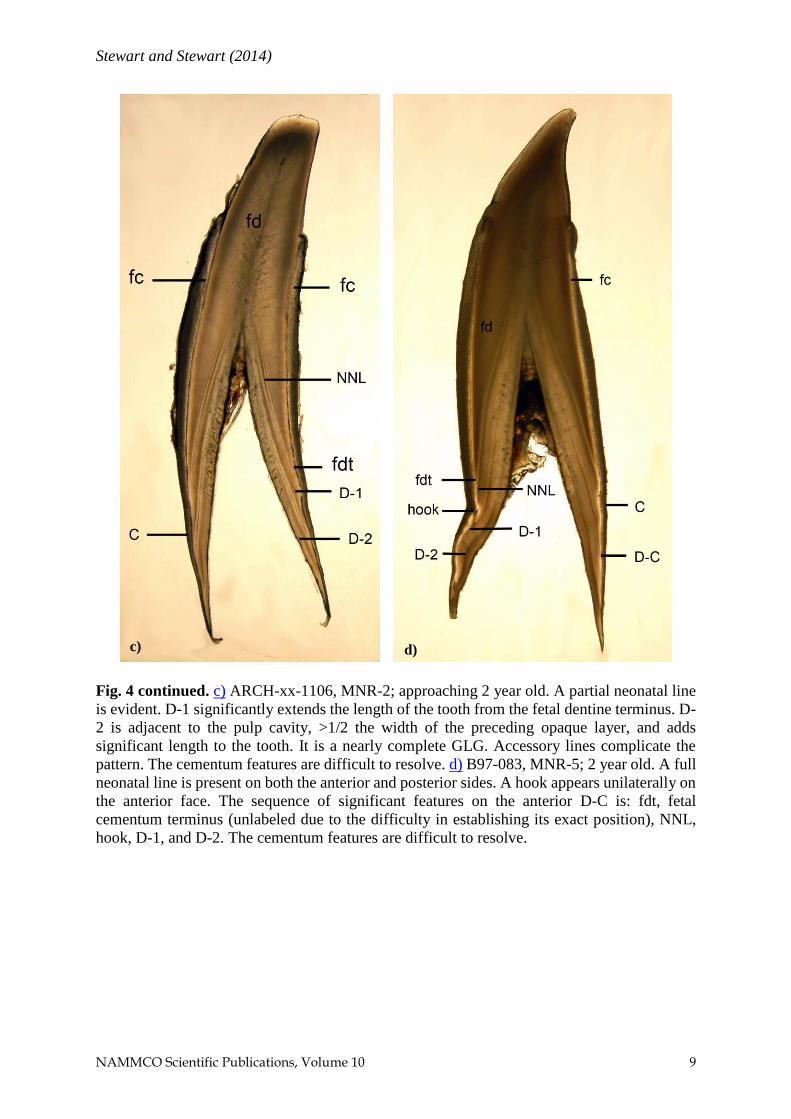
Fig. 4 continued. c) ARCH-xx-1106, MNR-2; approaching 2 year old. A partial neonatal line
is evident. D-1 significantly extends the length of the tooth from the fetal dentine terminus. D-
2 is adjacent to the pulp cavity, >1/2 the width of the preceding opaque layer, and adds
significant length to the tooth. It is a nearly complete GLG. Accessory lines complicate the
pattern. The cementum features are difficult to resolve. d) B97-083, MNR-5; 2 year old. A full
neonatal line is present on both the anterior and posterior sides. A hook appears unilaterally on
the anterior face. The sequence of significant features on the anterior D-C is: fdt, fetal
cementum terminus (unlabeled due to the difficulty in establishing its exact position), NNL,
hook, D-1, and D-2. The cementum features are difficult to resolve.
c) d)

Stewart and Stewart (2014)
NAMMCO Scientific Publications, Volume 10 10
cementum growth layer (variable) (Fig. 4d). The thickness of the early
postnatal dentine and postnatal cementum growth layers varies among beluga,
but the cementum is thinner. Despite beluga teeth being large generally, the
true median area which has the best clarity is still a very narrow zone. The
distinctiveness of the diagnostic features, in the prenatal to postnatal
transition area, likely is sensitive to the quality of the sections produced. The
variability in the neonatal line characteristics may reflect sectioning accuracy
over and above the intrinsic attributes of a beluga tooth.
As additional GLGs accumulate, other features appear. Nodes may occur at
the dentine-cementum interface (Fig. 5) and appear most often as thickened
areas where the opaque growth layers converge. If nodes are present in a
tooth, they typically develop after several GLGs and occur bilaterally.
Fig. 5. Nodes and pulp stones in the root tip area. ARRB01-1116,
MNR-5. Nodes are present at the dentine-cementum interface on
both sides of the section. Pulp stones obscure the GLG sequence
in the center portion of the dentine but a count can still be obtained
using the nodes.

Stewart and Stewart (2014)
NAMMCO Scientific Publications, Volume 10 11
Inclusions or pulp stones (Fig. 5) appear in a variety of shapes such as whorls,
circles, or tear-drops (Figs. 5 and 6) in the dentine, but not cementum. They
are seen in a number of odontocetes and form when odontoblasts dislodge
and become embedded in the developing dentine matrix (Benjamins 1999).
The frequency of occurrence of pulp stones increases with age but does not
correlate with either gender or stock (Benjamins 1999). Pulp stones may
distort the growth layers making discrimination of GLGs difficult.
COUNTING GLGS
The technical preparation of high quality, median longitudinal sections is
paramount to the proper assessment of age (Hohn and Fernandez 1999, Hohn
2009), otherwise both accuracy and precision may be affected. The clearest
growth layers and the maximum GLG counts are found in thin sections from
the median zone of the tooth. Sections that are offset even slightly from the
middle may not reflect the proper configuration of the cusp, possibly missing
fetal dentine or the growth layer adjacent to the pulp cavity (Fig. 6), and
distortion of GLGs may result in underestimates of age. For example, a
section that is off center either due to poor technique or unavoidable curvature
in the tooth will slice the cone-shaped GLG obliquely, not at right angles to
its circumference (Fig. 6a). Problematically, even in good median sections,
the last growth layer formed may differ among teeth of an individual. A broad
opaque growth layer may appear adjacent to the pulp cavity in one tooth and
a thin translucent growth layer may be seen adjacent to the pulp cavity in an
alternate tooth, so the age classes assigned would differ by one GLG if only
completed GLGs were recorded (Stewart 2012).
Beluga age estimates are based on identifying one translucent and one opaque
growth layer per GLG; one GLG cycle is deposited annually (Stewart et al.
2006). As mentioned previously, the first translucent dentine growth layer
adjacent to the fetal dentine is often difficult to identify. Readers typically cue
on and count the broader, opaque (dark) growth layers which end a GLG. For
the purposes of estimating age, the neonatal line (which is embedded in the
first opaque dentine growth layer) is not included individually in the GLG
count (birth = 0 age class; Hohn, 2009).
To determine the age of a beluga, one must consider not only the number of
GLGs counted but how the opposing factors of growth and wear, and the
presence of disruptive factors, affect the age estimate. The first step in
assessing a tooth section is to determine if fetal dentine is present, either in
its entirety or as a partial layer. Teeth selected for age estimates will have a
full complement of GLGs deposited in a beluga’s lifetime unless occlusal

Stewart and Stewart (2014)
NAMMCO Scientific Publications, Volume 10 12
Fig. 6. Comparison of an off-set section and a median section of a tooth containing pulp
stones. It helps interpretation to remember the section is a slice through the wall of a cone,
and GLGs in a section passing through the vertex of each will appear ˄-shaped, while
GLGs in a parallel section through any other plane (i.e., either side of the midline of the
tooth) will appear more ∩-shaped. a) AREP84-19, MNL-5; off-center section. In this off-
center section, the growth layers at the cusp are indistinct because they are ∩-shaped and
opaque dentine underlies translucent dentine. In addition, fetal dentine is absent, the root
tip is challenging to interpret, and the last growth layer deposited cannot be determined.
This section is not suitable for generating an age estimate. b) AREP84-19, MNL-5
alternate; median section. In this median section, the cone was sectioned near its vertex and
most of the opaque dentine lies at right angles to the viewed surface. The fetal dentine is
present and a translucent growth layer is apparent adjacent to some portions of the pulp
cavity. The growth layer pattern is challenging and, while nodes assist counting GLGs, the
age estimate is a minimum because some portions of the section cannot be read.
a) b)

Stewart and Stewart (2014)
NAMMCO Scientific Publications, Volume 10 13
wear has eroded the apical cusp below the level of fetal dentine. As occlusal
wear progresses (Fig. 7), usually the tip and then the wide, center portion of
the fetal dentine are worn away. Eventually, fetal dentine appears as two
separated bands laterally on the section, then only a small remnant remains as
a short trailing end next to the dentine-cementum interface at the cusp.
Finally, fetal dentine may be lost from a tooth altogether. Diagnostic features
such as the fetal dentine terminus, fetal cementum terminus, neonatal line,
and a hook are useful in identifying the prenatal to postnatal portion of a tooth.
Once fetal dentine is eroded totally, age estimates of a beluga must be
considered as a minimum age estimate. No correction factors for this loss of
accuracy are available currently.
The second step is to assess the growth layer adjacent to the pulp cavity (Figs.
5, 6, 8, 9). If a translucent growth layer is adjacent to the pulp cavity, the last
opaque layer identified finished a complete GLG cycle. If an opaque growth
layer is adjacent to the pulp cavity, then it is useful to assess whether it is <
½ or > ½ the length or width of the preceding opaque growth layer, to estimate
the proportion of the last GLG which has been deposited (Fig. 9).
The final step in assessing the tooth, concurrent with counting the GLGs, is
to evaluate areas which may be unreadable. If some GLGs cannot be counted,
then the final age estimate will be a minimum age only. Several factors may
limit the ability to count GLGs especially if the intrinsic quality of a tooth
section is poor. These factors include: (1) edge effects; (2) indistinct early-
formed GLGs; (3) compression of thin later-formed GLGs; (4) accessory
lines which are non-annual markers, and the (5) presence of inclusions or pulp
stones.
(1) Edge effects arise at the outer edge of dentine at the cusp and root tip,
and of the cementum due to light refraction at the edge of the section.
They can obscure the optical properties e.g. translucent vs. opaque, of
the last growth layer and are especially problematic if the last growth
layer formed is thin. Viewing the section from the reverse side may
help interpretation.
(2) Early-formed postnatal GLGs may be indistinct or confusing to
identify. There is an individual signature in beluga teeth whereby
repetitive features of the growth layers are evident as a pattern. In
young whales, the cadence of the growth layers has not been
established and any pattern is difficult to discern in dentine,
cementum, or both.
Stewart and Stewart (2014)
NAMMCO Scientific Publications, Volume 10 14
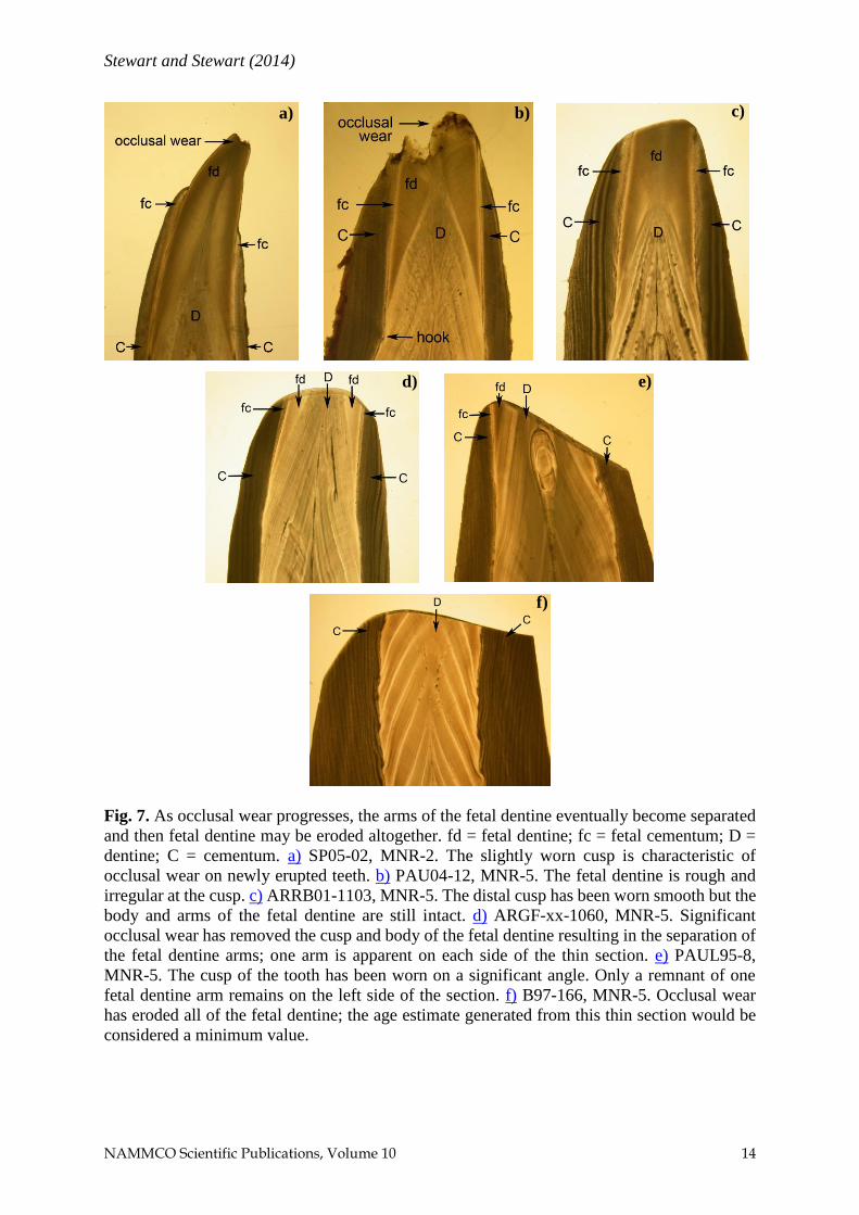
Fig. 7. As occlusal wear progresses, the arms of the fetal dentine eventually become separated
and then fetal dentine may be eroded altogether. fd = fetal dentine; fc = fetal cementum; D =
dentine; C = cementum. a) SP05-02, MNR-2. The slightly worn cusp is characteristic of
occlusal wear on newly erupted teeth. b) PAU04-12, MNR-5. The fetal dentine is rough and
irregular at the cusp. c) ARRB01-1103, MNR-5. The distal cusp has been worn smooth but the
body and arms of the fetal dentine are still intact. d) ARGF-xx-1060, MNR-5. Significant
occlusal wear has removed the cusp and body of the fetal dentine resulting in the separation of
the fetal dentine arms; one arm is apparent on each side of the thin section. e) PAUL95-8,
MNR-5. The cusp of the tooth has been worn on a significant angle. Only a remnant of one
fetal dentine arm remains on the left side of the section. f) B97-166, MNR-5. Occlusal wear
has eroded all of the fetal dentine; the age estimate generated from this thin section would be
considered a minimum value.
a) b) c)
d) e)
f)

Stewart and Stewart (2014)
NAMMCO Scientific Publications, Volume 10 15
Fig. 8. The last-formed growth layer adjacent to the pulp cavity is translucent. PAU04-12,
MNR-5. A translucent growth layer adjacent to the pulp cavity indicates the end of a GLG
cycle, so age is the total GLG count, assuming fetal dentine is present and all GLGs could be
counted. The opaque line between the last translucent growth layer and the pulp cavity is edge
effect. Accessory lines are apparent in the opaque growth layers.

Stewart and Stewart (2014)
NAMMCO Scientific Publications, Volume 10 16
Fig. 9. The last-formed growth layer adjacent to the pulp cavity is opaque. In (a) B95-545,
MNR-5, it is < ½ the preceding opaque growth layer both in width and length (seen in the
extension at the dentine-cementum interface). In (b) PAU04-03, MNR-5, the last-formed
opaque layer is > ½ the size of the previous opaque growth layer. Such estimates of the
proportion of the last GLG being formed may be used for different applications of age
estimates. Accessory lines within the overall pattern of GLG repetition, particularly in GLG-
3, are evident in (b).
a) b)

Stewart and Stewart (2014)
NAMMCO Scientific Publications, Volume 10 17
(3) Conversely, recently formed GLGs in older whales are often more
difficult to resolve due to the compression and thin nature of the
growth layers near the root tip.
(4) Accessory lines are non-annual layers that may confound GLG counts
(Fig. 8, 9b). Generally they are detected as disruptions in the overall
repetitive pattern of GLGs, which means they are also most
problematic in teeth of young whales in which there are too few GLGs
to make a dominant repetitive pattern.
(5) Pulp stones may disrupt the GLG pattern and make discerning GLGs
in their vicinity difficult (Figs. 5, 6). For the purpose of counting
GLGs, nodes are considered to be attributes of the dentine and may be
useful or necessary to produce age estimates if pulp stones or
inclusions are significant. Nodes may also be of use if the GLGs are
compressed, indistinct or ambiguous.
Generally, researchers address all of these issues by sectioning two teeth from
the same whale. The ideal candidates for sectioning are teeth that are straight,
unworn, undamaged, and large. Such teeth are uncommon among beluga but
comparing sections from more than one tooth allows the reader to select the
optimum tooth section that provides the most information. It may also be
possible to combine information between sections, for example if fetal
dentine is present in one but not the other and a distinctive mark appears in
both, a full age may be constructed by moving between legible areas of two
sections (NAMMCO in review).
CONCLUSION
Generally, the GLGs in beluga teeth can be challenging to interpret relative
to other marine mammals. The dentine GLGs may be complicated by
accessory layers and inclusions. Considerable variation of growth layer
clarity may occur within a tooth section, among teeth from the same jaw, and
among beluga from the same or different stocks. Understanding the biology
of beluga dentition and following standard protocols during tooth reading
sessions will improve accuracy and precision of age estimates. While final
age estimates are based on dentine GLG counts, a holistic interpretation of all
the attributes of a tooth can enhance the assessment of “age”. Subsequent
analysis of the animal’s sex, length, or date of collection may help resolve
ageing problems in specific whales but should not be done earlier in the age-
estimation process so as to minimize reader bias. It should be recognized,
however, that some whales cannot be aged by dentine GLG counts because
the teeth are too convoluted or damaged, or the material deposited is irregular
or indistinct.
Stewart and Stewart (2014)
NAMMCO Scientific Publications, Volume 10 18
Finally, there are two as yet unresolved issues concerning beluga age
estimates and their use in research and management. One lies with the beluga;
the other with the humans. Beluga have a protracted birth period, which may
vary by location, spanning May through August at least (Sergeant 1973,
Stewart and Stewart 1989). That means that at least the first GLG represents
different times and proportions of a year. For example, a beluga harvested in
July could be two months past a May birth or be one month away from its
first “birthday” in August. It is not known if the difference would be manifest
in different thicknesses of the first GLG and the layers would thenceforth be
synchronized, or if some other process operates. The human factor is the
application in which age estimates will be used. Some applications may
require only broad age categories while others need 1-year age class
resolution. Indeed, some studies may require refined date of harvest for part-
year age class increments.
Both issues require an understanding of the structures being examined and,
as Stewart (2012) noted, strict adherence to a protocol with detailed record
taking and record sharing. The preferred basis for designating an age class is
a complete description of all the dentine growth layers seen in a tooth. Then,
a user can choose how to assign an “age” using robust year classes or sensitive
subdivisions of the year classes.
ACKNOWLEDGEMENTS
The basis for this summary was a contract report prepared by B.E.S for L.
Loseto, DFO, Winnipeg, MB. We appreciate the funding support provided
and the permission to re-use material presented here, particularly some of the
images. J.B. Dunn contributed valuable technical assistance. We thank G.
Goodridge and L. Peters, Centennial Animal Hospital, Winnipeg, MB, for
their generous logistical support in obtaining the x-ray of the fetal beluga jaw.
The manuscript was improved by useful comments from A. Hohn and an
anonymous reviewer.
REFERENCES
Benjamins S (1999) Defining populations of the Beluga (Delphinapterus
leucas) using morphometry and ultrastructure of teeth. M.Sc. thesis,
University of Gröningen, Netherlands. 54 pp.
Brodie PF (1969) Mandibular layering in Delphinapterus leucas and age
determination. Nature. 221(5184):956–958 doi: http://dx.doi.org/10.
1038/221956a0
Brodie PF (1971) A reconsideration of aspects of growth, reproduction, and
behavior of the white whale (Delphinapterus leucas), with reference to

Stewart and Stewart (2014)
NAMMCO Scientific Publications, Volume 10 19
the Cumberland Sound, Baffin Island, population. J. Fish. Res. Board
Canada. 28(9):1309–1318 doi: http://dx.doi.org/10.1139/f71-198
Doan KH and Douglas CW (1953) Beluga of the Churchill region of Hudson
Bay. Bull. Fish. Res. Board Canada. 98:1–27
Douglas CW (1951) Report on beluga investigations at Churchill in 1950.
Fish. Res. Board Canada. Manuscript Rept. 485:1–35
Fay FH (1982) Ecology and biology of the Pacific walrus, Odobenus
rosmarus divergens, Illiger. North America Fauna No. 74:1–279
Heide-Jørgensen MP, Jensen J, Larsen AH, Teilmann J and Neurohr B (1994)
Age estimation of white whales (Delphinapterus leucas) from
Greenland. In: Born EW, Dietz R and Reeves RR (eds.) Studies of white
whales (Delphinapterus leucas) and narwhals (Monodon monoceros) in
Greenland and adjacent waters. Medd. Om Grøn., Biosci. 39:187–193
Hohn A (2009) Age estimation. In: Perrin WF, Wursig B and Thewissen JGM
(eds.) Encyclopedia of marine mammals. Second Edition. Academic
Press, San Diego, California. 11–17
Hohn AA and Fernandez S (1999) Biases in dolphin age structure due to age
estimation technique. Mar. Mamm. Sci. 15(4):1124–1132 doi: http://
dx.doi.org/10.1111/j.1748-7692.1999.tb00881.x
IWC—International Whaling Commission (1980) Report of the workshop.
In: Perrin WF and Myrick Jr AC (eds.) Age determination of toothed
whales and sirenians. Rep. Int. Whal. Comm. Special Issue 3.
International Whaling Commission, Cambridge, UK. 1-50
Ishiyama M (1987) Enamel structure in Odontocete whales. Scanning Micros.
1(3):1071–1079
Johnston DH, Joachim DG, Bachmann P, Kardong KV, Stewart REA, Dix
LM, Strickland MA and Watt ID (1987) Aging furbearers using tooth
structure and biomarkers. In: Novak M, Baker JA, Obbard ME and
Malloch B (eds.) Wild furbearer management and conservation in North
America. Ontario Ministry of Natural Resources, Toronto, ON. 228–243
Kleinenberg SE, Yablokov AV, Bel’Kovich BM and Tarasevich MN (1969)
Beluga (Delphinapterus leucas); investigation of the species. Israel Prog.
Sci. Translation, Jerusalem, No. TT-67-51345. 376 pp. (original
publication in Russian, Moscow, 1964)
Klevezal GA (1996) Recording structures of mammals. Determination of age
and reconstruction of life history. A.A. Balkema Publishers, Brookfield,
VT. 274 pp. (original publication in Russian, Moscow, 1998).
NAMMCO—North Atlantic Marine Mammal Commission (in review)
Report of the workshop on age estimation in beluga. NAMMCO Sci.
Publ. doi: http://dx.doi.org/10.7557/3.XXXX
Sergeant DE (1973) Biology of white whales (Delphinapterus leucas) in
western Hudson Bay. J. Fish. Res. Board Canada. 30(8):1065–1090 doi:
http://dx.doi.org/10.1139/f73-178

Stewart and Stewart (2014)
NAMMCO Scientific Publications, Volume 10 20
Sergeant DE (1981) On permissible exploitation rates of Monodontidae. Rep.
Int. Whal. Comm. 31:583–588
Stewart BE (2012) A technical report on methods for tooth preparation and
age estimates of beluga (Delphinapterus leucas). Can. Tech. Rep. Fish.
Aquat. Sci. 3020: xi-85 p.
Stewart BE, Innes S and Stewart REA (1998) Mandibular dental ontogeny of
ringed seals (Phoca hispida). Mar. Mamm. Sci. 14(2):221–231 doi:
http://dx.doi.org/10.1111/j.1748-7692.1998.tb00712.x
Stewart BE and Stewart REA (1989) Delphinapterus leucas. Mammal.
Species 336:1–8
Stewart REA and Stewart BE (1987) Dental ontogeny of harp seals, Phoca
groenlandica. Can. J. Zool. 65(6):1425–1434 doi: http://dx.doi.org
/10.1139/z87-225
Stewart REA, Stewart BE, Stirling I and Street E (1996) Counts of growth
layer groups in cementum and dentine in ringed seals (Phoca hispida).
Mar. Mamm. Sci. 12(3):383–401 doi: http://dx.doi.org/10.1111/j.1748-
7692.1996.tb00591.x
Stewart REA, Campana SE, Jones CM and Stewart BE (2006) Bomb
radiocarbon dating calibrates beluga (Delphinapterus leucas) age
estimates. Can. J. Zool. 84(12):1840–1852. doi: http://dx.doi.org/
10.1139/z06-182
Uhen MD (2009) Dental morphology, evolution of. In: Perrin WF, Wursig B
and Thewissen JGM Encyclopedia of marine mammals. Second Edition.
Academic Press, San Diego, California. 302–307