The bacterial and mitochondrial ribosomal A-site molecular switches ...
13
2654–2666 Nucleic Acids Research, 2008, Vol. 36, No. 8 Published online 16 March 2008 doi:10.1093/nar/gkn112 The bacterial and mitochondrial ribosomal A-site molecular switches possess different conformational substates Jiro Kondo and Eric Westhof* Architecture et Re ´ activite ´ de l’ARN, Universite ´ Louis Pasteur, Institut de Biologie Mole ´ culaire et Cellulaire, CNRS, 15 rue Rene ´ Descartes, 67084 Strasbourg, France Received January 5, 2008; Revised February 9, 2008; Accepted February 18, 2008 ABSTRACT The A site of the small ribosomal subunit partici- pates in the fidelity of decoding by switching between two states, a resting ‘off’ state and an active decoding ‘on’ state. Eight crystal structures of RNA duplexes containing two minimal decoding A sites of the Homo sapiens mitochondrial wild-type, the A1555G mutant or bacteria have been solved. The resting ‘off’ state of the mitochondrial wild-type A site is surprisingly different from that of the bacterial A site. The mitochondrial A1555G mutant has two types of the ‘off’ states; one is similar to the mitochondrial wild-type ‘off’ state and the other is similar to the bacterial ‘off’ state. Our present results indicate that the dynamics of the A site in bacteria and mitochondria are different, a property probably related to the small number of tRNAs used for decoding in mitochondria. Based on these structures, we propose a hypothesis for the molec- ular mechanism of non-syndromic hearing loss due to the mitochondrial A1555G mutation. INTRODUCTION Fidelity of decoding during protein biosynthesis can be divided into two steps; (i) initial selection and (ii) proof- reading (1,2). In the first step, the non-cognate tRNA is discriminated by the difference in free energy of base pairings between the codon and cognate tRNA anticodon. In the second step, the aminoacyl-tRNA decoding site (or the A site) acts as an RNA molecular switch upon recognition of the codon–anticodon pair and provokes an irreversible step of GTP hydrolysis. These two steps of decoding guarantee the high fidelity of translation. Crystal structures of various bacterial 30S and 70S ribosomes have revealed the molecular mechanisms of the proof- reading step at atomic level (3–10). When the cognate tRNA is delivered to the A site, the A-site molecular switch changes its conformation from an ‘off’ state to an ‘on’ state. These two local states are linked to the ‘open’ and ‘closed’ global states of the ribosome during decoding, as observed by Ramakrishnan and coworkers (1,2,6), but the connecting mechanisms are not fully described yet. In the ‘off’ state, two adenine residues, A1492 and A1493, do not make any interaction with codon–anticodon helix. On the other hand, in the ‘on’ state, these two adenines fully bulge out and come together with G530 in the shoulder domain of the 30S subunit to recognize the first two Watson-Crick base pair of codon–anticodon mini- helix and induce ribosomal transitions from the ‘open’ to the ‘closed’ forms (1,2,6). Recently, functional character- istics and tertiary structures of the A-site molecular switch in Homo sapiens cytoplasms have been revealed by X-ray analyses of RNA fragments containing the minimal A site (11), but those in H. sapiens mitochondria are still unknown. The secondary structure of the H. sapiens mitochondrial A site is very similar to that of the bacterial A site (Figure 1) (12). Differences are found at residue 1556 (1491 in bacterial numbering), which is a C in mitochon- dria and a G in bacteria, and at the base pair between 1494 and 1555 (1410 and 1490 in bacterial numbering), which is a CoA opposition in mitochondria and an A-U (66.5%) or a G=C (26.4%) base pair in bacteria (13). It is impor- tant to note that residue 1492 (1408 in bacterial number- ing) is an A in the H. sapiens mitochondrial as in bacterial A sites, while it is a G in the H. sapiens cytoplasmic A site (11,12). Because of the similarity in the secondary structure of the A site, it has long been believed that the H. sapiens mitochondrial A site has functional character- istics and tertiary structure similar to those of the bacterial A site. However, the structural components of the H. sapiens mitochondrial ribosome are noticeably different from *To whom correspondence should be addressed. Tel: +33 3 88 41 70 46; Fax: +33 3 88 60 18 22; Email: [email protected] ß 2008 The Author(s) This is an Open Access article distributed under the terms of the Creative Commons Attribution Non-Commercial License (http://creativecommons.org/licenses/ by-nc/2.0/uk/) which permits unrestricted non-commercial use, distribution, and reproduction in any medium, provided the original work is properly cited. Downloaded from https://academic.oup.com/nar/article-abstract/36/8/2654/2410228 by guest on 12 February 2018
Transcript of The bacterial and mitochondrial ribosomal A-site molecular switches ...

2654–2666 Nucleic Acids Research, 2008, Vol. 36, No. 8 Published online 16 March 2008doi:10.1093/nar/gkn112
The bacterial and mitochondrial ribosomalA-site molecular switches possess differentconformational substatesJiro Kondo and Eric Westhof*
Architecture et Reactivite de l’ARN, Universite Louis Pasteur, Institut de Biologie Moleculaire et Cellulaire,CNRS, 15 rue Rene Descartes, 67084 Strasbourg, France
Received January 5, 2008; Revised February 9, 2008; Accepted February 18, 2008
ABSTRACT
The A site of the small ribosomal subunit partici-pates in the fidelity of decoding by switchingbetween two states, a resting ‘off’ state and anactive decoding ‘on’ state. Eight crystal structuresof RNA duplexes containing two minimal decoding Asites of the Homo sapiens mitochondrial wild-type,the A1555G mutant or bacteria have been solved.The resting ‘off’ state of the mitochondrial wild-typeA site is surprisingly different from that of thebacterial A site. The mitochondrial A1555G mutanthas two types of the ‘off’ states; one is similar to themitochondrial wild-type ‘off’ state and the otheris similar to the bacterial ‘off’ state. Our presentresults indicate that the dynamics of the A site inbacteria and mitochondria are different, a propertyprobably related to the small number of tRNAs usedfor decoding in mitochondria. Based on thesestructures, we propose a hypothesis for the molec-ular mechanism of non-syndromic hearing loss dueto the mitochondrial A1555G mutation.
INTRODUCTION
Fidelity of decoding during protein biosynthesis can bedivided into two steps; (i) initial selection and (ii) proof-reading (1,2). In the first step, the non-cognate tRNA isdiscriminated by the difference in free energy of basepairings between the codon and cognate tRNA anticodon.In the second step, the aminoacyl-tRNA decoding site(or the A site) acts as an RNA molecular switch uponrecognition of the codon–anticodon pair and provokes anirreversible step of GTP hydrolysis. These two steps ofdecoding guarantee the high fidelity of translation. Crystalstructures of various bacterial 30S and 70S ribosomes
have revealed the molecular mechanisms of the proof-reading step at atomic level (3–10). When the cognatetRNA is delivered to the A site, the A-site molecularswitch changes its conformation from an ‘off’ state to an‘on’ state. These two local states are linked to the ‘open’and ‘closed’ global states of the ribosome during decoding,as observed by Ramakrishnan and coworkers (1,2,6), butthe connecting mechanisms are not fully described yet.In the ‘off’ state, two adenine residues, A1492 and A1493,do not make any interaction with codon–anticodon helix.On the other hand, in the ‘on’ state, these two adeninesfully bulge out and come together with G530 in theshoulder domain of the 30S subunit to recognize the firsttwo Watson-Crick base pair of codon–anticodon mini-helix and induce ribosomal transitions from the ‘open’ tothe ‘closed’ forms (1,2,6). Recently, functional character-istics and tertiary structures of the A-site molecularswitch in Homo sapiens cytoplasms have been revealedby X-ray analyses of RNA fragments containing theminimal A site (11), but those in H. sapiens mitochondriaare still unknown.
The secondary structure of theH. sapiens mitochondrialA site is very similar to that of the bacterial A site(Figure 1) (12). Differences are found at residue 1556(1491 in bacterial numbering), which is a C in mitochon-dria and a G in bacteria, and at the base pair between 1494and 1555 (1410 and 1490 in bacterial numbering), which isa CoA opposition in mitochondria and an A-U (66.5%)or a G=C (26.4%) base pair in bacteria (13). It is impor-tant to note that residue 1492 (1408 in bacterial number-ing) is an A in the H. sapiens mitochondrial as in bacterialA sites, while it is a G in the H. sapiens cytoplasmic A site(11,12). Because of the similarity in the secondarystructure of the A site, it has long been believed that theH. sapiens mitochondrial A site has functional character-istics and tertiary structure similar to those of the bacterialA site.
However, the structural components of the H. sapiensmitochondrial ribosome are noticeably different from
*To whom correspondence should be addressed. Tel: +33 3 88 41 70 46; Fax: +33 3 88 60 18 22; Email: [email protected]
� 2008 The Author(s)
This is an Open Access article distributed under the terms of the Creative Commons Attribution Non-Commercial License (http://creativecommons.org/licenses/
by-nc/2.0/uk/) which permits unrestricted non-commercial use, distribution, and reproduction in any medium, provided the original work is properly cited.
Downloaded from https://academic.oup.com/nar/article-abstract/36/8/2654/2410228by gueston 12 February 2018

those of the bacterial ribosome. The mitochondrialribosome is formed by a small 28S subunit and a large39S subunit. The small subunit consists of a 12S rRNAand 29 proteins (14–16), while the large subunit consists ofa 16S rRNA and 48 proteins (14,17,18). The 5S rRNA,which is present in the bacterial ribosome, is absent in themitochondrial ribosome. The ratio of protein to rRNAmass in the mitochondrial ribosome (2:1) is inverted fromthe ratio found in the bacterial ribosome (1:2). A surpris-ing feature of the H. sapiens mitochondrial system is theuse of an extended decoding mechanism, which allowstranslation of all 60 codons with only 22 tRNA species(19–23). Mammalian mitochondrial translation does notuse nucleus-encoded tRNAs, although mitochondrialimport of some tRNAs occurs in eukaryotic microorgan-isms (protozoa, fungi, algae), in some plants and in afew animals (24,25). In addition, tRNA modifications atthe wobble position of the anticodon, where post-transcriptionally modified nucleotides often play anessential role in the precise decoding of the genetic codein bacteria and eukaryotic cytoplasms (26), are less fre-quent in mitochondria (23,27). In other words, in contrastto Escherichia coli with 45 tRNA species (or 41 anti-codons) and H. sapiens cytoplasms with 46 tRNA species(28,29), mitochondria must gain flexibility (each tRNAdecodes two or four codons) in the decoding process(20–22) without loosing fidelity. Therefore, it is possiblethat mitochondria have unique proofreading mechanismin the decoding process to accommodate flexibility andfidelity.
The A1555G mutant of the H. sapiens mitochondrial Asite has been found in more than 120 families throughoutthe world and is one of the most common genetic causesof non-syndromic hearing loss (30–32). In the A1555Gmutant A site, the G1555 residue is expected to forma Watson-Crick base pair with the C1494 residue on theopposite strand rendering it closer to the bacterial A site(Figure 1).
In the present study, we have solved eight crystalstructures of RNA fragments containing two minimalA sites of the H. sapiens mitochondrial wild-type, theA1555G mutant or bacteria. Comparative structural ana-lyses provided us with insights into the decoding mecha-nisms in the mitochondrial and bacterial ribosomes. Theseobservations lead to a hypothesis for the molecularmechanism of non-syndromic hearing loss due to theA1555G mutation in the mitochondrial A site.
MATERIALS AND METHODS
Crystallization
Two internal loops of the bacterial A site, the H. sapiensmitochondrial A site or its A1555G mutant were insertedbetween Watson-Crick pairs in sequences designed to foldas a double helix (BACT, WT and MUT, respectively)(Supplementary Figures 1, 2 and 4). The RNA oligomerswere chemically synthesized by Dharmacon (Boulder, CO)and purified by HPLC and reverse phase chromatogra-phy. To resolve phase, bromine derivatives of WT andMUT with 5-bromouracil residues (BrU) at the centralstem were also synthesized (WT-Br, MUT-Br1 and MUT-Br2, respectively) (Supplementary Figures 3, 5–8). Beforecrystallization, 4mM paromomycin or tobramycin solu-tion containing 50mM sodium cacodylate buffer (pH 6.5)was prepared, and 2mM RNA solutions containing100mM sodium cacodylate (pH 6.5) and 25mM sodiumchloride were annealed by heating at 858C for 2minfollowed by slow cooling to 378C. Same volumes of RNAsolution and paromomycin or tobramycin solution weremixed at 378C and then cooled slowly to room tempera-ture (21–258C). Crystallizations were performed by thehanging-drop vapor diffusion method at 208C and 378Cby mixing 1 ml of RNA/aminoglycoside solution and 1 mlof crystallization solution containing 50mM sodiumcacodylate (pH 7.0), 0–2mM spermine tetrahydrochlor-ide, 50–200mM potassium chloride, 20–200mM stron-tium chloride, 0–5% (v/v) glycerol and 1–5% (v/v)2-methyl-2,4-pentanediol. Crystals suitable for X-rayexperiments were obtained after optimization of crystal-lization conditions. For the bacterial A-site RNA, theBACT-Co crystal was obtained in the condition contain-ing hexammine cobalt chloride (Table 1 and Supplemen-tary Figure 1). For the mitochondrial A-site RNA, twotypes of crystals (WT-K and WT-Br-K) were obtained inconditions containing potassium chloride (Table 1 andSupplementary Figures 2 and 3). And for the A1555Gmutant RNA, five types of crystals (MUT-K, MUT-Br1-Sr, MUT-Br1-K1, MUT-Br1-K2 and MUT-Br2-Co)were obtained in conditions containing potassium chlo-ride, strontium chloride or hexammine cobalt chloride(Table 1 and Supplementary Figures 4–8).
Data collection, structure determination and refinement
X-ray data of eight crystals were collected at 100K withsynchrotron radiation at the ID14-2, ID23-1 or BM30beamline in the European Synchrotron Radiation Facility(ESRF; Grenoble, France), or at the PX beamline in theSwiss Light Source (SLS; Villingen, Switzerland). Eachdataset was processed by using the program Crystalclear(Rigaku/MSC) or MOSFLM (33,34), and was scaled andmerged using Crystalclear or SCALA from the CCP4 suiteof crystallographic programs (35).The BACT-Co crystal has I4 spacegroup (Table 1),
which is different from any bacterial A-site crystalsobtained previously in conditions without hexamminecobalt chloride (P212121, P21 or P41) (36–41). Two crystalsof the mitochondrial A-site RNA, WT-K and WT-Br-K,have the same spacegroup P1 with similar unit cell
Figure 1. Secondary structures of the bacterial, H. sapiens mitochon-drial wild-type and its A1555G mutant A sites. The nucleotides differ-ent from those in the bacterial A site are colored in red. The G1555residue in the A1555G mutant is colored in blue.
Nucleic Acids Research, 2008, Vol. 36, No. 8 2655
Downloaded from https://academic.oup.com/nar/article-abstract/36/8/2654/2410228by gueston 12 February 2018

Table
1.Crystaldata,statisticsofdata
collectionsandstatisticsofstructure
refinem
ents
Bacterial
H.sapiensmitochondrialwildtype
H.sapiensmitochondrialA1555G
mutant
Crystalcode
BACT-C
oWT-K
WT-Br-K
MUT-K
MUT-Br1-Sr
MUT-Br1-K
1MUT-Br1-K
2MUT-Br2-C
oAminoglycoside
Paromomycin
Paromomycin
Paromomycin
Paromomycin
Paromomycin
Paromomycin
Tobramycin
Paromomycin
PDB
ID3BNL
3BNN
3BNO
3BNP
3BNQ
3BNR
3BNS
3BNT
Crystaldata
Space
group
I4P1
P1
P3121
C2
C2
C2
P64
Unit
cell(A
)(8)
a=
b=
53.6,
c=63.5
a=
34.6,b=
44.5,
c=52.4
a=
35.2,b=
45.5,
c=52.2
a=
b=
66.6,
c=57.6
a=
71.3,b=
76.1,
c=55.9
a=
71.2,b=
77.4,
c=56.3
a=
72.7,b=
77.1,
c=56.5
a=
b=
74.9,
c=22.5
�=
68.8,�=
71.3,
g=67.7
�=
71.4,�=
73.4,
g=74.0
�=
117.1
�=
115.1
�=
115.7
Za
12
21
22
20.5
Data
collection
Beamline
BM30ofESRF
PX
ofSLS
ID23-1
ofESRF
PX
ofSLS
ID23-1
ofESRF�
ID23-1
ofESRF
ID14-2
ofESRF
BM30ofESRF�
Wavelength
(A)
0.91636
0.9999
0.8856
0.9801
0.92070/0.92110/0.91625
0.91625
0.933
0.91950/0.91984/0.91637
Resolution(A
)36.8–2.6
39.6–2.0
29.0–2.35
57.7–2.7
49.8–2.0/49.8–2.0/49.8–2.0
51.0–2.1
35.4–1.9
37.4–2.3/37.4–2.4/37.4–2.3
Oftheoutershell(A
)2.7–2.6
2.1–2.0
2.4-2.35
2.9–2.7
2.1–2.0/2.1–2.0/2.1–2.0
2.2–2.1
2.0–1.9
2.4–2.3/2.5–2.4/2.4–2.3
Uniquereflections
5295
17270
11751
4263
17465/17512/17519
16388
21598
3368/2981/3374
Completeness(%
)99.9
97.0
98.0
99.4
97.4/97.4/97.4
99.8
97.4
100.0/99.9/99.9
Intheoutershell(%
)100.0
92.6
97.7
100.0
85.6/85.4/85.5
100.0
97.4
100.0/100.0/100.0
Rmergeb(%
)5.1
9.6
9.9
7.8
4.9/4.9/5.0
11.6
3.1
8.4/9.5/7.2
Intheoutershell(%
)28.6
36.9
39.0
36.4
28.0/33.2/37.3
37.8
37.8
30.4/26.9/25.7
Ranomc(%
)–
––
–5.1/4.0/4.5
––
6.6/6.5/5.2
Intheoutershell(%
)–
––
–24.6/29.0/33.1
––
19.0/16.0/15.5
Redundancy
7.1
2.7
7.7
9.9
3.5/3.5/3.5
6.9
3.8
9.9/9.9/9.9
Intheoutershell
7.3
2.6
8.0
10.2
2.6/2.6/2.6
7.1
3.8
10.5/10.5/10.5
Structure
refinem
ent
Resolutionrange(A
)36.0–2.6
39.6–2.0
36.9–2.35
40.0–2.7
49.8–2.0
51.0–2.1
35.4–1.9
37.4–2.3
Usedreflections
5278
17269
11718
4204
17514
16147
21594
3371
R-factord
(%)
22.0
23.8
22.6
23.1
22.9
21.5
24.7
21.7
Rfreee
25.1
26.4
25.5
27.0
26.9
24.5
26.0
25.5
Number
ofDNA
atoms
940
1788
1788
908
1942
1884
1884
464
Number
ofcations
8[Co(N
H3) 6]3+
––
–8Sr2
+,2K
+2K
+2K
+1[Co(N
H3) 6]3+,1Na+
Number
ofwater
38
114
31
13
191
183
102
55
Number
of
aminoglycoside
None
None
None
None
1(non-specific)
1(non-specific)
None
None
RMSD
Bondlength
(A)
0.004
0.005
0.005
0.005
0.004
0.004
0.004
0.004
Bondangles(8)
0.9
0.9
0.9
1.0
0.8
0.9
0.8
0.8
Improper
angles(8)
1.3
0.7
0.7
0.9
0.7
0.7
0.7
1.3
aNumber
ofdsR
NA
intheasymmetricunit.
bRmerge=
100��
hklj|Ihklj–<I h
klj>|/�
hklj<I h
klj>.
cRanom=
100��
hklj|Ihklj(+
)–I h
klj(�
)|/�
hklj[Ihklj(+
)+
I hklj(�
)].
dR-factor=
100��||Fo|–|F
c||/�|F
o|,where|F
o|and|F
c|are
optimallyscaledobserved
andcalculatedstructure
factoramplitudes,respectively.
eCalculatedusingarandom
setcontaining10%
ofobservationsthatwerenotincluded
throughoutrefinem
ent(58).
�Forphase
determinationwiththemultiple
anomalousdiffraction(M
AD)method,threedatasets
werecollectedwiththreewavelengths.Statisticsfrom
leftto
rightare
ofpeak,edgeandremote
data,respectively.
2656 Nucleic Acids Research, 2008, Vol. 36, No. 8
Downloaded from https://academic.oup.com/nar/article-abstract/36/8/2654/2410228by gueston 12 February 2018
dimensions, suggesting their isomorphism (Table 1). Threedifferent crystal forms were obtained for the A1555Gmutant-RNA, P3121, C2 and P64 (Table 1).
For MAD phasing using the anomalous scattering ofbromine atoms, X-ray data of MUT-Br1-Sr and MUT-Br2-Co were taken with three different wavelengths basedon XAFS measurements (Table 1). Initial phases wereestimated by the MADmethod using the program SOLVE(42) with figure-of-merit of 0.40 and 0.46, respectively.Electron densities, modified by solvent flattening with theprogram CNS (43), showed clearly the phosphate-ribosebackbone with the individual bases. The molecular struc-tures of MUT-Br1-Sr and MUT-Br2-Co were constructedon a graphic workstation with the program O (44). Initialphases of other crystals were determined by the MolecularReplacement method with the program AMoRe (45) usingthe bulk-solvent technique (46,47). The details of phasedeterminations will be discussed elsewhere.
The atomic parameters of each structure were refinedwith the program CNS (43) through a combination ofsimulated-annealing, crystallographic conjugate gradientminimization refinements and B-factor refinements,followed by interpretations of the omit map at everynucleotide residue. The statistics of structure refinementsare summarized in Table 1. Electron density indicatingspecific binding of aminoglycosides to the A site could notbe observed in any of the crystal structures, indicating thatcomplex formation did not occur under the presentconditions. In MUT-Br1-Sr and MUT-Br1-K1, a non-specific binding of paromomycin between two symmetri-cally related RNA duplexes was observed (SupplementaryFigures 5 and 6).
The adiabatic morphing calculations between the ‘off’and ‘on’ states of the A-site molecular switches wereperformed with the program CNS (43) using a pro-grammed input file morph_dist.inp (48,49). All figures weredrawn using the PyMOL Molecular Graphics system(2002) DeLano Scientific, San Carlos, CA (http://www.pymol.org).
Coordinates
The atomic coordinates have been deposited in the ProteinData Bank (PDB) with the ID codes 3BNL (BACT-Co),3BNN (WT-K), 3BNO (WT-Br-K), 3BNP (MUT-K),3BNQ (MUT-Br1-Sr), 3BNR (MUT-Br1-K1), 3BNS(MUT-Br1-K2) and 3BNT (MUT-Br2-Co).
RESULTS
Overview of the A-site structures
In all cases, the two RNA strands form a duplex(Supplementary Figures 1–8). At the center of theduplex, four contiguous Watson-Crick G=C base pairsare formed. In the case of bromine derivatives, two of fourG=C base pairs are replaced with the Watson-CrickA-BrU base pairs. Three Watson-Crick G=C base pairsclose the stem at both ends of the duplex. Some of theterminal overhanging residues at the 50-end (UU or C) areinvolved in crystal packing interactions and others aredisordered in the solvent channel.
The BACT-Co crystal contains an asymmetrical RNAduplex (Supplementary Figure 1). Two A sites in theduplex have almost identical conformations except for theA1492 residue (Supplementary Figure 9a), and are similarto the bacterial ‘off’ state with tucked-in A1492 and A1493found in the Thermus thermophilus 30S ribosomal particle[PDB ID: 1J5E in (3)].For the H. sapiens mitochondrial A site, two types of
crystals with P1 spacegroup, WT-K and WT-Br-K, wereobtained (Table 1). In each crystal, two RNA duplexes arein the asymmetric unit (Supplementary Figures 2 and 3).Therefore, a total of eight internal loops are observed.Although four of these eight A-site internal loops are freefrom any intermolecular interactions in crystalline stateand four others are involved in crystal packing, all of themhave identical conformations with a bulged-out residue,A1558 (A1493 in bacterial numbering), and a tucked-inresidue, A1557 (A1492 in bacterial numbering) (Supple-mentary Figure 9b), suggesting that there is no crystal-packing effect on this conformation of the A site. Sincethis conformation is different from the ‘on’ states of thebacterial and H. sapiens cytoplasmic A sites with bulged-out A1492 and A1493 [PDB ID: 1IBM in (5); 2FQN in(11)], it should correspond to the ‘off’ state of themitochondrial wild-type A site.For the A1555G mutant of the H. sapiens mitochon-
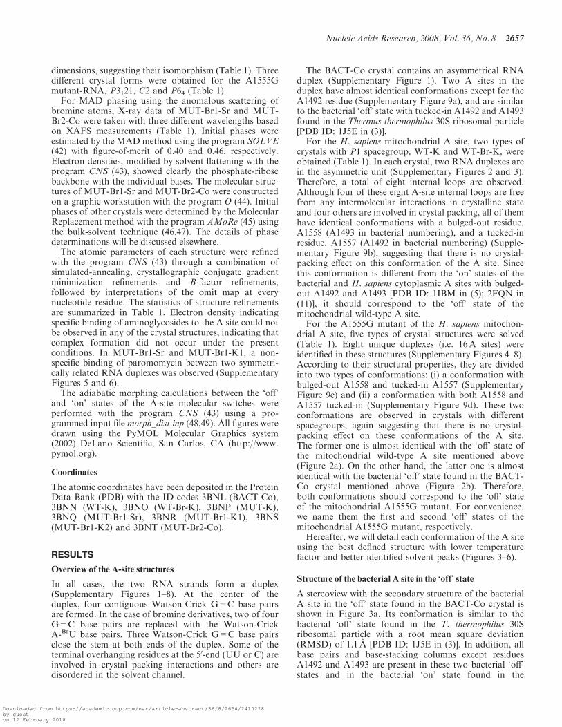
drial A site, five types of crystal structures were solved(Table 1). Eight unique duplexes (i.e. 16A sites) wereidentified in these structures (Supplementary Figures 4–8).According to their structural properties, they are dividedinto two types of conformations: (i) a conformation withbulged-out A1558 and tucked-in A1557 (SupplementaryFigure 9c) and (ii) a conformation with both A1558 andA1557 tucked-in (Supplementary Figure 9d). These twoconformations are observed in crystals with differentspacegroups, again suggesting that there is no crystal-packing effect on these conformations of the A site.The former one is almost identical with the ‘off’ state ofthe mitochondrial wild-type A site mentioned above(Figure 2a). On the other hand, the latter one is almostidentical with the bacterial ‘off’ state found in the BACT-Co crystal mentioned above (Figure 2b). Therefore,both conformations should correspond to the ‘off’ stateof the mitochondrial A1555G mutant. For convenience,we name them the first and second ‘off’ states of themitochondrial A1555G mutant, respectively.Hereafter, we will detail each conformation of the A site
using the best defined structure with lower temperaturefactor and better identified solvent peaks (Figures 3–6).
Structure of the bacterial A site in the ‘off’ state
A stereoview with the secondary structure of the bacterialA site in the ‘off’ state found in the BACT-Co crystal isshown in Figure 3a. Its conformation is similar to thebacterial ‘off’ state found in the T. thermophilus 30Sribosomal particle with a root mean square deviation(RMSD) of 1.1 A [PDB ID: 1J5E in (3)]. In addition, allbase pairs and base-stacking columns except residuesA1492 and A1493 are present in these two bacterial ‘off’states and in the bacterial ‘on’ state found in the
Nucleic Acids Research, 2008, Vol. 36, No. 8 2657
Downloaded from https://academic.oup.com/nar/article-abstract/36/8/2654/2410228by gueston 12 February 2018
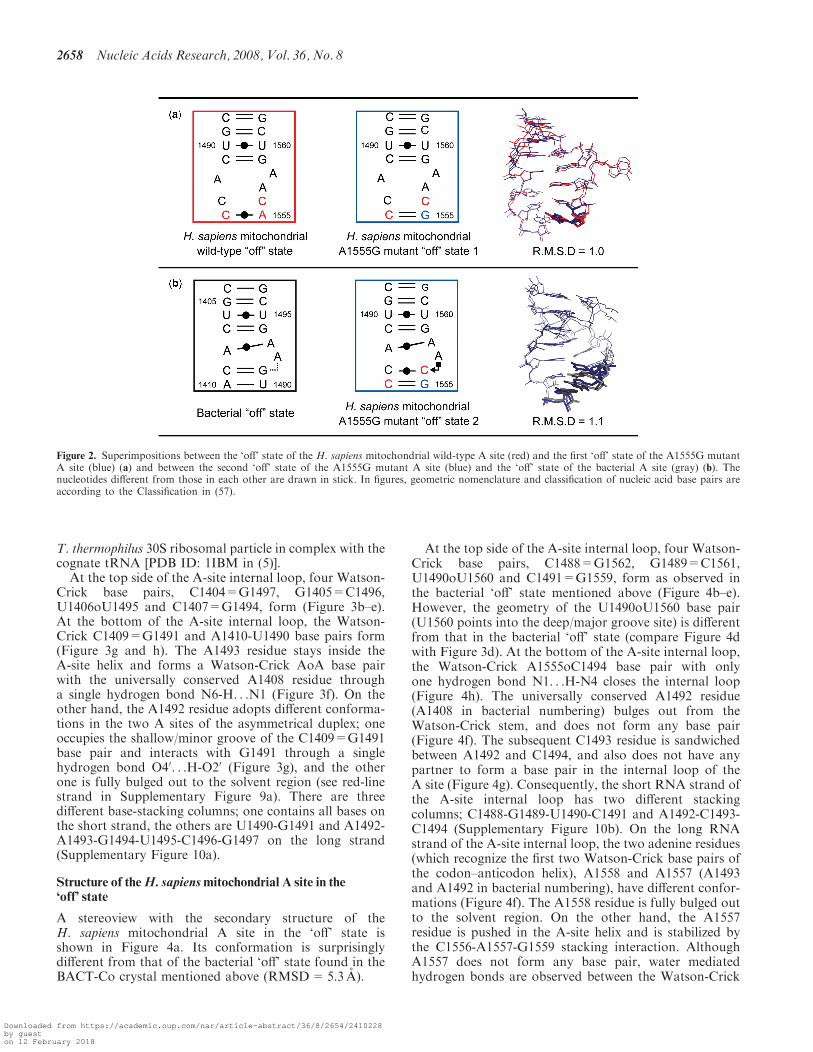
T. thermophilus 30S ribosomal particle in complex with thecognate tRNA [PDB ID: 1IBM in (5)].At the top side of the A-site internal loop, four Watson-
Crick base pairs, C1404=G1497, G1405=C1496,U1406oU1495 and C1407=G1494, form (Figure 3b–e).At the bottom of the A-site internal loop, the Watson-Crick C1409=G1491 and A1410-U1490 base pairs form(Figure 3g and h). The A1493 residue stays inside theA-site helix and forms a Watson-Crick AoA base pairwith the universally conserved A1408 residue througha single hydrogen bond N6-H. . .N1 (Figure 3f). On theother hand, the A1492 residue adopts different conforma-tions in the two A sites of the asymmetrical duplex; oneoccupies the shallow/minor groove of the C1409=G1491base pair and interacts with G1491 through a singlehydrogen bond O40. . .H-O20 (Figure 3g), and the otherone is fully bulged out to the solvent region (see red-linestrand in Supplementary Figure 9a). There are threedifferent base-stacking columns; one contains all bases onthe short strand, the others are U1490-G1491 and A1492-A1493-G1494-U1495-C1496-G1497 on the long strand(Supplementary Figure 10a).
Structure of theH. sapiensmitochondrial A site in the‘off’ state
A stereoview with the secondary structure of theH. sapiens mitochondrial A site in the ‘off’ state isshown in Figure 4a. Its conformation is surprisinglydifferent from that of the bacterial ‘off’ state found in theBACT-Co crystal mentioned above (RMSD=5.3 A).
At the top side of the A-site internal loop, four Watson-Crick base pairs, C1488=G1562, G1489=C1561,U1490oU1560 and C1491=G1559, form as observed inthe bacterial ‘off’ state mentioned above (Figure 4b–e).However, the geometry of the U1490oU1560 base pair(U1560 points into the deep/major groove site) is differentfrom that in the bacterial ‘off’ state (compare Figure 4dwith Figure 3d). At the bottom of the A-site internal loop,the Watson-Crick A1555oC1494 base pair with onlyone hydrogen bond N1. . .H-N4 closes the internal loop(Figure 4h). The universally conserved A1492 residue(A1408 in bacterial numbering) bulges out from theWatson-Crick stem, and does not form any base pair(Figure 4f). The subsequent C1493 residue is sandwichedbetween A1492 and C1494, and also does not have anypartner to form a base pair in the internal loop of theA site (Figure 4g). Consequently, the short RNA strand ofthe A-site internal loop has two different stackingcolumns; C1488-G1489-U1490-C1491 and A1492-C1493-C1494 (Supplementary Figure 10b). On the long RNAstrand of the A-site internal loop, the two adenine residues(which recognize the first two Watson-Crick base pairs ofthe codon–anticodon helix), A1558 and A1557 (A1493and A1492 in bacterial numbering), have different confor-mations (Figure 4f). The A1558 residue is fully bulged outto the solvent region. On the other hand, the A1557residue is pushed in the A-site helix and is stabilized bythe C1556-A1557-G1559 stacking interaction. AlthoughA1557 does not form any base pair, water mediatedhydrogen bonds are observed between the Watson-Crick
Figure 2. Superimpositions between the ‘off’ state of the H. sapiens mitochondrial wild-type A site (red) and the first ‘off’ state of the A1555G mutantA site (blue) (a) and between the second ‘off’ state of the A1555G mutant A site (blue) and the ‘off’ state of the bacterial A site (gray) (b). Thenucleotides different from those in each other are drawn in stick. In figures, geometric nomenclature and classification of nucleic acid base pairs areaccording to the Classification in (57).
2658 Nucleic Acids Research, 2008, Vol. 36, No. 8
Downloaded from https://academic.oup.com/nar/article-abstract/36/8/2654/2410228by gueston 12 February 2018
edge of A1557 and the O2P atom of A1492. As a result, allresidues on the long strand of the A-site loop exceptA1558 are involved in the same stacking column(Supplementary Figure 10b). To form these bulgedconformations, A1557 and A1558 adopt C20-endo sugarpuckers and have characteristic torsion angles around theP-O50 (�), C50-C40 (g) and O30-Pn+1 (�). Small � (928) and� (1218) angles and large g (1268) angle allow the A1558residue to protrude from the A-site helix into the solventregion, and small � (628) and g (508) angles and large� (3088) angle allow the A1557 residue to be bulged intothe A-site helix.
Structure of the A1555Gmutant of theH. sapiensmitochondrial A site in the first ‘off’ state
A stereoview with the secondary structure of the A1555Gmutant of the H. sapiens mitochondrial A site in the first‘off’ state is shown in Figure 5a. Its overall conformationis very similar to that of the ‘off’ state of the mitochondrialwild-type A site mentioned above (RMSD=1.0A)(Figure 2a). Only one difference between them is foundat the bottom of the A-site internal loop. As expected, aWatson-Crick G1555=C1494 base pair is observed in theA1555G mutant (Figure 5h) instead of the A1555oC1494
Figure 3. The bacterial A site in the ‘off’ state. (a) Secondary structure and stereoview. (b–h) Atomic details of each base pair of the A site. Threeuniversally conserved adenine residues, A1408, A1492 and A1493, are colored in orange, blue and red, respectively. Two nucleotides different fromthose in the mitochondrial A site, U1490 and G1491, are colored in cyan and green, respectively. The hydrogen bonds are represented by blackdashed lines.
Nucleic Acids Research, 2008, Vol. 36, No. 8 2659
Downloaded from https://academic.oup.com/nar/article-abstract/36/8/2654/2410228by gueston 12 February 2018

base pair in the wild-type (Figure 4h). As observed in thewild-type ‘off’ state, there are three stacking columns; onecontains all residues on the long strand except A1558, andothers are C1488-G1489-U1490-C1491 and A1492-C1493-C1494 on the short strand (Supplementary Figure 10c).
Structure of the A1555Gmutant of theH. sapiensmitochondrial A site in the second ‘off’ state
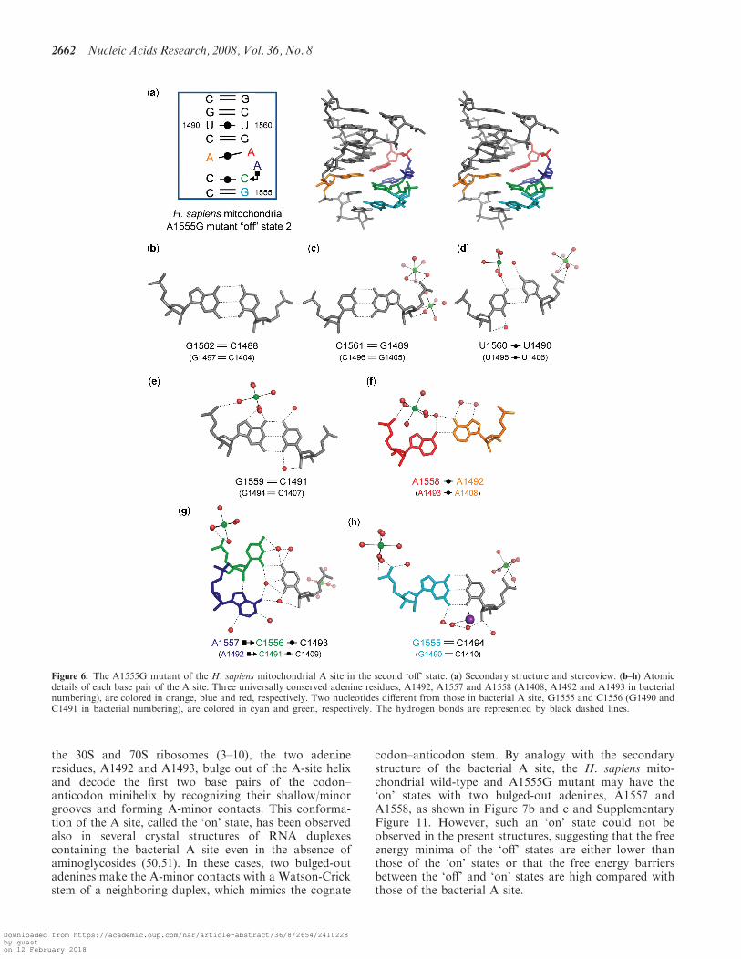
A stereoview with the secondary structure of the A1555Gmutant of the H. sapiens mitochondrial A site in the
second ‘off’ state is shown in Figure 6a. Its conformationis completely different from that of the first ‘off’ state ofthe A1555G mutant just discussed (RMSD=4.5 A). Onthe other hand, it is almost identical with the bacterial ‘off’state found in the BACT-Co crystal (RMSD=1.1 A)(Figure 2b). Differences between them are found only atthe bottom of the A-site internal loop. The C1493 residue(C1409 in bacterial numbering) forms a Watson-CrickC1493oC1556 base pair with only one hydrogen bondN4-H. . .O2 (Figure 6g) instead of the Watson-Crick C=Gbase pair in bacteria (Figure 3g). Since C1556 points into
Figure 4. The H. sapiens mitochondrial A site in the ‘off’ state. (a) Secondary structure and stereoview. (b–h) Atomic details of each base pair of theA site. Three universally conserved adenine residues, A1492, A1557 and A1558 (A1408, A1492 and A1493 in bacterial numbering), are colored inorange, blue and red, respectively. Two nucleotides different from those in the bacterial A site, A1555 and C1556 (A1490 and C1491 in bacterialnumbering), are colored in cyan and green, respectively. The hydrogen bonds are represented by black dashed lines.
2660 Nucleic Acids Research, 2008, Vol. 36, No. 8
Downloaded from https://academic.oup.com/nar/article-abstract/36/8/2654/2410228by gueston 12 February 2018

the deep/major groove side, there is a large space at theshallow/minor groove edge of C1556 occupied by A1557(A1492 in bacterial numbering). The A1557 residue formsa cis Hoogsteen/Sugar-edge base pair with C1556 througha direct hydrogen bond N7. . .H-O20 and a water-mediatedhydrogen bond N6-H. . .W. . .O2 (Figure 6g). Two water-mediated hydrogen bonds, N6-H. . .W. . .O2 andN6-H. . .W. . .N3, are observed between A1557 andC1493 (Figure 6g). At the bottom of the A-site helix, theWatson-Crick C1494=G1555 base pair forms (Figure 6h)instead of the Watson-Crick A-U base pair in bacteria(Figure 3h). Therefore, as observed in the bacterial ‘off’
state found in the BACT-Co crystal, there are threedifferent base-stacking columns. All bases on the shortstrand make one stacking column. On the other hand, thelong strand has two different base-stacking columns;G1555-C1556 and A1557-A1558-G1559-U1560-C1561-G1562 (Supplementary Figure 10d).
The ‘on’ states of theH. sapiensmitochondrial wild-typeand A1555Gmutant A sites
According to observations by Ramakrishnan andcoworkers on the bacterial A-site molecular switch on
Figure 5. The A1555G mutant of the H. sapiens mitochondrial A site in the first ‘off’ state. (a) Secondary structure and stereoview. (b–h) Atomicdetails of each base pair of the A site. Three universally conserved adenine residues, A1492, A1557 and A1558 (A1408, A1492 and A1493 in bacterialnumbering), are colored in orange, blue and red, respectively. Two nucleotides different from those in the bacterial A site, G1555 and C1556 (G1490and C1491 in bacterial numbering), are colored in cyan and green, respectively. The hydrogen bonds are represented by black dashed lines.
Nucleic Acids Research, 2008, Vol. 36, No. 8 2661
Downloaded from https://academic.oup.com/nar/article-abstract/36/8/2654/2410228by gueston 12 February 2018
the 30S and 70S ribosomes (3–10), the two adenineresidues, A1492 and A1493, bulge out of the A-site helixand decode the first two base pairs of the codon–anticodon minihelix by recognizing their shallow/minorgrooves and forming A-minor contacts. This conforma-tion of the A site, called the ‘on’ state, has been observedalso in several crystal structures of RNA duplexescontaining the bacterial A site even in the absence ofaminoglycosides (50,51). In these cases, two bulged-outadenines make the A-minor contacts with a Watson-Crickstem of a neighboring duplex, which mimics the cognate
codon–anticodon stem. By analogy with the secondarystructure of the bacterial A site, the H. sapiens mito-chondrial wild-type and A1555G mutant may have the‘on’ states with two bulged-out adenines, A1557 andA1558, as shown in Figure 7b and c and SupplementaryFigure 11. However, such an ‘on’ state could not beobserved in the present structures, suggesting that the freeenergy minima of the ‘off’ states are either lower thanthose of the ‘on’ states or that the free energy barriersbetween the ‘off’ and ‘on’ states are high compared withthose of the bacterial A site.
Figure 6. The A1555G mutant of the H. sapiens mitochondrial A site in the second ‘off’ state. (a) Secondary structure and stereoview. (b–h) Atomicdetails of each base pair of the A site. Three universally conserved adenine residues, A1492, A1557 and A1558 (A1408, A1492 and A1493 in bacterialnumbering), are colored in orange, blue and red, respectively. Two nucleotides different from those in bacterial A site, G1555 and C1556 (G1490 andC1491 in bacterial numbering), are colored in cyan and green, respectively. The hydrogen bonds are represented by black dashed lines.
2662 Nucleic Acids Research, 2008, Vol. 36, No. 8
Downloaded from https://academic.oup.com/nar/article-abstract/36/8/2654/2410228by gueston 12 February 2018

DISCUSSION
Bacteria have a ‘soft’ A-site molecular switch
For the bacterial A site, four conformations of the‘off’ states have been reported so far (SupplementaryFigure 11a) (11,52). Differences between them are foundonly at the two adenine residues, A1492 and A1493;(i) both are tucked-in, as observed in the presentBACT-Co crystal; (ii) only A1492 is bulged-out;(iii) only A1493 is bulged-out; (iv) both adenines arebulged-out but do not recognize the codon–anticodonstem. These multiple conformations of the bacterial ‘off’state suggest high flexibility of the bacterial A-sitemolecular switch. On the other hand, the bacterial ‘on’state is only observed in a single conformation, the onewith the two bulged-out adenines (Figures 7a and 8a andSupplementary Figure 11a). Since all base pairs and base-stacking columns except the A1492 and A1493 residuesare conserved in the bacterial ‘off’ and ‘on’ states, confor-mational adaptations between these two states can easilyoccur from any of the ‘off’ states (Figure 7a, Supplemen-tary Figure 11a and Supplementary movie ‘bacteria’).Recently, replica molecular dynamics simulations haveconfirmed that the free energy barrier between the ‘off’and ‘on’ states of the bacterial A site is sufficiently low foraa-tRNA binding to shift the equilibrium (53). For these
reasons, we suggest that bacteria present an energetically‘soft’ A-site molecular switch, which achieves high speedsof translation, probably at a slight cost of proofreadingaccuracy.
H. sapiensmitochondria have a ‘hard’ A-sitemolecular switch
In contrast to the flexible bacterial ‘off’ state, theH. sapiensmitochondrial ‘off’ state is only observed in a singleconformation, which is completely different from any ofthe bacterial ‘off’ state (Supplementary Figure 11). Byanalogy with the secondary structure of the bacterialA site, the H. sapiens mitochondrial wild-type may adoptthe ‘on’ state with two bulged-out adenines, A1557 andA1558, as shown in Figure 7b and SupplementaryFigure 11b. Superimposition between the mitochondrial‘off’ state and the crystal structure of the T. thermophilus70S ribosome in complex with tRNA and mRNA (8)suggests that the switching from the ‘off’ state to the ‘on’state needs a global conformational change (Figures 7b, 8band Supplementary movie ‘mitochondria-WT’). On theshort RNA strand of the A-site internal loop, the bulged-out A1492 and the subsequent C1493 and C1494 residues(1408, 1409 and 1410 in bacterial numbering) have to movewithin the loop in order to form the long base-stackingcolumn between all bases of the strand (Figure 7b). On the
Figure 7. Overview of the A-site molecular switches of the bacterial, mitochondrial wild-type and its A1555G mutant. Base-stacking columns arecolored in red, blue, orange and green, respectively. Bases, which are not involved in any stacking column, are colored in gray. Except whenindicated, all structures are crystallographically determined.
Nucleic Acids Research, 2008, Vol. 36, No. 8 2663
Downloaded from https://academic.oup.com/nar/article-abstract/36/8/2654/2410228by gueston 12 February 2018

long RNA strand, the tucked-in A1557 residue (A1492 inbacterial numbering) has to bulge out into the shallow/minor groove side, and the bulged-out A1558 residue(A1493 in bacterial numbering) has to move a longdistance towards the shallow/minor groove of the codon–anticodon stem (Figures 7b and 8b). Such conformationaltransitions are accompanied by drastic changes of torsionangles and sugar puckers. For example, the sugar puckersof A1557 and A1558 (A1492 and A1493 in bacterialnumbering) must be altered from the C20-endo to C30-endoconformations. Therefore, the free energy barrier betweenthe ‘off’ and ‘on’ states may be high. In other words,the mitochondrial A-site molecular switch may be energet-ically harder to achieve than the bacterial one. It is impor-tant to remember that the H. sapiens mitochondria with22 tRNA species must gain some flexibility (each tRNAdecodes two or four codons) in the decoding process(20–22) without loosing fidelity in contrast to E. coli with45 tRNA species (or 41 anticodons) and H. sapiens cyto-plasms with 46 tRNA species (28,29). However, mitochon-dria need to synthesize only 13 encoded protein speciesduring their long life cycle (23). It is thus possible thatmitochondria require a harder A-site molecular switch inorder to gain the necessary high accuracy of proofreadingprobably at the cost of translation speed.
Hypothesis for the molecular mechanism of non-syndromichearing loss due to the A1555Gmutation in themitochondrial A-site molecular switch
The A1555G mutant of the H. sapiens mitochondrialA site has two different ‘off’ states; one named the first
‘off’ state is almost identical with the mitochondrialwild-type ‘off’ state (Figure 2a), and the other onenamed the second ‘off’ state is very similar to the bacte-rial ‘off’ state found in the present BACT-Co crystal(Figure 2b). The latter conformation may be the inter-mediate state between the first ‘off’ state and the ‘on’ stateof the A1555G mutant, which could not be observed in themitochondrial wild-type. In other words, the A1555Gmutation renders the mitochondrial A-site molecularswitch close to the bacterial one. By analogy with thesecondary structure of the bacterial A site, the A1555Gmutant may have the ‘on’ state with bulged-out A1557and A1558, as shown in Figure 7c and SupplementaryFigure 11c. Conformational changes between the first andsecond ‘off’ states as well as between the second ‘off’ and‘on’ states may reversibly occur (see Supplementary movie‘mitochondria-A1555G’). Since the latter case would beeasier to occur than the former one, the A1555G mutantmolecular switch is closer to the bacterial A-site molecularswitch (compare Figure 7c with Figure 7a and Supple-mentary Figure 11c with Supplementary Figure 11a).As discussed above, although the decoding process inmitochondria is more flexible than that in bacteria, theproofreading step in mitochondria with the ‘hard’ A-sitemolecular switch may be more accurate than that inbacteria. However, the decoding process in mitochondriawith the A1555G mutation (containing the ‘soft’ bacterial-type A-site molecular switch) should become moreinaccurate. Since all 13 mitochondrial proteins aresubunits of the respiratory chain complexes and areinvolved in synthesis of ATP by oxidative phosphoryla-tion (23), non-syndromic hearing loss related to theA1555G mutation may be caused by interfering withenergy production through protein mistranslation indu-cing excess superoxide production resulting in oxidativedamage to mitochondria as proposed for aminoglycosideaction on mitochondria (54) and bacteria (55).
CONCLUSIONS
Due to the similarity of the secondary structure of theA site, it has been supposed that the functional character-istics and tertiary structure of the A-site molecular switchis basically conserved in bacteria, H. sapiens cytoplasmsand mitochondria. However, these three cell types arenoticeably different in their biological properties such aslife cycle, genome size, structural component of ribosomeand number of tRNA species. In our present and previouswork (11), we have shown how a small difference ofnucleotide sequences affects the dynamics of the A-sitemolecular switches underlying the decoding mechanismsadapted to their biological properties and environments.In addition, as observed in our previous work (56), it alsoaffects binding of aminoglycosides to the A-site molec-ular switches. The actual mechanisms and the couplingbetween the observed substates of the A sites and theglobal ribosomal movements are certainly rather complexand will require further techniques and experiments formore extensive unraveling.
Figure 8. Stereoviews of interactions among the A site, mRNA(orange) and tRNA (sky blue). (a) The T. thermophilus ribosome[PDBID: 2j00 in (8)]. (b) The H. sapiens mitochondrial A site super-posed on the T. thermophilus ribosomal A site. The A sites have the‘on’ and ‘off’ states in (a) and (b), respectively. Three universallyconserved adenine residues, A1492, A1557 and A1558 (A1408, A1492and A1493 in bacterial numbering), are colored in orange, blue andred, respectively. Two nucleotides different in the bacterial and mito-chondrial A sites, residues 1555 and 1556 (1490 and 1491 in bacterialnumbering), are colored in cyan and green, respectively.
2664 Nucleic Acids Research, 2008, Vol. 36, No. 8
Downloaded from https://academic.oup.com/nar/article-abstract/36/8/2654/2410228by gueston 12 February 2018
SUPPLEMENTARY DATA
Supplementary Data are available at NAR Online.
ACKNOWLEDGEMENTS
During part of this work, J.K. was supported by the JapanSociety for the Promotion of Science and the projectANR-O5-MIIM-026-01 from the Agence Nationale de laRecherche. We thank the European SynchrotronRadiation Facility and the Swiss Light Source forprovision of synchrotron radiation facilities and acknowl-edge the people of beamline ID14-2, ID23-1 and BM30 inESRF and PX in SLS. We are grateful to B. Francois forcrystallization and data collection of the MUT-K crystaland to A. Urzhumtsev for helpful discussions andsuggestions for solving structures. Funding to pay theOpen Access publication charges for this article wasprovided by the CNRS.
Conflict of interest statement. None declared.
REFERENCES
1. Ogle,J.M., Carter,A.P. and Ramakrishnan,V. (2003) Insights intothe decoding mechanism from recent ribosome structure.Trends Biochem. Sci., 28, 259–266.
2. Ogle,J.M., Carter,A.P. and Ramakrishnan,V. (2005) Structuralinsights into translation fidelity. Annu. Rev. Biochem., 74, 129–177.
3. Wimberly,B.T., Brodersen,D.E., Clemons,W.M.,Jr,Morgan-Warren,R.J., Carter,A.P., Vonrhein,C., Hartsch,T. andRamakrishnan,V. (2000) Structure of the 30S ribosomal subunit.Nature, 407, 327–339.
4. Carter,A.P., Clemons,W.M., Brodersen,D.E., Morgan-Warren,R.J.,Wimberly,B.T. and Ramakrishnan,V. (2000) Functional insightsfrom the structure of the 30S ribosomal subunit and its interactionswith antibiotics. Nature, 407, 340–348.
5. Ogle,J.M., Brodersen,D.E., Clemons,W.M.,Jr, Tarry,M.J.,Carter,A.P. and Ramakrishnan,V. (2001) Recognition of cognatetransfer RNA by the 30S ribosomal subunit. Science, 292, 897–902.
6. Ogle,J.M., Murphy,F.V., Tarry,M.J. and Ramakrishnan,V. (2002)Selection of tRNA by the ribosome requires a transition from anopen to a closed form. Cell, 111, 721–732.
7. Murphy,F.V.,4th and Ramakrishnan,V. (2004) Structure of apurine-purine wobble base pair in the decoding center of theribosome. Nat. Struct. Mol. Biol., 11, 1251–1252.
8. Selmer,M., Dunham,C.M., Murphy,F.V.,4th, Weixlbaumer,A.,Petry,S., Kelley,A.C., Weir,J.R. and Ramakrishnan,V. (2006)Structure of the 70S ribosome complexed with mRNA and tRNA.Science, 313, 1935–1942.
9. Dunham,C.M., Selmer,M., Phelps,S.S., Kelley,A.C., Suzuki,T.,Joseph,S. and Ramakrishnan,V. (2007) Structures of tRNAs withan expanded anticodon loop in the decoding center of the 30Sribosomal subunit. RNA, 13, 817–823.
10. Weixlbaumer,A., Murphy,F.V.,4th, Dziergowska,A., Malkiewicz,A.,Vendeix,F.A., Agris,P.F. and Ramakrishnan,V. (2007) Mechanismfor expanding the decoding capacity of transfer RNAs bymodification of uridines. Nat. Struct. Mol. Biol., 14, 498–502.
11. Kondo,J., Urzhumtsev,A. and Westhof,E. (2006) Two conforma-tional states in the crystal structure of the Homo sapienscytoplasmic ribosomal decoding A site. Nucleic Acids Res., 34,676–685.
12. Cannone,J.J., Subramanian,S., Schnare,M.N., Collett,J.R.,D’Souza,L.M., Du,Y., Feng,B., Lin,N., Madabusi,L.V.,MUller,K.M. et al. (2002) The comparative RNA web (CRW) site:an online database of comparative sequence and structureinformation for ribosomal, intron, and other RNAs. BMCBioinformatics, 3, 2.
13. Pfister,P., Hobbie,S., Vicens,Q., Bottger,E.C. and Westhof,E. (2003)The molecular basis for A-site mutations conferring aminoglycosideresistance: relationship between ribosomal susceptibility and X-raycrystal structures. Chembiochem, 4, 1078–1088.
14. O’Brien,T.W., Liu,J., Sylvester,J.E., Mougey,E.B., Fischel-Ghodsian,N., Thiede,B., Wittmann-Liebold,B. and Graack,H.(2000) Mammalian mitochondrial ribosomal proteins (4). Aminoacid sequencing, characterization, and identification of correspond-ing gene sequences. J. Biol. Chem., 275, 18153–18159.
15. Koc,E.C., Burkhart,W., Blackburn,K., Moseley,A., Koc,H. andSpremulli,L.L. (2000) A proteomics approach to the identificationof mammalian mitochondrial small subunit ribosomal proteins.J. Biol. Chem., 275, 32585–32591.
16. Suzuki,T., Terasaki,M., Takemoto-Hori,C., Hanada,T., Ueda,T.,Wada,A. and Watanabe,K. (2001) Proteomic analysis of themammalian mitochondrial ribosome. Identification of proteincomponents in the 28 S small subunit. J. Biol. Chem., 276,33181–33195.
17. Suzuki,T., Terasaki,M., Takemoto-Hori,C., Hanada,T., Ueda,T.,Wada,A. and Watanabe,K. (2001) Structural compensation for thedeficit of rRNA with proteins in the mammalian mitochondrialribosome. Systematic analysis of protein components of the largeribosomal subunit from mammalian mitochondria. J. Biol. Chem.,276, 21724–21736.
18. Koc,E.C., Burkhart,W., Blackburn,K., Moseley,A., Koc,H. andSpremulli,L.L. (2001) The large subunit of the mammalianmitochondrial ribosome. Analysis of the complement of ribosomalproteins present. J. Biol. Chem., 276, 43958–43969.
19. Anderson,S., Bankier,A.T., Barrell,B.G., de Bruijn,M.H.,Coulson,A.R., Drouin,J., Eperon,I.C., Nierlich,D.P., Roe,B.A.,Sanger,F. et al. (1981) Sequence and organization of the humanmitochondrial genome. Nature, 290, 457–465.
20. Barrell,B.G., Anderson,S., Bankier,A.T., de Bruijn,M.H., Chen,E.,Coulson,A.R., Drouin,J., Eperon,I.C., Nierlich,D.P., Roe,B.A.et al. (1980) Different pattern of codon recognition bymammalian mitochondrial tRNAs. Proc. Natl Acad. Sci. USA, 77,3164–3166.
21. Yokoyama,S. and Nishimura,S. (1995) Modified nucleosides andcodon recognition. In Soll,D. and Raj-Bhandary,U. (eds), tRNA:Structure, Biosynthesis and Function. ASM Press, Washington, DC,pp. 207–223.
22. Sengupta,S., Yang,X. and Higgs,P.G. (2007) The mechanisms ofcodon reassignments in mitochondrial genetic codes. J. Mol. Evol.,64, 662–688.
23. Florentz,C., Sohm,B., Tryoen-Toth,P., Putz,J. and Sissler,M. (2003)Human mitochondrial tRNAs in health and disease. Cell. Mol.Life Sci., 60, 1356–1375.
24. Schneider,A. and Marechal-Drouard,L. (2000) MitochondrialtRNA import: are there distinct mechanisms? Trends Cell Biol., 10,509–513.
25. Entelis,N.S., Kolesnilov,O.A., Martin,R.P. and Tarassov,I.A. (2001)RNA delivery into mitochondria. Adv. Drug Deliv. Rev., 49,199–215.
26. Sprinzl,M and Vassilenko,K.S. (2005) Compilation of tRNAsequences and sequences of tRNA genes. Nucleic Acids Res., 33,D139–D140.
27. Heckman,J.E., Sarnoff,J., Alzner-DeWeerd,B., Yin,S. andRajBhandary,U.L. (1980) Novel features in the genetic code andcodon reading patterns in Neurospora crassa mitochondria basedon sequences of six mitochondrial tRNAs. Proc. Natl Acad. Sci.USA, 77, 3159–3163.
28. Komine,Y., Adachi,T., Inokuchi,H. and Ozeki,H. (1990) Genomicorganization and physical mapping of the transfer RNA genes inEscherichia coli K12. J. Mol. Biol., 212, 579–598.
29. International Human Genome Sequencing Consortium. (2001)Initial sequencing and analysis of the human genome. Nature, 409,860–921.
30. Prezant,T.R., Agapian,J.V., Bohlman,M.C., Bu,X., Oztas,S.,Qiu,W.Q., Arnos,K.S., Cortopassi,G.A., Jaber,L., Rotter,J.I. et al.(1993) Mitochondrial ribosomal RNA mutation associated withboth antibiotic-induced and non-syndromic deafness. Nat. Genet., 4,289–294.
31. Hutchin,T.P. and Cortopassi,G.A. (2000) Mitochondrial defects andhearing loss. Cell. Mol. Life Sci., 57, 1927–1937.
Nucleic Acids Research, 2008, Vol. 36, No. 8 2665
Downloaded from https://academic.oup.com/nar/article-abstract/36/8/2654/2410228by gueston 12 February 2018

32. Fischel-Ghodsian,N. (2003) Mitochondrial deafness. Ear Hear., 24,303–313.
33. Leslie,A.G.W. (1992) Molecular data processing. In Moras,D.,Podjarny,A.D. and Thierry,J.C. (eds), Crystallography Computing 5,From chemistry to Biology Oxford University Press, Oxford,pp. 50–61.
34. Rossmann,M.G. and van Beek,C.G. (1999) Data processing. ActaCrystallogr., D55, 1631–1640.
35. Collaborative Computational Project, Number 4. (1994) The CCP4Suite: Programs for Protein Crystallography. Acta Crystallogr.,D50, 760–763.
36. Vicens,Q. and Westhof,E. (2001) Crystal Structure of ParomomycinDocked into the Eubacterial Ribosomal Decoding A Site. Structure,9, 647–658.
37. Vicens,Q. and Westhof,E. (2002) Crystal structure of acomplex between the aminoglycoside tobramycin and an oligonu-cleotide containing the ribosomal decoding a site. Chem. Biol., 9,747–755.
38. Vicens,Q. and Westhof,E. (2003) Crystal structure of geneticinbound to a bacterial 16S ribosomal RNA A site oligonucleotide.J. Mol. Biol., 326, 1175–1188.
39. Francois,B., Szychowski,J., Adhikari,S.S., Pachamuthu,K.,Swayze,E.E., Griffey,R.H., Migawa,M.T., Westhof,E. andHanessian,S. (2004) Antibacterial aminoglycosides with a modifiedmode of binding to the ribosomal-RNA decoding site. Angew.Chem. Int. Ed. Engl., 43, 6735–6738.
40. Francois,B., Russell,R.J.M., Murray,J.B., Aboul-ela,F.,Masquida,B., Vicens,Q. and Westhof,E. (2005) Crystal structures ofcomplexes between aminoglycosides and decoding A site oligonu-cleotides: role of the number of rings and positive charges in thespecific binding leading to miscoding. Nucleic Acids Res., 33,5677–5690.
41. Kondo,J., Francois,B., Russell,R.J., Murray,J.B. and Westhof,E.(2006) Crystal structure of the bacterial ribosomal decoding sitecomplexed with amikacin containing the g-amino-a-hydroxybutyryl(haba) group. Biochimie, 88, 1027–1031.
42. Terwilliger,T.C. and Berendzen,J. (1999) Automated MAD andMIR structure solution. Acta Crystallogr., D55, 849–861.
43. Brunger,A.T., Adams,P.D., Clore,G.M., DeLano,W.L., Gros,P.,Grosse-Kunstleve,R.W., Jiang,J.-S., Kuszewski,J., Nilges,M.,Pannu,N.S. et al. (1998) Crystallography & NMR system: a newsoftware suite for macromolecular structure determination.Acta Crystallogr., D54, 905–921.
44. Jones,T.A., Zou,J.Y., Cowan,S.W. and Kjeldgaard,M. (1991)Improved methods for building protein models in electron density
maps and the location of errors in these models. Acta Crystallogr.,A47, 110–119.
45. Navaza,J. (1994) AMoRe: an automated package for molecularreplacement. Acta Crystallogr., A50, 157–163.
46. Fokine,A. and Urzhumtsev,A. (2002) On the use of low-resolutiondata for translation search in molecular replacement. ActaCrystallogr., A58, 72–74.
47. Fokine,A., Capitani,G., Grutter,M.G. and Urzhumtsev,A. (2003)Bulk-solvent correction for fast translation search in molecularreplacement: service programs for AMoRe and CNS. J. Appl.Crystallogr., 36, 352–355.
48. Echols,N, Milburn,D. and Gerstein,M. (2003) MolMovDB: analysisand visualization of conformational change and structural flex-ibility. Nucleic Acids Res., 31, 478–482.
49. Krebs,W.G. and Gerstein,M. (2000) The morph server: a standar-dized system for analyzing and visualizing macromolecular motionsin a database framework. Nucleic Acids Res., 28, 1665–1675.
50. Shandrick,S., Zhao,Q., Han,Q., Ayida,B.K., Takahashi,M.,Winters,G.C., Simonsen,K.B., Vourloumis,D. and Hermann,T.(2004) Monitoring molecular recognition of the ribosomal decodingsite. Angew Chem. Int. Ed. Engl., 43, 3177–3182.
51. Zhao,F., Zhao,Q., Blount,K.F., Han,Q., Tor,Y. and Hermann,T.(2005) Molecular recognition of RNA by neomycin and a restrictedneomycin derivative. Angew Chem. Int. Ed. Engl., 44, 5329–5334.
52. Kondo,J. and Westhof,E. (2007) Structural comparisons betweenprokaryotic and eukaryotic ribosomal decoding A sites free andcomplexed with aminoglycosides. In Arya,D.P. (ed), Aminoglycosideantibiotics, From Chemical Biology to Drug Discovery. Wiley-Interscience, Hoboken, NJ, pp. 209–223.
53. Sanbonmatsu,K.Y. (2006) Energy landscape of the ribosomaldecoding center. Biochimie, 88, 1053–1059.
54. Hutchin,T. and Cortopassi,G. (1994) Proposed molecular andcellular mechanism for aminoglycoside ototoxicity. Antimicrob.Agents Chemother., 38, 2517–2520.
55. Kohanski,M.A., Dwyer,D.J., Hayete,B., Lawrence,C.A. andCollins,J.J. (2007) A common mechanism of cellular death inducedby bactericidal antibiotics. Cell, 130, 797–810.
56. Kondo,J., Francois,B., Urzhumtsev,A. and Westhof,E. (2006)Crystal structure of the Homo sapiens cytoplasmic ribosomaldecoding site complexed with apramycin. Angew. Chem. Int. Ed.Engl., 45, 3310–3314.
57. Leontis,N.B. and Westhof,E. (2001) Geometric nomenclature andclassification of RNA base pairs. RNA, 7, 499–512.
58. Brunger,A.T. (1992) Free R value: a novel statistical quantity forassessing the accuracy of crystal structure. Nature, 355, 472–475.
2666 Nucleic Acids Research, 2008, Vol. 36, No. 8
Downloaded from https://academic.oup.com/nar/article-abstract/36/8/2654/2410228by gueston 12 February 2018