THE AUDITORY SYSTEM - Weebly
8
1 SPECIAL SENSES: THE AUDITORY SYSTEM REVISION OF PHYSICS: WAVES A wave is an oscillation of power, sound waves have two main characteristics: amplitude , which is the maximum displacement or the power of a wave and it represents the intensity, and frequency (the inverse of wave length) which represents the pitch of the sound.(Figure 1) Any sound or any letter represents the intensity of the sound as in speaking in a low volume, low intensity sounds have low amplitude. A higher voice has high amplitude (intensity). The difference between the letters, for example B and V, is in the frequency . Two different sounds have different frequencies. Saying a letter in a low voice has a low intensity and a certain frequency, when saying the same letter again in a higher voice -> this is the same frequency but with a higher intensity. An average human distinguishes frequencies as low as 20Hertz and up to 20 KHz (20,000 Hz). EAR ANATOMY (FIGURE 2) First there is the auricle in the outer ear that collects the sounds, any cut would affect the sound collection but not too much. And due to the curves inside it, the other function is sound localization in vertical dimension. (It does not calculate the position; the central parts in the brain with the help of its reflections do the calculation of the vertical dimension. There is also the auditory canal which ends at the tympanic membrane whose function is to convert sound waves to vibration. The middle ear: the most important part here is the three bones of hearing which will receive the vibration from the tympanic membrane and transfer them to the cochlea. Figure 1
Transcript of THE AUDITORY SYSTEM - Weebly
1
SPECIAL SENSES:
THE AUDITORY SYSTEM
REVISION OF PHYSICS: WAVES
A wave is an oscillation of power, sound waves have two main characteristics: amplitude, which is the
maximum displacement or the power of a wave and it represents the intensity, and frequency (the inverse of
wave length) which represents the pitch of the sound.(Figure 1)
Any sound or any letter represents the intensity of the
sound as in speaking in a low volume, low intensity sounds have
low amplitude. A higher voice has high amplitude (intensity).
The difference between the letters, for example B and V, is in
the frequency. Two different sounds have different
frequencies. Saying a letter in a low voice has a low intensity
and a certain frequency, when saying the same letter again in a
higher voice -> this is the same frequency but with a higher
intensity.
An average human distinguishes frequencies as low as 20Hertz and up to 20 KHz (20,000 Hz).
EAR ANATOMY(FIGURE 2)
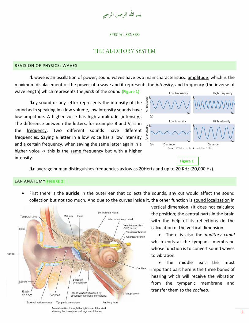
First there is the auricle in the outer ear that collects the sounds, any cut would affect the sound
collection but not too much. And due to the curves inside it, the other function is sound localization in
vertical dimension. (It does not calculate
the position; the central parts in the brain
with the help of its reflections do the
calculation of the vertical dimension.
There is also the auditory canal
which ends at the tympanic membrane
whose function is to convert sound waves
to vibration.
The middle ear: the most
important part here is the three bones of
hearing which will receive the vibration
from the tympanic membrane and
transfer them to the cochlea.
Figure 1
2
Those three bones are fixed in their place by ligaments in a way to allow a lever system because the
vibration of sounds will arrive by air vibration and when going to the cochlea, since the cochlea is filled
with fluid that has a higher resistance, the vibration must be transferred in a higher power, so there
must be an amplification of the signal and that is the function of the lever system in the three bones in
addition to the area difference in site between the tympanic membrane and the Stapedius muscle that
is in contact with the oval window of the cochlea.
Thereof, the most important function of the three bones in the middle ear is amplification of the signal
through it. Another function for the middle ear is that the Eustachian tube that connects the middle ear with
the pharynx equalizes the pressure so that the outside pressure is the same as the inside pressure. The idea is
that the tympanic membrane is not a stiff membrane so when going to the dead sea for example, the outside
pressure will be higher and it will push the membrane inside and stretch it, and in this case the sound cannot
vibrate it easily so the pressure must be equalized around it by letting the outside pressure inside through the
larynx and the Eustachian tube. This can be achieved by chewing a gum, opening the mouth or yawning. All
these movements allow the Eustachian tube to open and equalize the pressure difference.
Note: in children, the Eustachian tube is wider and shorter and this opens the way to infections of the
middle ear.
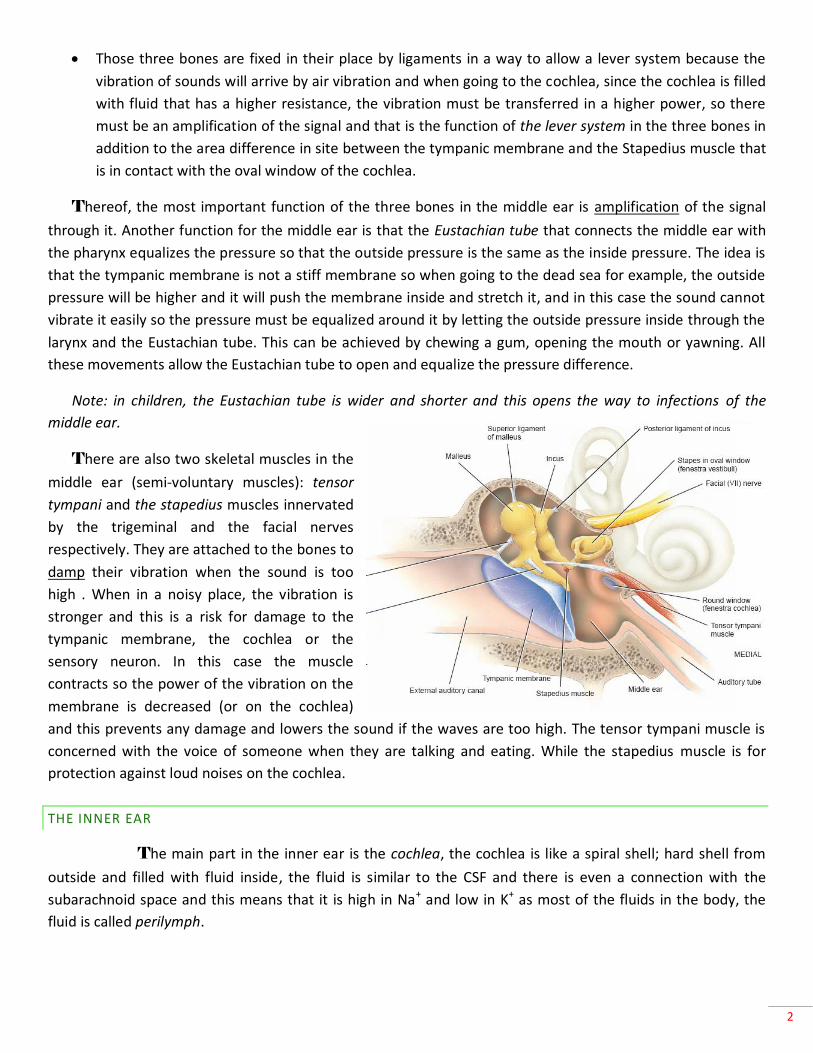
There are also two skeletal muscles in the
middle ear (semi-voluntary muscles): tensor
tympani and the stapedius muscles innervated
by the trigeminal and the facial nerves
respectively. They are attached to the bones to
damp their vibration when the sound is too
high . When in a noisy place, the vibration is
stronger and this is a risk for damage to the
tympanic membrane, the cochlea or the
sensory neuron. In this case the muscle
contracts so the power of the vibration on the
membrane is decreased (or on the cochlea)
and this prevents any damage and lowers the sound if the waves are too high. The tensor tympani muscle is
concerned with the voice of someone when they are talking and eating. While the stapedius muscle is for
protection against loud noises on the cochlea.
THE INNER EAR
The main part in the inner ear is the cochlea, the cochlea is like a spiral shell; hard shell from
outside and filled with fluid inside, the fluid is similar to the CSF and there is even a connection with the
subarachnoid space and this means that it is high in Na+ and low in K+ as most of the fluids in the body, the
fluid is called perilymph.
Figure 2

3
There is another tube located in the center of the space inside the cochlea forming a canal,
called cochlear duct. It divides the compartment of the cochlea into two compartments: upper and lower.
This duct is continuous along the entire cochlea except at the end, it ends before that and
because of that the fluid in the upper compartment is continuous with the fluid in the lower. The separating
duct also has a fluid in its space called
endolymph. This space and its fluid is a closed
space, not coming in contact with any other part,
it is also very high in K+ and low in Na+.
Now, any vibration will be transferred
through the three bones and the last one is the
stapes which transfers the vibrations to the
cochlea is pushing the oval window of the
cochlea, then the vibration the stapes will carry
on to the fluid inside, and that is how the
vibration is transferred to the cochlea. Pushing a
fluid in a closed space is hard so there must be a
vent for the vibration to enter a compartment,
now the vibration will enter from the stapes through an oval window (the entrance gate).(Figure 3.)At the end
of the cochlea on the other side there is another window covered by a membrane called round window (Exit
gate). Vibrations enters through one window and exit through the other, and they must travel along the entire
cochlea since the duct is making a route inside the cochlea from the upper to the lower compartments.
In brief, vibrations:
THE PHYSIOLOGY PART
The sensory part is in the cochlea is the
spiral organ or Organ of Corti (located
between the vestibular duct -scala
vestibulai- and the tempanic duct -scala
tempani) , it consists of three main parts.
Around the duct are two septa (separating
the duct from the cochlear compartments),
the upper septum is solid and does not
vibrate - we consider it a solid immobile
structure for simplicity- , (Figure 4.)The
Arrive at the tympanic
membrane
Amplified in the three bones
Goes in the fluid through the oval
window
Then through the entire
cochlea to the round window
Figure 3
Figure 4
4
lower is elastic and is called basilar membrane which is one part of the organ of Corti where neuronal
receptors are found, their function is to receive the vibrations and are called hair cells, because they look as
cells with spiky hair. They have cilias up that push against the third part which is a thick heavy gelatinous
membrane called tectorial membrane.
So the organ of Corti is a basilar elastic membrane on which the hair cells are sitting and their extensions
support the tectorial membrane. What happens is that when there is a vibration in the fluid, it will vibrate the
elastic basilar membrane so the membrane will go up and down and push the hair cells with it (up and down),
the tectorial membrane is also pushed but it is heavy so the hairs will bend right and left. For simplicity, we
will consider that it will bend to one direction, and this bending causes opening of K+ channels. Hair cells have
mechanical-gated potassium channels that open when the hairs are bent, allowing the potassium from the
endolymph to enter the cell and depolarize it. A depolarization to a certain level will open voltage-gated
calcium channels, so calcium will enter and it will allow the vesicles to release the neurotransmitter.
Again, vibration in the fluid -> moves the basilar membrane -> bending of cells -> opening of potassium
channels -> depolarization -> calcium enters -> release of NTs -> action potential in the next cell (In the spiral
ganglia) the second order neuron
This is the way of the reception but it should be able to detect both the frequency and intensity. How does
this happen?
DETECTION OF INTENSITY
A high-intensity sound will move the membrane higher because the vibration is bigger, so the bending
and the opening of channels will be more, therefore there are more neurotransmitter release and higher
frequency of firing.
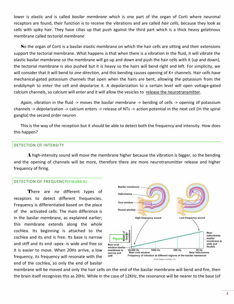
DETECTION OF FREQUENCY(FIGURE 5)
There are no different types of
receptors to detect different frequencies.
Frequency is differentiated based on the place
of the activated cells. The main difference is
in the basilar membrane, as explained earlier;
this membrane extends along the whole
cochlea. Its beginning is attached to the
cochlea and its end is free. Its base is narrow
and stiff and its end -apex- is wide and free so
it is easier to move. When 20Hz arrive, a low
frequency, its frequency will resonate with the
end of the cochlea, so only the end of basilar
membrane will be moved and only the hair cells on the end of the basilar membrane will bend and fire, then
the brain itself recognizes this as 20Hz. While in the case of 12KHz, the resonance will be nearer to the base (of
Figure 5

5
the basilar membrane) and the cells there will fire, after that the brain detects this as 12KHz. (Illustrated in
Figure5.) note : each 1 mm of the basilar membrane responds to a certain frequency .
Note: a difference of around 100Hz can be distinguished as separate signals for the humans .
In the organ of Corti, there are two types of hair cells: inner hair cells at the front and each cell is in
one line, and outer hair cells that are close together. Inner hair cells receive type I neurons, each hair cell
synapses with four to five neurons. This gives better resolution of sound in term of frequency. In outer hair
cells, each neuron receives from six to seven hair cells and this decreases the discrimination of frequencies but
its advantage is when there is a low vibration, the outer hair cells are easier to summate. At low intensity, we
do not hear in a good discrimination because we hear mainly by outer hair cells. This type of neurons is type II.
Inner hair cells In one line By type I neurons One cell synapses with many neurons
Outer hair cells Compressed together By type II neurons Many cells synapse with one neuron
The neurons are mainly bipolar type of neurons; one terminal will synapse with hair cells, cell bodies
are in the spiral ganglia and the other end will go centrally to the brainstem to the cochlear nucleus in the
medulla. The cochlear nucleus has two main divisions, anterior and posterior cochlear nucleus.
Note both Intensity and frequency should be carried all the way up and preserved on the way . frequency
differs depending on which cell has been activated so that there should be different frequencies all the way
from the basilar membrane to the cortex. In that, the axons will distribute themselves in a tonotopic
organization that is preserved all the way of all connections from basilar membrane to the cochlear nucleus to
the thalamus to the cortex. Both
anterior and posterior cochlear nucleus
will take all the tones and all the
frequencies organized in that particular
way.
The adjacent picture shows the
location of the cochlear nucleus in the
medulla (Left: the posterior. Right: part
of the posterior and part of the
anterior.)(Figure 6.)
From there, it will go to the
central pathway where it will be divided
into two pathways, - we have two ears
to help in sound localization – and for
that, the information must be collected
from the two ears -> this is the binaural pathway. The other pathway is characterized to preserve the
information, this will be the pathway that receives from one ear, it is the monaural pathway.
Figure 6: Location of the cochlear nucleus.

6
THE TWO-EAR PATHWAY
To differentiate the localization, one must compare two things. For example: we know that a sound
came from the right side and not the left side and in an angle of 10° not 90° because it the sound was
higher in the right ear and it arrived there faster than the left. So this discrimination helps in localization by
comparing time and intensity. What helps in both of them is the superior olive . The choclear nucleus and
more specifically the anterior choclear nucleas will project into the superior olive complex; the superior
olive complex has a medial part which distinguishes the time and a lateral part that distinguishes the
intensity.
For time: take two neurons (each from a side) and check the time difference between them, here the
synapses should not affect this so each superior olive will take a direct connection from the anterior
cochlear nucleus ipsilaterally and contralaterally, then they articulate in a specific circuit to check the time
difference.
For intensity: we want the difference in
both, take two: one is activating and the
other is inhibiting so that only the difference
remains in this case, the ipsilateral will
receive a direct activating connection while
the contralateral will send inhibitory
connection through inhibitory interneurons
(indirect synapses).
Then the information from both of them
will ascend to the lateral lemniscus to
the inferior colliculus and synapse at the
lateral and the outer part of inferior
colliculus and then to the thalamus
(medial geniculate body) then to the
auditory cortex (primary and secondary
auditory cortex ) . Crossing fibers will
pass through something called the
trapezoid body(Figure 7)which is in the
upper part of the medulla where
auditory information decussates.
Now since both the right and the left superior olives receive from the two ears and the information
will go up, any lesion rostral to the cochlear nucleus will not lead to deafness in one side. This information
at a basilar level is correct, but there is actually a difference in the loss because of the monaural pathway.
Figure 7

7
For now, consider that deafness will not happen in one side but a lesion in the cochlear nucleus or the
nerve or the cochlea will cause deafness in that side.
THE MONAURAL PATHWAY
The information in this pathway travels mainly from the posterior cochlear nucleus without any
interference. In binaural pathway, there were a lot of crossings and a lot of comparisons so the discrete time
and the discrete intensity differed and changed depending on the difference between them not on the
discrete time or discrete intensity, only the difference. This information can help to know the order and the
time for each frequency to get the full information and this should be preserved and this is why the posterior
cochlear nucleus will send – without any synapses – directly up to the contralateral inferior colliculus to the
thalamus to the cortex. The information that is not that much analyzed and processed will go to the primary
cortex (the redlines in figure 7), while the processed information with localization will go to the secondary
auditory pathway. In the inferior colliculus, the monaural is in the center while the binaural is in the periphery,
the monaural in the anterior division and binaural
in the posterior division. One goes to the primary
and the other to the secondary.
The pictures below show the location of
the superior olive (8) and the medial
geniculate nucleus (9).
Figure 10
8
(Figure 10)The primary auditory complex is found on the medial-to-superior part of temporal lobe, called
the transverse gyri of Heschl and its number is 41. Next to it is 42 which is the secondary auditory cortex. The
tonotopic organization will be found from the basilar membrane through the cochlear nucleus through all
pathways till it reaches the cortex. The columns will remain organized, each column will be specified to a
particular frequencies. Usually there are two columns, one receives from the two ears and both are excitatory
and the other will have excitatory from the contralateral ear and inhibitory from the ipsilateral ear. In figure
10, the 50Hz has two columns, one is excitatory and excitatory and the other is excitatory and inhibitory and
this will help in processing and will reach the secondary cortex.
Remember the process of adaptation and damping that help us in tolerating loud sounds.
There is a process called feedback loop or descending loop that does selective amplification. This
detection starts from the cortex that determines which particular sound will be
amplified, and descend to the cochlear nucleus then to the cells. First occurs
sensitization of type I fibers in that frequency, the main thing is an efferent
pathway from the cortex to the choclear cells and directly from them to the outer
hair cells to move them in that their hair will bring the tectorial membrane closer to
the hair cells only in that area to be amplified and also we cause more sensitization to the basal membrane ,
this all is called selective amplification characteristic of cochlea or the descending loop of the cochlea. And this
concluded the lecture.
Written by
Waseem Kamal
Figure 9