The art of intraoperative glioma identification
7
MINI REVIEW published: 30 July 2015 doi: 10.3389/fonc.2015.00175 Edited by: André O. Von Bueren, University Medical Center Goettingen, Germany Reviewed by: Ganesh Rao, M.D. Anderson Cancer Center, USA Vinesh Puliyappadamba, University of Alabama, USA Kristian Aquilina, Great Ormond Street Hospital for Children NHS Trust, UK Eelco Hoving, University Hospital Groningen, Netherlands *Correspondence: Christopher B. Shields, Norton Neuroscience Institute, Norton Healthcare, 210 East Gray Street, Suite 1102, Louisville, KY 40202, USA [email protected] Specialty section: This article was submitted to Neuro-Oncology, a section of the journal Frontiers in Oncology Received: 21 May 2015 Accepted: 14 July 2015 Published: 30 July 2015 Citation: Zhang ZZ, Shields LBE, Sun DA, Zhang YP, Hunt MA and Shields CB (2015) The art of intraoperative glioma identification. Front. Oncol. 5:175. doi: 10.3389/fonc.2015.00175 The art of intraoperative glioma identification Zoe Z. Zhang 1 , Lisa B. E. Shields 2 , David A. Sun 2 , Yi Ping Zhang 2 , Matthew A. Hunt 1 and Christopher B. Shields 2,3 * 1 Department of Neurosurgery, University of Minnesota, Minneapolis, MN, USA, 2 Norton Neuroscience Institute, Norton Healthcare, Louisville, KY, USA, 3 Department of Anatomical Sciences and Neurobiology, University of Louisville School of Medicine, Louisville, KY, USA A major dilemma in brain-tumor surgery is the identification of tumor boundaries to max- imize tumor excision and minimize postoperative neurological damage. Gliomas, espe- cially low-grade tumors, and normal brain have a similar color and texture, which poses a challenge to the neurosurgeon. Advances in glioma resection techniques combine the experience of the neurosurgeon and various advanced technologies. Intraoperative methods to delineate gliomas from normal tissue consist of (1) image-based navigation, (2) intraoperative sampling, (3) electrophysiological monitoring, and (4) enhanced visual tumor demarcation. The advantages and disadvantages of each technique are discussed. A combination of these methods is becoming widely accepted in routine glioma surgery. Gross total resection in conjunction with radiation, chemotherapy, or immune/gene therapy may increase the rates of cure in this devastating disease. Keywords: glioma, tumor, resection, technique, intraoperative Introduction The purpose of brain-tumor resection is to maximize tumor removal while sparing healthy tissue. The extent of resection is a key prognostic factor; however, complete tumor resection is often not possible. Improvements in survival time, functional recovery, and tumor recurrence rates are associated with increasing extents of safe resection in older patients (≥60 years) (1). Due to the imprecise correlation between pre-operative images and intraoperative anatomy as well as poor differentiation of low-grade glioma from normal tissue in non-eloquent areas, substantial tumor volume may remain postoperatively. The frequency of residual tumor following surgery is surprisingly high, leading to rapid disease recurrence (2, 3). To avoid these shortcomings, better delineation of normal from tumor tissue intraoperatively could improve the clinical outcomes following tumor removal surgeries. Delineation tools and methods have been designed and improved continuously to increase the chance of total tumor resection and to decrease normal tissue damage adjacency of important structures. Intraoperatively, certain gliomas may be vaguely suggested by their physical differences, such as natural color or the dissimilar firmness of the tumor tissues. The usefulness of these physical signs for recognition of glioma also depends on the location and extension. If the glioma is superficially located, the affected gyri may become distended, edematous, discolored, or possess different vascularity. These signs are less obvious in deep-seated lesions. Traditionally, excision of gliomas relies on the neurosurgeon’s ability to detect slight variations in cortical topography; however, even the experienced surgeon may be unable to detect the changes. Because the limited differentiable capabilities of naked eyes and hands of the surgeons, advanced Frontiers in Oncology | www.frontiersin.org July 2015 | Volume 5 | Article 175 1
Transcript of The art of intraoperative glioma identification
MINI REVIEWpublished: 30 July 2015
doi: 10.3389/fonc.2015.00175
Edited by:André O. Von Bueren,
University Medical Center Goettingen,Germany
Reviewed by:Ganesh Rao,
M.D. Anderson Cancer Center, USAVinesh Puliyappadamba,
University of Alabama, USAKristian Aquilina,
Great Ormond Street Hospital forChildren NHS Trust, UK
Eelco Hoving,University Hospital Groningen,
Netherlands
*Correspondence:Christopher B. Shields,
Norton Neuroscience Institute, NortonHealthcare, 210 East Gray Street,
Suite 1102, Louisville, KY 40202, [email protected]
Specialty section:This article was submitted to
Neuro-Oncology, a section of thejournal Frontiers in Oncology
Received: 21 May 2015Accepted: 14 July 2015Published: 30 July 2015
Citation:Zhang ZZ, Shields LBE, Sun DA,
Zhang YP, Hunt MA and Shields CB(2015) The art of intraoperative
glioma identification.Front. Oncol. 5:175.
doi: 10.3389/fonc.2015.00175
The art of intraoperativeglioma identificationZoe Z. Zhang1, Lisa B. E. Shields2, David A. Sun2, Yi Ping Zhang2, Matthew A. Hunt1 andChristopher B. Shields2,3*
1 Department of Neurosurgery, University of Minnesota, Minneapolis, MN, USA, 2 Norton Neuroscience Institute, NortonHealthcare, Louisville, KY, USA, 3 Department of Anatomical Sciences and Neurobiology, University of Louisville School ofMedicine, Louisville, KY, USA
A major dilemma in brain-tumor surgery is the identification of tumor boundaries to max-imize tumor excision and minimize postoperative neurological damage. Gliomas, espe-cially low-grade tumors, and normal brain have a similar color and texture, which posesa challenge to the neurosurgeon. Advances in glioma resection techniques combinethe experience of the neurosurgeon and various advanced technologies. Intraoperativemethods to delineate gliomas from normal tissue consist of (1) image-based navigation,(2) intraoperative sampling, (3) electrophysiological monitoring, and (4) enhanced visualtumor demarcation. The advantages and disadvantages of each technique are discussed.A combination of these methods is becoming widely accepted in routine glioma surgery.Gross total resection in conjunction with radiation, chemotherapy, or immune/genetherapy may increase the rates of cure in this devastating disease.
Keywords: glioma, tumor, resection, technique, intraoperative
Introduction
The purpose of brain-tumor resection is to maximize tumor removal while sparing healthy tissue.The extent of resection is a key prognostic factor; however, complete tumor resection is oftennot possible. Improvements in survival time, functional recovery, and tumor recurrence ratesare associated with increasing extents of safe resection in older patients (≥60 years) (1). Due tothe imprecise correlation between pre-operative images and intraoperative anatomy as well aspoor differentiation of low-grade glioma from normal tissue in non-eloquent areas, substantialtumor volume may remain postoperatively. The frequency of residual tumor following surgery issurprisingly high, leading to rapid disease recurrence (2, 3).
To avoid these shortcomings, better delineation of normal from tumor tissue intraoperativelycould improve the clinical outcomes following tumor removal surgeries. Delineation tools andmethods have been designed and improved continuously to increase the chance of total tumorresection and to decrease normal tissue damage adjacency of important structures. Intraoperatively,certain gliomas may be vaguely suggested by their physical differences, such as natural color or thedissimilar firmness of the tumor tissues. The usefulness of these physical signs for recognition ofglioma also depends on the location and extension. If the glioma is superficially located, the affectedgyri may become distended, edematous, discolored, or possess different vascularity. These signs areless obvious in deep-seated lesions.
Traditionally, excision of gliomas relies on the neurosurgeon’s ability to detect slight variations incortical topography; however, even the experienced surgeon may be unable to detect the changes.Because the limited differentiable capabilities of naked eyes and hands of the surgeons, advanced
Frontiers in Oncology | www.frontiersin.org July 2015 | Volume 5 | Article 1751

Zhang et al. Art of intraoperative glioma identification
imaging methods and identification assays are in continual devel-opment for glioma delineation. Various technologies may detectsubtle differences between the glioma and normal tissue using,such modalities as ultrasound (US), computed tomography (CT),nuclear magnetic resonance imaging, functionally by electrophys-iological assays (4), and biologically by assessments on protein,lipid, DNA, ion channel, vascular permeability, autofluorescence,and metabolism alterations. In this review, the representativemethods and tools for differentiating glioma from normal CNStissue are discussed.
Intraoperative Delineation of Gliomas
Image-Based NavigationDelineation of gliomas is initiated during pre-operative surgicalplanning. Gross tumor delineation is achieved using imagingtechnologies based on US (reflecting ultrasonic waves), com-puted tomographic x-ray, and magnetic resonance of tissue. Pre-operative and intraoperative imaging confers anatomic landmarksthat help determine the surgical approach.
MRI NavigationMRI navigation is an essential tool in surgical planning. BothCT and MRI are used for surgical navigation; however, MRI ispreferred due to superior visualization of both the tumor andthe normal anatomy in most cases. MRI neuronavigation assiststhe surgeon in determining the optimal approach andmonitoringthe extent of tumor resection intraoperatively. Several intraoper-ative systems are available, such as Brainlab (Feldkirchen, Ger-many), CranialMap (Stryker, Kalamazoo, MI, USA), StealthSta-tion (Medtronic, Minneapolis, MN, USA), each differing in hard-ware and software design. Neuronavigation systems incorporatemultiple registration techniques to bring the imaging informationinto the surgical field to assist surgical guidance. Regardless ofthe pros and cons of the various systems, frameless navigationmay be superior to frame-based ones because image distortionmay occur in an area of interest situated adjacent to the frame(5). High-field MRI coupled with stereotactic neuronavigationprovides anatomical and functional guidance in glioma surgery(6–8).Diffusion tensor imaging defineswhitematter fiber tracts toassist in identifying areas of tumor infiltration and reducing dam-age to normal tracts during tumor excision (9). Neuronavigationwith multimodal imaging data, such as structural and metabolicdata, fiber tracking, and 3D visualization, has been proposed tooptimize the safety and extent of tumor resection (10).
Based on information obtained from pre-operative images, theaccuracy of navigation may be influenced by brain sagging due tohead position, CSF drainage, and the effects of tissue resection.Neuronavigation using intraoperative MRI imaging may avoidpotential errors caused by tissue shift (11, 12). Adaptation ofMRI scans to the surgical suite increases the accuracy of tumordelineation and the extent of glioma resection. The incidence ofcomplete tumor resection was significantly higher using intraop-erative MRI with no increase in neurological deficits comparedto conventional surgery (p= 0.023) (13, 14). The intraoperativeMRI is inconvenient due to its bulky coil that encroaches onlimited surgical space, and its strong magnetic field requires MRI
compatible tools and supplies when surgery is performed in themagnetic field (15, 16). Although the ioMRI scanner in whichthe patient is fixed in one position has several advantages (noneed to move the patient during the operation, which could leadto potential anesthetic complications, a shorter time to obtainimages), it requires non-ferro-magnetic instruments that are notas robust as conventional neurosurgical instruments. Most avail-able ioMRI scans require that the patient be moved from thesurgical site located outside the magnetic field into the intraop-erative scanner. Alternatively, the MR scanner may be moved tothe patient who remains in the surgical site. These methods delaythe surgery while the patient (or MRI scanner) is moved to thelocation in which images will be performed. However, having thesurgical site remote from the scanner is advantageous insofar asconventional neurosurgical instrumentsmay be used and a higherpower MR scan is usually available. Intraoperative MRI may alsobe limited in tumor delineation during surgery in that surgeryitself may induce contrast enhancement. This enhancement maymake identification of residual tumor difficult or give the appear-ance of residual tumor rather than normal brain. A techniquethat improves repeat intraoperative imaging includes iron oxidenanoparticles (17).
Ultrasound NavigationUltrasound navigation is cost-effective and has been used intra-operatively for decades (18). US navigation is a real-time methodfor delineating solid, cystic, and necrotic tissues based on theirdifferent acoustic impedances and reflection coefficients. Three-dimensional ultrasonography has been widely adapted for place-ment of catheters, needles, or other instruments into sites ofabnormal tissue (19). US is optimal for accurate placement of nee-dles for glioma biopsy. Under its guidance, the trajectory and thedepth of the biopsy needle may easily be planned. IntraoperativeUS imaging also helps to determine the surgical corridor and toguide tumor resection based on the size, shape, and localizationof lesions in situ in real-time (20, 21). Newly developed lineararray intraoperativeUShas a significantly superior ability to detectresidual glioma compared to conventional intraoperative US. Lin-ear array US rivals ioMR scans in identifying residual tumor;however, ioMRI has a higher specificity and lower sensitivity (22).
Contrast-enhanced US is used in neurosurgery to assist in thedelineation of tumor margins. It also identifies afferent and effer-ent blood vessels as well as the perfusion patterns of the tumor,which is particularly valuable in highly vascular tumors (23, 24).Gross tumor resection of intracerebral high-grade tumors wasachieved in 21/22 (95.5%) of patients with little morbidity usinghigh-frequency US (hfioUS) (25). This method allowed detaileddiscrimination between normal, pathological, and edematous tis-sue in all patients.
The true border of the tumor is usually larger than that depictedby ultrasonography. In a study of 101 supratentorial glioma cases,intraoperative MRI proved to be more accurate in tumor delin-eation than intraoperative ultrasonography (26). The drawback ofUS is the inability to provide both high resolution and deep pene-tration. Improving resolution of ultrasonographymay increase theprecision of tumor delineation; however, signal accuracy deterio-rates when identifying deep-seated tumors (27, 28).
Frontiers in Oncology | www.frontiersin.org July 2015 | Volume 5 | Article 1752

Zhang et al. Art of intraoperative glioma identification
Intraoperative SamplingThis method produces the greatest diagnostic accuracy althoughinformation from specimens from a single patient is spatially andtemporally discontinuous. The final diagnosis, particularly forunusual tumors, may be delayed for several days and requires thatspecimens be placed together. Precision of tumor identificationincreases with the number of tissue samples obtained. Tumordifferentiation is based on molecular, cellular, and structuraldifferences between gliomas and normal tissue.
Mass Spectrometry-Based AnalysisMass spectrometry molecular analysis is faster and more accuratecompared to histological evaluation. The diagnosis is based ontumor-specific molecules and analysis of oncometabolites. Usingdesorption electrospray ionization mass spectrometry (DESI-MS), differences of lipids and their concentration between gliomasand normal brain are used to confirm the tumor type and todefine tumor margins (29, 30). Due to its ability to rapidly analyzetissue samples, intraoperative mass spectrometry may confirmbrain-tumor pathology (30–32).
Electrophysiological Monitoring (Mapping)Dielectric properties of human gliomas and surrounding tissuesmay be measured by a network analyzer using a coaxial linecapacitive sensor. The permittivity and conductivity of tumorsis 30% higher than surrounding normal tissue (33). These elec-trophysiological differences have not been used clinically to dif-ferentiate glioma types. Current electrophysiological methodsused for intraoperative tumor delineation are indirect and defineboundaries of normal circuitry adjacent to the glioma. Intra-operative functional mapping by electro-cortical stimulation ormagnetoencephalography guides supratentorial glioma resection,especially valuable when the tumor is in proximity to eloquentbrain. Functional mapping by applying lower electrical stimuli ateither the cortical or subcortical levels identifies the eloquent areasbefore and during glioma resection, which improves the accuracyof tumor resection, reduces neurological deficits, and prolongssurvival (34–39).
Although this method provides indirect evidence of tumordemarcation, its immediate feedback to the surgeon may avoidpotential risks. A major shortcoming of this technique is thatthe patient must be awake to assess sensorimotor, language, cal-culation, and semantic function, especially for surgery on thedominant hemisphere (40).
Visually Enhanced Methods for TumorDemarcationTumor visualization may be enhanced using specially designedmicroscopes with filters capable of detecting fluorescent lightemission with or without enhancement. The differentiationbetween tumor and normal tissue depends on the uptake ofspecific enhancing agents, metabolism, and the production ofdetectable bio-products by tumor. These differences are visual-ized under the surgical microscope equipped with appropriatefilters. This is a promising strategy for real-time tumor discrim-ination in situ. Dye-based methods have been investigated butwere unacceptable for clinical use due to inadequate color contrast
between tumor and normal brain tissue (41). New microscopeimaging technologies and novel enhancing agents continue toevolve, which provide a sharper delineation between tumor andnormal brain tissue.
5-Aminolevulinic Acid5-Aminolevulinic acid (5-ALA) is an accepted method thatenhances glioma visualization under blue light. The metabolismof 5-ALA is dependent on heme biosynthetic pathways. Sincegliomas are deficient in ferrochelatase enzyme, accumulation ofthe fluorophore protoporphyrin IX exhibits strong fluorescencein gliomas compared to surrounding brain tissue under bluelight. Intraoperative 5-ALA enables more complete resection ofmalignant glioma leading to greater survival (42–46).
Confocal MicroscopyConfocal microscopy combined with chemical-induced fluores-cence may provide non-invasive histological images. This is alsoreferred to as optical sectioning and allows visualization of thetumor in one plane. This technology reduces fluorescent lightscatter, thus, resulting in higher resolution and contrast images,with in vivomagnification up to 1000× (47). Intraoperative visual-ization of brain tumors is greatly enhanced by new technologies of5-ALAwith confocalmicroscopy that is of importance not only forresection of high-grade gliomas but also for intraoperative visual-ization of anaplastic foci in a tumor initially suspected of being lowgrade (48). Fluoroscein injected intraoperatively and visualizedwith white light, use of fluorescein filters attached to the micro-scope, and confocal endomicroscopy demonstrate an increasedsuccess in near total removal of tumor. Intraoperative confocalmicroscopy is a safe, effective, and convenient method to visualizecellular 5-ALA-induced tumor fluorescence within LGGs and atthe brain-tumor interface, which may aid in obtaining a greaterextent of resection (49–51).
MicrospectrofluorometerMicrospectrofluorometer measures the fluorescence spectra todistinguish tumor from normal tissue microscopically in vivo. Asthe 5-ALA method visualizes the tumor directly, the microspec-trofluorometer utilizes a readout device (spectrometer or spectro-scope) to differentiate the fluorescence emission from the glioma.This dye-free method identifies tumors based on the glioma’sunique autofluorescence that is distinct from normal brain. Undercertain pathological processes, the autofluorescent properties oftissues change due to oncogenesis (52). Time-resolved fluores-cence spectroscopy records the decay profiles of the autofluores-cence that improves the sensitivity on tumor delineation. Undermicrospectrofluorometry, the normal white matter showed twopeaks of fluorescence emission at 390 and 460 nm; the 390-nm emission peak was absent or reduced from the gliomas (53,54). The spectrofluorometry method is accurate in differenti-ating between tumor and normal tissue. Tumor discriminationmay be enhanced by certain agents, such as chloro-aluminumphthalocyanine tetrasulfonate (55).
Other Tumor Delineating MethodsOther tumor delineating methods: (1) Fluorescence lifetime imag-ing microscopy (FLIM) allows the demarcation of tumor from
Frontiers in Oncology | www.frontiersin.org July 2015 | Volume 5 | Article 1753

Zhang et al. Art of intraoperative glioma identification
normal tissue. Tumor metabolism is different from that of nor-mal tissue. At a wavelength band emission of 460± 25 nm cor-responding to NADH/NADPH fluorescence, malignant gliomasexhibit a weaker fluorescence intensity (p< 0.05) and a longerduration (p< 0.005) (56). (2) Tumor Paint BLZ-100 is a chloro-toxin ligand conjugated with indocyanine green that has a higheraffinity for gliomas. The indocyanine green labeled tumor tissuecan be identified vividly under near-infrared (NIR) lighting (57).(3) A IRDye 800CW-RGD peptide in tumor delineation servesas a probe to bind integrin receptors that are overexpressed inmalignant gliomas for angiogenesis and growth. 800CW-RGDpeptide-enhanced tumors may be visualized using near-infraredIRDye (58). (4) A dye-loaded polyacrylamide nanoparticles coatedF3 peptide in tumor demarcation produces tight binding on thetumor surface receptor nucleolin. The affinity binding of dye-loaded nanoparticles with glioma helps to visualize the affectedtissue from normal brain (59, 60). (5) Triple-modality magneticresonance imaging–photoacoustic Raman imaging nanoparticleshave been studied experimentally as a molecular strategy to delin-eate tumors. Intravenous injection of these nanoparticles intoglioblastoma-bearing animals allows sharp tumor delineation dueto specific retention of the nanoparticles by tumor cells (61). (6)Stimulated Raman scattering (SRS) microscopy can differentiatetumors fromnormal brain based on diverse Raman spectra, whichreflects the histoarchitectural and biochemical variations of thetumor. SRS serves as a label-free technique for tumor demarcationin vivo by which the revealed tumor margins correlate well with
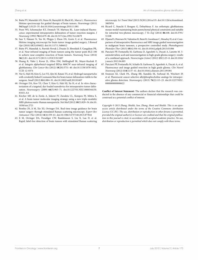
histological assessment (kappa = 0.98) (Figure 1) (60, 62, 63). (7)Two-photon or multi-photon fluorescence microscopy has beenutilized for tumor margin visualization. The advantage of usinginfrared lighting avoids the phototoxicity of tissues exposed tolight in the surgical field (64).
Discussion
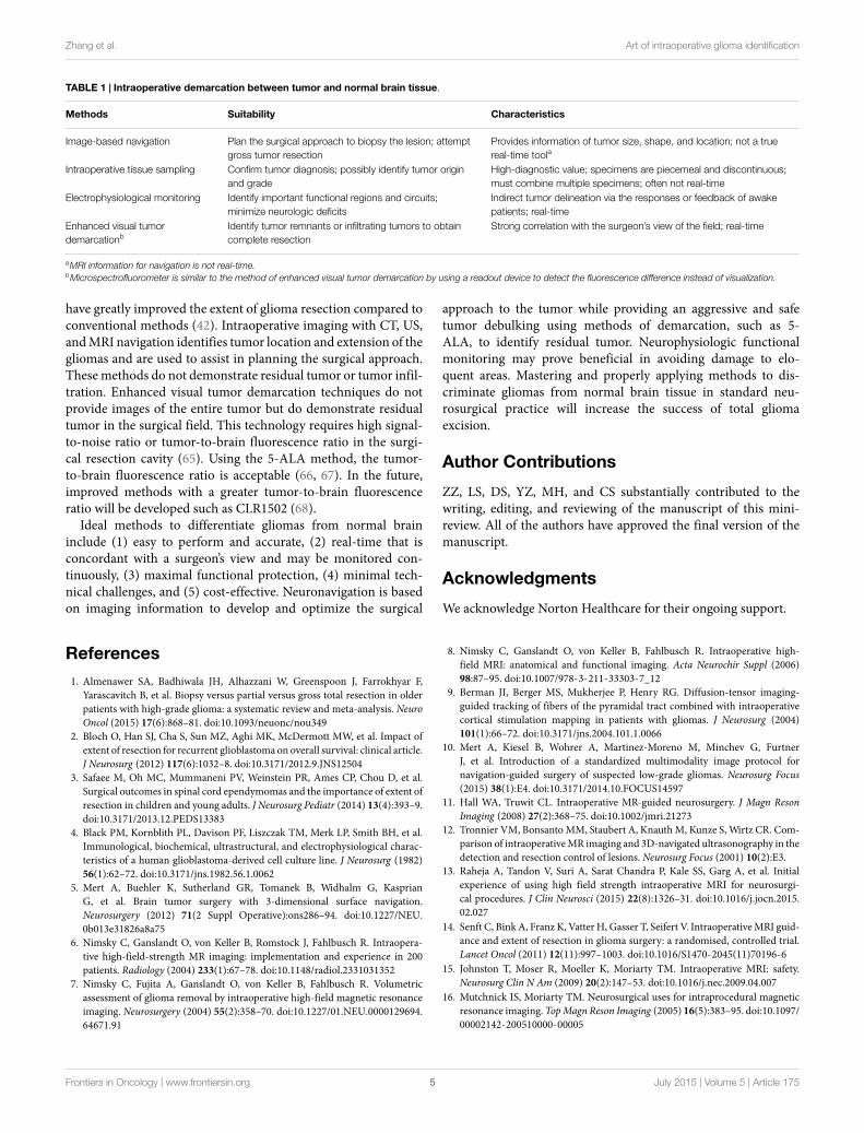
The ultimate goal of brain-tumor surgery is to attain maximumtumor resection and to achieve the greatest survival with minimalneurological deficits. In addition, it is important to obtain a precisepathological diagnosis of the cellular origins and grade of thetumor. The surgery reduces the neurological symptoms caused bytumor compression/invasion and tumor-associated tissue edema.The key issue for glioma surgery is to obtain intraoperative tumordelineation. Since the texture and color of gliomas, especiallylow-grade tumors, are similar to normal brain tissue, accuratetumor identification remains a challenge for neurosurgeons. Tra-ditionally, the determination of tumor margins depends on theexperience of the neurosurgeon to sense subtle differences incolor, texture, and surface vascularity of the glioma. Several tech-nologies used intraoperatively to delineate tumor margins havebeen developed and continue to rapidly evolve. Current availabletechnologies that assist with intraoperative demarcation betweentumor and normal brain tissue are listed in Table 1.
Each innovative technology has its advantages and limitations.In a recent review, advanced intraoperative delineation techniques
FIGURE 1 | In vivo stimulated Raman scattering microscopy imagesof human glioblastoma multiforme xenografts. (A) The arachnoidaland pial vessels on the surface of the normal brain were clearly identified inthe standard bright field and stimulated Raman scattering images.Visualization of the tumor was undetectable under standard operativeconditions. Certain regions of brain tissue that appeared grossly normalunder bright field microscopy revealed extensive tumor infiltration on
stimulated Raman scattering microscopy. The dashed line represents thetumor margin which was observed both biochemically and structurally.(B,C) The glioblastoma multiforme xenograft tissue was blue and cellular.(D) Normal axonal processes and vascular patterns were noted innon-infiltrated (normal) cortex. [Permission obtained from the publisherAmerican Association for the Advancement of Science (AAAS); Ji et al.(63)].
Frontiers in Oncology | www.frontiersin.org July 2015 | Volume 5 | Article 1754
Zhang et al. Art of intraoperative glioma identification
TABLE 1 | Intraoperative demarcation between tumor and normal brain tissue.
Methods Suitability Characteristics
Image-based navigation Plan the surgical approach to biopsy the lesion; attemptgross tumor resection
Provides information of tumor size, shape, and location; not a truereal-time toola
Intraoperative tissue sampling Confirm tumor diagnosis; possibly identify tumor originand grade
High-diagnostic value; specimens are piecemeal and discontinuous;must combine multiple specimens; often not real-time
Electrophysiological monitoring Identify important functional regions and circuits;minimize neurologic deficits
Indirect tumor delineation via the responses or feedback of awakepatients; real-time
Enhanced visual tumordemarcationb
Identify tumor remnants or infiltrating tumors to obtaincomplete resection
Strong correlation with the surgeon’s view of the field; real-time
aMRI information for navigation is not real-time.bMicrospectrofluorometer is similar to the method of enhanced visual tumor demarcation by using a readout device to detect the fluorescence difference instead of visualization.
have greatly improved the extent of glioma resection compared toconventional methods (42). Intraoperative imaging with CT, US,andMRI navigation identifies tumor location and extension of thegliomas and are used to assist in planning the surgical approach.Thesemethods do not demonstrate residual tumor or tumor infil-tration. Enhanced visual tumor demarcation techniques do notprovide images of the entire tumor but do demonstrate residualtumor in the surgical field. This technology requires high signal-to-noise ratio or tumor-to-brain fluorescence ratio in the surgi-cal resection cavity (65). Using the 5-ALA method, the tumor-to-brain fluorescence ratio is acceptable (66, 67). In the future,improved methods with a greater tumor-to-brain fluorescenceratio will be developed such as CLR1502 (68).
Ideal methods to differentiate gliomas from normal braininclude (1) easy to perform and accurate, (2) real-time that isconcordant with a surgeon’s view and may be monitored con-tinuously, (3) maximal functional protection, (4) minimal tech-nical challenges, and (5) cost-effective. Neuronavigation is basedon imaging information to develop and optimize the surgical
approach to the tumor while providing an aggressive and safetumor debulking using methods of demarcation, such as 5-ALA, to identify residual tumor. Neurophysiologic functionalmonitoring may prove beneficial in avoiding damage to elo-quent areas. Mastering and properly applying methods to dis-criminate gliomas from normal brain tissue in standard neu-rosurgical practice will increase the success of total gliomaexcision.
Author Contributions
ZZ, LS, DS, YZ, MH, and CS substantially contributed to thewriting, editing, and reviewing of the manuscript of this mini-review. All of the authors have approved the final version of themanuscript.
Acknowledgments
We acknowledge Norton Healthcare for their ongoing support.
References1. Almenawer SA, Badhiwala JH, Alhazzani W, Greenspoon J, Farrokhyar F,
Yarascavitch B, et al. Biopsy versus partial versus gross total resection in olderpatients with high-grade glioma: a systematic review and meta-analysis. NeuroOncol (2015) 17(6):868–81. doi:10.1093/neuonc/nou349
2. Bloch O, Han SJ, Cha S, Sun MZ, Aghi MK, McDermott MW, et al. Impact ofextent of resection for recurrent glioblastoma on overall survival: clinical article.J Neurosurg (2012) 117(6):1032–8. doi:10.3171/2012.9.JNS12504
3. Safaee M, Oh MC, Mummaneni PV, Weinstein PR, Ames CP, Chou D, et al.Surgical outcomes in spinal cord ependymomas and the importance of extent ofresection in children and young adults. J Neurosurg Pediatr (2014) 13(4):393–9.doi:10.3171/2013.12.PEDS13383
4. Black PM, Kornblith PL, Davison PF, Liszczak TM, Merk LP, Smith BH, et al.Immunological, biochemical, ultrastructural, and electrophysiological charac-teristics of a human glioblastoma-derived cell culture line. J Neurosurg (1982)56(1):62–72. doi:10.3171/jns.1982.56.1.0062
5. Mert A, Buehler K, Sutherland GR, Tomanek B, Widhalm G, KasprianG, et al. Brain tumor surgery with 3-dimensional surface navigation.Neurosurgery (2012) 71(2 Suppl Operative):ons286–94. doi:10.1227/NEU.0b013e31826a8a75
6. Nimsky C, Ganslandt O, von Keller B, Romstock J, Fahlbusch R. Intraopera-tive high-field-strength MR imaging: implementation and experience in 200patients. Radiology (2004) 233(1):67–78. doi:10.1148/radiol.2331031352
7. Nimsky C, Fujita A, Ganslandt O, von Keller B, Fahlbusch R. Volumetricassessment of glioma removal by intraoperative high-field magnetic resonanceimaging. Neurosurgery (2004) 55(2):358–70. doi:10.1227/01.NEU.0000129694.64671.91
8. Nimsky C, Ganslandt O, von Keller B, Fahlbusch R. Intraoperative high-field MRI: anatomical and functional imaging. Acta Neurochir Suppl (2006)98:87–95. doi:10.1007/978-3-211-33303-7_12
9. Berman JI, Berger MS, Mukherjee P, Henry RG. Diffusion-tensor imaging-guided tracking of fibers of the pyramidal tract combined with intraoperativecortical stimulation mapping in patients with gliomas. J Neurosurg (2004)101(1):66–72. doi:10.3171/jns.2004.101.1.0066
10. Mert A, Kiesel B, Wohrer A, Martinez-Moreno M, Minchev G, FurtnerJ, et al. Introduction of a standardized multimodality image protocol fornavigation-guided surgery of suspected low-grade gliomas. Neurosurg Focus(2015) 38(1):E4. doi:10.3171/2014.10.FOCUS14597
11. Hall WA, Truwit CL. Intraoperative MR-guided neurosurgery. J Magn ResonImaging (2008) 27(2):368–75. doi:10.1002/jmri.21273
12. Tronnier VM, BonsantoMM, Staubert A, KnauthM, Kunze S, Wirtz CR. Com-parison of intraoperativeMR imaging and 3D-navigated ultrasonography in thedetection and resection control of lesions. Neurosurg Focus (2001) 10(2):E3.
13. Raheja A, Tandon V, Suri A, Sarat Chandra P, Kale SS, Garg A, et al. Initialexperience of using high field strength intraoperative MRI for neurosurgi-cal procedures. J Clin Neurosci (2015) 22(8):1326–31. doi:10.1016/j.jocn.2015.02.027
14. Senft C, Bink A, Franz K, Vatter H, Gasser T, Seifert V. IntraoperativeMRI guid-ance and extent of resection in glioma surgery: a randomised, controlled trial.Lancet Oncol (2011) 12(11):997–1003. doi:10.1016/S1470-2045(11)70196-6
15. Johnston T, Moser R, Moeller K, Moriarty TM. Intraoperative MRI: safety.Neurosurg Clin N Am (2009) 20(2):147–53. doi:10.1016/j.nec.2009.04.007
16. Mutchnick IS, Moriarty TM. Neurosurgical uses for intraprocedural magneticresonance imaging.TopMagn Reson Imaging (2005) 16(5):383–95. doi:10.1097/00002142-200510000-00005
Frontiers in Oncology | www.frontiersin.org July 2015 | Volume 5 | Article 1755
Zhang et al. Art of intraoperative glioma identification
17. Hunt MA, Bago AG, Neuwelt EA. Single-dose contrast agent for intraoperativeMR imaging of intrinsic brain tumors by using ferumoxtran-10. AJNR Am JNeuroradiol (2005) 26(5):1084–8.
18. Gerganov VM, Samii A, Akbarian A, Stieglitz L, Samii M, Fahlbusch R. Relia-bility of intraoperative high-resolution 2D ultrasound as an alternative to high-field strength MR imaging for tumor resection control: a prospective compara-tive study. J Neurosurg (2009) 111(3):512–9. doi:10.3171/2009.2.JNS08535
19. Homapour B, Bowen JE, Want EJ, O’Neill K, Apostolopoulos V, Nandi D,et al. Intra-operative, real-time, three-dimensional ultrasound assisted position-ing of catheters in the microdialysis of glial tumours. J Clin Neurosci (2010)17(4):506–10. doi:10.1016/j.jocn.2009.06.022
20. Reinacher PC, Van Velthoven V. Intraoperative ultrasound imaging: practicalapplicability as a real-time navigation system. Acta Neurochir Suppl (2003)85:89–93. doi:10.1007/978-3-7091-6043-5_12
21. Unsgaard G, Selbekk T, BrostrupMuller T, Ommedal S, Torp SH,Myhr G, et al.Ability of navigated 3D ultrasound to delineate gliomas and metastases – com-parison of image interpretations with histopathology. Acta Neurochir (Wien)(2005) 147(12):1259–69. doi:10.1007/s00701-005-0624-1
22. Coburger J, Scheuerle A, Kapapa T, Engelke J, Thal DR, Wirtz CR, et al.Sensitivity and specificity of linear array intraoperative ultrasound in glioblas-toma surgery: a comparative study with high field intraoperative MRI andconventional sector array ultrasound. Neurosurg Rev (2015) 38(3):499–509.doi:10.1007/s10143-015-0627-1
23. Prada F, Perin A, Martegani A, Aiani L, Solbiati L, Lamperti M, et al. Intra-operative contrast-enhanced ultrasound for brain tumor surgery. Neurosurgery(2014) 74(5):542–52. doi:10.1227/NEU.0000000000000301
24. Ritschel K, Pechlivanis I, Winter S. Brain tumor classification on intraoper-ative contrast-enhanced ultrasound. Int J Comput Assist Radiol Surg (2015)10(5):531–40. doi:10.1007/s11548-014-1089-6
25. Serra C, Stauffer A, Actor B, Burkhardt JK, Ulrich NH, Bernays RL, et al.Intraoperative high frequency ultrasound in intracerebral high-grade tumors.Ultraschall Med (2012) 33(7):E306–12. doi:10.1055/s-0032-1325369
26. Talos IF, Zou KH, Ohno-Machado L, Bhagwat JG, Kikinis R, Black PM,et al. Supratentorial low-grade glioma resectability: statistical predictive analysisbased on anatomic MR features and tumor characteristics. Radiology (2006)239(2):506–13. doi:10.1148/radiol.2392050661
27. Selbekk T, Jakola AS, Solheim O, Johansen TF, Lindseth F, Reinertsen I,et al. Ultrasound imaging in neurosurgery: approaches to minimize surgicallyinduced image artefacts for improved resection control. Acta Neurochir (Wien)(2013) 155(6):973–80. doi:10.1007/s00701-013-1647-7
28. Solheim O, Selbekk T, Jakola AS, Unsgard G. Ultrasound-guided operations inunselected high-grade gliomas – overall results, impact of image quality andpatient selection. Acta Neurochir (Wien) (2010) 152(11):1873–86. doi:10.1007/s00701-010-0731-5
29. Eberlin LS, Norton I, Orringer D, Dunn IF, Liu X, Ide JL, et al. Ambientmass spectrometry for the intraoperative molecular diagnosis of human braintumors. Proc Natl Acad Sci U S A (2013) 110(5):1611–6. doi:10.1073/pnas.1215687110
30. Santagata S, Eberlin LS, Norton I, Calligaris D, Feldman DR, Ide JL, et al.Intraoperative mass spectrometry mapping of an onco-metabolite to guidebrain tumor surgery. Proc Natl Acad Sci U S A (2014) 111(30):11121–6. doi:10.1073/pnas.1404724111
31. Calligaris D, Norton I, Feldman DR, Ide JL, Dunn IF, Eberlin LS, et al. Massspectrometry imaging as a tool for surgical decision-making. J Mass Spectrom(2013) 48(11):1178–87. doi:10.1002/jms.3295
32. Huang J, Gholami B, Agar NY, Norton I, HaddadWM, Tannenbaum AR. Clas-sification of astrocytomas and oligodendrogliomas from mass spectrometrydata using sparse kernel machines. Conf Proc IEEE Eng Med Biol Soc (2011)2011:7965–8. doi:10.1109/IEMBS.2011.6091964
33. Lu Y, Li B, Xu J, Yu J. Dielectric properties of human glioma and surroundingtissue. Int J Hyperthermia (1992) 8(6):755–60. doi:10.3109/02656739209005023
34. Chang EF, Clark A, Smith JS, Polley MY, Chang SM, Barbaro NM, et al.Functional mapping-guided resection of low-grade gliomas in eloquent areasof the brain: improvement of long-term survival. Clinical article. J Neurosurg(2011) 114(3):566–73. doi:10.3171/2010.6.JNS091246
35. DeWitt Hamer PC, Robles SG, Zwinderman AH, Duffau H, BergerMS. Impactof intraoperative stimulation brain mapping on glioma surgery outcome: ameta-analysis. J Clin Oncol (2012) 30(20):2559–65. doi:10.1200/JCO.2011.38.4818
36. Duffau H, Denvil D, Lopes M, Gasparini F, Cohen L, Capelle L, et al. Intraoper-ative mapping of the cortical areas involved in multiplication and subtraction:an electrostimulation study in a patient with a left parietal glioma. J NeurolNeurosurg Psychiatry (2002) 73(6):733–8. doi:10.1136/jnnp.73.6.733
37. Duffau H, Lopes M, Arthuis F, Bitar A, Sichez JP, Van Effenterre R, et al.Contribution of intraoperative electrical stimulations in surgery of low gradegliomas: a comparative study between two series without (1985-96) and with(1996-2003) functional mapping in the same institution. J Neurol NeurosurgPsychiatry (2005) 76(6):845–51. doi:10.1136/jnnp.2004.048520
38. Sanai N, Martino J, Berger MS. Morbidity profile following aggressive resectionof parietal lobe gliomas. J Neurosurg (2012) 116(6):1182–6. doi:10.3171/2012.2.JNS111228
39. Tarapore PE, Findlay AM, Honma SM, Mizuiri D, Houde JF, Berger MS,et al. Language mapping with navigated repetitive TMS: proof of techniqueand validation. Neuroimage (2013) 82:260–72. doi:10.1016/j.neuroimage.2013.05.018
40. Yordanova YN, Moritz-Gasser S, Duffau H. Awake surgery for WHO grade IIgliomas within “noneloquent” areas in the left dominant hemisphere: toward a“supratotal” resection. Clinical article. J Neurosurg (2011) 115(2):232–9. doi:10.3171/2011.3.JNS101333
41. Moore GE. Fluorescein as an agent in the differentiation of normal and malig-nant tissues. Science (1947) 106(2745):130–1. doi:10.1126/science.106.2745.130-a
42. Barbosa BJ, Mariano ED, Batista CM, Marie SK, Teixeira MJ, Pereira CU, et al.Intraoperative assistive technologies and extent of resection in glioma surgery:a systematic review of prospective controlled studies. Neurosurg Rev (2014)38(2):217–26. doi:10.1007/s10143-014-0592-0
43. Margetis K, Rajappa P, Tsiouris AJ, Greenfield JP, Schwartz TH. Intraoperativestereotactic injection of indigo carmine dye to mark ill-defined tumor margins:a prospective phase I-II study. J Neurosurg (2015) 122(1):40–8. doi:10.3171/2014.9.JNS14113
44. Pichlmeier U, Bink A, Schackert G, Stummer W. Resection and sur-vival in glioblastoma multiforme: an RTOG recursive partitioning analy-sis of ALA study patients. Neuro Oncol (2008) 10(6):1025–34. doi:10.1215/15228517-2008-052
45. Schucht P, Seidel K, Beck J, Murek M, Jilch A, Wiest R, et al. Intraoper-ative monopolar mapping during 5-ALA-guided resections of glioblastomasadjacent to motor eloquent areas: evaluation of resection rates and neu-rological outcome. Neurosurg Focus (2014) 37(6):E16. doi:10.3171/2014.10.FOCUS14524
46. Stummer W, Rodrigues F, Schucht P, Preuss M, Wiewrodt D, NestlerU, et al. Predicting the “usefulness” of 5-ALA-derived tumor fluores-cence for fluorescence-guided resections in pediatric brain tumors: a Euro-pean survey. Acta Neurochir (Wien) (2014) 156(12):2315–24. doi:10.1007/s00701-014-2234-2
47. Zehri AH, Ramey W, Georges JF, Mooney MA, Martirosyan NL, Preul MC,et al. Neurosurgical confocal endomicroscopy: a review of contrast agents,confocal systems, and future imaging modalities. Surg Neurol Int (2014) 5:60.doi:10.4103/2152-7806.131638
48. Widhalm G. Intra-operative visualization of brain tumors with 5-aminolevulinic acid-induced fluorescence. Clin Neuropathol (2014) 33(4):260–78. doi:10.5414/NP300798
49. Acerbi F, Cavallo C, Broggi M, Cordella R, Anghileri E, Eoli M, et al.Fluorescein-guided surgery for malignant gliomas: a review. Neurosurg Rev(2014) 37(4):547–57. doi:10.1007/s10143-014-0546-6
50. Sanai N, Snyder LA, Honea NJ, Coons SW, Eschbacher JM, Smith KA, et al.Intraoperative confocal microscopy in the visualization of 5-aminolevulinicacid fluorescence in low-grade gliomas. J Neurosurg (2011) 115(4):740–8.doi:10.3171/2011.6.JNS11252
51. Shields LB, Choucair AK. Management of low-grade gliomas: a review ofpatient-perceived quality of life and neurocognitive outcome.World Neurosurg(2014) 82(1–2):e299–309. doi:10.1016/j.wneu.2014.02.033
52. Bottiroli G, Croce AC, Locatelli D, Nano R, Giombelli E, Messina A, et al. Braintissue autofluorescence: an aid for intraoperative delineation of tumor resectionmargins.Cancer Detect Prev (1998) 22(4):330–9. doi:10.1046/j.1525-1500.1998.CDOA34.x
53. Butte PV, Fang Q, Jo JA, Yong WH, Pikul BK, Black KL, et al. Intraoper-ative delineation of primary brain tumors using time-resolved fluorescencespectroscopy. J Biomed Opt (2010) 15(2):027008. doi:10.1117/1.3374049
Frontiers in Oncology | www.frontiersin.org July 2015 | Volume 5 | Article 1756
Zhang et al. Art of intraoperative glioma identification
54. Butte PV,Mamelak AN, NunoM, Bannykh SI, Black KL,Marcu L. Fluorescencelifetime spectroscopy for guided therapy of brain tumors. Neuroimage (2011)54(Suppl 1):S125–35. doi:10.1016/j.neuroimage.2010.11.001
55. Poon WS, Schomacker KT, Deutsch TF, Martuza RL. Laser-induced fluores-cence: experimental intraoperative delineation of tumor resection margins. JNeurosurg (1992) 76(4):679–86. doi:10.3171/jns.1992.76.4.0679
56. Sun Y, Hatami N, Yee M, Phipps J, Elson DS, Gorin F, et al. Fluorescencelifetime imaging microscopy for brain tumor image-guided surgery. J BiomedOpt (2010) 15(5):056022. doi:10.1117/1.3486612
57. Butte PV, Mamelak A, Parrish-Novak J, Drazin D, Shweikeh F, Gangalum PR,et al. Near-infrared imaging of brain tumors using the tumor paint BLZ-100to achieve near-complete resection of brain tumors. Neurosurg Focus (2014)36(2):E1. doi:10.3171/2013.11.FOCUS13497
58. Huang R, Vider J, Kovar JL, Olive DM, Mellinghoff IK, Mayer-Kuckuk P,et al. Integrin alphavbeta3-targeted IRDye 800CW near-infrared imaging ofglioblastoma. Clin Cancer Res (2012) 18(20):5731–40. doi:10.1158/1078-0432.CCR-12-0374
59. Nie G, Hah HJ, Kim G, Lee YE, QinM, Ratani TS, et al. Hydrogel nanoparticleswith covalently linked Coomassie blue for brain tumor delineation visible to thesurgeon. Small (2012) 8(6):884–91. doi:10.1002/smll.201101607
60. Orringer DA, Koo YE, Chen T, Kim G, Hah HJ, Xu H, et al. In vitro charac-terization of a targeted, dye-loaded nanodevice for intraoperative tumor delin-eation. Neurosurgery (2009) 64(5):965–71. doi:10.1227/01.NEU.0000344150.81021.AA
61. Kircher MF, de la Zerda A, Jokerst JV, Zavaleta CL, Kempen PJ, Mittra E,et al. A brain tumor molecular imaging strategy using a new triple-modalityMRI-photoacoustic-Ramannanoparticle.NatMed (2012) 18(5):829–34. doi:10.1038/nm.2721
62. Bentley JN, Ji M, Xie XS, Orringer DA. Real-time image guidance for braintumor surgery through stimulated Raman scattering microscopy. Expert RevAnticancer Ther (2014) 14(4):359–61. doi:10.1586/14737140.2013.877844
63. Ji M, Orringer DA, Freudiger CW, Ramkissoon S, Liu X, Lau D, et al.Rapid, label-free detection of brain tumors with stimulated Raman scattering
microscopy. Sci Transl Med (2013) 5(201):201ra119. doi:10.1126/scitranslmed.3005954
64. Ricard C, Stanchi F, Rougon G, Debarbieux F. An orthotopic glioblastomamouse model maintaining brain parenchymal physical constraints and suitablefor intravital two-photon microscopy. J Vis Exp (2014) 86:108. doi:10.3791/51108
65. Eljamel S, PetersenM,Valentine R, Buist R,GoodmanC,MoseleyH, et al. Com-parison of intraoperative fluorescence and MRI image guided neuronavigationin malignant brain tumours, a prospective controlled study. PhotodiagnosisPhotodyn Ther (2013) 10(4):356–61. doi:10.1016/j.pdpdt.2013.03.006
66. Panciani PP, Fontanella M, Garbossa D, Agnoletti A, Ducati A, Lanotte M. 5-aminolevulinic acid and neuronavigation in high-grade glioma surgery: resultsof a combined approach. Neurocirugia (Astur) (2012) 23(1):23–8. doi:10.1016/j.neucir.2012.04.003
67. Panciani PP, Fontanella M, Schatlo B, Garbossa D, Agnoletti A, Ducati A, et al.Fluorescence and image guided resection in high grade glioma. Clin NeurolNeurosurg (2012) 114(1):37–41. doi:10.1016/j.clineuro.2011.09.001
68. Swanson KI, Clark PA, Zhang RR, Kandela IK, Farhoud M, Weichert JP,et al. Fluorescent cancer-selective alkylphosphocholine analogs for intraoper-ative glioma detection. Neurosurgery (2015) 76(2):115–23. doi:10.1227/NEU.0000000000000622
Conflict of Interest Statement: The authors declare that the research was con-ducted in the absence of any commercial or financial relationships that could beconstrued as a potential conflict of interest.
Copyright © 2015 Zhang, Shields, Sun, Zhang, Hunt and Shields. This is an open-access article distributed under the terms of the Creative Commons AttributionLicense (CC BY). The use, distribution or reproduction in other forums is permitted,provided the original author(s) or licensor are credited and that the original publica-tion in this journal is cited, in accordance with accepted academic practice. No use,distribution or reproduction is permitted which does not comply with these terms.
Frontiers in Oncology | www.frontiersin.org July 2015 | Volume 5 | Article 1757