The AAO Forum for Osteopathic Thought...
36
The AAO JOURNAL JOURNAL Tradition Shapes the Future Volume 18 Number 4 December 2008 Forum for Osteopathic Thought Official Publication of the American Academy of Osteopathy ® Contemplations on the Art of OMT After Thirty Years of Practice Karen M. Steele, DO, FAAO Presents the 2008 Northup Lecture Page 9...
Transcript of The AAO Forum for Osteopathic Thought...

The AAO
JOURNAL JOURNAL Tradition Shapes the Future Volume 18 Number 4 December 2008
Forum for Osteopathic Thought
Official Publication of the American Academy of Osteopathy®
Contemplations on the Art of OMT After Thirty Years of Practice
Karen M. Steele, DO, FAAO Presents the 2008 Northup Lecture
Page 9...

Correction to "Improvement of L4-L5 disc positioning following treatment with orthotics used to cor-rect gait dysfunction and level the sacral base", AAOJ Vol. 18, No. 3 The original article displayed an image on page 22 was dated incorrectly. The images on the right side was mislabeled and should read Figure 2.MRI of lumbosacral spine taken on July 19th, 2007. This correction is made to maintain the integrity and scholarship of this article in particular and The AAO Journal in general. Fortunately, the date confirms no intervening treatment alluded to on page 23 paragraph one.
American Academy of Osteopathy® is your voice . . .
. . . in teaching, advocating, and researching the science, art and philosophy of osteopathic medicine, emphasizing the integration of osteopathic principles, practices and manipulative treatment in patient care.
The AAO Membership Committee invites you to join the American Academy of Osteopathy® as a 2008-2009 member. AAO is your professional organization. It fosters the core principles that led you to choose to become a Doctor of Osteopathy. For just $4.53 a week (less than a large specialty coffee at your favorite coffee shop or just 65 cents a day (less than a bottle of water), you can become a member of the specialty professional organization dedicated to the core principles of your profession! Your membership dues provide you with A national advocate for osteopathic manipulative
medicine (including appropriate reimbursement for OMM services) with osteopathic and allopathic profes-sionals, public policy makers, the media and the pub-lic.
Referrals of patients through the Find a Physician tool on the AAO website as well as calls to the AAO office.
Discounts on quality educational programs provided by AAO at its Annual Convocation and workshops.
New online courses (coming soon). Networking opportunities with your peers. Discounts on publications in the AAO Bookstore.
Watch for details about our new online market place coming soon!
Free subscription to the AAO Journal published four times annually.
Free subscription to the new AAO online member newsletter.
Access to the active members section of the AAO website which will be enhanced in the coming months to include many new features including resource links, job bank, and much more.
Discounts in advertising in AAO publications, on the website, and at AAO’s Convocation.
Access to the American Osteopathic Board of Neuromusulosketal Medicine—the only existing certifying board in manual medicine in the medical world today.
Maintenance of an earned Fellowship program to recognize excellence in the practice of osteopathic manipulative medicine.
Promotion of research on the efficacy of osteopathic medicine.
Supporting the future of the profession through the Undergraduate American Academy of Osteopathy on college campuses.
Your professional dues are deductible as a business expense.
Please watch your mail for membership dues renewal invoices. If you have any questions regarding membership or renewal of membership, please contact Kelli Bowersox, Director of Member Donor Relations, at 317/879-1881 or [email protected].
Thank you for supporting the American Academy of Osteopathy®
PAGE 2 A A O J O U R N A L

Contributors 4
2009 AAO Course Calendar (January-May) 6
CME Certification of Home Study Forms 32
2008 Journal Index 33
Editorial View from the Pyramids: Raymond J. Hruby, DO, FAAO 5
Regular features: Dig On: Richard L. Van Buskirk, DO, PhD, FAAO 6
Original Contribution Retrospective Study of a Peer Assessment Education Encounter in the Development of Osteopathic Clinical Skills Michael D. Lockwood, DO, FCA, Eric J. Snider, DO, and Michael Chipman, OMS IV
12
The detection and recording of cranial rhythmic impulse in acupuncture points using Surface Scanning Laser Displacement Meter and its signifi-cance. Krishnahari S. Pribadi, MD, ABPN Diplomat
20
Advertising Rates: Size of Ad:
Full Page $600 placed one(1) time $575 placed two (2) times $550 placed four (4) times
7 ½ x 9½
Half Page $400 placed one(1) time $375 placed two (2) times $350 placed four (4) times
7½ x 4¾
Third Page $300 placed one(1) time $275 placed two (2) times $250 placed four (4) times
2¼ x 4¾
Fourth Page $200 placed one(1) time $175 placed two (2) times $150 placed four (4) times
3⅓ x 4¾
Professional Card $60 3½ x 2
Classified $1.00 per word
Advertising Rates for The AAO Journal. Offi-cial Publication of the American Academy of Osteopathy® The AAO and AOA affiliate organizations and members of the Academy are entitled to a 20% discount on advertising in this Journal. Call the American Academy of Osteopathy® (317)879-1881 for more information. Subscriptions: $60.00 per year (USA) $78.00 per year (International)
3500 DePauw Boulevard Suite 1080
Indianapolis, IN 46268 P: (317) 879-1881 F: (317) 879-0563
www.academyofosteopathy.org
American Academy of Osteopathy® Guy A. DeFeo, DO.........................................President George J. Pasquarello, DO, FAAO…...President-Elect Harriet O’Connor, CFRE, CAE…..Executive Director
Editorial Advisory Board Raymond J. Hruby, DO, FAAO Denise K. Burns, DO Stephen M. Davidson, DO Eileen L. DiGiovanna, DO, FAAO Eric J. Dolgin, DO William J. Garrity, DO Stefan L. J. Hagopian, DO Hollis H. King, DO, PhD, FAAO John McPartland, DO Stephen F. Paulus, DO, MS Paul R. Rennie, DO, FAAO Mark E. Rosen, DO
The AAO Journal is not responsible for statements made by any contributor. Although all advertising is expected to conform to ethical medical standards, acceptance does not imply endorsement by this journal. Opinions expressed in The AAO Journal are those of authors or speakers and do not necessarily reflect viewpoints of the editors or official policy of the American Academy of Osteopathy® or the institu-tions with which the authors are affiliated, unless specified. Cover Photos: Courtesy of Karen Ayers Academic Graphic Artist/Photographer, West Vir-ginia School of Osteopathic Medicine.
PAGE 3 V O L U M E 1 8 , I S S U E 4
2008 Northup memorial lecture
Contemplations on the Art of OMT After Thirty Years of Practice Karen M. Steele, DO, FAAO
9
THE AAO
Official Publication of the American Academy of Osteopa-thy®
OURNAL J Forum for Osteopathic
The mission of the American Academy of Osteopathy®
is to teach, advocate, and research the science, art and phi-losophy of osteopathic medicine, emphasizing the integra-tion of osteopathic principles, practices and manipulative treatment in patient care.
Tradition Shapes the Future · Volume 18 Number 4 · December 2008
In This Issue:
The AAO Journal Raymond J. Hruby, DO, FAAO.........Interim Editor-in-Chief Harriet O’Connor, CFRE, CAE…………Supervising Editor Jennifer Taylor, MPH, CHES……………. Managing Editor The AAO Journal is the official publication of the American Academy of Osteopathy®. Issues are published in March, June, September, and December each year. Third-class postage paid at Carmel, IN. Postmaster: send address changes to: American Academy of Osteopathy® 3500 DePauw Blvd., Suite 1080, Indianapolis, IN., 46268 Phone: 317-879-1881; Fax: (317) 879-0563; www. academyofosteopathy.org
Student Physician
Healing Hands: Using Osteopathic Manipulative Treatment to Address Visceral Structures through Somatovisceral Reflexes: A Case Study in Gas-troesophageal Reflux Disease Beau Branyon, OMS III
29

PAGE 4 A A O J O U R N A L
Krishnahari Pribadi, MD, ABPN Diplomate “The detection and recording of cranial rhythmic impulse in acupuncture points using Surface Scanning Laser Displace-ment Meter and it’s significance.” Osteopathy in the cranial field continues to be controversial in some circles, yet at the same time is probably one of the most researched areas in the profession. In recent years instrumentation using Doppler technology, strain gauges, and now the Surface Scanning Laser Displacement Meter have emerged as instruments that could be validated for objective measurement of the cranial rhythmic impulse, thus allowing far better research in this area than ever before possible. Dr. Pribadi uses such technology to measure correlations between the cranial rhythmic impulse and acupuncture meridiens to illustrate interconnections between these two systems. Karen Steele, DO, FAAO, Professor and Associate Dean for Osteopathic Medical Education, West Virginia School of Os-teopathic Medicine “Contemplations on the Art of OMT After Thirty Years of Prac-tice.” In the 2008 Northup Lecture, Dr. Steele talks about the art of osteopathic practice, and specifically addresses what she feels are unique attributes that DOs bring to patient care. All this is based on her reflections on thirty years of practice in osteopathic medi-cine. She uses the art of pottery making as a metaphor to illustrate her lessons learned over the years. Her discussion provides valu-able insight into how we can all strive to better master the art of osteopathic practice. Michael Lockwood, DO, Eric Snider, DO, and Michael Chip-man, OMS IV, Department of Osteopathic Manipulative Medicine, Kirksville College of Osteopathic Medicine “Retrospective Study of a Peer Assessment Education Encoun-ter in the Development of Osteopathic Clinical Skills.” The word “doctor” comes from the Latin verb “docere”, meaning “to teach”. At one time or another most of us were taught that as doctors an important part of our job is to teach: to educate pa-tients, for example, and to educate future osteopathic physicians. The authors of this article describe a clinical educational experi-ence that allows first and second year osteopathic medical stu-dents to teach each other, to learn from each other, and at the same time to improve and validate their osteopathic manipulative treatment skills.
Contributors
Regular Features:
DIG ON. Richard L. Van Buskirk, DO, PhD, FAAO Muscle Impulse, A Muscle Energy Variant This paper describes a new osteopathic manipulative method, Muscle Impulse. The technique appears to be a variant of the Muscle Energy Technique but is faster and requires less resis-tance from the operator and less effort from the patient.
AAO Seeks New AAOJ Editor
and Associate Editor
In July of 2008, Robert C. Clark, DO experienced an un-expected illness. Recognizing that the need to rest more, most will agree that Dr. Clark’s family and practice come first and will understand that his volunteer activities must be curtailed, preventing him from having sufficient time to serve out his current three-year term (calendar years 2007-2009) as Editor of the AAO Journal, Dr. Clark notified AAO President Guy A. DeFeo, DO that he would end his service with the September 2008 edition of the AAOJ.
Dr. DeFeo invites interested AAO members to submit a cover letter and curriculum vitae to the AAO Board of Trus-tees, who have the responsibility for appointment of the jour-nal editor and associate editor. Assuming there will be multi-ple qualified candidates, the Board may wish to conduct inter-views at the 2009 AAO Convocation in Little Rock, Arkansas. Candidates should submit a cover letter and CV to AAO head-quarters, postmarked no later than Friday February 15, 2009. In the cover letter, the candidates should explain why they wish to serve in this important post and document professional experience that would qualify them for the job.
The AAOJ Editor and Associate Editor serves without compensation. He/she is appointed to a three-year term and charged with the responsibility of publishing a quarterly jour-nal within the annual budget appropriated by the Board of Trustees. The Editor will be responsible for soliciting contri-butions, submitting them to peer review, and selecting the final material for publication. The Journal Editor will have recourse to the advice and assistance of the Editorial Advisory Board, the Publications Committee and the AAO executive director. The Editor works with the AAO staff, particularly the Com-munications Director, to write various columns, proof blueline drafts, and meet publication deadlines, i.e. the first day of March, June, September, and December. While there is con-siderable latitude given to the Journal Editor, the Board of Trustees will expect continued evolution of this professional journal with the long range goal to have it included in the Na-tional Library of Medicine's Index Medicus.
Send cover letter and CV to: American Academy of Osteopathy®
3500 DePauw Blvd., Suite 1080 Indianapolis, IN 46268
Fax: 3171879-1881 E-mail: [email protected]

PAGE 5 V O L U M E 1 8 , I S S U E 4
“Just when I thought I was out…they pull me back in.” Thus I am reminded of this quote, uttered by Don Michael Cor-leone (a.k.a. Al Pacino) in the 1990 film “The Godfather: Part III” (along with my apologies for ending a sentence with a preposition!). As some of you already know, I was the founding editor of The AAOJ and remained so for 10 years. At that point the editorship was passed to Anthony Chila, DO, FAAO, and more recently to Robert Clark, DO. Recently, Dr. Clark in-formed us that he needed to step down as the Editor-in-Chief of the Journal for personal reasons. He sent us a statement about his situation, which reads as follows:
“The September 2008 issue is my last as the editor in chief. In July, I had an unexpected illness. Although mercifully short, it has had some lingering effects. I do not have the stamina that I had before. Fortunately, that is improving albeit slowly. I have been forced to examine my life and prioritize my activities. For the past three years my practice has been half time. This gave me time for various profes-sional and personal activities. With the need to rest more, there were too many things in my personal life that were not getting done. Most will agree that my family and practice come first and will understand that my volunteer activities must be curtailed. Based on that premise, I have resigned as editor in chief of The AAO Journal”.
We wish Dr. Clark all the best, hope that he is doing well, and thank him for the outstanding job he did as Editor-in-Chief of the Journal these past years. When all this occurred, I was contacted by the AAO leader-ship and asked if I would step in as interim Editor-in-Chief in order to make sure The AAOJ stayed on track and we met publi-cation deadlines on time. I agreed to do so, and the AAO lead-ership is planning a strategy for the future of The AAO Journal. While all of this is going on, I also recently stepped down from my full time position as Chair of the OMM Department at the Western University College of Osteopathic Medicine of the Pacific (COMP), and am now working on only a part-time basis at the University. Although I do have lots of other plans for my time, this does allow me some room to take on the AAOJ Edi-torship without too much difficulty. My part-time activities at COMP primarily involve doing faculty development with the newly hired OMM faculty and the Predoctoral Teaching Fel-lows, and doing some curriculum development and analysis for the department. I do plan on doing some other writing and pub-lishing, but all in due time. An unusual thing (perhaps an omen of sorts?) happened to me while working on this issue of The AAOJ. I started writing this editorial page while at the COMP campus the other day, and while there I stopped into the OMM department library to look at some of the past issues of the Journal for inspiration. The very first issue I picked up was from December 1998 -- exactly 10 years ago! Do you remember what was in that issue? I didn’t either. Here is a listing of the major articles published in that issue: “The Early History of Osteopathy”, by Dennis Dowling, DO, CSPOMM (who has since earned his FAAO);
“How Does our Profession Get 100,000 Cases of Influenza Re-ported?”, by Deborah M. Heath, DO, and Albert F. Kelso, PhD; The Thomas L. Northup Lecture: “The Tree and the Wind: A Fable of Osteopathic Growth and Destruction”, by Eileen L. Di-Giovanna, DO, FAAO; “Is Human Cerebrospinal Fluid Reab-sorbed by Lymph? Lymph Drainage Therapy (LDT) and Manual Drainage of the Central Nervous System”, by Bruno Chikly, MD; and “Evolving Strategies for Management of Various Mus-culoskeletal Disorders: Evidence-Based Approaches and Be-yond”, by Wolfgang G. Gilliar, DO. In my editorial page in that same issue I discussed osteo-pathic unity. I have always been impressed at how the leaders and members of the AAO can always come together in times of need, much like now when there is a need to step in and keep the Journal a growing, vibrant entity within our organization. I made note of the fact that we could all learn some valuable information about unity by observing flock of geese flying in formation. I quoted an excerpt from an address first given by anthropologist Angeles Arrien and published in the newsletter of the Maryland Association of Extension Home Economists. The excerpt is as follows:
Fact 1: As each bird flaps its wings, it creates a uplift for the bird following. By flying in a “V” formation, the whole flock adds 71% greater flying range than if one bird flew alone. Lesson 1: People who share a common direction and sense of community can get where they are going quicker and easier because they are traveling on the strength of one another. Fact 2: Whenever a goose falls out of formation, it suddenly feels the drag and resistance of trying to fly alone and quickly gets back into formation to take advantage of the lifting power of the bird immediately in front. Lesson 2: If we have as much sense as geese, we will stay in formation with those who are ahead of where we want to go and be willing to accept their help as well as give ours to oth-ers. Fact 3: When the lead goose gets tired, it rotates back into for-mation and another goose flies the point position. Lesson 3: It pays to take turns doing the hard tasks and sharing leadership. Fact 4: The geese in formation honked from behind to encour-age those up front to keep up their speed. Lesson 4: We need to make sure our honking from behind is encouraging and not something else. Fact 5: When a goose gets sick wounded or shot down, two geese drop out of formation and follow it down to help and protect it. They stay with it until it is able to fly again or dies. Then they launch on their own, with another formation, or catch up with their flock. Lesson 5: If we have as much sense as geese, we, too, will stand by each other in difficult times as well as when we are strong.
Lessons from geese. We can still learn from them, as much now as back then.
From the Pyramids
Raymond J. Hruby

PAGE 6 A A O J O U R N A L
WHY Choose AAO? Highest quality CME that can be found anywhere.
Personal focus on your education with low table trainer to student ratios of 1:8!
Participants are educated to view function as related to the structure of the body and how to arrive at diagnoses, which include critical areas of somatic dysfunction needing treatment.
AAO courses provide a coding and reimbursement component to bring you up-to-date on current billing procedures.
Development of palpatory and manual treatment skills with colleagues who are the most sought after teachers of musculoskeletal medicine and palpatory diagnosis in the world.
Every introductory course reviews osteopathic history as well as em-phasizing osteopathic principles, philosophy and palpatory diagnosis--the key elements to successful implementation of manual and muscu-loskeletal medicine.
Upcoming Course Offerings
Enhance your Education! Develop your Skills! Earn CME Credit!
December 5-7, 2008 Osteopathic Approach to Cranial Nerve Dysfunction ala Barral Pomona, California 24 CME* This new course will cover the functional asso-ciations between the cranial nerves, the sutures, the dura, and the brain. By directly palpating the peripheral branches of cranial nerves, we can evaluate/palpate them for abnormal tension.
January 9‐11, 2009 Fundamentals of OMM Ft. Lauderdale, Florida 20 CME* This course will review indications, contraindi-cations, documentation and coding for osteo-pathic manual medicine; indications for treat-ment, approach to common conditions, inte-grating OMT into your clinical practice.
January 23‐25, 2009 Fundamentals of OMM Glendale, Arizona 20 CME* This course will review indications, contraindi-cations, documentation and coding for osteo-pathic manual medicine; indications for treat-ment, approach to common conditions, inte-grating OMT into your clinical practice.
February 28‐March 1, 2009 Dr. Fulford’s Basic Percussion: A Systematic Approach to the Whole Body Tucson, Arizona 15 CME* This level III course is based on the life work of Robert C. Fulford, DO in osteopathy, life en-ergy, vibration, and the use of the percussion vibrator. This basic course explores these con-cepts using material that Dr. Fulford developed during his lifetime.
For more information or to register for any of these programs, please visit:
www.academyofosteopathy.org
May 1‐3, 2009 Evidenced-Based Manual Medicine: A Prob-lem Oriented Approach Pomona, California 20 CME* This is a level I didactic and hands-on labora-tory course designed for the practicing clini-cian, educators or health professional students, who wish to better understand the evidence-based manual medicine approach to patients with musculoskeletal problems.
May 15‐17, 2009 The Still Technique Stratford, New Jersey 20 CME* This level III course will cover the history of the Still Technique, its loss and recovery; iden-tify the underlying method of the Still tech-nique; learn segmented diagnostic techniques that are shared by this technique with HVLA and muscle energy technique as well as those unique to the Still technique.
American Academy of Osteopathy®
Diana L. Finley, CMP, Education Director 3500 DePauw Blvd, Suite 1080
Indianapolis, IN 46268 P: (317) 879-1881 F: (317) 879-0563
Email: [email protected]
March 25, 2009 Progressive Inhibition of Neuromusculoskeletal Structures (PINS) Little Rock, Arkansas 6 CME* This level I course, developed by Dennis J. Dowling, DO, FAAO, is a system of diagnosis and treatment in which the osteopathic practi-tioner locates two related points and sequen-tially applies inhibitory pressure along a series of related points.
March 29‐31, 2009 Osteopathic Considerations in the Foregut with an Emphasis on Gastroesophageal Re-flux Disease Little Rock, Arkansas 20 CME* This new level IV course will explore the fore-gut and its most common medical condition, gastroesophageal reflux disease (GERD).
March 26-29, 2009 AAO Annual Convocation Basic Mechanisms of Osteopathy: Balancing the Neuroendocrine Immune System Little Rock, Arkansas 26.5 CME* The annual Convocation will re-discover the role of science as a key to balancing physio-logical function through the use of osteopathic manipulative treatment.
*The AAO has requested that the AOA Council on Continuing Medical Education approve this program for (the identified number of) hours of AOA Category 1-A CME credits.

Dig On: Muscle Impulse, A Muscle Energy Variant
Quite unexpectedly, I have stumbled onto another new method for manipulating musculoskeletal restrictions or somatic dysfunctions. I was instrumental in the recovery and redevelop-ment of a manipulative method originally used by Andrew Tay-lor Still14,15. Therefore, it is with more than a little bemusement that I have once again come across a manipulative method that seems sufficiently different from others developed by our pro-fession that I believe it deserves a new name. The initial discovery of this seemingly new manipulative method came while treating the tensor fascia lata and iliotibial (IT) band. Although the Still Technique is quite powerful in treating a wide variety of tissue restrictions, it too is not always successful. One place where it sometimes fails to demonstrate efficacy is in treatment of the marked tenderness over the femo-ral greater trochanter. If the gluteal muscles and their tendons have been successfully treated and tenderness over the greater trochanter remains, I typically treat the tensor fascia lata and IT band using Still Technique. If that resolves the tenderness in the tensor fascia lata and IT band but the greater trochanter remains tender to palpation, I begin to entertain the possibility that it represents a trochanteric bursitis. Rather than jumping to conclu-sions, I typically will attempt to treat it with muscle energy or counterstrain. If that fails, I then conclude that a trochanteric bursitis is the source of the problem and treat the patient accord-ingly. Unfortunately, the counterstrain position of ease for the ten-sor fascia lata and IT band (marked abduction of the leg at the hip) would sometimes cause tension and pain in the antagonist muscles, particularly adductor brevis and longus. One time, rather than stopping and treating these antagonists with Still Technique, I asked the patient to attempt to adduct the leg while I continued to hold the position of ease for the tensor fascia lata. In the past, I had simply followed the muscle energy formula, progressively abducting the leg three times after a voluntary adduction against resistance for 7 to 10 seconds. In this particu-lar case, the patient was older and not in good shape. I knew from experience with her that she would have trouble sustaining any muscle action. I simply asked her to try to push with her leg briefly three times. She pushed for about a second three times and I did not try to further abduct the leg between pushes. Much to my surprise her pain in her adductors stopped and I was able to complete the counterstrain for the tensor fascia lata. When I evaluated her hip adductor muscles there was no evidence of somatic dysfunction. Without thinking too much about it, I began to use this pulse technique whenever I was performing a Counterstrain maneuver and had to deal with pain in the patient’s antagonist muscles. I did not realize that I was doing anything unusual until a student who was rotating in my office asked me about it. At that point, I began to look at it as a variant of muscle energy, and further evaluate its properties. One thing that rapidly became apparent was that, like muscle energy, three successive activations of the muscles being treated
Richard L. Van Buskirk were necessary and sufficient to produce a release. If I was using the technique as a muscle energy variant rather than as an adjunct to counterstrain, I would bring the af-fected tissue to its restriction and then hold it in place while the patient mounted three very brief impulses away from the barrier. Only after the third impulse I would then passively move the tissue through where the barrier had been. Typically the amount of release past the restriction was similar to the result if the pa-tient had produced three prolonged isometric activations fol-lowed each time by the operator moving the tissue toward the “new barrier”. Similar to muscle energy the new technique would fully resolve any and all signs and symptoms of somatic dysfunction (asymmetry of presentation and range of motion, tissue texture changes, and tenderness if present). I began to show this new variation of the muscle energy technique. The physicians and students who saw it and tried it felt it was enough different that it deserved its own name. There-fore, I have taken to calling it muscle impulse. At this point I do not know if others have also found this variant. However I can find no reference to it. In some ways muscle impulse is similar to T. J. Ruddy’s “rapid resistive duction” technique12. This early manipulative method is considered a precursor to the muscle energy technique developed by Fred Mitchell, Sr., DO11. However, Ruddy’s tech-nique involved holding a tight muscle at its’ restrictive barrier and having the patient (or the operator if the patient was unable to cooperate) mount a series of very rapid miniature contractions toward the barrier at the basal heart rate (up to 20 contractions at 60 per minute) while holding the tissue rigidly in place. The method was focused on strengthening a muscle rather than re-moving restrictions. As it is currently conceived, muscle impulse follows the fol-lowing protocol:
Identify the tissue showing signs of somatic dysfunction. Like muscle energy (and as well counterstrain and Still technique) the technique can treat a wide variety of tissues including muscles and tendons, ligaments, joints, and vertebrae33,4,10,11.
Identify the tissue restriction. The more accurate the de-lineation of the “barrier” the better the results. In this regard, it is very similar to both muscle energy and high velocity low amplitude (HVLA).3,7,8,9,10,11,16
Bring the tissue to its restriction and hold it in place. Have the patient produce three very brief attempts to move
the tissue towards its ease (away from the restriction). I typically give the instructions “Push, Stop, Push, Stop, Push, Stop” as fast as I can say them. This does not allow the development of any real muscle force. Ideally the tissue should be monitored to ensure that the patient is in fact making an isometric impulse.
As soon as the patient has made the last isometric impulse the physician carries the tissue passively through the area of prior restriction. As in muscle energy, HVLA
PAGE 7 V O L U M E 1 8 , I S S U E 4

CME QUIZ The purpose of the quiz found on page 32 is to provide a con-venient means of self-assessment for your reading of the scien-tific content in the “Muscle Impulse, A Muscle Energy Variant” by Richard L. Van Buskirk, DO, PhD, FAAO. Answer each of the questions listed. The correct answers will be published in the March 2009 issue of The AAOJ. To apply for Category 2 -B CME credit, transfer your answers to the AAOJ CME Quiz Application Form answer sheet on page 32 The AAO will record the fact that you submitted the form for Category 2 -B CME credit and will forward your test results to the AOA Division of CME for documentation. You must have a 70% accuracy to order to receive CME credits.
PAGE 8 A A O J O U R N A L
9. McConnell CP. The Practice of Osteopathy, 2nd Edition. The Hammond Press, 1900.
10. Mitchell Fred L. Jr., and Mitchell, P.K.G. The Muscle Energy Manual, Vols. 1,2,3, MET Press, East Lansing, MI, 1995.
11. Mitchell Fred L. Jr., Moran, P.S., and Pruzzo, N.A. An Evaluation and Treatment Manual of Osteopathic Muscle Energy Procedures. Mitchell, Moran, and Pruzzo, Associates, Valley Park, MO, 1979.
12. Ruddy TJ. Osteopathic rapid rhythmic resistive technic. Academy of Ap-plied Osteopathy Yearbook, 1962, 23-31.
13. Van Buskirk RL. Nocioceptive reflexes and the somatic dysfunction: a model. JAOA, 90 (1990), 792-809.
14. Van Buskirk RL. A manipulative technique of Andrew Taylor Still JAOA, 96 (1996), 597-602.
15. Van Buskirk RL. Still Technique Manual, Applications of a Rediscovered Technique of Dr. Andrew Taylor Still, Second Edition. American Academy of Osteopathy, Indianapolis, IN, 2007.
16. Walton WJ. Textbook of Osteopathic Diagnosis and Technique Proce-dures. The American Academy of Osteopathy, Newark, OH, 1972.
Accepted for Publication: October 2008
After January, 2009 please address correspondence to: Richard L. Van Buskirk, DO, PhD, FAAO 2900 S. Tamiami Trail Sarasota, FL, 34239 Email: [email protected]
and Still technique there is no sense of remaining bar-rier and the tissue shows its full normal range of mo-tion.
Muscle Impulse differs from muscle energy in several important details:
Muscle energy takes approximately 35 seconds to execute per tissue treated according to the original proto-col.3,10,11 I have heard others discuss doing the isomet-ric activation for less than 10 seconds per activation, but never for as brief a period as that used in muscle impulse (approximately one second). Thus, the whole treatment of a restricted tissue in muscle impulse takes no more than four seconds.
Muscle energy has the operator move the tissue into the barrier after each isometric activation.3,10,11 Muscle impulse holds the tissue rigidly at the original restric-tion until all three impulses are finished, then moves the tissue through what had been the restriction.
Muscle energy requires the operator to carefully try to get the patient to use as little force as possible during the isometric phase.3,10,11 Often this is a losing battle and the patient actually moves the tissue non-isometrically to at least some degree. This also puts some strain on the operator to try to hold the tissue in place. Muscle Impulse involves such a brief activation of the muscle that only minimal force is actually generated by the patient. In this regard it is more similar to the opto-kinetic activation sometimes utilized in the cervical spine. There is no strain on the operator.
As with most osteopathic manipulative methods there is no easily defined mechanism of action. In part of course that is because there is no rigorously tested hypothesis as to the genera-tion or maintenance of the somatic dysfunction. Lacking that, there are many hypotheses both as to the causes of somatic dys-function and the mechanism of action of manipulative methods in relieving somatic dysfunction. I tend to lean toward an inte-grated mechanism that includes both neural elements and fascial elements13,15 as does Fred Mitchell Jr., DO10, although most texts delineating putative mechanisms underlying Muscle En-ergy tend to focus on neural mechanisms to the extent they dis-cuss mechanisms at all3,8,11. Clearly the rapidity with which resolution occurs points to at least a partially neural mechanism, whether involving the nociceptor13,15 or muscle spindle.1,2,5,6
References: 1. Denslow JS, and Hassett. The central excitatory state associated with pos-
tural abnormalities. J. Neurophysiol, 5 (1944). 393-402. 2. Denslow JS, Korr IM, and Krems AD. Quantitative studies of chronic
facilitation in human motoneuron pool. Am J Physiol, 105 (1947). 229-38. 3. Greenman PE. Principles of Manual Medicine. Third Edition. Lippincott
Williams & Wilkins, Baltimore. 2003. 4. Jones LH. Strain & Counterstrain. American Academy of Osteopathy,
Newark, OH. 1981. 5. Korr IM. The neural basis of the osteopathic lesion. JAOA, 47 (1947). 191-
198. 6. Korr IM. Proprioceptors and somatic dysfunction. JAOA, 75 (1975). 638-
650. 7. Kappler RE. and Jones JM. Thrust (High-Velocity Low-Amplitude). In:
Robert C. Ward (ed.), Foundations for Osteopathic Medicine, 2nd Edition. Lippincott Williams & Wilkins, Philadelphia, PA 20030. 852-880.
8. Kuchera M and Kuchera WM. Osteopathic Principles in Practice, 2nd Edition, KCOM Press, Kirksville, MO. 1991.

Contemplations on the Art of OMT After Thirty Years of Practice
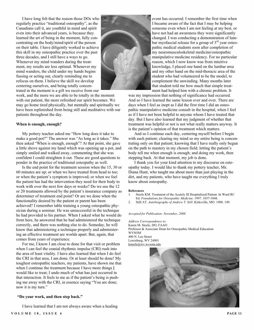
For a few moments today, I would like to talk a bit about the art of osteopathic manipulative treatment, OMT, osteopathy, or traditional osteopathy, as you may call it. I have chosen to ex-plore those attributes within ourselves which we must bring to the patient interaction when providing OMT to our patients. And, I have chosen to use the metaphor of the art of pottery in this treatise, as I am a novice potter and still easily fall into the “beginners mind” of this art. Both osteopathy and pottery deal with dirt and the divine, and either can be a metaphor for the other. I learned osteopathy first and much later became a novice potter. The lessons I have learned and gifts I have received from osteopathy that I would like to ponder today are summed up by the following phrases:
Do not expect to be good for a really, really, long time. Center yourself first. When is enough, enough? Do your work, and then step back.
Do not expect to be good for a really, really long time. I have always felt like I was sculpting when doing OMT, starting with a vision of the underlying anatomy as I would try to gracefully, purposely, and firmly move the tissues and ener-gies under my hands, leading the body toward a more functional balance. So it seems natural that I would eventually want to try working with clay. I had not thrown very many pots before my teacher began encouraging me to buy my own pottery wheel and kiln. I had been renting space and time in her studio, so this rec-ommendation would not have come from a perspective of finan-cial gain. I would throw a few tiny pots which she would in-clude with other tiny pots into a student bisque firing. I would then apply the glazes to my tiny pots and she would put them through a second student firing. The resultant tiny pots brought me great joy.... for a while. Then I wanted to explore with other clays and other glazes. The results were less predictable, and sometimes catastrophic. I learned why she put student work in a firing separate from her professional work – uneven pots ex-plode in the kiln when the heat is absorbed differentially across uneven pot walls! And any pieces on that same shelf are in jeop-ardy of being shattered from flying shrapnel. I learned that glazes applied too thickly run and then fuse to the kiln shelf. I learned that red glazes make everything else in the kiln red, and that glazes which are not a good match for the clay pit, leaving unglazed areas which make the pot unsafe for using with food. After I had the awful experience of a whole shelf of my work explode because I could not make the hard decision to discard a faulty piece, and went ahead and fired it with other pieces in my kiln, she said to me “Now you are truly a potter”! After I had the soft red terra cotta clay melt and fuse to the kiln shelf because I would fired it at Cone 6 rather than Cone 06, it
altered my respect for the characteristics of the clay body. And it reminded me of the power of logarithmic tables. Now, when I look at a pot, I “see” with more than just my eyes. I see the beauty of the clay and glazes which have been selected to form a functional and beautiful piece. I also know the strength of the clay that has been used; I see the lines of the potter’s fingertips making the swirl in the bottom of the bowl; I know if the glaze chosen was well suited to the clay body of the piece; and I ap-preciate the asymmetries that make this piece unique and hand-made rather than poured and casted. And I am still very much a beginner in the art of pottery. Is it not the same for the practice of osteopathy, after we have been at it a while? We evaluate a patient and we can see what the end result will likely be, with and without our treat-ment. We do not really know how we know, be we know there is a short leg, or emotional trauma underlying the patient’s symptom. We can sense the age of our patient when they in-curred the contributing trauma or illness. Some of us can see damaged internal tissue, or sense vibration aberrations in the patient. Based on our cognitive and intuitive knowledge, we envision what we believe could be the desired end result – a child who can efficiently coordinate their suck and swallow mechanism; a teenager who can run without knee pain; better respiratory status in a midlife adult hospitalized with pneumo-nia; or peace in an elder facing end of life issues. We then for-mulate a treatment plan, knowing that we will reevaluate at every visit, and we will be willing to allow the status of the pa-tient at follow up to alter our original plan. We learn how to “dose” our osteopathic manipulative treatments, through lessons from our teachers – those more experienced in this skill, and our patient teachers, who are the stricter of the two. Early in my osteopathic practice, I had an elder return to me with greater pain after a vigorous treatment to a frail body. I then really knew that older bodies need gentler handling. I had over treated her and put her to bed for a few days. Fifteen years later, after many more lessons learned about dosing OMT, I discussed the ma-nipulative prescription concept in detail in my chapter “Treatment of the Acutely Ill Hospitalized Patient” in the first edition of Foundations for Osteopathic Medicine1. I saw a woman in her early 40’s for recurrent headaches, whose medical work up was benign. I finally thought to have her stand up and performed a postural examination. I found a significant postural strain which, when addressed, provided relief in her symptoms. Early in my career, I had children improve in their middle ear functioning after 8 or 10 treatments, for which the parents and I were delighted. Then I learned the secret of the diaphragm in children’s problem, and when I began to routinely access and treat the diaphragm, the children got better much more quickly. Now I have learned to trust my “intuition” that there is “something” at a given level of my patient’s being, a problem of “mind, matter or motion”.2
Karen M. Steele
PAGE 9 V O L U M E 1 8 , I S S U E 4
2008 Northup Lecture

There is a saying that smooth seas never made a great mari-ner. We do not become a master in traditional osteopathy from reading books, or from listening to our wise elders. We must “go to sea” and really experience the dance of healing with our patients, and then follow up and see how their body responded. It is that feedback over time from many patient interactions which hones our skills. I remember Dr. John Harakal at a Fac-ulty Development Seminar sponsored by the Sutherland Cranial Teaching Foundation expressing alarm and distress that there were young osteopathic practitioners who were teaching cranial osteopathy with only five years of experience. He thought it was preposterous! We must have experience before we can antici-pate the end. Hence my teacher’s exclamation “Now you are a true potter” when all my pots on one shelf were shattered. It was more than just disappointment that I had lost a few pots. The time making those pots had not been wasted because I had in-creased my skill by making them. It was the visceral reaction in me because I had been told this could happen, I knew better, and I did it anyway. After that experience I really knew to carefully select which pots go on to be fired. It takes 60 months to obtain 5 years of experience, which for most skills, is a minimum amount of time to become competent – not highly skilled, just competent. Anticipating the end is the gestalt of experience that a seasoned osteopathic practitioner brings to every patient interaction. I remember Dr. Robert Ful-ford remarking late in his life, that he was amazed at what he had learned in the previous few years. The better we can predict the end and the paths our patients will likely follow to get there, the more efficiently we can help guide them toward health. Center yourself first Everyone knows that in the art of pottery, centering is the hardest part. Centering is at the center of pottery – and of osteopathy. But what is centering? And how do we learn to center ourselves? We learn from our wise ones, and from our patients, while we keep trying – forever. I have learned in pottery to wedge the clay well; form it into approxi-mately the shape I want this pot to take; start the wheel; and then sit and wait. I feel the clay in my hands; watch and feel the wheel in its hypnotizing rhythm; raise my arms; and only when my whole being is focused on making the clay sit at the center of the spin-ning wheel do I forcefully throw the clay onto the wheel. It is the same with an interac-tion with a patient. We enter the room and begin interviewing the patient, at first nonverbally, and then with our questions. We decide if OMT would likely be benefi-cial to that patient, and if so, we obtain their consent. Then we center ourselves, and align ourselves with our patient. When there is nothing else in our mind except that patient’s body/mind/spirit, and how we will enter into a dance of healing with them, then we begin our treatment. I learned from the potter’s wheel a new found respect for the intentional decision to become focused on the task at hand, and let nothing else enter my thoughts. I learned to concentrate only
on the speed of the wheel, the wetness and consistency of the clay, and the position of my hands in relation to the clay. I re-member noticing the tension in my triceps muscle, as I learned to pull the clay toward me, and against the centrifugal force of the wheel. I have utilized this same concept when doing OMT, where I am not just pushing or pulling a bone or fascial band, but balancing that body from within, allowing it to function more fully, gracefully, and comfortably. I have found this skill to be crucial in being able to treat the “rich and famous”. I re-member my first time I treated my then Department Chairper-son, Dr. Mike Kuchera, when I was a resident. I was so nervous that I was shaking, even though I had known him for a long time, as we had graduated only a year apart from KCOM a little more than 10 years prior, and his wife and I had been friends before I ever even met Dr. Mike. What he received was an ar-ticulatory treatment! Now I am much better able to simply inter-act with the center of the being on my table. I am not tied to the outcome – only to doing my best for that treatment. I can treat the wealthy, or famous, or everyday person with the same level of intent and skill. I am freed from feeling that those who travel hours or days to come to me for treatment deserve more. What they deserve is my best, which is not necessarily more. I am freed from ego-centric caring what the outcome of “my” treat-ment will be. So I do not feel the pressure of “proving” osteopa-thy to a skeptic who is giving it a try. All I have control over is what I bring to that interaction, and the patient then does with it what he or she will. It is freeing, to be centered. But it is not necessarily easy or automatic. Surely, it is much easier and auto-matic with practice, but I find it still requires intention on a
regular basis. And where does this centering come from? Again, taking the metaphor of throwing a clay pot, after I had learned to throw the clay into the center of the spinning wheel, then came the centering. I was using much hand strength to mould the clay, while pulling it toward me so it would not spin off the wheel. My hands got very tired, but my lumps of clay were still not centered. When I would open them, they were lopsided. They would not be pots I would put into a kiln to fire – as I learned the hard way. So, they were thrown into the clay recycling bucket, and I would try centering another pot. I remember the “aha!” moment when I finally really understood where the strength comes from in centering. And it was not that I needed more hand strength. It was strength from my core being – from my solar plexus. I learned this when my teacher put her hands over mine on my small wedge of clay, and centered my clay through
my hands. She certainly had strength in her hands, but her hand muscles were gentle. Her strength was a tension in every muscle and tendon in her arms, and shoulders, and upper back, strength-ened by her abdomen and lower back and the very center of her being. I got it. “What you need to do pottery is hand strength and concentration”, my teacher said when I first began pottery lessons. That was the “concentration” part she was talking about! And I use that same lesson with my patients, of centering myself before I ever begin to treat a patient, from the very center of my being, and with my whole being.
PAGE 10 A A O J O U R N A L
PAGE 11 V O L U M E 1 8 , I S S U E 4
I have long felt that the reason those DOs who regularly practice “traditional osteopathy”, as the Canadians call it, are youthful in mind and spirit even into their advanced years, is because they learned the art of being in the moment, fully con-centrating on the body/mind/spirit of the person on their table. I have diligently worked to achieve this skill in my osteopathic practice over the past three decades, and I still have a ways to go. Whenever my mind wanders during the treat-ment, my results are less optimal. Whenever my mind wanders, the child under my hands begins fussing or acting out, clearly reminding me to refocus on them. I believe the skill we develop centering ourselves, and being totally concen-trated in the moment is a gift we receive from our work, and the more we are able to be totally in the moment with our patient, the more refreshed our spirit becomes. We may go home tired physically, but mentally and spiritually we have been replenished from being still and meditative with our patients throughout the day. When is enough, enough? My pottery teacher asked me “How long does it take to make a good pot?” The answer was “As long as it takes.” She then asked “When is enough, enough”? At that point, she gave a little shove against my hand which was opening up a pot, and simply smiled and walked away, commenting that she was confident I could straighten it out. These are good questions to ponder in the practice of traditional osteopathy as well. Is the end point for the treatment session when the 15, 30 or 60 minutes are up; or when we have treated from head to toe; or when the patient’s symptom is improved; or when we feel the patient has had the intervention they need for their body to work with over the next few days or weeks? Do we use the 12 or 20 treatments allowed by the patient’s insurance company as determiner of treatment end point? Or are we done when the functionality desired by the patient or parent has been achieved? I remember table training a young osteopathic phy-sician during a seminar. He was unsuccessful in the technique he had provided to his partner. When I asked what he would do from here, he answered that he had administered the technique correctly, and there was nothing else to do. Someday, he will know that administering a technique properly and administer-ing an effective treatment are worlds apart. But, again, that comes from years of experience. For me, I know I am close to done for that visit or problem when I can feel the cranial rhythmic impulse (CRI) rush into the area of least vitality. I have also learned that when I do feel the CRI in that area, I am done. Or at least should be done! My toughest osteopathic teachers, my patients, have shown me that when I continue the treatment because I have more things I would like to treat; I undo much of what has just occurred in that interaction. It feels to me as if the patient’s being is push-ing me away with the CRI, in essence saying “You are done; now it is my turn.” “Do your work, and then step back.” I have learned that I am not always aware when a healing
event has occurred. I remember the first time when I became aware of the fact that I may be helping someone even when I am not feeling at my best, or have not had an awareness they were significantly changed. I was conducting a demonstration of lum-bar myofascial release for a group of 3rd year osteo-pathic medical students soon after completion of my neuromusculoskeletal medicine/osteopathic manipulative medicine residency. For no particular reason, which I now know was from intuitive knowledge, I placed one hand on the lumbar area and my other hand on the mid-thoracic area of the student who had volunteered to be the model, to complement the unwinding. Many months later that student told me how much that simple treat-ment had helped him with a chronic problem. It
was my impression that nothing of significance had occurred. And so I have learned the same lesson over and over. There are days when I feel as inept as I did the first time I did an osteo-pathic manipulative medicine consult in the hospital, when I feel as if I have not been helpful to anyone whom I have treated that day. But I have also learned that my judgment of whether that treatment was helpful or not is not what really matters anyway. It is the patient’s opinion of that treatment which matters. And so I continue each day, centering myself before I begin with each patient; clearing my mind so my entire being is concen-trating only on that patient; knowing that I have really only begun on the path to mastery in my chosen field; letting the patient’s body tell me when enough is enough; and doing my work, then stepping back. At that moment, my job is done. I thank you for your kind attention to my discourse on oste-opathy today. I would like to thank my pottery teacher, Ms. Diana Hunt, who taught me about more than just playing in the dirt, and my patients, who have taught me everything I truly know about osteopathy. References 1. Steele KM. Treatment of the Acutely Ill Hospitalized Patient. In Ward RC
Ed. Foundations for Osteopathic Medicine. 1997. 1037-1048. 2. Still AT. Autobiography of Andrew T. Still. Kirksville, MO. 1908. 149. Accepted for Publication: November, 2008 Address Correspondence to: Karen M. Steele, DO, FAAO Professor & Associate Dean for Osteopathic Medical Education WVSOM 400 N. Lee Street Lewisburg, WV 24901 [email protected]
Abstract Background Because primary care physicians see a large number of patients with musculoskeletal problems, osteopathic clinical skills are important for successful patient care. We have incorporated a peer assessment of osteopathic medical students dubbed a “real patient encounter” or “RPE” as an educational exercise. A real patient encounter (where students carry out selected aspects of medical history taking, physical examination, differential diag-nosis, treatment, and evidence-based medicine) performed by second year osteopathic medical students using first year medi-cal students as patients was studied to determine if the encounter has benefit in developing osteopathic clinical skills. Methods First and second year osteopathic medical students at A.T. Still University - Kirksville College of Osteopathic Medicine partici-pated in a pseudo patient-physician exercise, dubbed real patient encounter, for the Osteopathic Theory and Methods course in the spring quarter of 2006. Osteopathic medical students were evaluated and treated with osteopathic manipulation to evaluate levels of skill and provide a peer assessment and feedback. The first year students presented as patients with mostly self-selected musculoskeletal and/or related organ system medical problems for evaluation and osteopathic treatment by the second year stu-dents. Second year student doctors documented areas of somatic dysfunction and pain levels experienced by the first year student patients before administering osteopathic treatment. For extra credit, the first year students completed a post-encounter elec-tronic questionnaire evaluating second year student perform-ance. Percentages were calculated to assess the data. Results Second year medical students identified 3.9±1.1 (mean±SD) different regions of somatic dysfunction with a range of 1 to 7 body regions out of 9 reported with somatic dysfunction in indi-vidual first year students. Using a standard, 11-point analog pain scale, first year students reported a level of pain of 2.7±1.8, with levels ranging from 0 to 8. Using a standard Likert scale to rate the care given by the second year students, first year students reported satisfaction ranging from 71% to 97% in various cate-gories. Conclusions Results seem to indicate that peer encounters called RPEs are valid academic exercises that are well received by student pa-tients and doctors, thereby validating the osteopathic clinical skills taught at colleges of osteopathic medicine.
Background It is sometimes argued that osteopathic medical students do
not have access to a sufficient number of clinic patients during the first two years of training and, therefore, fail to gain the medical experience, basic problem solving skills, and pattern recognition necessary for the successful development of primary care skills. While multiple educational modalities exist to teach osteopathic clinical skills and to build autonomous learning skills, all teaching tools have intrinsic limitations and do not adequately develop all necessary physician level skills. For in-stance, problem-based learning and the case-based format pro-vide increments of improvement only in certain skill sets, such as understanding the nature of a diagnosis or clinical presenta-tion by a complex example.1, 2 The development and use of com-puterized interactive educational formats are valuable, espe-cially for developing differential diagnosis skills. The almost universal tool of the standardized patient represents a uniform methodology for ascertaining minimal competence in certain arenas and has been shown to be reliable and useful in the devel-opment of physician behaviors.3 The standardized patient format has been adopted by the National Board of Osteopathic Medical Examiners (NBOME) as one of the testing formats for assess-ment of osteopathic physicians. When coupled with self-assessment, the feedback from the standardized patient process can provide an objective method for application of certain ascer-tainable standards.3
However, while standardized patient encounters offer stu-dents opportunities for the development of osteopathic clinical skills, they primarily assess the process (correct diagnosis and development of patient interaction skills) rather than the specific outcome required for successful intervention (correct diagnosis and treatment of the patient performed in a professional man-ner). For that reason, one could argue that standardized patient encounters advance education in Bloom’s hierarchy of the cog-nitive domain4 to the “application” level. Bloom’s application level denotes a successful process rather than a successful or meaningful outcome. Because the same standardized patient is used for multiple procedures, a given standardized patient en-counter lacks the necessary component of a patient-unique, out-come-driven modality. By definition, standardized patients are expected to be uniform in their hypothetical pseudophysical examination responses, laboratory values, and formulated his-tory. The special, testable encounter lacks patient-unique vari-ability as the same standardized patient presenting the same dis-ease process is used for each student physician. Additionally, standardized patients cannot be used to assess a student’s osteo-pathic clinical skills. Standardized patients “simulate” certain disease states with predictable patterns of somatic dysfunction associated with organ system problems: the student physician is evaluated on the correct palpatory location of dysfunction rather than determining the areas of dysfunction, but each individual is unique. Another drawback for the use of standardized patients in
PAGE 12 A A O J O U R N A L
Retrospective Study of a Peer Assessment Education Encoun-ter in the Development of Osteopathic Clinical Skills Michael Lockwood* §, Eric Snider*, Michael Chipman* *These authors contributed equally to this work. §Corresponding author

PAGE 13 V O L U M E 1 8 , I S S U E 4
the development of osteopathic clinical skills is that standard-ized patients cannot be subjected to all modes of osteopathic treatment (especially direct action high-velocity, low-amplitude thrusting techniques) from different student physi-cians. Since the development of osteopathic clinical skills re-quires the practice of osteopathic manipulative techniques, new standardized patients would be required for multiple encoun-ters.
Students are given the opportunity to improve their clinical skills in their osteopathic skills laboratories, where they evalu-ate and treat their fellow classmates. However, these encoun-ters may not be considered by the students as representative of actual patient encounters or as a means of advancing clinical skills. Other means are required for convincing students that their fellow classmates are valuable and convenient resources for developing clinical skills. Thus, the formality and realism of the standardized patient encounter can be combined with the treatment of fellow classmates to create a “real patient encoun-ter” (RPE). RPEs involve students performing selected aspects of medical history taking, physical examination, differential diagnosis, treatment, and evidence-based medicine. The expec-tations of the exercise include real (not contrived or scripted) patient-identified problems mandating relevant history to be recorded in the Subjective section of a SOAP note, accurate physical examination findings to be recorded in the Objective section, diagnostic conclusions to be recorded in the clinical Assessment section, and a coherent treatment program to be recorded in the Plan section. While the setting is educational, the encounter promotes desired physician skill and treatment components. The RPE also provides management of clinical skills development in an observed educational setting among a patient population with actual medical issues and, therefore, can provide educators with additional information on skill set development which is not possible in standardized patient en-counters.3
To verify that this educational tool is beneficial for the de-velopment of osteopathic clinical skills, the RPE for the Osteo-pathic Theory and Methods course at A.T. Still University - Kirksville College of Osteopathic Medicine (ATSU-KCOM) in the spring quarter of 2006 was analyzed where second year students were the physician and first year students were the patient. An extra credit post-encounter electronic questionnaire was completed by the first year students to ascertain levels of satisfaction with the second year students’ performance.
Methods
The local institutional review board (IRB) reviewed this proposed retrospective study. As all identifying information was changed to protect anonymity and students were not re-quired to divulge medical history items they felt were sensi-tive, IRB exemption was granted.
Two RPEs are part of the assigned coursework for Osteo-pathic Theory and Methods; data for this study was obtained from the second RPE from the course. All RPEs are observed by an ATSU-KCOM faculty member (supervising physician) from the Department of Osteopathic Manipulative Medicine and take place during scheduled class laboratory periods. Dur-ing this RPE, the second year medical students who performed in the role of physician were provided a review of the patient history in a modified COPMAP (chief complaint, onset, pro-gression, modifying factors, associated symptoms, and previ-
ous occurrences) format. This COPMAP was used for completing the HPI (history of present illness) portion of the medical history. Included in the history form were an analog pain scale, front and back pain diagrams, and a review of systems. The first year stu-dent patients indicated only those current medical problems (mostly musculoskeletal in nature) they felt comfortable divulg-ing. It was possible for a student patient to list no current com-plaints. The second year medical students focused on the primary complaint of the patient with the expectation that the osteopathic neuromusculoskeletal examination would be integrated. They were also expected to identify and address any particular prob-lems reported by the patient with respect to visceral disease. So-matic dysfunction regions analyzed were the head, cervical, tho-racic, lumbar, ribs, pelvis, sacrum, upper extremities, and lower extremities. Designation of areas identified with somatic dysfunc-tion typically includes two or more of four criteria: 1) local tissue responses and texture changes, such as temperature, sudomotor activity, confluent rubbery texture, and segmental muscle hyper-tonicity; 2) local hyperalgesia; 3) altered end motor range and end point barrier feel; and 4) asymmetry of anatomic structures. Organ system complaints analyzed by region were the head and neck, cardiovascular, respiratory, gastrointestinal, genitourinary, endocrine, skin, musculoskeletal, and neurological. Before the appropriate osteopathic medical treatment was administered by the student, the student physicians’ findings were confirmed by the supervising physician. At any time during the encounter, the supervising physician could interrupt the encounter and address clinical deficiencies of the student physician, but they were also available to answer questions and provide help when requested to do so by the student.
In general, the second year students were free to choose the osteopathic treatment modalities they felt would be most advanta-geous for their patients, but that were also consistent with their individual skill set development. Proposed treatment methods were only modified if the patient had an underlying condition or specific reason for requiring a particular technique or methodol-ogy. After completing the osteopathic treatment component, the supervising physician reevaluated and critiqued the treatment phase of the encounter so that the student physician had immedi-ate feedback for improving their clinical skills. During this stage, the faculty member might direct the student physician to further treat an area considered to have remaining somatic dysfunction. Occasionally, the faculty member, rather than the student, cor-rected the remaining somatic dysfunction to reinforce positive treatment effects and provide additional instruction on an individ-ual basis. An effort was made not to undermine the confidence of the student physician.
The second year student physician ended the encounter by providing the first year student patient further treatment sugges-tions, information on lifestyle modifications, instructions on stretching techniques, and follow-up patient care. The final course requirement for the second year student was to write a progress note using the SOAP (subjective, objective, assessment, plan) format. The SOAP note was analyzed for information re-garding identified somatic dysfunction by region, patient com-plaints by body region and organ system, and reported level of pain to determine if the student physician could correctly identify areas of dysfunction and as a means of evaluating the develop-ment of their osteopathic clinical skills. Means and standard de-viations were calculated for these three variables.
Using a standard Likert scale, an extra credit, voluntary, elec-

tronic post-encounter survey consisting of 20 questions in seven categories with additional space for written comments was completed by the first year students. A list of the questions comprising the online survey is appended to this manuscript as an additional file. Students provided their name to receive the extra credit, but the survey tool did not correlate respondent name with actual responses, thus preserving anonymity. On the survey, students indicated how strongly they agreed or dis-agreed with questions concerning the second year student phy-sician’s performance in the seven categories of osteopathic manipulative medicine, general medical knowledge, interper-sonal and communication skills, patient care, patient education, professionalism, and overall performance. Some of the ques-tions specifically addressed patient comfort, treating areas of complaint, treatment of associated areas, overall organization, and the integration of osteopathic findings with the history and physical examination. For the purposes of data analysis, the questions that students marked as “strongly agree” or “agree” were combined into one category. Questions marked as “disagree” and “strongly disagree” were also combined into one category. Additionally, data analysis included a neutral category.
Data was collected on the number of participants and their ages. Percentages were calculated based on specific number of responses and total sample size.
Results
One hundred and sixty-six first year osteopathic medical students and 173 second year osteopathic medical students participated in the RPE. Due to unequal numbers of students, some first year students were treated twice (although non-student volunteer patients were recruited when possible). Four first year students’ data were excluded from subsequent analy-ses because of incomplete data acquisition, leaving a cohort of 162 student patients. Participants were aged 20 to 37 years.
Student Physician Findings Analysis of the second year medical students’ SOAP notes
identified 3.9±1.1 (mean±SD) different regions of somatic dys-function for the first year student patients with a range of 1 to 7 body regions out of 9 reported with somatic dysfunction. For each patient, since more than one region could be diagnosed with somatic dysfunction, second year students could possibly diagnose somatic dysfunctions in each of the 10 body regions, causing the total percentage of the measured data for all re-gions to be greater than 100% (percentages are calculated per region). Figure 1 shows of the 162 patients, 70 (43%) had so-matic dysfunction in the head region, 109 (67%) in the cervi-cal, 140 (86%) in the thoracic, 77 (48%) in the lumbar, 67 (41%) in the ribs, 63 (39%) in the pelvis, 44 (27%) in the sa-crum, 27 (17%) in the upper extremities, and 41 (25%) in the lower extremities.
Further analysis of the second year students’ SOAP notes yielded a total number of areas of somatic dysfunction among the student patients. Figure 2 reveals of the possible 9 areas of somatic dysfunction evaluated, <1% out of 162 patients (1) had only 1 area of somatic dysfunction, 9 (6%) had 2 areas of dys-function, 49 (30%) had 3 areas of dysfunction, 58 (36%) had 4 areas of dysfunction, 33 (20%) had 5 areas of dysfunction, 9 (6%) had 6 areas of dysfunction, and 3 (2%) had 7 areas of dysfunction. No patients had 0, 8, or 9 areas of dysfunction.
The first year students reported their self-selected areas of complaint (mostly musculoskeletal) by body region and organ system. For each patient, more than one body region and organ system could be a source of complaint so that the total percent-age of the measured data for all regions and organ systems could be greater than 100% (percentages are calculated per body region/organ system). Shown in Figure 3, of the 162 stu-dent patients, 68 (42%) complained of head and neck pain, 1 (<1%) of cardiovascular pain, 10 (6%) of respiratory pain, 5 (3%) of gastrointestinal pain, 1 (<1%) of skin pain, and 32 (20%) of neurological pain. Although included as possible sources of complaint, there were no reported complaints of genitourinary or endocrine pain. One hundred and fifty-eight (98%) of the student patients had a complaint of musculoskele-tal pain which when combined with complaints in other organ systems show a large number of untreated or under treated medical issues in this population.
PAGE 14
Figure 3 - Percentage of Number of Self-Reported Student Patient Complaints by Body Region and Organ Systems (n=162)
Figure 1 - Percentage of Diagnosed Somatic Dysfunction by Region (n=162)
Figure 2 - Percentage of Total Number of Areas of Somatic Dys-function in Student Patients (n=162)

Self-reported pain levels of the first year students prior to examination were quantified on a standard 11-point analog pain scale. Figure 4 shows of the 162 students only 134 re-corded a pain score. Those first year students reported 2.7±1.8 level of pain with reported pain levels ranging from 0 to 8. Fur-ther, 13 (10%) reported a score of 0 or no pain, 22 (16%) a pain score of 1, 35 (26%) a pain score of 2, 28 (21%) a pain score of 3, 14 (10%) a pain score of 4, 11 (8%) a pain score of 5, 6 (4%) a pain score of 6, 3 (2%) a pain score of 7, and 2 (<2%) a pain score of 8. There were no reported pain scores of 9 or 10.
Post-encounter Survey Of the 166 first year student participants of the RPE, 162
had complete data and 142 (86%) responded to the post-encounter survey for extra credit. Ninety-two of those students (65%) added written comments to their post-encounter survey. A few sample comments are included in this section to provide examples of the first year student patients’ peer evaluations. Since this was a survey, there was a combination of subjective and objective responses. For example, under professionalism, objective items included “arrives on time” and “wore his/her lab coat” while subjective items included “took adequate time with history and physical examination” and “overall cleanli-ness.” Responses were obtained for 99% of all questions in the seven categories relating to osteopathic manipulative medicine, general medical knowledge, interpersonal and communication skills, patient care, patient education, professionalism, and overall performance. Favorable student responses in all catego-ries ranged from 71% to 97% with some variability from 1% to 23% in the neutral component. The unfavorable responses ranged from 2% to 6%.
The osteopathic manipulative medicine category had the lowest satisfaction (71%) and highest neutral response (23%) of the entire survey for the question relating to whether or not the student physician resolved the patient’s current somatic dysfunction. One student wrote that they marked a neutral re-sponse for this question because “my pain is chronic” and is not easily resolved by treatment. A couple more students also qualified their written comments in relation to a chronic medi-cal condition. Negative comments addressing the second year students’ osteopathic clinical skills primarily addressed student doctor lack of confidence: “The student doctor did not seem to be comfortable or knowledgeable about all of the techniques that needed to be done in order to treat my problems.” How-ever, a few first year students specifically stated that the stu-
dent physicians asked for help from the supervising physician, which enabled the student to successfully treat the patient. Fi-nally, a few students wrote that the student physicians found more somatic dysfunction than expected as illustrated by one student who wrote, “She did a very good job and found much more dysfunction than I thought I had”.
Discussion
We acknowledge there are intrinsic limitations of this exer-cise and notable problems of bias, including incomplete medi-cal history, knowledge that the encounter was primarily educa-tional rather than medical care, non-selection of a personal physician, and knowledge of level of knowledge of the student physicians. Despite this, results of this study seem to indicate that RPEs do have benefit in developing the osteopathic clini-cal skills of osteopathic medical students. Second year student physicians were able to identify areas of somatic dysfunction in the first year student patients. In some instances, the student physicians identified more areas of dysfunction than expected by the first year students. Notably, the second year students reported a large percentage of first year students with somatic dysfunction of the cervical (67%) and thoracic (86%) spine, which may reflect first year student postural habits with study-ing and neuromuscular reflex from organ dysfunction. It may also reflect areas the second year students have developed more confidence with diagnosing and treatment skills. From the data it is clear that a substantial number of student patients had untreated pain and other medical issues. It is noteworthy that some of the first year students rated their pain levels rather high on an analog pain scale. Investigation into the affected organ systems identified complaints with 98% having a com-plaint of a musculoskeletal nature. Since the medical history was a voluntary self-disclosure, some visceral illness may have been interpreted by the second year students as musculoskeletal when, in reality, the identified dysfunction may have been the result of visceral disease. These results reinforce for both groups of students that their colleagues are a valuable resource for developing clinical skills, especially since the RPE tends to highlight that even healthy patients can have somatic dysfunc-tion. The post-encounter survey completed by first year stu-dents generally indicated the positive benefit of this experi-ence.
There is a growing movement within the osteopathic pro-fession to improve medical education, particularly as concerns the attainment of primary care skills,5,6 and to focus on osteo-pathic palpation and treatment skills. While standardized pa-tients and patient simulators provide some clinical skill devel-opment, RPEs observed and critiqued by faculty members re-main the optimal means of learning clinical skills.7 Addition-ally, RPEs may have intrinsic value in the development of pa-tient care behaviors consistent with the Stillian paradigm.8 Presentation of this topic is temporally and philosophically congruent with efforts to inculcate professionalism, advance integrity, and demonstrate efficacy of osteopathic treatments at the colleges of osteopathic medicine.
In a literature search, it was found that no other osteopathic medical school has published information or results regarding the usage of a RPE-type tool in the training of entire classes of medical students. However, some studies have specifically looked at teaching students osteopathic techniques. Boulet et al. argue that student osteopathic manipulative treatment
PAGE 15 V O L U M E 1 8 , I S S U E 4
Figure 4 - Percentage of Self-reported Pain of Student Patients on a Standard Analog Pain Scale (n=134)

dent. Because the RPEs are an activity of the Department of Osteopathic Manipulative Medicine, there may also be a bias among the second year students in favor of diagnosing muscu-loskeletal problems.
In the future, there are some improvements which will be implemented to enhance the RPE experience and to provide better peer assessment and feedback. For instance, the follow-up surveys will include a posttreatment pain scale as well as measurements regarding activities of daily living and associated functions. The addition of these measures will help correlate the effectiveness and duration of the treatment with the treatment given and also provide invaluable feedback for the student phy-sician concerning the efficacy of their osteopathic treatment.
Conclusions
This study has shown that second year student physicians display the ability to diagnose and treat somatic dysfunction with a high level of peer satisfaction. Results of this and other RPEs can provide useful pedagogical information for medical educators and osteopathic medical students, and be a valuable addition to other educational modes commonly employed at colleges of osteopathic medicine. In this study, the RPE pro-vided useful information about skill set development and had a degree of patient care realism that would not otherwise be actu-alized. RPEs add a level of clinical reality for osteopathic medi-cal students where each individual patient presents unique so-matic dysfunction and real complaints. By participating in these encounters, first and second year medical students can more easily comprehend that their fellow classmates do typically have significant somatic dysfunction worthy of intervening with osteopathic manipulative treatment.
There appears to be a growing development of expectations within the osteopathic curriculum directed towards student competence in the seven core competencies and skill sets as a prerequisite for success on COMLEX Level I, II, III, and the PE component. It seems clear to us that the inculcation of behav-iors in osteopathic manipulative medicine at the medical student level should lead to best practice patterns and advance the best medical care and practices consistent with the osteopathic para-digm. The educational benefit of the palpate and be palpated phenomenon, treat and be treated dictum seems to be reinforced by these real patient encounters performed by groups of stu-dents acting as patient and physician.
Competing interests
The authors declare that they have no competing interests.
Authors’ contributions Dr. Lockwood is the corresponding and primary author,
involved in every aspect of the study and manuscript prepara-tion. Dr. Snider is a coauthor and substantially aided in analysis of data as well as preparation and revision of the manuscript. Student-physician Chipman was involved with data entry, data analysis, and statistical computation. All authors read and ap-proved the final manuscript.
Acknowledgements
The authors wish to thank Deborah Goggin, MA, Scientific Writer, ATSU Office of Research Support, for her invaluable assistance with technical writing; and Patty Lyons, Administra-tive Assistant, ATSU-KCOM Department of Osteopathic Ma-
(OMT) can be assessed using trained osteopathic physician raters and an objective rating tool.9 Steele et al. showed that osteopathic medical students increased their confidence in their ability to do OMT by participating in a student OMT clinic.10 Unlike the current study, neither of these studies assessed effi-cacy of the student-performed OMT, so comparison is limited.
Even though medical students are generally considered to be healthy, results of this study indicated that the first year stu-dents had detectable somatic dysfunction. It should be noted that the presence or lack of dysfunction is not solely dependent on general health status although it can sometimes be an indica-tion of visceral illness. Further, the age range of the student patients was 20 to 37; therefore, the high values of self-rated pain also seem reasonable.11 When comparing a comparable age group from another study,12 the pain reported by our first year medical students was similar. However, the other study used the presence of low back pain as a marker for somatic dysfunction as opposed to an analog pain scale, thus representing a limita-tion of this comparison. Further, medical students may spend a disproportionate amount of time studying and may not take enough time for recreation, proper diet, or sleep, which may increase the number of complaints and make it even more diffi-cult to compare the two studies. The observed pain scores in this study, however, do indicate a number of untreated body regions of somatic dysfunction in the first year medical student population. Typically, pain levels found in this range and above in similar populations affect quality of life, activities of daily living, and personal relationships as shown by Diepenmaat et al. in their study of an adolescent student population.13
Based on the first year students’ post-encounter survey re-sponses, students indicated that, overall, the second year student physicians provided good quality of care. Patient care and pro-fessionalism were also rated highly. The first year students scored the student physicians lower in categories involving de-livery of osteopathic manipulative treatment and patient educa-tion. This may indicate a potential for curricular improvement by devoting more educational resources to these two areas. The lower satisfaction in the osteopathic manipulative treatment category is actually a relatively high patient satisfaction score and may be more reflective of those first year students with ongoing and chronic medical conditions for whom regular OMT would be more effective. For some, it may also be indica-tive of those student physicians who were less confident in their osteopathic knowledge base and required assistance from the supervising physician. The lower patient education score likely reflects less experience base and the lack of insight to provide patient education to resolve problems and prevent recurrences.
It is acknowledged that there is inherent bias in the RPE. First year students indicated only those medical conditions they felt comfortable divulging, and while some student physicians were able to detect areas of somatic dysfunction not indicated by the student patient, a lack of disclosure in the presenting complaint may have affected the student physician’s ability to fully recognize the implications of their physical findings in this educational encounter. Further, due to slightly unequal numbers of first and second year students, some first year students were treated twice by different student physicians. In this instance, the second year student physician may have lost the full benefit of the encounter because the student patient had recently been treated. Also, the daily interaction between the two groups may decrease the novelty of the encounter for the second year stu-
PAGE 16 A A O J O U R N A L

nipulative Medicine, for her excellent work on manuscript preparation.
References 1. Steadman RH, Coates WC, Huang YM, et al. Simulation-based training is superior to problem-based learning for the acquisition of critical assessment and management skills. Crit Care Med. 2006. 34. 151-157 2. Block EF, Lottenberg L, Flint L, Jakobsen J, Liebnitzky D. Use of a human patient simulator for the advanced trauma life support course. Am Surg. 2002. 68. 648-651 3. Aamodt CB, Virtue DW, Dobbie AE. Trained standardized patients can train their peers to provide well-rated, cost-effective physical exam skills training to first-year medical students. Fam Med. 2006. 38.326-329 4. Bloom B. Major categories in the taxonomy of educational objectives. Avail-able at: http.//www.iun.edu/~edusew/docs/2b_Tax_Cog_Domain.pdf. Accessed June 8, 2006 5. Gimpel JR. Getting “beyond the barriers” in reforming osteopathic medical education. J Am Osteopath Assoc. 2007. 107.270-275 6. Clawson DK, Jackson DW, Ostergaard DJ. It’s past time to reform the mus-culoskeletal curriculum. Acad Med. 2001. 76.709-710 7. Holmboe ES. Faculty and the observation of trainees’ clinical skills. prob-lems and opportunities. Acad Med. 2004. 79.16-22 8. Rogers FJ, D’Alonzo GE, Glover JC, et al. Proposed tenets of osteopathic medicine and principles for patient care. J Am Osteopath Assoc. 2002. 102.63-65 9. Boulet JR, Gimpel JR, Dowling DJ, Finley M. Assessing the ability of medi-cal students to perform osteopathic manipulative treatment techniques. J Am Osteopath Assoc. 2004. 104.203-211 10. Steele KM, Baker HH, Boxwell GF, Steele-Killeen S. Community-based osteopathic manipulative medicine student clinic changes in curriculum and student confidence levels. J Am Osteopath Assoc. 2005. 105.503-513 11. Papageorgiou AC, Croft PR, Ferry S, Jayson MI, Silman AJ. Estimating the prevalence of low back pain in the general population. Evidence from the South Manchester Back Pain Survey. Spine. 1995. 20.1889-1894 12. Snider KT, Johnson JC, Snider EJ, Degenhardt BF. Increased incidence and severity of somatic dysfunction in chronic low back pain. J Am Osteopath Assoc. In press 13. Diepenmaat AC, van der Wal MF, de Vet HC, Hirasing RA. Neck/shoulder, low back, and arm pain in relation to computer use, physical activity, stress, and depression among Dutch adolescents. Pediatrics. 2006. 117.412-416
Accepted for Publication. October, 2008 Address Correspondence to: Michael D. Lockwood, DO, FCA Kirksville College of Osteopathic Medicine A.T. Still University of Health Sciences 800 W. Jefferson St. Kirksville, MO 63501 [email protected] Eric J. Snider, DO Kirksville College of Osteopathic Medicine A.T. Still University of Health Sciences 800 W. Jefferson St. Kirksville, MO 63501 [email protected] Michael Chipman, OMS IV [email protected]
PAGE 17 V O L U M E 1 8 , I S S U E 4
Osteopathic Manipulative Medicine
1. The student physician was able to resolve my current somatic dys-function component. 2. The student physician performed the osteopathic manipulative treatment in a manner that was reasonably comfortable for me. 3. The student’s osteopathic manipulative treatment was thorough - he/she performed treatment including the primary area of complaint and associated areas. 4. The student physician performed the treatment in an organized fashion with smooth transition between techniques.
General Medical Knowledge
1. The student physician adequately reviewed my history, identifying key areas which required further history. 2. The student physician asked follow-up questions to determine de-tails about my chief complaint. 3. The student physician exhibited adequate general medical knowl-edge.
Interpersonal and Communication Skills
1. The student physician presented himself/herself with professional speech and actions, including introducing himself/herself and asking questions in a polite, straightforward manner using appropriate lan-guage. 2. The student really listened to what I was saying, picked up on my nonverbal behavior, and asked follow-up questions to get more detail.
Patient Care
1. The student physician performed a proper physical examination based on my chief complaint and other aspects of my medical history. 2. The student physician successfully integrated osteopathic manipu-lative medicine elements into my physical examination.
Patient Education
1. The student physician addressed lifestyle changes that may be im-portant to my health or chief complaint. 2. The student physician adequately educated me with respect to my general medical condition. 3. The student adequately educated me with respect to the osteopathic manipulative treatment or other osteopathic principles and practice.
Professionalism
1. The student physician managed his/her time well including arriv-ing on time as well as devoting adequate and appropriate time for the elements of history, physical examination, and treatment. 2. The student physician was professional, competent, thorough, fo-cused, decisive, and organized. 3. The student doctor acted in a professional, competent, thorough, focused, decisive, and organized manner. 4. The student physician exhibited professional appearance, such as wearing his/her laboratory coat, overall cleanliness, and items of per-sonal grooming, jewelry, and hair color.
Overall
1. I was satisfied with the quality of care I was given. Based on this experience, I would trust my medical care to this doctor and follow his/her suggestions. 2. I would trust this student doctor with the care of one of my family members in the future.
Post-Encounter Survey

PAGE 18 A A O J O U R N A L
RES. NO. 44 A/08 ACTION: APPROVED SUBJECT: AMENDMENT TO BOARD ELIGIBILITY REQUIRE-MENT FOR AOA BOARD CERTIFICATION SUBMITTED BY: Bureau of Osteopathic Specialists In July 2008, the House of Delegates approved the following resolu-tion: WHEREAS, in February 2008, the AOA Board of Trustees approved
the BOS recommendation to amend the AOA Board Eligibility process; and
WHEREAS, in April 2008, the BOS reviewed the approved Board of Trustees action and determined that a date of implementation had not been noted within the above stated process; and
WHEREAS, the BOS feels that appropriate notification of this signifi-cant change to the board eligibility process must be made prior to implementation; now, therefore be it
RESOLVED that the AOA Board of Trustees approve the BOS rec-ommendation to have the new board eligibility process become effective July 1, 2009; and, be it further
RESOLVED that the effective date be widely communicated through The JAOA, The DO, the AOA Websites and through special notification to all osteopathic medical schools, osteopathic train-ing programs and all specialty affiliates.
Explanatory Statement: To ensure that the profession and interested parties are aware of the newly approved AOA board eligibility process before it comes effective, the BOS Assembly voted to have that proc-ess become effective July 1, 2009 to ensure at least a one year window of notification of the change. Reference: pp. 240-243
RES. NO. B28 M/08 ACTION: APPROVED SUBJECT: AMENDMENT TO BOARD ELIGIBILITY REQUIREMENT FOR AOA BOARD CERTIFICATION SUBMITTED BY: Bureau of Osteopathic Specialists In February 2008, the Board of Trustees approved the following resolution: WHEREAS, in November 2007 the Bureau of Osteopathic Specialists
(BOS) reviewed a proposal from the BOS Executive Committee on revising the current BOS policy on board eligibility; and
WHEREAS, this proposal was set forth to address the issue of whether the process to obtain initial certification should be limited to a finite amount of time to obtain that certification; and
WHEREAS, it is conceivable that a certification candidate, under the cur-rent policy, could remain in the certification process on an indefinite basis; and
WHEREAS, such a process was felt to not be in the best interest of protect-ing the public and the objectives of the Quality Movement; and
WHEREAS, several AOA Certifying Boards already have a policy that limits how long a candidate can remain in the certification process; and
WHEREAS, the BOS voted to create a global BOS policy that would limit the amount of time in which a candidate must complete the certifica-tion process before losing that privilege permanently; now, therefore, be it
RESOLVED, that the AOA Board of Trustees approve the BOS recommen-dation of having candidates complete the entire process within the six-years of initial board eligibility with an opportunity for candidates to petition their certifying board to reenter the process one additional time, and be it further
RESOLVED, that the Board of Trustees also approve the BOS recommen-dation to also not allow further efforts to become certified if candidates do not complete the certification process at the conclusion of the reen-try process.
(Continued RES. NO. B28 M/08 Page 2 - Amendment to Board Eligibility Requirement for AOA Board Certification ) Proposal to revise the current BOS Board Eligibility Process 1. A candidates for certification will have six years to be board eligible and complete the certification process. 2. At the end of six years of board eligibility, if the candidates has not obtained final certification, the candidate may petition the board to renter the certification process. The board will grant the candidate the ability to renter the process. The candidate must begin at the beginning of the process and must start at the next available administration of the exam. The candidate will have two attempts to pass each step of the examination process. If a failure of any of the steps occur the candidate must repeat that failure at the next available administration. 3. After exhausting the above process the candidate is not eligible to continue the process. 4. In order for a candidate to be eligible to reenter the certification process a candidate must re-petition the board. The board will establish criteria that must be met prior to granting re entry. The re entry process needs to be submitted and approved by the SRC of the BOS. The applicant upon approval of the board will follow the same process as outlined in number 2. If the candidate is unsuccessful in this attempt, there will be no further opportunities to become certified. 5. Certifying boards may have more stringent requirements in the limitation of time in which a candidate for certification must have complete the entire certification process. Explanatory Statement: The BOS Assembly voted to approve the above revision to the board eligibility process in light of its move towards continu-ous certification. The proposed policy will ensure that candidates would not remain board eligible or in the certification process indefinitely. Such a policy change would be consistent with the BOS mission to help in the protection of the public by ensuring that candidates for certification complete the process within set parameters. Those not completing the process within the stipulated time would no longer be allowed to enter the certification process. If the above recommendations are approved, the BOS will submit specific language to be approved for inclusion in the BOS Handbook at the July 2008 meeting of the AOA Board of Trustees. Reference: pp. 224-226

PAGE 19 V O L U M E 1 8 , I S S U E 4
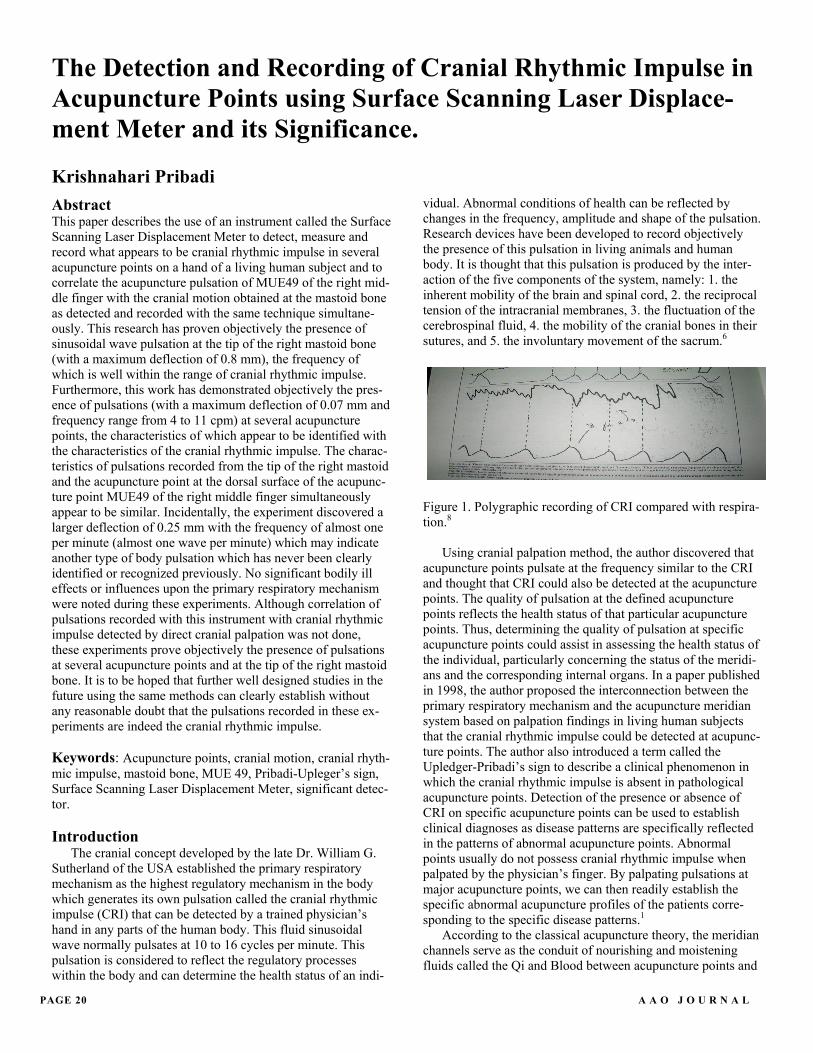
Abstract This paper describes the use of an instrument called the Surface Scanning Laser Displacement Meter to detect, measure and record what appears to be cranial rhythmic impulse in several acupuncture points on a hand of a living human subject and to correlate the acupuncture pulsation of MUE49 of the right mid-dle finger with the cranial motion obtained at the mastoid bone as detected and recorded with the same technique simultane-ously. This research has proven objectively the presence of sinusoidal wave pulsation at the tip of the right mastoid bone (with a maximum deflection of 0.8 mm), the frequency of which is well within the range of cranial rhythmic impulse. Furthermore, this work has demonstrated objectively the pres-ence of pulsations (with a maximum deflection of 0.07 mm and frequency range from 4 to 11 cpm) at several acupuncture points, the characteristics of which appear to be identified with the characteristics of the cranial rhythmic impulse. The charac-teristics of pulsations recorded from the tip of the right mastoid and the acupuncture point at the dorsal surface of the acupunc-ture point MUE49 of the right middle finger simultaneously appear to be similar. Incidentally, the experiment discovered a larger deflection of 0.25 mm with the frequency of almost one per minute (almost one wave per minute) which may indicate another type of body pulsation which has never been clearly identified or recognized previously. No significant bodily ill effects or influences upon the primary respiratory mechanism were noted during these experiments. Although correlation of pulsations recorded with this instrument with cranial rhythmic impulse detected by direct cranial palpation was not done, these experiments prove objectively the presence of pulsations at several acupuncture points and at the tip of the right mastoid bone. It is to be hoped that further well designed studies in the future using the same methods can clearly establish without any reasonable doubt that the pulsations recorded in these ex-periments are indeed the cranial rhythmic impulse. Keywords: Acupuncture points, cranial motion, cranial rhyth-mic impulse, mastoid bone, MUE 49, Pribadi-Upleger’s sign, Surface Scanning Laser Displacement Meter, significant detec-tor. Introduction The cranial concept developed by the late Dr. William G. Sutherland of the USA established the primary respiratory mechanism as the highest regulatory mechanism in the body which generates its own pulsation called the cranial rhythmic impulse (CRI) that can be detected by a trained physician’s hand in any parts of the human body. This fluid sinusoidal wave normally pulsates at 10 to 16 cycles per minute. This pulsation is considered to reflect the regulatory processes within the body and can determine the health status of an indi-
vidual. Abnormal conditions of health can be reflected by changes in the frequency, amplitude and shape of the pulsation. Research devices have been developed to record objectively the presence of this pulsation in living animals and human body. It is thought that this pulsation is produced by the inter-action of the five components of the system, namely: 1. the inherent mobility of the brain and spinal cord, 2. the reciprocal tension of the intracranial membranes, 3. the fluctuation of the cerebrospinal fluid, 4. the mobility of the cranial bones in their sutures, and 5. the involuntary movement of the sacrum.6
Figure 1. Polygraphic recording of CRI compared with respira-tion.8
Using cranial palpation method, the author discovered that acupuncture points pulsate at the frequency similar to the CRI and thought that CRI could also be detected at the acupuncture points. The quality of pulsation at the defined acupuncture points reflects the health status of that particular acupuncture points. Thus, determining the quality of pulsation at specific acupuncture points could assist in assessing the health status of the individual, particularly concerning the status of the meridi-ans and the corresponding internal organs. In a paper published in 1998, the author proposed the interconnection between the primary respiratory mechanism and the acupuncture meridian system based on palpation findings in living human subjects that the cranial rhythmic impulse could be detected at acupunc-ture points. The author also introduced a term called the Upledger-Pribadi’s sign to describe a clinical phenomenon in which the cranial rhythmic impulse is absent in pathological acupuncture points. Detection of the presence or absence of CRI on specific acupuncture points can be used to establish clinical diagnoses as disease patterns are specifically reflected in the patterns of abnormal acupuncture points. Abnormal points usually do not possess cranial rhythmic impulse when palpated by the physician’s finger. By palpating pulsations at major acupuncture points, we can then readily establish the specific abnormal acupuncture profiles of the patients corre-sponding to the specific disease patterns.1
According to the classical acupuncture theory, the meridian channels serve as the conduit of nourishing and moistening fluids called the Qi and Blood between acupuncture points and
The Detection and Recording of Cranial Rhythmic Impulse in Acupuncture Points using Surface Scanning Laser Displace-ment Meter and its Significance.
Krishnahari Pribadi
PAGE 20 A A O J O U R N A L
jects using different methodologies such polygraphic recording, frequency oscillator technique, mechanical transducer, elec-tronic transducer, etc.8 Unfortunately, none of these techniques can be used to detect and record the cranial rhythmic impulse at acupuncture points as most of the pick-off sensors are too large to be applied to an acupuncture point. We need to have a micro-sensor that can detect the motion of the acupuncture point with-out detection of other waves or movements in the surrounding tissue such as skin, nail, or bone. In 2006, the author had the luck of being introduced to an instrument called the Surface Scanning Laser Displacement Meter which is capable of meas-uring and recording surface displacement as low as 0.01 mi-cron.
This paper describes the use of this instrument to detect, measure and record what appears to be cranial rhythmic im-pulse in several acupuncture points on a hand of a living human subject and to correlate this acupuncture pulsation with the cra-nial motion obtained at the mastoid bone as detected and re-corded with the same technique simultaneously. Unfortunately, because of the cost, the author was not able to secure the instru-ment and be present during the experiments and had to be happy with designing several experiments to be conducted by a technician (not trained in osteopathy in the cranial field or acu-puncture medicine) who happened to have access to the use of this instrument located in another country. These preliminary experiments were done to study if the instrument could be used to record cranial rhythmic impulse in a living human subject at specific acupuncture points and a selected mastoid bone and to study and correlate the characteristics of the recording. Once, the experiments establish the applicability of the instrument in measuring and recording cranial rhythmic impulse in living human subjects, more elaborate and well designed research experiments will be proposed and conducted to evaluate the validity, reliability, sensitivity and accuracy of the instrument in detecting, measuring and recording cranial rhythmic impulse in living human subjects non-invasively in the cranial bones, sac-ral bone, teeth, nail, hair, and acupuncture points and other structures in the body.
The author hopes that once it is established that the instru-ment can be used to non-invasively record cranial rhythmic impulse in living human subjects, we will develop a medical device that can be used clinically to diagnose cranial strain le-sions and to monitor cranial osteopathic treatment process as well as a diagnostic device that can measure and record the cra-nial rhythmic impulse pattern at acupuncture points to help es-tablish medical diagnosis as well as monitoring acupuncture treatment and designing acupuncture therapy plan. Theoreti-cally, this instrument can also be used to detect any forces such as electromagnetic, light, physical, bio-energy, herbal, gems, homeopathic remedies, chemical drugs, toxins, etc., or even cosmic radiation in the outer space, or even mental and spiritual forces that may affect the primary respiratory mechanism in a negative or positive way. Finally, the objective recording of cranial rhythmic impulse in acupuncture points can thus support my hypothesis regarding the interconnection between the pri-mary respiratory mechanism and the acupuncture meridian sys-tem as well as scientific evidence that the acupuncture meridian system does exist and the anatomical structure in the human (and animal) body could be then scientifically established.
the internal organs served by the specific meridians.2 It has been demonstrated that the acupuncture points possess electri-cal phenomena as objectively measured by electrical instru-ments that record electrical conductivity and electrical current. Usually, acupuncture points have higher electrical conductivity then the surrounding skin. Studies have shown that “fluctuations in cutaneous electric current over acupuncture points on certain channels coincided with variations in the func-tional activity of those internal organs associated with the chan-nel.”3 It is suggested further that “the channels are a kind of axis for the biological electricity of the body, or an electric cur-rent, a special kind of electron or electron “bundle” which passes electromagnetic waves along a fixed course; that the electric phenomena of the skin reflect the electromagnetic field within the body, and just as the magnetic field within the body corresponds to the cosmos, changes in the cosmos may then be reflected in fluctuations in the electrical activity along the chan-nels; and the Qi in traditional Chinese medicine is the equiva-lent of modern electromagnetic phenomena, etc.”4
The idea that there is interconnection between the primary respiratory system and the acupuncture meridian system is fur-ther supported by the fact that when an acupuncture needle is inserted into a specific abnormal acupuncture point (for exam-ple Li 3 in liver disease) the cranial rhythmic impulse palpated at the head or any body parts will suddenly cease for up to 20 minutes. It will usually return at the completion of acupuncture treatment usually immediately prior to the removal of the nee-dle. Furthermore, the disappearance of the cranial rhythmic impulse coincides with the “Qi sensation” obtained by the op-erator inserting the needle. This phenomenon is exactly the same as the still-point phenomenon obtained by still point tech-niques introduced by Upledger to manipulate and stimulate the primary respiratory mechanism (CV technique, sacral and leg technique).5 Cranial manipulation techniques to correct abnor-mal cranial strain patterns usually also induce “still-point” in which no cranial rhythmic impulse is detected by the operator’s hands before the completion of the procedures.6 The cessation of cranial rhythmic impulse also occurs during Somato- Emo-tional Release in Craniosacral Therapy as developed by Upledger when significant emotions are being released and discharged when access to trauma memory is activated by a specific body position mimicking the original physical trauma. Upledger coined the term “the significance detector” to de-scribe the signaling function of the cranial rhythmic impulse during treatment. When cranial rhythmic impulse suddenly stops during “therapeutic image” it usually signals that a sig-nificant stimulus (such as a visual image, a voice, a feeling or a sensation) is just about to enter awareness and there is “something good is happening inside”. By exploring this sig-nificant image, idea, or sensation through dialogue to solve par-ticular health issues that the person is experiencing, the cranial rhythmic impulse will return much stronger than before after completion of the treatment process.7 Thus, it appears that dur-ing “still-point” induced by therapeutic procedures (such as cranial manipulation, somato-emotional release, therapeutic image, acupuncture)that affect and work through the primary respiratory mechanism, significant therapeutic processes are happening signaling systematic changes occurring within the primary respiratory mechanism.
Several devices and techniques have been developed to re-cord cranial rhythmic impulse in living animals and human sub-
PAGE 21 V O L U M E 1 8 , I S S U E 4
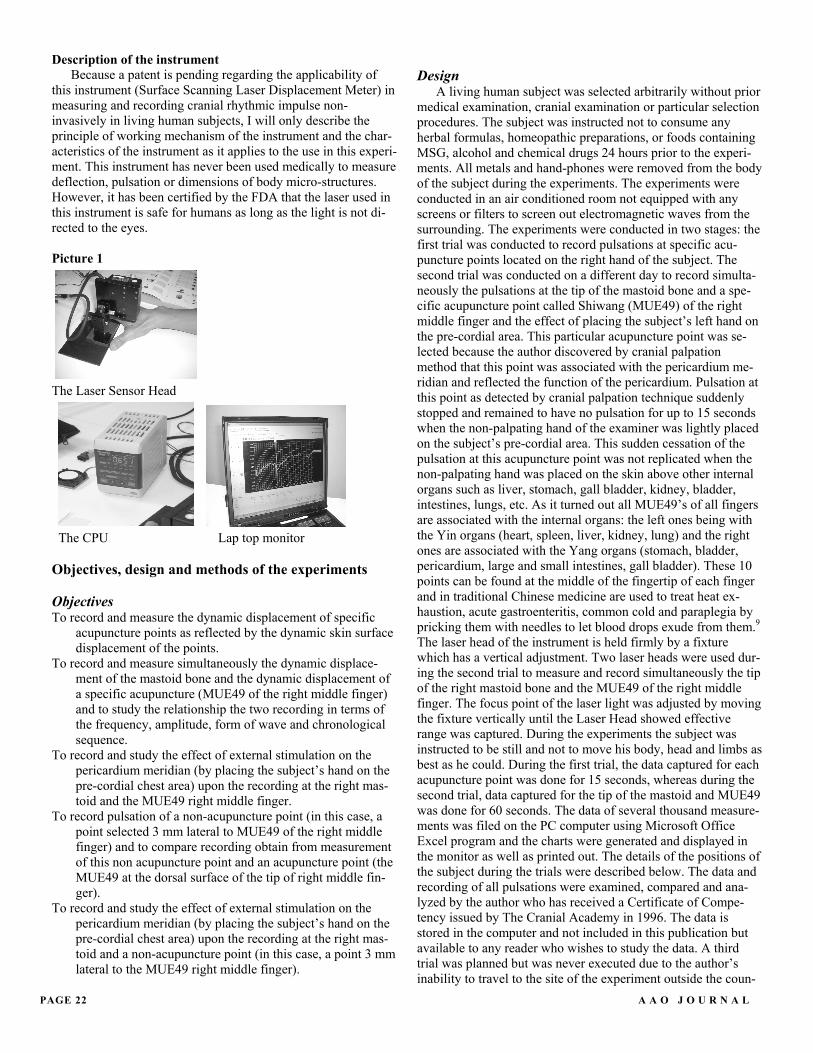
Description of the instrument Because a patent is pending regarding the applicability of
this instrument (Surface Scanning Laser Displacement Meter) in measuring and recording cranial rhythmic impulse non-invasively in living human subjects, I will only describe the principle of working mechanism of the instrument and the char-acteristics of the instrument as it applies to the use in this experi-ment. This instrument has never been used medically to measure deflection, pulsation or dimensions of body micro-structures. However, it has been certified by the FDA that the laser used in this instrument is safe for humans as long as the light is not di-rected to the eyes.
Picture 1
The Laser Sensor Head
The CPU Lap top monitor Objectives, design and methods of the experiments Objectives To record and measure the dynamic displacement of specific
acupuncture points as reflected by the dynamic skin surface displacement of the points.
To record and measure simultaneously the dynamic displace-ment of the mastoid bone and the dynamic displacement of a specific acupuncture (MUE49 of the right middle finger) and to study the relationship the two recording in terms of the frequency, amplitude, form of wave and chronological sequence.
To record and study the effect of external stimulation on the pericardium meridian (by placing the subject’s hand on the pre-cordial chest area) upon the recording at the right mas-toid and the MUE49 right middle finger.
To record pulsation of a non-acupuncture point (in this case, a point selected 3 mm lateral to MUE49 of the right middle finger) and to compare recording obtain from measurement of this non acupuncture point and an acupuncture point (the MUE49 at the dorsal surface of the tip of right middle fin-ger).
To record and study the effect of external stimulation on the pericardium meridian (by placing the subject’s hand on the pre-cordial chest area) upon the recording at the right mas-toid and a non-acupuncture point (in this case, a point 3 mm lateral to the MUE49 right middle finger).
Design
A living human subject was selected arbitrarily without prior medical examination, cranial examination or particular selection procedures. The subject was instructed not to consume any herbal formulas, homeopathic preparations, or foods containing MSG, alcohol and chemical drugs 24 hours prior to the experi-ments. All metals and hand-phones were removed from the body of the subject during the experiments. The experiments were conducted in an air conditioned room not equipped with any screens or filters to screen out electromagnetic waves from the surrounding. The experiments were conducted in two stages: the first trial was conducted to record pulsations at specific acu-puncture points located on the right hand of the subject. The second trial was conducted on a different day to record simulta-neously the pulsations at the tip of the mastoid bone and a spe-cific acupuncture point called Shiwang (MUE49) of the right middle finger and the effect of placing the subject’s left hand on the pre-cordial area. This particular acupuncture point was se-lected because the author discovered by cranial palpation method that this point was associated with the pericardium me-ridian and reflected the function of the pericardium. Pulsation at this point as detected by cranial palpation technique suddenly stopped and remained to have no pulsation for up to 15 seconds when the non-palpating hand of the examiner was lightly placed on the subject’s pre-cordial area. This sudden cessation of the pulsation at this acupuncture point was not replicated when the non-palpating hand was placed on the skin above other internal organs such as liver, stomach, gall bladder, kidney, bladder, intestines, lungs, etc. As it turned out all MUE49’s of all fingers are associated with the internal organs: the left ones being with the Yin organs (heart, spleen, liver, kidney, lung) and the right ones are associated with the Yang organs (stomach, bladder, pericardium, large and small intestines, gall bladder). These 10 points can be found at the middle of the fingertip of each finger and in traditional Chinese medicine are used to treat heat ex-haustion, acute gastroenteritis, common cold and paraplegia by pricking them with needles to let blood drops exude from them.9
The laser head of the instrument is held firmly by a fixture which has a vertical adjustment. Two laser heads were used dur-ing the second trial to measure and record simultaneously the tip of the right mastoid bone and the MUE49 of the right middle finger. The focus point of the laser light was adjusted by moving the fixture vertically until the Laser Head showed effective range was captured. During the experiments the subject was instructed to be still and not to move his body, head and limbs as best as he could. During the first trial, the data captured for each acupuncture point was done for 15 seconds, whereas during the second trial, data captured for the tip of the mastoid and MUE49 was done for 60 seconds. The data of several thousand measure-ments was filed on the PC computer using Microsoft Office Excel program and the charts were generated and displayed in the monitor as well as printed out. The details of the positions of the subject during the trials were described below. The data and recording of all pulsations were examined, compared and ana-lyzed by the author who has received a Certificate of Compe-tency issued by The Cranial Academy in 1996. The data is stored in the computer and not included in this publication but available to any reader who wishes to study the data. A third trial was planned but was never executed due to the author’s inability to travel to the site of the experiment outside the coun-
PAGE 22 A A O J O U R N A L

Recording of Trial Number 1 Objective: to measure and record pulsations at several acupunc-ture points located on the right hand of a living human subject ( Large Intestine 3, Tripple Burner 3, and Small Intestine 3) 1. Recording of pulsation at an acupuncture point ( LI 3) on the right hand.
Graphic 1: Pulsation at LI3 ( first recording)
Graphic 2: Pulsation at LI3 ( second recording) 2. Recording of pulsation at an acupuncture point (TB3) on the
right hand.
Graphic 3: Pulsation at TB3 ( first recording)
try because of financial constraints. Methods A. Cranial Rhythmic Impulse Measurement Procedures – First Trial Objective To record and measure the dynamic displacement of specific
acupuncture points as reflected by the dynamic skin sur-face displacement of the points.
The first trial consists of taking measurements of the dynamic displacements of 3 specific acupuncture points, namely: LI – 3, TB – 3 and SI – 3 located on the right hand of a living human subject.
Picture 2
Picture 2. Locations of LI 3, TB-3 and SI 3 on the right hand Steps 1. Place Hand underneath the Laser Head, the Laser Head is
held firmly by a Fixture which has a vertical adjustment. 2. Test 1, Measurement taken on LI-3. Locate Laser Light
onto LI-3, adjust the focus point by moving the fixture vertically until the Laser Head shows effective range is captured.
3. Start measurement by activating a start button on the PC Terminal.
4. Data was captured for around 15 sec. 5. Save Data on PC terminal. 6. Measurements were taken twice for all 3 locations
(Experiments: 1,2,3,4,5,6).
PAGE 23 V O L U M E 1 8 , I S S U E 4

3. To record pulsation of a non- acupuncture point (in this case, a point selected 3 mm lateral to the selected acupuncture point)and to compare recording obtain from measurements of this non acupuncture point and an acupuncture point (the MUE49 at the dorsal surface of the tip of right middle fin-ger).
To record and study the effect of external stimulation on the pericardium meridian (by placing the subject’s left hand on the pre-cordial chest area) upon the recording at the right mastoid and the MUE49 right middle finger.
Steps 1. The measuring unit of the instrument is placed above the
right mastoid bone of an individual. The individual lays on his left side and the right mastoid bone is located. The right mastoid bone is located behind the right ear as the most prominent triangular bone exactly behind the ear. The instru-ment is mounted on a static support. The individual is in-structed as best as he can not to move his head, body, arms, hands and fingers. The point selected is the tip of the right mastoid bone and is marked by a reflecting paint from a sil-ver marker. Then, the dynamic displacements of the selected point are measured by the instrument and recorded serially for 60 seconds. The serial recording graph is displayed on the screen and printed out. A second measuring unit is placed above the MUE49 point of the right middle finger. The MUE49 point is located ex-actly at the center of an imagined line drew from the mid-point of the central border (border closer to the wrist) of the nail of the right middle finger and the midpoint of the digital crease between the last and the middle carpal bones of the middle finger. The right hand is placed in front of his body on a flat surface such as the flat surface of the table where the subject lays with the dorsal part faced above. The instru-ment is mounted on a static support above the selected point. The individual is instructed as best as he can not to move his head, his body, his arms, hands and fingers. The point se-lected is marked by a reflecting paint from a silver marker. Then, the dynamic displacements of the selected point are measured by the instrument and recorded serially for 60 sec-onds. The recording graph is then recorded and printed out.
Picture 3
Picture 3. Location of MUE49
Graphic 4: Pulsation at TB3 ( second recording ) 3. Recording of pulsation at an acupuncture point (SI3) on the
right hand
Graphic 5: Pulsation at SI 3 ( first recording)
Graphic 6: Pulsation at SI 3 ( second recoding) B. Cranial Rhythmic Impulse Measurement Procedures – Second Trial Objectives 1. To record and measure the dynamic displacement of the
mastoid bone as reflected by the dynamic skin surface dis-placement of a point selected on one of the mastoid bone.
2. To record and measure simultaneously the dynamic displace-ment of the mastoid bone and the dynamic displacement of a specific acupuncture (MUE49 of the right middle finger) and to study the relationship the two recording in terms of the frequency, amplitude, form of wave and chronological se-quence.
MUE-49
PAGE 24 A A O J O U R N A L

PAGE 25
2. Immediately after completing the step 1, have the subject touch his chest skin above his heart (by simply placing his left palm on the left breast: Repeat Step 1 for another minute simultaneously.
3. After a rest of 5 minutes with left hand off the chest area, then shift the measuring instrument above the respective MUE49 to measure pulsation at a non-acupuncture point about 3 mm off the respective MUE49 to the right (toward the right little finger) and simultaneously measure pulsation at the point of the right mastoid bone.
Recording of Trial Number 2 1. To record and measure simultaneously the dynamic displace-
ments of the tip of the right mastoid and the MUE49 of the right middle finger
Graphic 1: Pulsation at MUE49 of the right middle finger.
Graphic 2: Pulsation at the tip of the right mastoid 1. To record the effect of external stimulation (by placing the
subject’s right hand lightly on his precordial area) on the pericardium (circulation) meridian upon the recording at the right mastoid and the MUE49 right middle finger.
Graphic 3: Pulsation at MUE49 of the right middle finger during placement of right hand on precordial area.
Graphic 4: Pulsation at the tip of right mastoid during placement of right hand on precordial area. STEP 3: To compare recording obtain from measurements of a non acupuncture point (a point selected 3 mm lateral to MUE49 of right middle finger) and the tip of the right mastoid with the right hand off the precordial area
Graphic 5: Pulsation at a non-acupuncture point 3 mm off MUE49 with left hand off the precordial area.
Graphic 6: Pulsation at the tip of right mastoid with left hand off the precordial area.
V O L U M E 1 8 , I S S U E 4

PAGE 26 A A O J O U R N A L
C. Cranial Rhythmic Impulse Measurement Procedures – Third Trial (this trial has never been done as the au-thor was not able to travel to the location of the experi-ment outside the country) Objective
To verify if the recording produced is identified and similar with the CRI obtained clinically by a certified cranial osteo-pathic physician on the same individual. Step
While the pulsation at the tip of the right mastoid bone of the subject is measured and recorded by the instrument, the CRI at the head is palpated by a certified Cranial Osteopathic Physician who records the finding regarding the frequency, amplitude, and wave-forms. This recording is compared with the recording ob-tained by the instrument. Discussions The recording of pulsations at the three acupuncture points (namely: LI – 3, TB – 3 and SI – 3 located on the right hand of a living human subject) distinctly demonstrates large sinusoidal waveforms of different shapes and frequencies mixed with more rapid smaller sharp pulsations. Variations of patterns of the sinu-soidal waveforms are noted even at the same point during the first and second recording for each acupuncture point. The shapes, frequencies and amplitudes of pulsations at the three acupuncture points (located at different meridians, namely: large intestine, triple burner and small intestine) are also different. During the first recording of point L1, the maximum deflection is 0. 3 mm and the frequency of the sinusoidal wave is approxi-mately 4 cycle per minute, whereas second recording of the same point reveals maximum deflection of 0.07 mm with the frequency being 10 cycles per minute (there are 2.5 sinusoidal waves per 15 seconds ). This figure correspondents exactly with the normal CRI frequency (10 to 16 cycles per minute). The fre-quency of the tiny pulsation of the second recording of LI 3 is approximately 80 per minute (approximately 20 tiny pulsations per 15 seconds) which is in the range of cardiac pulsation. Un-fortunately, we did not record the CRI frequency and radial pulse of the subject at the same time. The tiny pulsation recorded from TB 3 demonstrates frequency at 68 beats per minute (17 beats per 15 seconds). Whereas the frequency of the tiny pulsa-tion recorded from SI 3 is approximately 112 per minute (28 tiny waves per 15 seconds), again within the range of the frequency of cardiac pulsation. This indicates that the subject experienced acceleration of his heart rate during this experiment. Distinct sinusoidal waves are identified in first recording of SI 3 at the frequency of 12 cycles per minute (or 3 waves per 15 seconds) and with maximum deflection of 0.04 mm. The second recoding of SI 3 reveals one half sinusoidal wave followed by absence of sinusoidal waves with a maximum deflection of 0.,15 mm. The first recoding of TB 3 shows frequency of sinusoidal waves at 4 cycles per minute (1 wave per 15 seconds) and maximum deflec-tion of 0.07 mm, whereas second recording indicates frequency at 6 cpm (1.5 waves per 15 seconds) and a maximum deflection of 0.05 mm. In summary, the first experiment clearly demonstrates sinu-soidal waves at three acupuncture points at frequencies ranging
from 4 to 12 cycles per minute (within the frequency range of cranial rhythmic impulse) and deflections ranging from 0.05 mm to 0.15 mm. Tiny sharp pulsations are noted with the frequencies ranging from 68 beats to 112 beats per minute, within the range of the frequencies of cardiac pulsation. If we accept the hypothe-sis that acupuncture pulsations at acupuncture points reflect functioning of organs or processes within the organs, this experi-ment reveals that the subject was experiencing variable function-ing of the three meridians: large intestine, triple burner (or endo-crine) and small intestine. LI 3 (Sanjian) is the transporting point of large intestine and is indicated for the treatment of tooth ache, sore throat, trigeminal neuralgia, painful eyes, malaria and the inflammation of dorsum of the hand. TB 3 (Zhongzu) is the transporting point of the Tripple Burner (facilitates the circula-tion of Qi and benefits the ear) and is indicated to treat deaf mu-tism, tinnitus, deafness, headache, neuralgia, etc.). SI 3 (Houxi) is the transporting point of the small intestine and the meeting point of the governing channel on the small intestine and relaxes the muscle channels, opens the governing channels and clears the Spirit. It is indicated in malaria, seizures, psychosis, hysteria, intercostals neuralgia, night sweats, stiff neck, low back pain and deaf mutism. Unfortunately, we do not have the medical record or clinical history of the subject to enable us to correlate these findings with clinical data. Capturing the data around 60 seconds for each experiment during the second trial clearly eliminates the sharp pulsations “cardiac pulsations” and amplifies the sinusoidal waves “cranial rhythmic impulse” of the recording of pulsations obtained from the tip of the right mastoid and the right MUE49 of the right middle finger. The first experiment clearly demonstrates the similarities of the frequencies, shapes and phases of the pulsa-tions obtained from the tip of the right mastoid and the right MUE49. A larger deflection of 0.25 mm with the frequency of almost one per minute (almost one wave per minute) is noted on the first recording obtained from the acupuncture point MUE49. This may indicates another type of body pulsation which has never been clearly identified or recognized previously. Perhaps it reflects the whole body pulsation as the body swells and shrinks at the frequency of one per minute in response to cosmic fluctua-tion. The frequency of acupuncture pulsation at MUE49 is about 11 per minute, exactly the same as the number for the frequency pulsation at the tip of the right mastoid bone. This clearly is within the normal frequency range of normal cranial rhythmic impulse which is between 10 to 16 cycles per minute. The maxi-mum deflection of the MUE49 pulsation is 0.07 mm, whereas the tip of the right mastoid deflects approximately 0.8 mm (almost ten times of the acupuncture deflection). This wide de-flection indicates that the pulsation recorded at the tip of the right mastoid was most likely associated with bone movements (in this case the right mastoid) rather than pulsation of an acu-puncture point located at the tip of the mastoid bone. Although we did not correlate this pulsation with cranial rhythmic impulse palpated at the cranium of the subject clinically, we can be sure that this recorded pulsation is identified with the cranial rhyth-mic impulse. This experiment clearly demonstrates the presence of CRI at an acupuncture point as the acupuncture pulsation shows exactly the same frequency, waveforms, and phases as the recording of pulsation obtained simultaneously with the same instrument (using a second sensor) from the tip of the right mas-toid. The instrument indeed was able to detect and record objec-tively pulsation at the tip of the right mastoid which appears to

be the cranial rhythmic impulse of the subject. It appears that the pulsation recorded at the MUE49 preceded the pulsation at that tip of the right mastoid by approximately a half second (which appears to correspondent with clinical finding: simultaneous cranial palpation of the right MUE49 and the tip of right mastoid bone demonstrates time difference). This finding supports the idea that there is movement of fluids within the acupuncture meridian and the primary respiratory mechanism, the direction being from the acupuncture points, meridian channels and to the primary respiratory mechanism (tip of the right mastoid) and back. The second experiment of the second trial reveals different characteristics of pulsations at the acupuncture point (MUE49) and the tip of the right mastoid. Immediately, after the left palm of the subject was placed on the pre-cordial chest area, a sudden cessation of the acupuncture pulsation and an absence of this pulsation for almost 60 seconds appeared which was followed by a sudden large displacement of 0.4 mm distance. This was accompanied by sharp waves of 16 cycles per minute recorded at the tip of the right mastoid bone which was also followed abruptly by a negative deflection of 3 mm distance (sudden ex-ternal rotation of the right temporal bone). This can be inter-preted that the placement of the left palm on the pre-cordial area blocks the flow of fluids within the pericardium meridian which causes accumulation of cerebrospinal fluid within the primary respiratory mechanism as evidenced by acceleration of the cra-nial rhythmic impulse (from 11 cycles per minute to 16 cycles per minute). Again, it supports the contention that there is inter-connection between the two fluid space systems. The sudden wide deflections recorded at both points signifies an escape phe-nomenon: the mounting pressure within the fluid space of the two systems overcame the fluid restriction at the pericardium area exerted by placing of the hand on the pre-cordial area. This phenomenon occurs also during induction of still-point by put-ting the primary respiratory mechanism into extreme extension with the CV4, sacral compression and leg techniques introduced by Upledger. Thus, by simply placing a hand on the skin surface of an organ one can induce a still-point and improves the pri-mary respiratory mechanism! This can be simply demonstrated clinically by monitoring the CRI at the head while placing the non-monitoring hand on the skin surface of various organs suc-cessively. Thus, the interconnection between the primary respi-ratory mechanism, internal organs and the acupuncture meridian system is clearly established. The third experiment of the second trial clearly shows that a point on the skin 3 mm off the MUE49 pulsates at a frequency higher than the acupuncture point itself (14 cpm as opposed to 11 cpm) of the same subject. Puzzlingly, simultaneous recording of the tip of the right mastoid reveals the compete absence of pulsation interspersed with about 8 wide deflections (which can be artifacts produced by sudden movements of the head of the subject because of fatigue). The process of recording of a non-acupuncture point (perhaps a point at the wall of a meridian channel?) appears to be accompanied by a prolonged still point of the primary respiratory mechanism. Perhaps the laser light used in this measuring process stimulates the primary respiratory mechanism to undergo a still-point without concomitant cessa-tion of the acupuncture pulsation, thus demonstrating the sensi-tivity of the system to light energy as well as the connection be-tween the meridian channel and the primary respiratory mecha-nism. However, measuring pulsations at acupuncture points us-
ing this instrument does not cause a still –point of the CRI at the head. Indeed, clinical cranial palpation of the walls of meridian channels demonstrates higher frequencies of channel pulsations compared to frequencies of pulsations at the acupuncture points and the head. Perhaps, the walls of meridian channels are formed by cells that pulsate at higher frequencies which function to pre-vent fluids from escaping from the channels, and to direct and pump the fluids within the channels to move along the courses of the channels. The presence of the walls of the meridian chan-nels may not be apparent on dissected tissues of dead human subjects or animals, just as cranial rhythmic impulse is not ob-tainable in dead human subjects or animals. This phenomenon (higher frequencies of pulsations of the channel walls) can be exploited to detect and map out the anatomy structure of the meridian channels by recording the pulsations of channels using this instrument. It appears that other structures of the skin do not posses pulsations at higher frequencies than the frequency of CRI unless they are functionally abnormal. Summary, significance, and scientific values of these findings This preliminary research has proven objectively the pres-ence of sinusoidal wave pulsation at the tip of the right mastoid bone (with a maximum deflection of 0.8 mm), the frequency of which is well within the range of cranial rhythmic impulse. Fur-thermore, this work has demonstrated objectively the presence of pulsations (with a maximum deflection of 0.07 mm and fre-quency range from 4 to 11 cpm) at several acupuncture points, the characteristics of which appear to be identified with the char-acteristics of the cranial rhythmic impulse. Incidentally, the ex-periment discovered a larger deflection of 0.25 mm with the frequency of almost one per minute (almost one wave per min-ute) which may indicate another type of body pulsation which has never been clearly identified or recognized previously. The instrument is sensitive and specific enough to detect and record pulsations at acupuncture points and at the skin surface of a bone (in this case the right mastoid bone) without picking up other body pulsations or waves (such as respiration, muscular contrac-tions, organ movements, etc.) with the exception of cardiac pul-sation. Capturing the data around 60 seconds for each experi-ment during the second trial clearly eliminates the sharp pulsa-tions “cardiac pulsations” and amplifies the sinusoidal waves “cranial rhythmic impulse” of the recorded pulsations. No sig-nificant bodily ill effects or influences upon the primary respira-tory mechanism were noted during the experiments. However, a prolonged still point occurred while the instrument was measur-ing the pulsation of a point located approximately 3 mm off the acupuncture point, which may well be the channel wall of a me-ridian or the wall of an acupuncture point. This point exhibited a pulsation at a higher frequency then the acupuncture point itself. Placing a hand on the corresponding organ (pericardium in this case) induced cessation of pulsation at the acupuncture point related to the organ and stimulated the primary respiratory mechanism with acceleration of the pulsation at the tip of the right mastoid bone. This phenomenon was followed by larger sudden deflections of both points. Although correlation of pulsa-tions recorded with this instrument with cranial rhythmic im-pulse detected by direct cranial palpation was not done, these experiments prove objectively the presence of pulsations at sev-
PAGE 27 V O L U M E 1 8 , I S S U E 4

eral acupuncture points and at the tip of the right mastoid bone. It is to be hoped that further well designed researches in the fu-ture using the same methods can clearly establish without any reasonable doubt that the pulsations recorded in these experi-ments are indeed the cranial rhythmic impulse. The interconnection of the primary respiratory mechanism and the acupuncture meridian system demonstrates that the high-est body regulatory system is established through fluid wave pulsations generated by these two systems which travel along the peripheral nerves and meridian channels which are con-nected to the spinal cord and the brain. It is the author’s conten-tion that information from one to the other system (and the brain and spinal cord) and all organs (and cells)is transmitted via the fluids that travel from the primary respiratory mechanism system (the cerebrospinal fluid ), through the nerves and exude at the nerve endings and enter the thousands of acupuncture points to follow the courses of all meridians and organs and back to to the primary respiratory mechanism system. The meridian channels not only contain “the Qi and the Blood and the corresponding Yin and Yang energies” as explicated by the traditional Chinese medicine, but may also contain the cerebrospinal fluid (and all the physical, chemical and electrical components) generated by the brain via the interconnection of the two systems. Thus, this theory explains the efficacies as well as the wide clinical appli-cation of cranial osteopathic manipulation and acupuncture treat-ment in various maladies of human beings and animals as well. These two systems are sensitive to physical, chemical, electrical as well as light energy and other form of bio-energies. The physical force responsible for pumping the meridian channel fluids to travel in the meridian channels is none but the pumping action of the primary respiratory mechanism delivered by the pulsatile movement of the primary respiratory mechanism known as the “cranial rhythmic impulse”. Physical restrictions in the primary respiratory mechanism may cause slowing of fluids (and bio-energy) movements within the meridian channels and abnormal functioning of acupuncture points may cause abnormal functioning on the primary respiratory mechanism. These ex-periments may have established the anatomical presence of the primary respiratory mechanism (and its pulsation) and the acu-puncture meridian system (and its pulsation) and their intercon-nection. Future work may yield more data and findings to sup-port this hypothesis. This Surface Scanning Laser Displacement Meter may well be the instrument capable of detecting minute pulsations of the two systems and their anatomical parts and structures and may have applications in research and clinical work in cranial oste-opathy and acupuncture medicine and eventually may help un-ravel the secrets of the working of human anatomy and physiol-ogy in health and diseases. This instrument may prove to be use-ful in mapping out the anatomy structure of the acupuncture me-ridian system, namely the meridian channels and their routes and acupuncture points and their interconnections and the specific physiological regulatory functions of all acupuncture points. Perhaps, more meridians and acupuncture points and their spe-cific regulatory roles will be discovered further. References 1. Pribadi K. Psychosynergisis, A Synthesis of Osteopathic Medicine, Acu-
puncture Medicine and Homeopathy in Psychiatry. AAOJ.8:1.29-35.
2. O’Connor J and Bensky. Acupuncture, A Comprehensive Text. Seattle. Eastland Press, 1987 pp. 34
3. ibid, pp. 106-107 4. ibid, p. 107 5. Upledger, J. E. and Vredevoogd, J. D.: Craniosacral Therapy, Seattle.
Eastland Press, 1987 6. Magoun HJ. Osteopathy in the cranial field. Idaho: The Cranial Academy,
1976. 7. Upledger, Somato Emotional Release and Beyond, Seattle: Eastland Press,
1988, pp. 132-135 8. Frymann V M.A Study of the Rhytmic Motions of the Living Cranium.
JAOA. Vol 70. May 1971. pp. 928-945 9. O’Connor J and Bensky. Acupuncture, A Comprehensive Text. Seattle.
Eastland Press, 1987, p. 386 and p. 615
* The author thanks to Mr. Edwin Purwanto, who introduced him to this instru-ment and proposed that this instrument could be used to detect cranial rhythmic impulse in living human subjects and to Mr. Cheng Choong Foo, who intelli-gently and carefully conducted all the experiments under the author’s direction. Without their contributions, this work could have never been done. Accepted for Publication: October, 2008
Address Correspondence to: Krishnahari S. Pribadi, MD, ABPN Diplomat General Holistic Medicine Practice Jalan Borneo 8, Timur, Zip Code 16419 Jawa Barat, Indonesia
PAGE 28 A A O J O U R N A L

Healing Hands: Using Osteopathic Manipulative Treatment to Address Visceral Structures through Somatovisceral Reflexes: A Case Study in Gastroesophageal Reflux Disease
The field of osteopathy is a total system of health care which pro-fesses and teaches the osteopathic philosophy based upon four key principles. 1. The body is a unit. 2. It has its own self-protecting and regulating mechanisms. 3. Structure and function are reciprocally interrelated. 4. Treatment considers the preceding three principles. Perhaps no other field of study within osteopathic medicine epito-mizes these principles more clearly than somatovisceral reflexes and the application of these connections to achieve optimal treat-ment for the patient.
The interrelationship of structure and function is vividly por-trayed by the interplay between the somatic and visceral struc-tures. Propriocepter input from somatic dysfunction may facili-tate a cord segment. If that cord segment is also the site of the cell bodies for sympathetic outflow to a visceral structure, that par-ticular visceral structure’s function may be affected by excessive sympathetic tone. The exchange from somatic sensory to visceral sympathetic outflow occurs by way of the facilitated cord seg-ment and is called the somatovisceral reflex.2
According to Irvin M. Korr, Phd, facilitated spinal cord seg-ments encourage and support physiological, hormonal and bio-chemical conditions which increase a patient’s complications and slow or inhibit a patient’s recovery from dysfunction or disease. The classic terminology is that the facilitated segment acts as a “neurologic lens”, focusing stressor input upon target viscera.2
Research by Reflex Neurophysiologist Michael Patterson has found evidence that early osteopathic manipulation of somatic dysfunction might be a form of preventative medicine. Some of his research on animals has fostered the following conclusions. 1. Persistent musculoskeletal dysfunction affecting a certain
spinal cord segment can affect the function of an internal organ related to that same spinal cord segment for its inner-vation.
2. Removal of the musculoskeletal irritation of dysfunction allows the related internal organ to return to its normal func-tion.
If the dysfunction of the musculoskeletal system remains long enough, it apparently “burns a memory pattern” within the central nervous system so that when the initial irritating musculoskeletal influence is removed, the original dysfunction not only continues but grows in severity.2 The research literature has established a clear interconnection between somatic dysfunction and visceral pathology. The well trained osteopathic physician is equipped with the knowledge and skill to assess and treat visceral pathology with manipulation. The objective is to find the somatic dysfunction which correlates to the visceral pathology and treat the lesion in order to re-establish normal function within the viscera before the pathologic “memory pattern” sets in.
Case This is a retrospective case study of a now 58-year-old white female professor presenting with chronic pain in the neck and upper back secondary to excessive reading and computer work required of a college professor. The patient reported that OMT was therapeutic for her chronic pain. The patient’s history in-cludes a fracture of the mid-thoracic spine in her teens. She could not recall the exact spinal vertebrae involved in the frac-ture. She later developed Gastro-esophageal reflux disease (GERD) in 2005. She was prescribed Prilosec and Protonix for her GERD. At the time of her diagnosis of GERD she had pro-found epigastric pain and described an effortless return of gas-tric contents into the pharynx without nausea, retching, or ab-dominal contractions. The Prilosec and Protonix gave some re-lief, but the symptoms continued to persist. Her past medical history also consisted of mitral valve prolapsed diagnosed in 1968 at age 20. She currently takes Inderal for her heart condi-tion. Family history consists of heart valve disease including mother, father and sister. Physical Exam (structural evaluation)
WT: 172 BP: 110/70 HR: 62 Resp: 12 Cervicals: OA SLRR Right levator scapula tightness Thoracics: T4-T6 SRRL Lumbars: unremarkable Sacrum: mild posterior right sacroiliac tenderpoint Pelvis: Right innominate anterior Ribs: TART changes at the T5 and rib 5
Diagnosis
When the patient presented for her first visit on 8/05, she had recently been diagnosed with GERD by her primary care physi-cian. She had somatic dysfunction located at the OA and tho-racic spine. She also had TART changes at T5 and rib 5. Therapy and Clinical Course
The patient received treatment of the above mentioned areas once a month over a 14-month period. Treatment consisted of condylar decompression to normalize parasympathetic tone. Soft tissue and rib raising techniques were used around the area of T5-T9 to normalize sympathetic tone. This was accompanied by muscle energy techniques to address patterns of segmental facilitation and their resulting somatic dysfunction. Celiac Gan-glion release was also used to address the collateral sympathetic ganglion in order to further normalize hypersympathetic outflow to the upper gastrointestinal tract.
The patient responded well to osteopathic manipulative treatment (OMT). She reported drastic improvement of her GERD symptoms since the beginning of her therapy. She has
Beau Branyon
PAGE 29 V O L U M E 1 8 , I S S U E 4

also decreased the number of prescription medications used for GERD. She now takes only a low dose of Nexium. The patient will continue to follow up monthly with the OMT physician for maintenance on chronic pain secondary to work posture. Special emphasis will continue on the areas of somatic dysfunction re-lated to the somatovisceral regulation of her GERD symptoms. Review of the Literature
Gastroesophageal Reflux Disease (GERD) is defined as symptoms or mucosal damage produced by the abnormal reflux of gastric contents into the esophagus. The cardinal symptoms associated with GERD are heartburn and regurgitation. Sympa-thetic innervation to the upper gastrointestinal tract including the esophagus and stomach originates from T5-T9 forming the Greater Splanchnic Nerve which synapses at the Celiac Gan-glion. The effects of increased sympathetic innervation to the gastrointestinal tract include increased vascular tone leading to decreased oxygen and nutrients to tissues and decreased peristal-sis leading to constipation. Parasympathetic innervation to the lower 2/3 of the esophagus and the stomach is provided by the vagus nerve (CN X). Increased parasympathetic activity in-creases acid secretion and the rate of gut peristalsis.
Understanding the areas of origin of autonomic outflow is of the utmost importance when one takes into consideration the somatovisceral connection within the patient. Akio Sato, MD, PhD (1970-1990) performed experiments with rats and cats and was able to show that somatic nerve stimulation can affect car-diovascular and GI tract depending on which area of the soma is stimulated and the strength of stimulation. After completing his research, Sato concluded,”…the functions of various visceral organs can be influenced by a proper cutaneous stimulation as a result of the somatosympathetic or the somatoparasympathetic reflexes. I hope this knowledge is used to carry out similar ex-perimentation in other mammals and that finally this knowledge will be clinically useful in altering the visceral function of hu-mans.”5 The experiments of Friedman, PhD, Hudson, DO, and Young, DO further confirmed the somatovisceral connection. In these studies the paravertebral muscles of dogs were stimulated both mechanically and electrically. Stimulation in the area of T3-T7 produced duodenal contractions of high amplitude.1
Some of the earliest research on the connection between the soma and viscera was carried out during the first half of the 20th century by Louisa Burns, DO and her coworkers and assistants. They used thousands of animals including rats, rabbits, guinea pigs, goats, dogs, and others. They would produce spinal lesions within the animal similar to those found in humans. The find-ings were remarkable. Lesioning T4 would produce and increase in heart rate and a disturbance of cardiac rhythm, unless the le-sion was reduced. After reduction of the lesion, function of the viscera was returned to normal.4 In the gastrointestinal tract it was found that lesions to T4-T5 resulted in hyperemia of the gastric mucosa and excessive formation of gastric acid. Lesion-ing of T7 was followed by vascular changes in the stomach.4
Some areas of the stomach were injected, while other areas blood supply was decreased, secondary to an imbalance of sym-pathetic and parasympathetic outflow to the tissue. These find-ings have clearly shown that the osteopathic spinal lesion can result locally in a lesion in the nervous system and reflexively in visceral pathology. The research also shows that correcting so-matic dysfunction can have a therapeutic affect on correspond-ing viscera.
Discussion In the case of the 58-year-old professor, it is very interesting that she had a history of a fracture to the mid thoracic spine in her teenage years. This trauma could have set in motion a so-matic dysfunction that after years of degeneration preceded to facilitate a cord segment (T5-T9) corresponding to the sympa-thetic outflow to the upper gastrointestinal tract. With this idea in mind, and recalling the theories of Reflex Neurophysiologist Michael Patterson, PhD, it stands to reason that correcting the somatic dysfunction in the areas of sympathetic and parasympa-thetic outflow will improve visceral function in the correspond-ing area. Treatment was focused on the area of T5-T9 in order to nor-malize sympathetic tone to the upper gastrointestinal tract. Theoretically treatment of this area should balance vascular tone leading to the arrival of increased oxygen and nutrients at the tissue. Addressing this area will also contribute to normalizing gut motility. Treatment was further focused upon the Occipto-atlantal joint where the vagus exits on its course to supply para-sympathetic innervation to the upper GI tract. Treating this re-gion should normalize parasympathetic outflow causing a de-crease in the amount of gastric acid produced in the stomach and optimizing gut motility. This affect is of great importance in treating GERD. The patient’s response to therapy solidifies the connection between the soma and viscera. Her steady improvement has contributed to increasing patient satisfaction. OMT has contrib-uted to a reduction in the use of pharmaceuticals in this case as well. Clearly OMT is extremely beneficial as adjunctive care in the treatment of GERD. Conclusion and Summary The intimate connection between the soma and viscera is the ultimate verification that structure and function are indeed recip-rocally interrelated. One hundred years of quality research has proven this connection between somatic dysfunction and vis-ceral pathology. In this case, a 58-year-old professor with severe gastroesophageal reflux reported to the OMT Clinic for chronic back pain in the thoracic region. At her New Patient visit her GERD symptoms required two medications for control. After 14 months of OMT the patient reported improvement of her GERD symptoms. She now requires only one medication to control her GERD symptoms. Therapy focused on somatic dysfunction in the T5-T9 area to address sympathetics to the upper GI tract and the OA joint to address the parasympathetics to the same area.
From the early days of Osteopathy the truth of the visceroso-matic connection has been used to help many patients. The words of prominent early osteopath, Hazzard in his 1899 text-book, Principles of Osteopathy say it best:
“In our treatment of a spine there are two points which we may take into consideration; two objects which we may have in view. In the first place, we may wish to treat the spine itself. In the second place, we may wish to reach, by treating the centers along the spine, the viscera to which these nerves run.”3
PAGE 30 A A O J O U R N A L

Rare 1899 Osteopathic Novel Reprinted for All Osteopathic Physicians and Supporters To Read!
-Crutches For Sale-
Discover the truth about Osteopathy during the early days of the profession, from people who actually experienced it first hand. This is the only real novel written about Osteopathy in over 100 years!
**This timely novel, profoundly and prophetically written in 1899 at the birth of the Osteopathic Profession, will enter-tain and enlighten you about the seemingly perpetual strug-gle that Osteopathy has sustained. There are 363 pages of pure Osteopathy for you to enjoy, just as A.T. Still and others lived it! **Cost: $19.99 plus $6.00 postage. **To order Crutches For Sale, contact the publisher, Kudzu House, at (770)-894-4226 Monday - Wednesday 9-5. Credit Cards and PayPal accepted. For further information or a summary of the novel, please e-mail [email protected].
PAGE 31 V O L U M E 1 8 , I S S U E 4
AAO Journal Editor, Happy Birthday Dr. Mitchell! I recently had the opportunity to direct the Muscle Energy Masters course sponsored by the AAO in Glendale, Arizona. This was the second time that faculty with over 100 years of experience teaching Muscle Energy were gathered to present the evolution of this re-markable approach to osteopathic manipulation. Philip E. Greenman, DO, FAAO and Edward G. Stiles, DO, FAAO had first received a comprehensive tutorial with Fred Mitchell, Sr., DO in 1960. They were joined at AZCOM with Carl Steele, DO who taught with Fred Mitchell, Jr., DO at MSUCOM since 1982. This incredible grouping each taught their approaches in lectures and labs to enthusiastic recipients. I, myself, was amazed at the insights I garnered from having three masters truly collaborating. There was such respect and each attentively took in the other’s lecture and then engaged in asking questions and sharing “a-has” as the material sifted through their osteopathic minds. I thank Dr. Mitchell for the conversation we had when this conference was first discussed. I could not agree with him more that Mus-cle Energy is one of the great gifts we have to share with the osteopathic profession. Its solid grounding in the biomechanics of the hu-man body, the simplicity of its physiological basis for operation, and the ease in which one can utilize it in clinical situations is ex-tremely valuable. I wish that every DO could have the chance to experience a course like this one just held in November. Minimally, I would recommend that educators-academicians and preceptors alike make the effort to attend CME that enlivens this approach. I write to honor Dr. Mitchell and congratulate him on his 80th year. His knowledge and generosity will forever guide my hands in caring for my patients.
Osteopathically yours, Stephanie Waecker, DO
Bibliography 1. Friedman MHF, Hudson WP, and Young GS. “Gastrointestinal reflexes of
visceral and somatic origin:” JAOA; Vol 78; No 12; August 1979: 903-904 2. Kuchera, W.A., Kuchera, M.L., Osteopathic Principles in Practice (2nd
edition revised), 1994:6,34 3. Mein, A.E., Richards, D.G., McMillin, D.L., McPartland, J.M., Nelson,
C.D., “Physiological Regulation Through Manual Therapy:” Physical Medicine and Rehabilitation: State of the Art Reviews; Vol 14; No. 1; Feb 2000: 27-42
4. Robuck, S.V., “The Relation of the Osteopathic Somatic Lesion to Vis-ceral Pathology, The A.D. Becker Memorial Address, 1950:” JAOA; Vol. 6; No. 1; Feb 1951:321-324
5. Sato, A.; “The modulation of visceral functions by somatic afferent activ-ity”: Jpn J Physiology; 1987; 37:1-17.
Accepted for Publication: October, 2008

AMERICAN OSTEOPATHIC ASSOCIATION CONTINUING MEDICAL EDUCATION
This CME Certification of Home Study Form is intended to document individual review of articles in the Journal of the American Academy of Osteopathy under the criteria described for Category 2-B CME credit.
Name of Article: Muscle Impulse, A Muscle Energy Variant Author(s): Richard L. Van Buskirk, DO, PhD, FAAO Publication: Journal of the American Academy of Osteopathy, Volume 18, No.4, September 2008, pp. 7-8
Mail this page with your quiz answers to: American Academy of Osteopathy®
3500 DePauw Blvd, Suite 1080 Indianapolis, IN 46268
Complete the quiz below by circling the correct answer and mail to the AAO. The AAO will forward your completed test results to the AOA. You must have a 70% accuracy in order to receive CME credits.
Answer sheet to December 2008 AAOJ CME quiz will appear in the March 2009 issue.
1. Which sequence best describes the Muscle Impulse method?
A. Position at ease, have the patient push toward the bar-rier, carry the tissue away from the barrier.
B. Position at restriction, have the patient push toward the barrier, carry the tissue away from the barrier.
C. Position at restriction, have the patient push away from the barrier, carry the tissue away from the bar-rier.
D. Position at restriction, have the patient push away from the barrier, carry the tissue through the barrier.
E. Position at ease, have the patient push away from the barrier, carry the tissue through the barrier.
2. Which of the following manipulative methods is/are most
similar to Muscle Impulse? A. Muscle Energy B. HVLA C. Still technique D. Counterstrain E. Both A and B
3. What is the optimum number of impulses initiated by the patient in Muscle Impulse?
A. 1 B. 2 C. 3 D. 4 E. 5
4. Which of the following tissues is/are considered targets
for the Muscle Impulse technique when there is somatic dysfunction?
A. Muscle B. Tendon C. Vertebra D. Joints E. All of the above F. None of the above
5. The Muscle Impulse technique is distinguished from
Muscle Energy by A. The direction of the patient’s effort B. The rapidity of the technique C. The operator’s push into the barrier D. The number of efforts the patient makes E. The positioning of the patient initially
PAGE 32 A A O J O U R N A L
September 2008 AAOJ CME quiz answers: Osteopathic Medicine and the Geriatric Patient
1. D 2. B 3. E 4. A 5. C 6. D
Improvement of L4-L5 disc positioning following treatment with orthotics used to correct gait dys-function and level the sacral base. 1. D 2. E 3. D 4. C 5. A 6. E
June 2008 AAOJ CME quiz answers: 1. E 2. E 3. A 4. C 5. E 6. B 7. C 8. A 9. A 10. C
OURNAL J THE AAO
Official Publication of the American Academy of Osteopathy®
CME CERTIFICATION OF HOME STUDY FORM
This is to certify that I, _________________________________, Please print full name
READ the following article for AOA CME credits.
Category 2-B credit may be granted for this article.
00_______________ _________________________ AOA No. College, Year of Graduation Signature ______________________________________________ Street Address __________________________________________ City, State, Zip _________________________________________ FOR OFFICE USE ONLY Category: 2-B Credits ________ Date: __________________ AAO No. 00____________________________________________ Physician’s Name _______________________________________

2008 Journal Index By Author Krystal Batchelor, OMS III Migraine and OMT; Volume 18, No 1, March 2008, pp. 30-31 Beau Branyon, OMS III Healing Hands: Using Osteopathic Manipulat-ive Treatment to Address Visceral Structures through Somatovisceral Reflexes: A Case Study in Gastroesophageal Reflux Disease; Volume 18, No 4, December 2008, pp. 29-31 Denise K. Burns, DO The Cranial Plunger; Volume 18, No 2, June 2008, pp. 9-18 Lelita Carter, OMS IV Improvement of L4-L5 Disc Positioning Fol-lowing Treatment of Orthotics Used to Correct Gait Dysfunction and Level the Sacral Base; Volume 18, No 3, September 2008, pp. 21-23 Michael Chipman, OMS IV Retrospective Study of a Peer Assessment Edu-cation Encounter in the Development of Osteo-pathic Clinical Skills; Volume 18, No 4, De-cember 2008, pp. 12-17 Robert C. Clark, DO Book Review: Biodynamic Craniosacral Ther-apy; Volume 18, No 1, March 2008, p. 32 Book Review: Bottom Line’s Ultimate Heal-ing; World’s Greatest Treasure of Health Se-cretes Volume II More Ultimate Healing; Vol-ume 18, No 2, June 2008, p. 37 Book Review: Dylogic Syndrome; Volume 18, No 2, June 2008, p. 37 Book Review: Functional Morphology, the Dynamic Wholeness of the Human Organism; Volume 18, No 3, September 2008, p. 28 Zachary J. Comeaux, DO, FAAO Dig On: Even though you’re choking on it, it’s all in your mind” – an osteopathic perspective on Globus Hystericus; Volume 18, No 1, March 2008, pp. 6-7 United States Osteopathic Education; The Challenge of Globalization; Volume 18, No 3, September 2008, pp. 11-15 David R. Essig-Beatty, DO Book Review: Fire on the Prairie: The Life and Times of Andrew Taylor Still, Founder of Os-teopathic Medicine; Volume 18, No 1, March 2008, p. 32
David B. Fuller, DO, FAAO A Comparison of Swedenborg’s and Suther-land’s Descriptions of Brain, Dural Membrane, and Cranial Bone Motion; Volume 18, No 2, June 2008, pp. 20-29 Russell G. Gamber, DO Migraine and OMT; Volume 18, No 1, March 2008, pp. 30-31 Raymond J. Hruby, DO, FAAO An Osteopathic Approach to Visceral Disease: A Case of Recurrent Urinary Tract Infection; Volume 18, No 2, June 2008, pp. 30-33 Osteopathic Medicine and the Geriatric Patient; Volume 18, No 3, September 2008, pp. 16-20 Austin L. Jones, OMS IV Osteopathic Manipulative Treatment in Preg-nancy and Augmentation of Labor: A Case Report; Volume 18, No 1, March 2008, pp. 27-29 Hollis H. King DO, PhD, FAAO 2007 Northup Memorial Lecture Osteopathy: In Good Hands (and I don’t mean Allstate®); Volume 18, No 1, March 2008, pp. 21-26 Christine Lerma, BS, OMS-V An Osteopathic Approach to Visceral Disease: A Case of Recurrent Urinary Tract Infection; Volume 18, No 2, June 2008, pp. 30-33 James A. Lipton, DO, FAAO Improvement of L4-L5 Disc Positioning Fol-lowing Treatment of Orthotics Used to Correct Gait Dysfunction and Level the Sacral Base; Volume 18, No 3, September 2008, pp. 21-23 Michael D. Lockwood, DO Osteopathic Manipulative Treatment in Preg-nancy and Augmentation of Labor: A Case Report; Volume 18, No 1, March 2008, pp. 27-29 Retrospective Study of a Peer Assessment Edu-cation Encounter in the Development of Osteo-pathic Clinical Skills; Volume 18, No 4, De-cember 2008, pp. 12-17 Thomas M. McCombs, DO Dig On: “Poiseuille’s Panacea”: a New Direc-tion in Osteopathic Manipulation of the Tho-rax; Volume 18, No 2, June 2008, p. 5 Michael K. Mesisca, OMS-IV An Osteopathic Approach to Visceral Disease: A Case of Recurrent Urinary Tract Infection Volume 18, No 2, June 2008, pp. 30-33
Thomas L. Northup, DO From the Archives: Modern Foot Problems Volume 18, No 2, June 2008, pp. 6-8 Stephen F. Paulus, DO From the Archives: Independence and Interde-pendence; Volume 18, No 3, September 2008, pp. 6-7 Krishnahari Pribadi, MD, ABPN Diplomate The Detection and Recording of Cranial Rhythmic Impulse in Acupuncture Points Us-ing Surface Scanning Laser Displacement Meter and its Significance; Volume 18, No 4, December 2008, pp. 20-28 Donald Sielhl, DO, FACOS, FAAO, FAOAO From the Archives: Management of Postural Imbalance in Orthopedic Patients (Leg Length Inequality); Volume 18, No 3, September 2008, pp. 9-10 Eric J. Snider, DO Retrospective Study of a Peer Assessment Edu-cation Encounter in the Development of Osteo-pathic Clinical Skills; Volume 18, No 4, De-cember 2008, pp. 12-17 Margaret A. Sorrell, DO Autism Spectrum Disorder; Volume 18, No 1, March 2008, pp. 11-19 Karen M. Steele, DO, FAAO 2008 Northup Lecture Contemplations of the Art of OMT After Thirty Years of Practice; Volume 18, No 4, December 2008, pp. 9-11 Sarah Towne, DO Dig On: “Poiseuille’s Panacea”: a New Direc-tion in Osteopathic Manipulation of the Tho-rax; Volume 18, No 2, June 2008, p. 5 Michael Treece, MD Dig On: “Poiseuille’s Panacea”: a New Direc-tion in Osteopathic Manipulation of the Tho-rax; Volume 18, No 2, June 2008, p. 5 Richard L. Van Buskirk, DO, FAAO Dig On: Muscle Impulse, A Muscle Energy Variant; Volume 18, No 4, December 2008, pp. 7-8 By Subject Autism Autism Spectrum Disorder; Margaret A. Sorrell, DO; Volume 18, No 1, March 2008, pp. 11-19 Book Review Biodynamic Craniosacral Therapy; Robert C. Clark, DO; Volume 18, No 1, March 2008, p. 32
PAGE 33 V O L U M E 1 8 , I S S U E 4

PAGE 34 A A O J O U R N A L
D. Lockwood, DO; Volume 18, No 1, March 2008, pp. 27-29 Healing Hands: Using Osteopathic Manipulat-ive Treatment to Address Visceral Structures through Somatovisceral Reflexes: A Case Study in Gastroesophageal Reflux Disease; Beau Branyon, OMS III; Volume 18, No 4, December 2008, pp. 29-31 Migraine and OMT; Krystal Batchelor, OMS 3 and Russell G. Gamber, DO; Volume 18, No 1, March 2008, pp. 30-31 Original Contribution The Cranial Plunger; Denise K. Burns, DO; Volume 18, No 2, June 2008, pp. 9-18 Retrospective Study of a Peer Assessment Education Encounter in the Development of Osteopathic Clinical Skills; Michael D. Lock-wood, DO, Eric J. Snider, DO, Michael Chip-man, OMS IV; Volume 18, No 4, December 2008, pp. 12-17 United States Osteopathic Education; the Chal-lenge of Globalization; Zachary J. Comeaux, DO, FAAO; Volume 18, No 3, September 2008, pp. 11-15 Scientific Paper A Comparison of Swedenborg’s and Suther-land’s Descriptions of Brain, Dural Membrane, and Cranial Bone Motion; David B. Fuller, DO, FAAO; Volume 18, No 2, June 2008, pp. 20-29 Student Physician An Osteopathic Approach to Visceral Disease: A Case of Recurrent Urinary Tract Infection; Christine Lerma, BS, OMS-V, Michael K. Mesisca, OMS-IV, Raymond J. Hruby, DO, FAAO; Volume 18, No 2, June 2008, pp. 30-33 Osteopathic Manipulative Treatment in Preg-nancy and Augmentation of Labor: A Case Report; Austin L. Jones, OMS 4 and Michael D. Lockwood, DO; Volume 18, No 1, March 2008, pp. 27-29 Surface Scanning Laser The detection and recording of cranial rhyth-mic impulse in acupuncture points using Sur-face Scanning Laser Displacement Meter and its significance; Krishnahari Pribadi, MD, ABPN Diplomate; Volume 18, No 4, Decem-ber 2008, pp. 20-28 Thorax “Poiseuille’s Panacea”: a New Direction in Osteopathic Manipulation of the Thorax; Tho-mas M. McCombs, DO, Sarah Towne, DO, and Michael Treece, MD; Volume 18, No 2, June 2008, p. 5
pedic Patients (Leg Length Inequality); Donald Sielhl, DO, FACOS, FAAO, FAOAO; Volume 18, No 3, September 2008, pp. 9-10 Modern Foot Problems; Thomas L. Northup, DO; Volume 18, No 2, June 2008, pp. 6-8 Gastroesophageal Reflux Disease Healing Hands: Using Osteopathic Manipulat-ive Treatment to Address Visceral Structures through Somatovisceral Reflexes: A Case Study in Gastroesophageal Reflux Disease; Beau Branyon, OMS III; Volume 18, No 4, December 2008, pp. 29-31 Geriatric Patient Osteopathic Medicine and the Geriatric Patient; Raymond J. Hruby, DO, FAAO, MS; Volume 18, No 3, September 2008, pp. 16-20 Heel Lifts Improvement of L4-L5 Disc Positioning Fol-lowing Treatment of Orthotics Used to Correct Gait Dysfunction and Level the Sacral Base; James A. Lipton, DO, FAAO, Lelita Carter, OMS IV; Volume 18, No 3, September 2008, pp. 21-23 Leg Length Inequality From the Archives: Management of Postural Imbalance in Orthopedic Patients (Leg Length Inequality); Donald Sielhl, DO, FACOS, FAAO, FAOAO; Volume 18, No 3, September 2008, pp. 9-10 Migraine Migraine and OMT; Krystal Batchelor, OMS 3 and Russell G. Gamber, DO; Volume 18, No 1, March 2008, pp. 30-31 Muscle Energy Muscle Impulse, A Muscle Energy Variant; Richard L. Van Buskirk, DO, FAAO; Volume 18, No 4, December 2008, pp. 7-8 Northup Lecture 2007 Northup Memorial Lecture Osteopathy: In Good Hands (and I don’t mean Allstate®); Hollis H. King DO, PhD, FAAO; Volume 18, No 1, March 2008, pp. 21-26 2008 Northup Lecture Contemplations of the Art of OMT After Thirty Years of Practice; Karen M. Steele, DO, FAAO; Volume 18, No 4, December 2008, pp. 9-11 OMT 2008 Northup Lecture Contemplations of the Art of OMT after Thirty Years of Practice; Karen M. Steele, DO, FAAO; Volume 18, No 4, December 2008, pp. 9-11 Osteopathic Manipulative Treatment in Preg-nancy and Augmentation of Labor: A Case Report; Austin L. Jones, OMS 4 and Michael
Bottom Line’s Ultimate Healing; World’s Greatest Treasure of Health Secretes Volume II. More Ultimate Healing; Robert C. Clark, DO; Volume 18, No 2, June 2008, p. 37 Dylogic Syndrome; Robert C. Clark, DO; Vol-ume 18, No 2, June 2008, p. 37 Fire on the Prairie: The Life and Times of An-drew Taylor Still, Founder of Osteopathic Medicine; David R. Essig-Beatty, DO; Volume 18, No 1, March 2008, p. 32 Functional Morphology, the Dynamic Whole-ness of the Human Organism; Robert C. Clark, DO; Volume 18, No 3, September 2008, pp. 28 Case History Healing Hands: Using Osteopathic Manipulat-ive Treatment to Address Visceral Structures through Somatovisceral Reflexes: A Case Study in Gastroesophageal Reflux Disease; Beau Branyon, OMS III; Volume 18, No 4, December 2008, pp. 29-31 Cranial Motion A Comparison of Swedenborg’s and Suther-land’s Descriptions of Brain, Dural Membrane, and Cranial Bone Motion; David B. Fuller, DO, FAAO; Volume 18, No 2, June 2008, pp. 20-29 The detection and recording of cranial rhyth-mic impulse in acupuncture points using Sur-face Scanning Laser Displacement Meter and its significance; Krishnahari Pribadi, MD, ABPN Diplomate; Volume 18, No 4, Decem-ber 2008, pp. 20-28 Dig On Even though you’re choking on it, it’s all in your mind” – an osteopathic perspective on Globus Hystericus; Zachary J. Comeaux, DO, FAAO; Volume 18, No 1, March 2008, pp. 6-7 Muscle Impulse, a Muscle Energy Variant; Richard L. Van Buskirk, DO, FAAO; Volume 18, No 4, December 2008, pp. 7-8 “Poiseuille’s Panacea”: a New Direction in Osteopathic Manipulation of the Thorax; Tho-mas M. McCombs, DO, Sarah Towne, DO, and Michael Treece, MD; Volume 18, No 2, June 2008, p. 5 Foot From the Archives: Modern Foot Problems; Thomas L. Northup, DO; Volume 18, No 2, June 2008, pp. 6-8 From the Archives Independence and Interdependence; Stephen F. Paulus, DO; Volume 18, No 3, September 2008, pp. 6-7 Management of Postural Imbalance in Ortho-

Instructions to Authors The American Academy of Osteopathy® (AAO) Journal is a peer-reviewed publication for disseminating information on the science and art of osteopathic manipulative medicine. It is directed toward osteopathic physi-cians, students, interns and residents, and particularly toward those physicians with a special interest in osteopathic manipulative treatment.
The AAO Journal welcomes contributions in the following categories:
Original Contributions: Clinical or applied research, or basic science re-search related to clinical practice.
Case Reports: Unusual clinical presentations, newly recognized situations or rarely reported features.
Clinical Practice: Articles about practical applications for general practitio-ners or specialists.
Special Communications: Items related to the art of practice, such as poems, essays and stories.
Letters to the Editor
Comments on articles published in The AAO Journal or new information on clinical topics. Letters must be signed by the author(s). No letters will be published anonymously, or under pseudonyms or pen names.
Book Reviews
Reviews of publications related to osteopathic manipulative medicine and to manipulative medicine in general.
Note
Contributions are accepted from members of the AOA, faculty members in osteopathic medical colleges, osteopathic residents and interns and students of osteopathic colleges. Contributions by others are accepted on an individual basis.
Submission:
Submit all papers (in word format) to:
American Academy of Osteopathy 3500 DePauw Blvd, Suite 1080 Indianapolis, IN 46268 Email: [email protected]
Editorial Review
Papers submitted to The AAO Journal may be submitted for review by the Editorial Board. Notification of acceptance or rejection usually is given within three months after receipt of the paper; publication follows as soon as possible thereafter, depending upon the backlog of papers. Some papers may be rejected because of duplication of subject matter or the need to establish priorities on the use of limited space.
Requirements for manuscript submission:
Manuscript
1. Type all text, references and tabular material using upper and lower case, doublespaced with one-inch margins. Number all pages consecutively.
2. Submit original plus two copies. Retain one copy for your files.
3. Check that all references, tables and figures are cited in the text and in numerical order.
4. Include a cover letter that gives the author’s full name and address, tele-phone number, institution from which work initiated and academic title or position.
5. Manuscripts must be published with the correct name(s) of the author(s). No manuscripts will be published anonymously, or under pseudonyms or pen names.
6. For human or animal experimental investigations, include proof that the
project was approved by an appropriate institutional review board, or when no such board is in place, that the manner in which informed consent was obtained from human subjects.
7. Describe the basic study design; define all statistical meth-ods used; list measurement instruments, methods, and tools used for independent and dependent variables.
8. In the “Materials and Methods” section, identify all inter-ventions that are used which do not comply with approved or standard usage.
FLOPPY, CD-ROM or DVD
We encourage and welcome a floppy, CDROM, or DVD con-taining the material submitted in hard copy form. Though we prefer receiving materials saved in rich text format on a CD-ROM or via Email, materials submitted in paper format are ac-ceptable.
Abstract
Provide a 150-word abstract that summarizes the main points of the paper and its conclusions.
Illustrations
1. Be sure that illustrations submitted are clearly labeled.
2. Photos and illustrations should be submitted as a 5” x 7” glossy black and white print with high contrast. On the back of each photo, clearly indicate the top of the photo. If pho-tos or illustrations are electronically scanned, they must be scanned in 300 or higher dpi and saved in .jpg format.
3. Include a caption for each figure.
Permissions
Obtain written permission from the publisher and author to use previously published illustrations and submit these letters with the manuscript. You also must obtain written permission from patients to use their photos if there is a possibility that they might be identified. In the case of children, permission must be ob-tained from a parent or guardian.
References
1. References are required for all material derived from the work of others. Cite all references in numerical order in the text. If there are references used as general source material, but from which no specific information was taken, list them in alphabetical order following the numbered journals.
2. For journals, include the names of all authors, complete title of the article, name of the journal, volume number, date and inclusive page numbers. For books, include the name(s) of the editor(s), name and location of publisher and year of publication. Give page numbers for exact quotations.
Editorial Processing
All accepted articles are subject to copy editing. Authors are responsible for all statements, including changes made by
the manuscript editor. No material may be reprinted from The AAO Journal without the written permission of the editor and the author(s).
PAGE 35 V O L U M E 1 8 , I S S U E 4
3500 DePauw Boulevard, Suite 1080 Indianapolis, IN 46268 ADDRESS SERVICE REQUESTED
NON-PROFIT ORG. U.S. POSTAGE
PAID PERMIT #14
CARMEL, INDIANA