OSTEOPATHIC PRINCIPLES AND PRACTICE: OMT IN 2018 · 2018-10-12 · Osteopathic Philosophy and...
23
OSTEOPATHIC PRINCIPLES AND PRACTICE: OMT IN 2018 Walter B. Flesner III, D.O. Medical Director, ICPR, Cape Coral Florida. Past President, Florida Osteopathic Medical Association 1996-1997. Jon P. Burdzy, D.O., C.P.E., F.A.A.F.P. Family Physician, Physician’s Primary Care of SW Florida
Transcript of OSTEOPATHIC PRINCIPLES AND PRACTICE: OMT IN 2018 · 2018-10-12 · Osteopathic Philosophy and...

OSTEOPATHIC PRINCIPLES AND PRACTICE: OMT IN 2018
Walter B. Flesner III, D.O. Medical Director, ICPR, Cape Coral Florida. Past President, Florida Osteopathic Medical Association 1996-1997.
Jon P. Burdzy, D.O., C.P.E., F.A.A.F.P. Family Physician, Physician’s Primary Care of SW Florida
Objectives● At the conclusion of this lecture, the
participant should be able to: ● discuss types of somatic dysfunction: ● acute vs. chronic, primary vs. secondary
● recognize TART findings ● be familiar with different OMT modalities ● discuss the -25 modifier ● review hands on skill lab with Drs. Burdzy
and Flesner

Osteopathic Philosophy and Osteopathic Manipulative Medicine
● One of seven core competencies taught in Osteopathic Medical Schools
● Tenets of Osteopathic Medicine ● The body is a unit; the person is a unit of body, mind, and spirit. ● The body is capable of self-regulation, self-healing, and health
maintenance. ● Structure and function are reciprocally interrelated. ● Rational treatment is based upon an understanding of the basic
principles of body unity, self-regulation, and the interrelationship of structure and function.
● Founded by Andrew Taylor Still, MD (1828-1917) https://osteopathic.org/about/leadership/aoa-governance-documents/tenets-of-osteopathic-medicine/

Somatic Dysfunction: Structure, Function, and Dysfunction
● Somatic Dysfunction: Impaired or altered function of related components of the somatic (body framework) system: skeletal, arthrodial and myofascial structures, and their related vascular, lymphatic, and neural elements. Somatic dysfunction is treatable using osteopathic manipulative treatment.
● The positional and motion aspects of somatic dysfunction are best described using at least one of three parameters: 1). The position of a body part as determined by palpation and referenced to its adjacent defined structure, 2). The directions in which motion is freer, and 3). The directions in which motion is restricted.
● Glossary of Osteopathic Terminology, 2011, p.53
Somatic Dysfunction (Easy)
● Somatic Dysfunction - abnormal function of a body part treatable by OMT
● Described by: where it’s felt in relation to it’s neighboring part, where is moves easier, where it doesn’t want to move

Barrier Concept - Somatic Dysfunction in a Nutshell
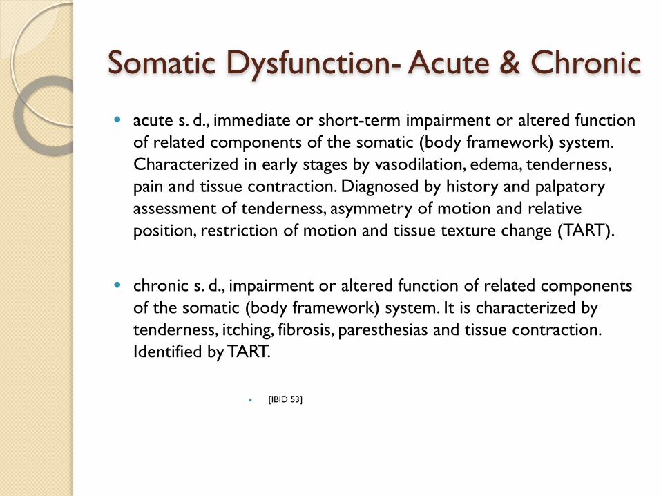
Somatic Dysfunction- Acute & Chronic● acute s. d., immediate or short-term impairment or altered function
of related components of the somatic (body framework) system. Characterized in early stages by vasodilation, edema, tenderness, pain and tissue contraction. Diagnosed by history and palpatory assessment of tenderness, asymmetry of motion and relative position, restriction of motion and tissue texture change (TART).
● chronic s. d., impairment or altered function of related components of the somatic (body framework) system. It is characterized by tenderness, itching, fibrosis, paresthesias and tissue contraction. Identified by TART.
● [IBID 53]

Acute vs. Chronic (Easy)
● Acute - short term abnormality. Flushed, swollen (or contracted), tender, painful. TART present
● Chronic - long present abnormality. Tender, itchy, numb, fibrous, contracted. TART present

Somatic Dysfunction - Primary and Secondary● primary s. d., 1. The somatic dysfunction that maintains a total pattern of
dysfunction, including other secondary dysfunctions. 2. The initial or first somatic dysfunction to appear temporally.
● secondary s. d., somatic dysfunction arising either from mechanical or neurophysiologic response subsequent to or as a consequence of other etiologies.
● type I s. d., a group curve of thoracic and/or lumbar vertebrae in which the freedoms of motion are in neutral with sidebending and rotation in opposite directions with maximum rotation at the apex (rotation occurs toward the convexity of the curve) based upon the Principles of Fryette.
● type II s. d., thoracic or lumbar somatic dysfunction of a single vertebral unit in which the vertebra is significantly flexed or extended with sidebending and rotation in the same direction (rotation occurs into the concavity of the curve) based upon the Principles of Fryette.
● [IBID]
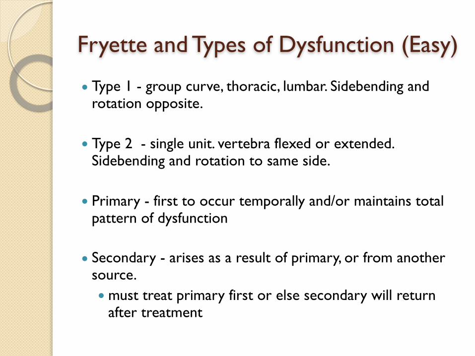
Fryette and Types of Dysfunction (Easy)
● Type 1 - group curve, thoracic, lumbar. Sidebending and rotation opposite.
● Type 2 - single unit. vertebra flexed or extended. Sidebending and rotation to same side.
● Primary - first to occur temporally and/or maintains total pattern of dysfunction
● Secondary - arises as a result of primary, or from another source. ● must treat primary first or else secondary will return
after treatment

Facilitation● spinal facilitation: 1. The maintenance of a pool of neurons (e.g.,
premotor neurons, motor neurons or preganglionic sympathetic neurons in one or more segments of the spinal cord) in a state of partial or subthreshold excitation; in this state, less afferent stimulation is required to trigger the discharge of impulses. 2. A theory regarding the neurophysio- logical mechanisms underlying the neuronal activity associated with somatic dysfunction. 3. Facilitation may be due to sustained increase in afferent input, aberrant patterns of afferent input, or changes within the affected neurons themselves or their chemical environment. Once established, facilitation can be sustained by normal central nervous system (CNS) activity.
● [IBID. 55]
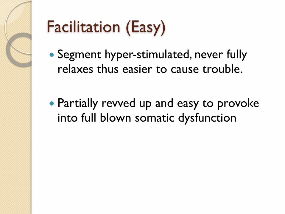
Facilitation (Easy)
● Segment hyper-stimulated, never fully relaxes thus easier to cause trouble.
● Partially revved up and easy to provoke into full blown somatic dysfunction
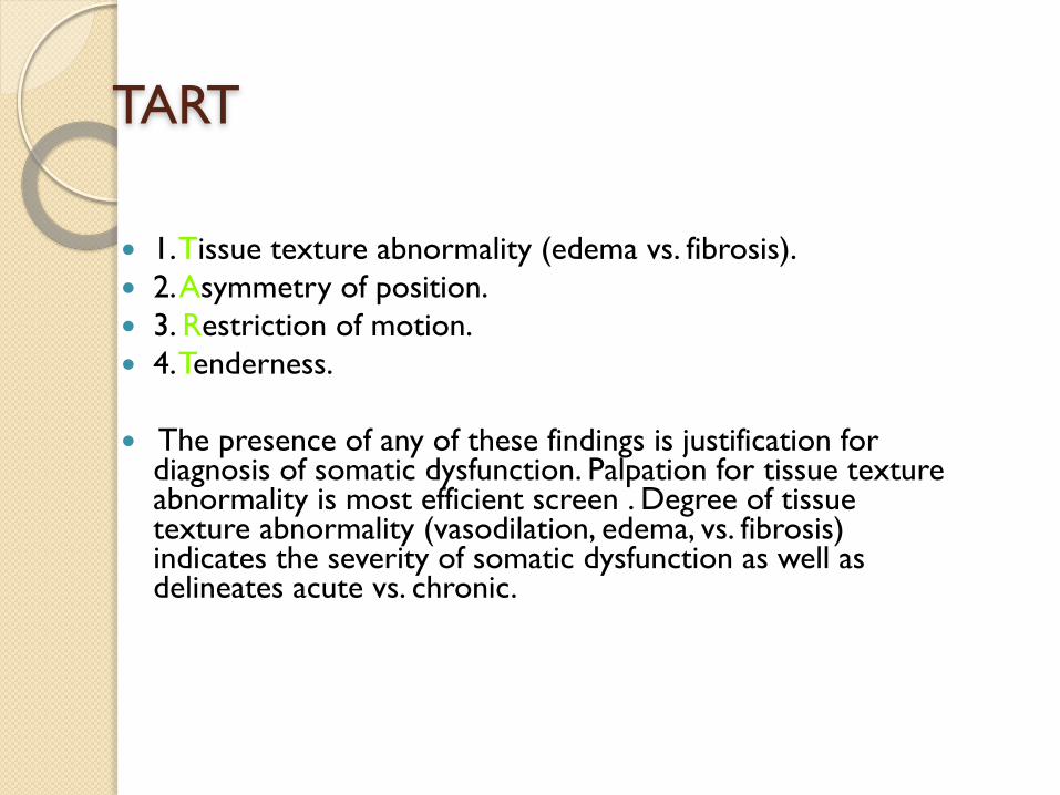
TART
● 1. Tissue texture abnormality (edema vs. fibrosis). ● 2. Asymmetry of position. ● 3. Restriction of motion. ● 4. Tenderness.
● The presence of any of these findings is justification for diagnosis of somatic dysfunction. Palpation for tissue texture abnormality is most efficient screen . Degree of tissue texture abnormality (vasodilation, edema, vs. fibrosis) indicates the severity of somatic dysfunction as well as delineates acute vs. chronic.

Diagnosis: Somatic Dysfunction
● Mechanical somatic dysfunction demonstrates asymmetry of motion stressed in paradigm of muscle energy OMT techniques and restriction of motion stressed in paradigm of articular dysfunction OMT techniques.
● Tenderness should not be confused with pain. Pain is patient subjective; tenderness is discomfort elicited on palpation by physician , objective finding- involuntary: muscle twitch or facial wince. Tenderness may be used to confirm tissue texture abnormality.
● Motion restriction places compensatory stress upon adjacent structures with resultant pain- which may be adjacent or distant, such as intervertebral cervical muscle spasms causing adjacent cervical facet pain or distant upper extremity cervical radiculopathy.

Exam
● Tissue texture abnormality and tenderness in absence of findings of mechanical restriction of motion is indicative of secondary somatic dysfunction of reflex origin: ◦ X-ray, CT, MRI, EMG, Ultrasound to diagnose and find pain generator such as facet
disease, spinal cord or nerve root impingement for appropriate referral to Interventionalist or Surgeon.
● Cervical exam: Supine ● Thoracic exam: Seated ● Lumbosacal exam: Prone or seated ● Standing structural exam: Inspection- Check for pelvic side shift, observation for
lateral-rotational curves, i.e. scoliosis, observation of AP curves; loss of lordosis cervical and lumbar vs. flattened spine, loss of kyphosis thoracic vs. increased- buffalo hump.
● Palpation- for tissue texture abnormality layered-superficial vs. deep. ● Segmental-Regional Exam: ROM- Flexion-extension, Left and right side bending ,
left and right rotation, restricted regionally vs. segmental. ● Practice, Repetition, Experience- will improve diagnostic skills.

Clinical Example● Patient presents with dysesthesia of palmar surface of right hand
consistent with presumptive diagnosis of carpal tunnel syndrome.
● Upper extremity complaints often are linked to upper thoracic somatic dysfunction. ● Although involved somatosensory innervation of the median
nerve should result in paravertebral findings from C6 to T1, findings are more typically encountered in upper thoracic region.
● Sympathetic innervation of the upper extremity arises from T1 to T4. ● Autonomic nerves contain afferent as well as efferent neurons,
therefore sympathetic afferent neurons from the right upper extremity are capable of facilitation in the upper thoracic region lateralizing to the right.

Example - continued● Facilitation can result from spinal dysfunction or spinal
segmental dysfunction with facilitation can result from postural accommodation to pelvic unleveling.
● Spinal segmental facilitation has been shown to result in segmentally related soft tissue edema, adding edema to the carpal tunnel.
● Response of axial musculoskeletal system to force of gravity is usually asymmetric. Most individuals have unequal leg length with resultant pelvic unleveling.
● Therefore most everyone is predisposed to compensatory group and segmental spinal dysfunction and stresses from activities of daily work and activities, trauma- micro & macro. It is understandable that musculoskeletal problems are so widespread.
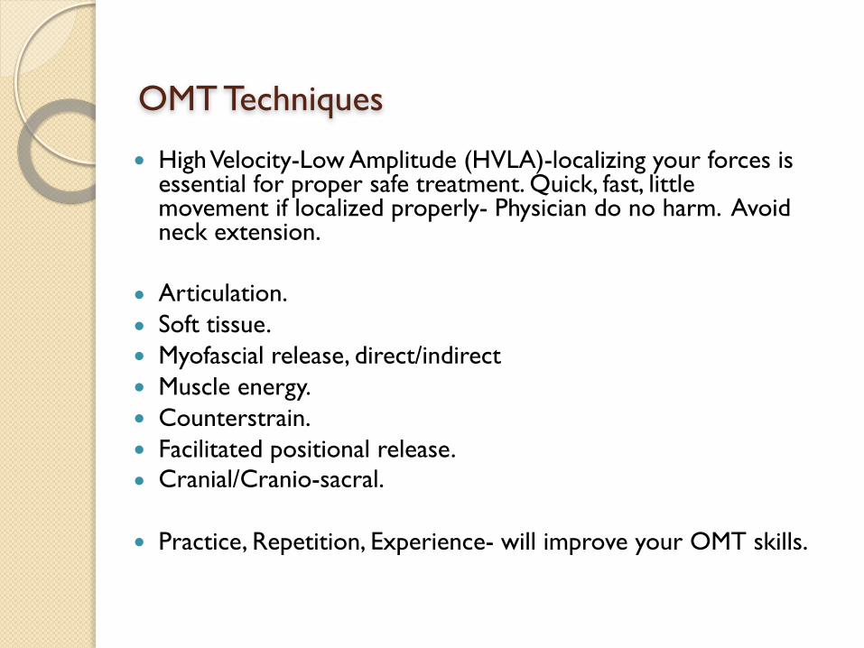
OMT Techniques
● High Velocity-Low Amplitude (HVLA)-localizing your forces is essential for proper safe treatment. Quick, fast, little movement if localized properly- Physician do no harm. Avoid neck extension.
● Articulation. ● Soft tissue. ● Myofascial release, direct/indirect ● Muscle energy. ● Counterstrain. ● Facilitated positional release. ● Cranial/Cranio-sacral.
● Practice, Repetition, Experience- will improve your OMT skills.
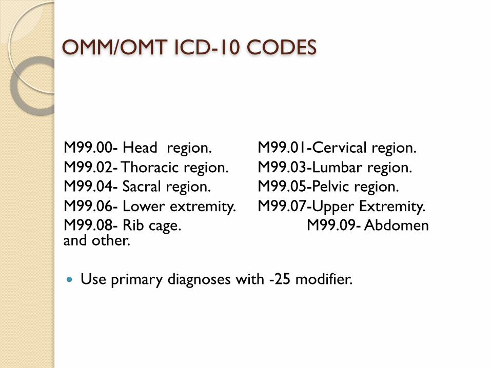
OMM/OMT ICD-10 CODES
M99.00- Head region. M99.01-Cervical region. M99.02- Thoracic region. M99.03-Lumbar region. M99.04- Sacral region. M99.05-Pelvic region. M99.06- Lower extremity. M99.07-Upper Extremity. M99.08- Rib cage. M99.09- Abdomen and other.
● Use primary diagnoses with -25 modifier.
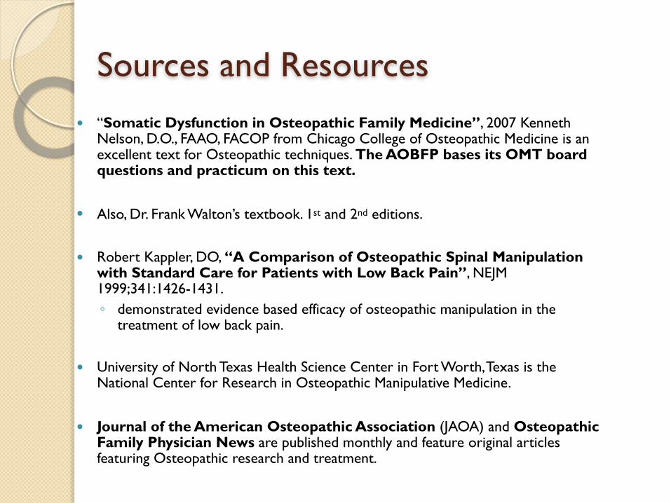
Sources and Resources● “Somatic Dysfunction in Osteopathic Family Medicine”, 2007 Kenneth
Nelson, D.O., FAAO, FACOP from Chicago College of Osteopathic Medicine is an excellent text for Osteopathic techniques. The AOBFP bases its OMT board questions and practicum on this text.
● Also, Dr. Frank Walton’s textbook. 1st and 2nd editions.
● Robert Kappler, DO, “A Comparison of Osteopathic Spinal Manipulation with Standard Care for Patients with Low Back Pain”, NEJM 1999;341:1426-1431. ◦ demonstrated evidence based efficacy of osteopathic manipulation in the
treatment of low back pain.
● University of North Texas Health Science Center in Fort Worth, Texas is the National Center for Research in Osteopathic Manipulative Medicine.
● Journal of the American Osteopathic Association (JAOA) and Osteopathic Family Physician News are published monthly and feature original articles featuring Osteopathic research and treatment.

Sources and Resources
● Glossary of Osteopathic Terminology, AACOM, 2011, https://www.aacom.org/docs/default-source/insideome/got2011ed.pdf
● Alexander S. Nicholas, D.O, FAAQ, and Evan A. Nicholas, D.O., Atlas of Osteopathic Techniques, LWW, 2015.
● Phillip Greenman, DO, FAAO, Principles of Manual Medicine, LWW, 1996
● Robert Savarese, DO, OMT Review, 3rd Edition, 2003
● Andrew Taylor Still
◦ The Philosophy and Mechanical Principles of Osteopathy ◦ Philosophy of Osteopathy ◦ Autobiography

OMT Hands on Skills Lab
Jon P. Burdzy, DO
Walter B. Flesner III, DO

Any Questions? Thank you!