Temperature and pH effects on biophysical and ... · Keywords: self-assembling; peptide RADA16-I;...
11
Journal of Peptide Science J. Pept. Sci. (2008) Published online in Wiley InterScience (www.interscience.wiley.com). DOI: 10.1002/psc.988 Temperature and pH effects on biophysical and morphological properties of self-assembling peptide RADA16-I ‡ ZHAOYANG YE, a § HANGYU ZHANG, a § HANLIN LUO, a § SHUNKANG WANG, a QINGHAN ZHOU, a XINPENG DU, a CHENGKANG TANG, a LIYAN CHEN, a JINGPING LIU, a YING-KANG SHI, b ER-YONG ZHANG, b RUTLEDGE ELLIS-BEHNKE c,d and XIAOJUN ZHAO a * a Institute for NanoBiomedical Technology and Membrane Biology, West China Hospital, Sichuan University, Science Park No 1, Ke Yuan 4th St., Gao Peng Road, Hi-tech Industrial Development Zone, Chengdu, 610041, Sichuan, China b West China Medical School, West China Hospital, Sichuan University, Guo Xue Xiang 37, Chengdu, 610041, Sichuan, China c Department of Brain & Cognitive Sciences 46-6007, Massachusetts Institute of Technology, 77 Massachusetts Avenue of Technology, MA 02139-4307, USA d Department of Anatomy, Li Ka Shing Faculty of Medicine, University of Hong Kong, 1/F Laboratory Block, 21 Sassoon Road, Pokfulam, Hong Kong SAR, China Received 26 August 2007; Revised 23 October 2007; Accepted 31 October 2007 Abstract: It has been found that the self-assembling peptide RADA 16-I forms a β-sheet structure and self-assembles into nanofibers and scaffolds in favor of cell growth, hemostasis and tissue-injury repair. But its biophysical and morphological properties, especially for its β-sheet and self-assembling properties in heat- and pH-denatured conditions, remain largely unclear. In order to better understand and design nanobiomaterials, we studied the self-assembly behaviors of RADA16-I using CD and atomic force microscopy (AFM) measurements in various pH and heat-denatured conditions. Here, we report that the peptide, when exposed to pH 1.0 and 4.0, was still able to assume a typical β-sheet structure and self-assemble into long nanofiber, although its β-sheet content was dramatically decreased by 10% in a pH 1.0 solution. However, the peptide, when exposed to pH 13.0, drastically lost its β-sheet structure and assembled into different small-sized globular aggregates. Similarly, the peptide, when heat-denatured from 25 to 70 ° C, was still able to assume a typical β-sheet structure with 46% content, but self-assembled into small-sized globular aggregates at much higher temperature. Titration experiments showed that the peptide RADA16-I exists in three types of ionic species: acidic (fully protonated peptide), zwitterionic (electrically neutral peptide carrying partial positive and negative charges) and basic (fully deprotonated peptide) species, called ‘super ions’. The unordered structure and β-turn of these ‘super ions’ via hydrogen or ionic bonds, and heat Brownian motion under the above denatured conditions would directly affect the stability of the β-sheet and nanofibers. These results help us in the design of future nanobiomaterials, such as biosensors, based on β-sheets and environmental changes. These results also help understand the pathogenesis of the β-sheet-mediated neuronal diseases such as Alzheimer’s disease and the mechanism of hemostasis. Copyright 2008 European Peptide Society and John Wiley & Sons, Ltd. Keywords: self-assembling; peptide RADA16-I; biophysical and morphological properties; hemostasis INTRODUCTION The design, synthesis and fabrication of biological nanomaterials, such as nanofibers and scaffolds based on a ‘bottom-up’ self-assembly peptide system, have attracted a great deal of attention from biomaterials and nanobiotechnology researchers for their poten- tial applications in nanobiotechnology and biomedical engineering [1–21]. For example, the 16-residue pep- tide, RADARADARADARADA (RADA16-I), was designed and synthesized by mimicking the amphiphilic segment * Correspondence to: Xiaojun Zhao, Institute for NanoBiomedical Technology and Membrane Biology, West China Hospital, Sichuan University, Science Park No 1, Ke Yuan 4th St., Gao Peng Road, Hi- tech Industrial Development Zone, Chengdu, 610041, Sichuan, China; e-mail: [email protected]; [email protected] ‡ This article is part of the Special Issue of the Journal of Peptide Science entitled ‘‘Peptides in Nanotechnology’’. § These authors contributed equally to this work. of EAEAKAKAEAEAKAKA (EAKA 16-II) of the nat- urally occurring yeast Zuotin protein. This ionic- complementary peptide contains a regular repeat of alternating hydrophobic and hydrophilic amino acids, including positively and negatively charged residues being arranged alternatively (Figure 1). RADA 16-I is able to self-assemble into scaffolds, composed of interwoven nanofibers with ∼100-nm mesh pores, in a way that mimics naturally occurring extracellular matrix (ECM), thus supporting cell and tissue growth, and consequently tissue-injury repair. For example, this peptide matrix has been shown to support rat hippocampal cell cultures, allowing for pro- liferation and differentiation, as well as synaptogenesis, in the RADA scaffold [5]. In addition, the nanomate- rial has also been reported to repair the optic tract in Chinese hamsters and act as an effective hemostatic medicine [22]. Copyright 2008 European Peptide Society and John Wiley & Sons, Ltd.
Transcript of Temperature and pH effects on biophysical and ... · Keywords: self-assembling; peptide RADA16-I;...
Journal of Peptide ScienceJ. Pept. Sci. (2008)Published online in Wiley InterScience (www.interscience.wiley.com). DOI: 10.1002/psc.988
Temperature and pH effects on biophysical andmorphological properties of self-assembling peptideRADA16-I‡
ZHAOYANG YE,a § HANGYU ZHANG,a § HANLIN LUO,a § SHUNKANG WANG,a QINGHAN ZHOU,a XINPENG DU,a
CHENGKANG TANG,a LIYAN CHEN,a JINGPING LIU,a YING-KANG SHI,b ER-YONG ZHANG,b
RUTLEDGE ELLIS-BEHNKEc,d and XIAOJUN ZHAOa*a Institute for NanoBiomedical Technology and Membrane Biology, West China Hospital, Sichuan University, Science Park No 1, Ke Yuan 4th St.,Gao Peng Road, Hi-tech Industrial Development Zone, Chengdu, 610041, Sichuan, Chinab West China Medical School, West China Hospital, Sichuan University, Guo Xue Xiang 37, Chengdu, 610041, Sichuan, Chinac Department of Brain & Cognitive Sciences 46-6007, Massachusetts Institute of Technology, 77 Massachusetts Avenue of Technology, MA02139-4307, USAd Department of Anatomy, Li Ka Shing Faculty of Medicine, University of Hong Kong, 1/F Laboratory Block, 21 Sassoon Road, Pokfulam, HongKong SAR, China
Received 26 August 2007; Revised 23 October 2007; Accepted 31 October 2007
Abstract: It has been found that the self-assembling peptide RADA 16-I forms a β-sheet structure and self-assembles intonanofibers and scaffolds in favor of cell growth, hemostasis and tissue-injury repair. But its biophysical and morphologicalproperties, especially for its β-sheet and self-assembling properties in heat- and pH-denatured conditions, remain largely unclear.In order to better understand and design nanobiomaterials, we studied the self-assembly behaviors of RADA16-I using CD andatomic force microscopy (AFM) measurements in various pH and heat-denatured conditions. Here, we report that the peptide,when exposed to pH 1.0 and 4.0, was still able to assume a typical β-sheet structure and self-assemble into long nanofiber,although its β-sheet content was dramatically decreased by 10% in a pH 1.0 solution. However, the peptide, when exposedto pH 13.0, drastically lost its β-sheet structure and assembled into different small-sized globular aggregates. Similarly, thepeptide, when heat-denatured from 25 to 70 °C, was still able to assume a typical β-sheet structure with 46% content, butself-assembled into small-sized globular aggregates at much higher temperature. Titration experiments showed that the peptideRADA16-I exists in three types of ionic species: acidic (fully protonated peptide), zwitterionic (electrically neutral peptide carryingpartial positive and negative charges) and basic (fully deprotonated peptide) species, called ‘super ions’. The unordered structureand β-turn of these ‘super ions’ via hydrogen or ionic bonds, and heat Brownian motion under the above denatured conditionswould directly affect the stability of the β-sheet and nanofibers. These results help us in the design of future nanobiomaterials,such as biosensors, based on β-sheets and environmental changes. These results also help understand the pathogenesis of theβ-sheet-mediated neuronal diseases such as Alzheimer’s disease and the mechanism of hemostasis. Copyright 2008 EuropeanPeptide Society and John Wiley & Sons, Ltd.
Keywords: self-assembling; peptide RADA16-I; biophysical and morphological properties; hemostasis
INTRODUCTION
The design, synthesis and fabrication of biologicalnanomaterials, such as nanofibers and scaffolds basedon a ‘bottom-up’ self-assembly peptide system, haveattracted a great deal of attention from biomaterialsand nanobiotechnology researchers for their poten-tial applications in nanobiotechnology and biomedicalengineering [1–21]. For example, the 16-residue pep-tide, RADARADARADARADA (RADA16-I), was designedand synthesized by mimicking the amphiphilic segment
* Correspondence to: Xiaojun Zhao, Institute for NanoBiomedicalTechnology and Membrane Biology, West China Hospital, SichuanUniversity, Science Park No 1, Ke Yuan 4th St., Gao Peng Road, Hi-tech Industrial Development Zone, Chengdu, 610041, Sichuan, China;e-mail: [email protected]; [email protected]‡ This article is part of the Special Issue of the Journal of PeptideScience entitled ‘‘Peptides in Nanotechnology’’.§ These authors contributed equally to this work.
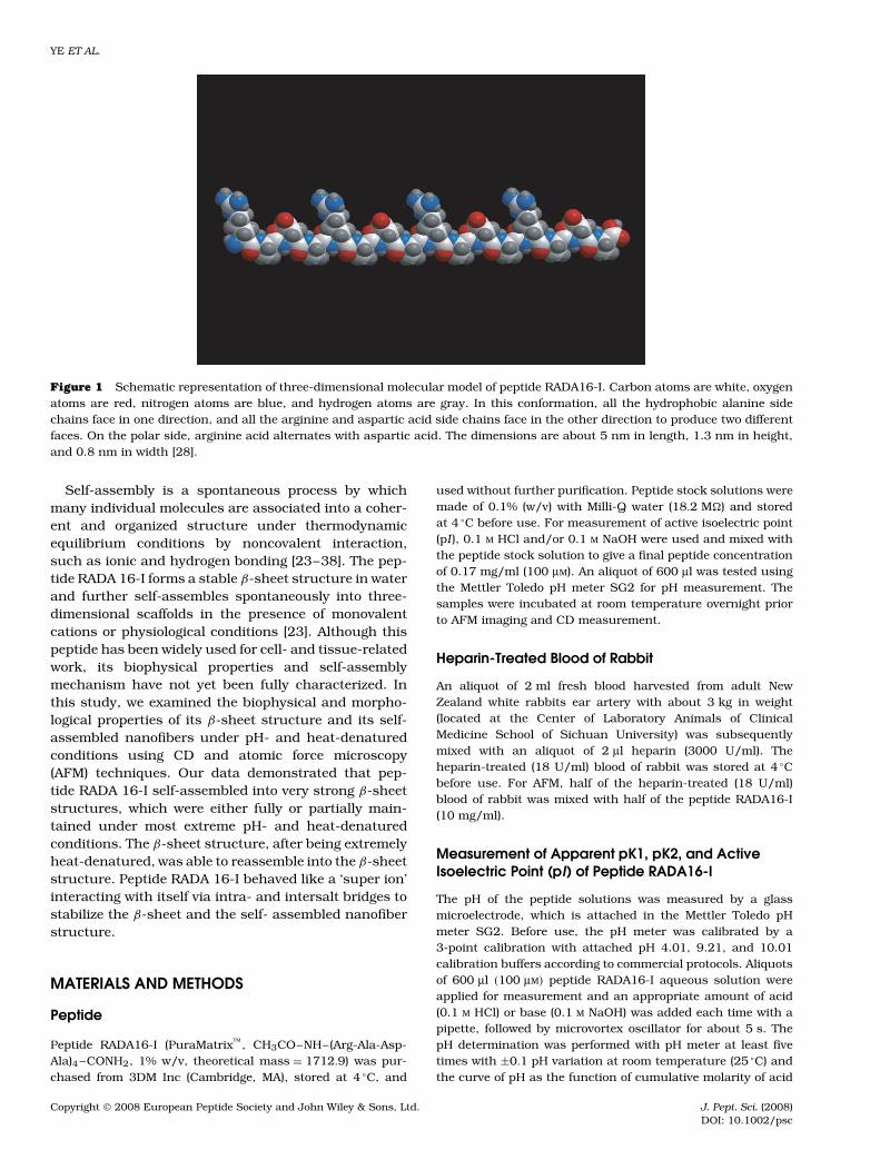
of EAEAKAKAEAEAKAKA (EAKA 16-II) of the nat-urally occurring yeast Zuotin protein. This ionic-complementary peptide contains a regular repeat ofalternating hydrophobic and hydrophilic amino acids,including positively and negatively charged residuesbeing arranged alternatively (Figure 1).
RADA 16-I is able to self-assemble into scaffolds,composed of interwoven nanofibers with ∼100-nmmesh pores, in a way that mimics naturally occurringextracellular matrix (ECM), thus supporting cell andtissue growth, and consequently tissue-injury repair.For example, this peptide matrix has been shown tosupport rat hippocampal cell cultures, allowing for pro-liferation and differentiation, as well as synaptogenesis,in the RADA scaffold [5]. In addition, the nanomate-rial has also been reported to repair the optic tract inChinese hamsters and act as an effective hemostaticmedicine [22].
Copyright 2008 European Peptide Society and John Wiley & Sons, Ltd.
YE ET AL.
Figure 1 Schematic representation of three-dimensional molecular model of peptide RADA16-I. Carbon atoms are white, oxygenatoms are red, nitrogen atoms are blue, and hydrogen atoms are gray. In this conformation, all the hydrophobic alanine sidechains face in one direction, and all the arginine and aspartic acid side chains face in the other direction to produce two differentfaces. On the polar side, arginine acid alternates with aspartic acid. The dimensions are about 5 nm in length, 1.3 nm in height,and 0.8 nm in width [28].
Self-assembly is a spontaneous process by whichmany individual molecules are associated into a coher-ent and organized structure under thermodynamicequilibrium conditions by noncovalent interaction,such as ionic and hydrogen bonding [23–38]. The pep-tide RADA 16-I forms a stable β-sheet structure in waterand further self-assembles spontaneously into three-dimensional scaffolds in the presence of monovalentcations or physiological conditions [23]. Although thispeptide has been widely used for cell- and tissue-relatedwork, its biophysical properties and self-assemblymechanism have not yet been fully characterized. Inthis study, we examined the biophysical and morpho-logical properties of its β-sheet structure and its self-assembled nanofibers under pH- and heat-denaturedconditions using CD and atomic force microscopy(AFM) techniques. Our data demonstrated that pep-tide RADA 16-I self-assembled into very strong β-sheetstructures, which were either fully or partially main-tained under most extreme pH- and heat-denaturedconditions. The β-sheet structure, after being extremelyheat-denatured, was able to reassemble into the β-sheetstructure. Peptide RADA 16-I behaved like a ‘super ion’interacting with itself via intra- and intersalt bridges tostabilize the β-sheet and the self- assembled nanofiberstructure.
MATERIALS AND METHODS
Peptide
Peptide RADA16-I (PuraMatrix, CH3CO–NH–(Arg-Ala-Asp-Ala)4 –CONH2, 1% w/v, theoretical mass = 1712.9) was pur-chased from 3DM Inc (Cambridge, MA), stored at 4 °C, and
used without further purification. Peptide stock solutions weremade of 0.1% (w/v) with Milli-Q water (18.2 M�) and storedat 4 °C before use. For measurement of active isoelectric point(pI ), 0.1 M HCl and/or 0.1 M NaOH were used and mixed withthe peptide stock solution to give a final peptide concentrationof 0.17 mg/ml (100 µM). An aliquot of 600 µl was tested usingthe Mettler Toledo pH meter SG2 for pH measurement. Thesamples were incubated at room temperature overnight priorto AFM imaging and CD measurement.
Heparin-Treated Blood of Rabbit
An aliquot of 2 ml fresh blood harvested from adult NewZealand white rabbits ear artery with about 3 kg in weight(located at the Center of Laboratory Animals of ClinicalMedicine School of Sichuan University) was subsequentlymixed with an aliquot of 2 µl heparin (3000 U/ml). Theheparin-treated (18 U/ml) blood of rabbit was stored at 4 °Cbefore use. For AFM, half of the heparin-treated (18 U/ml)blood of rabbit was mixed with half of the peptide RADA16-I(10 mg/ml).
Measurement of Apparent pK1, pK2, and ActiveIsoelectric Point (pI) of Peptide RADA16-I
The pH of the peptide solutions was measured by a glassmicroelectrode, which is attached in the Mettler Toledo pHmeter SG2. Before use, the pH meter was calibrated by a3-point calibration with attached pH 4.01, 9.21, and 10.01calibration buffers according to commercial protocols. Aliquotsof 600 µl (100 µM) peptide RADA16-I aqueous solution wereapplied for measurement and an appropriate amount of acid(0.1 M HCl) or base (0.1 M NaOH) was added each time with apipette, followed by microvortex oscillator for about 5 s. ThepH determination was performed with pH meter at least fivetimes with ±0.1 pH variation at room temperature (25 °C) andthe curve of pH as the function of cumulative molarity of acid
Copyright 2008 European Peptide Society and John Wiley & Sons, Ltd. J. Pept. Sci. (2008)DOI: 10.1002/psc

TEMPERATURE AND PH EFFECTS ON SELF-ASSEMBLING PEPTIDE RADA16-I
or base was determined. The apparent pK1, pK2, and activepI of peptide RADA16-I were all determined, according to therelated formulae described in textbook.
CD Measurement
All CD spectroscopy was performed on the peptide (100 µM)solutions (pure water and 0.1 M HCl or 0.1 M NaOH) that wereincubated at 4 °C overnight. CD data were gathered on an Avivmodel 400 spectrophotometer with 0.001–0.2 millidegreessensitivity and a 2-mm pathlength quartz cuvette (HellmaStandard Cuvet 110QS (Quartz Suprasil)). The supportedparameters: λ = 260–190 nm, �λ (bandwidth) = 1 nm, timeconstant = 1 s, using 3-times scans for average. Be sure tocheck the baseline CD signals of the empty cuvette and recorda baseline spectrum of the cuvette containing only pure waterunder identical conditions.
CD data were corrected by conversion to mean-residueellipticity to account for slight differences in molecular weightand concentration. The effects of pH and temperature onpeptide structure were determined by taking CD scans atdifferent pH (1.0, 4.0, 7.0, and 9.5) and different temperaturesranging from 25 to 80 °C.
The position and the relative magnitude of the minimum-mean-residue ellipticity were monitored to evaluate theβ-sheet structures and their relative β-sheet content of peptideRADA16-I at various environments.
In order to fully investigate the effect of pH and tempera-ture on secondary structures and self-assembling nanofiberformation of peptide RADA16-I, a free software CDPro (http://lamar.colostate.edu/∼sreeram/CDPro/main.html) wasapplied to estimate secondary-structure contents. The pep-tide secondary-structure fractions at various environmentswere calculated by the modified Contin method (CONTINLLprogram) with comparison to selected reference proteins set(IBasis7 (SDP48), λ = 240–190 nm). The calculated CD spec-tral data were compared with the experimental CD spectrumwith little difference (data not shown), and the amount ofthe root-mean-square deviation (RMSD) and the normalized-mean-square deviation (NMRSD) between the both are closeto 0.1 and zero (Tables 2 and 3), respectively, suggesting thatCONTINLL program and SDP48 fitting are suitable for CDanalyses of peptide RADA16-I.
Atomic Force Microscopy (AFM)
An aliquot of approximately 5 µl of the peptide (0.17 mg/ml)solution, heparin-treated (18 U/ml) blood, or heparin-treated(18 U/ml) blood mixture (1 : 1 v/v) with peptide RADA16-I (10 mg/ml), was evenly placed on the surface (about10 mm × 10 mm square or 15-mm diameter) of a freshlycleaved mica surface. Each sample was left on the mica forabout 30 s and then rinsed with aliquots of 100 µl of Milli-Qwater to remove unattached peptides or blood cells. The sampleon the mica surface was then air-dried for AFM observation.
AFM was performed with a SPI4000 Probe Station & SPA-400 SPM Unit (Seiko Instruments Inc., Chiba, Japan) usingthe dynamic force mode. The images utilized a 20-µm scanner(400) and an Olympus Si-DF20 microcantilever as well as aSi tip of radius 10 nm, rectangular base 200.00 µm, with tipheight of 10.00 µm and spring constant Kz of 12.00 N/m. Thecantilever’s free resonance frequency (Fo) was 127.00 kHz,
and the applied frequency (the working point) was set on thelower side of the resonance frequency (the left side of the QCurve), accompanied by the cyclic contact mode. All of themeasurements were performed in ambient air. Height imageswere recorded with 512 × 512 pixels resolution, in which thebrightness of morphology increases as a function of heightand the main parameters of AFM observation were as follows:Amp. Ref. (amplitude reference): −0.15 to −0.35, I Gain/PGain (integral gain/proportional gain): 0.10–0.33/0.05–0.15,Amplitude: ∼1 V, scan speed: 0.5–1.5 Hz. All image data setswere subjected to first-order flattening, in order to correct forthe tilt of the selected area and cancel any distortion (reversal)of causeless drifting or vibration (creeping) of the scanner. TheAFM observation of each sample was repeated at least fourtimes.
To quantitatively analyze the data, a large amount of theimages has been examined in order to find the surface coverageof nanofibers, clusters and branches and further calculate thelength of nanofibers. In higher magnification images, the widthand height of nanofibers can be determined using SPI3800N-offline image analysis program for win2000 software, alsocalled as ‘SPIWin’.
Phase-imaging AFM can simultaneously monitor the changeof the phase lag of the oscillation frequency of microcantileverwith different visualized pixels, when performing height-imaging AFM at the tapping mode. One of distinguishedcharacterizations for phase imaging is its high sensitivityfor the change of morphology [39]. Therefore, phase-imagingAFM has been employed to discriminate overlapping betweenblood cells and peptide RADA16-I in order to detect themorphological change of blood cells surface. The red andwhite blood cells are identified by their conventional scanningelectron microscopy (SEM) images described in textbook.
RESULTS
Measurement of the Apparent pK1, pK2, and theIsoelectric Point (pI) of Peptide RADA16-I
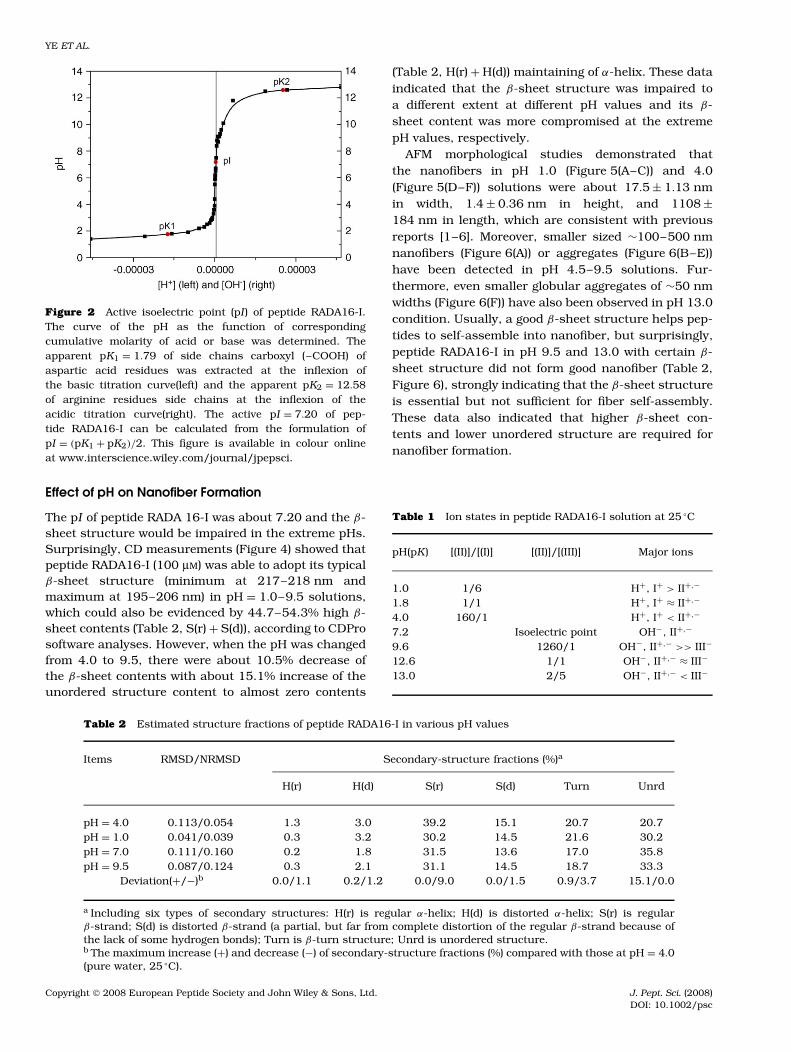
The molecular model of peptide RADA16-I is shownin Figure 1. This peptide contains four acidic residuesof aspartic acid (pI = 2.77, pKR = 3.65), four alkalineresidues of arginine (pI = 10.76, pKR = 12.48), andeight alanine (pI = 6.00) residues. The titration exper-iment showed that this peptide has a similar titrationcurve, when compared to that of a general nonpolaramino acid. An abrupt change in its apparent pI ofabout 7.20 was detected (Figure 2). Further analysisindicated that the apparent pK of four acidic aspar-tic acid residues was defined as pK1 ≈ 1.79, which ismuch lower than pKR (3.65) of aspartic acid with about100 times active acidic ion concentration increase. Theapparent pK2 of four alkaline arginine residues wasabout 12.58 with little alkaline enhancement mainlybecause of high stability of the strong basic guani-dinium substituent. The different ions states (Table 1,and Figure 3) of peptide RADA16-I are distinguished bythe apparent pK1, pK2, and pI.
Copyright 2008 European Peptide Society and John Wiley & Sons, Ltd. J. Pept. Sci. (2008)DOI: 10.1002/psc
YE ET AL.
Figure 2 Active isoelectric point (pI ) of peptide RADA16-I.The curve of the pH as the function of correspondingcumulative molarity of acid or base was determined. Theapparent pK1 = 1.79 of side chains carboxyl (–COOH) ofaspartic acid residues was extracted at the inflexion ofthe basic titration curve(left) and the apparent pK2 = 12.58of arginine residues side chains at the inflexion of theacidic titration curve(right). The active pI = 7.20 of pep-tide RADA16-I can be calculated from the formulation ofpI = (pK1 + pK2)/2. This figure is available in colour onlineat www.interscience.wiley.com/journal/jpepsci.
Effect of pH on Nanofiber Formation
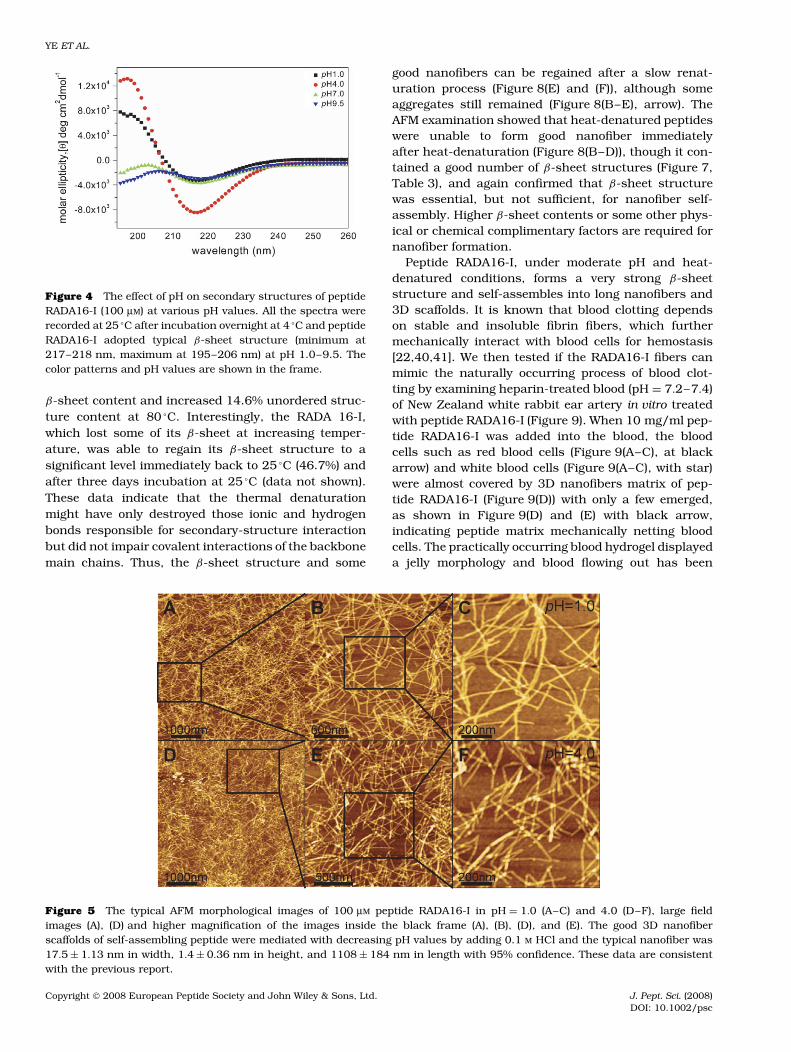
The pI of peptide RADA 16-I was about 7.20 and the β-sheet structure would be impaired in the extreme pHs.Surprisingly, CD measurements (Figure 4) showed thatpeptide RADA16-I (100 µM) was able to adopt its typicalβ-sheet structure (minimum at 217–218 nm andmaximum at 195–206 nm) in pH = 1.0–9.5 solutions,which could also be evidenced by 44.7–54.3% high β-sheet contents (Table 2, S(r) + S(d)), according to CDProsoftware analyses. However, when the pH was changedfrom 4.0 to 9.5, there were about 10.5% decrease ofthe β-sheet contents with about 15.1% increase of theunordered structure content to almost zero contents
(Table 2, H(r) + H(d)) maintaining of α-helix. These dataindicated that the β-sheet structure was impaired toa different extent at different pH values and its β-sheet content was more compromised at the extremepH values, respectively.
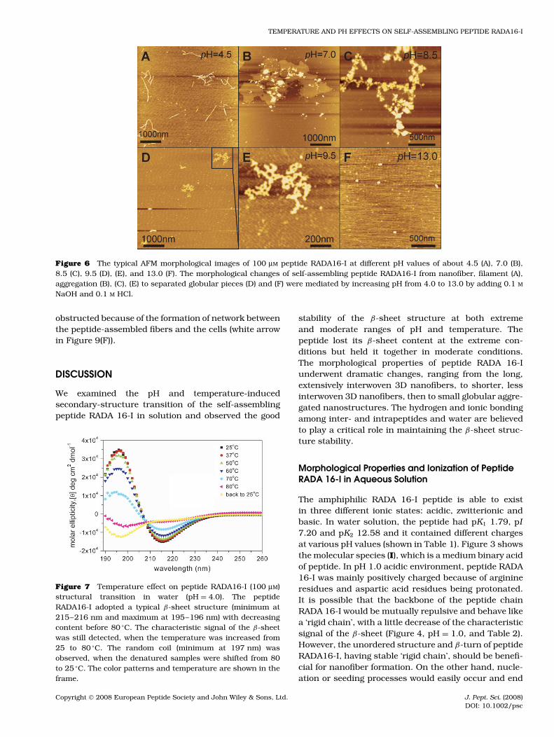
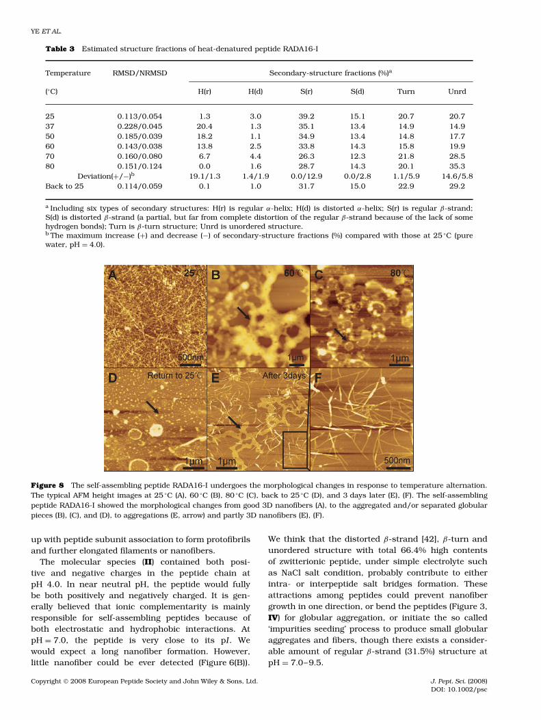
AFM morphological studies demonstrated thatthe nanofibers in pH 1.0 (Figure 5(A–C)) and 4.0(Figure 5(D–F)) solutions were about 17.5 ± 1.13 nmin width, 1.4 ± 0.36 nm in height, and 1108 ±184 nm in length, which are consistent with previousreports [1–6]. Moreover, smaller sized ∼100–500 nmnanofibers (Figure 6(A)) or aggregates (Figure 6(B–E))have been detected in pH 4.5–9.5 solutions. Fur-thermore, even smaller globular aggregates of ∼50 nmwidths (Figure 6(F)) have also been observed in pH 13.0condition. Usually, a good β-sheet structure helps pep-tides to self-assemble into nanofiber, but surprisingly,peptide RADA16-I in pH 9.5 and 13.0 with certain β-sheet structure did not form good nanofiber (Table 2,Figure 6), strongly indicating that the β-sheet structureis essential but not sufficient for fiber self-assembly.These data also indicated that higher β-sheet con-tents and lower unordered structure are required fornanofiber formation.
Table 1 Ion states in peptide RADA16-I solution at 25 °C
pH(pK) [(II)]/[(I)] [(II)]/[(III)] Major ions
1.0 1/6 H+, I+ > II+,−
1.8 1/1 H+, I+ ≈ II+,−
4.0 160/1 H+, I+ < II+,−
7.2 Isoelectric point OH−, II+,−
9.6 1260/1 OH−, II+,− >> III−
12.6 1/1 OH−, II+,− ≈ III−
13.0 2/5 OH−, II+,− < III−
Table 2 Estimated structure fractions of peptide RADA16-I in various pH values
Items RMSD/NRMSD Secondary-structure fractions (%)a
H(r) H(d) S(r) S(d) Turn Unrd
pH = 4.0 0.113/0.054 1.3 3.0 39.2 15.1 20.7 20.7pH = 1.0 0.041/0.039 0.3 3.2 30.2 14.5 21.6 30.2pH = 7.0 0.111/0.160 0.2 1.8 31.5 13.6 17.0 35.8pH = 9.5 0.087/0.124 0.3 2.1 31.1 14.5 18.7 33.3
Deviation(+/−)b 0.0/1.1 0.2/1.2 0.0/9.0 0.0/1.5 0.9/3.7 15.1/0.0
a Including six types of secondary structures: H(r) is regular α-helix; H(d) is distorted α-helix; S(r) is regularβ-strand; S(d) is distorted β-strand (a partial, but far from complete distortion of the regular β-strand because ofthe lack of some hydrogen bonds); Turn is β-turn structure; Unrd is unordered structure.b The maximum increase (+) and decrease (−) of secondary-structure fractions (%) compared with those at pH = 4.0(pure water, 25 °C).
Copyright 2008 European Peptide Society and John Wiley & Sons, Ltd. J. Pept. Sci. (2008)DOI: 10.1002/psc
TEMPERATURE AND PH EFFECTS ON SELF-ASSEMBLING PEPTIDE RADA16-I
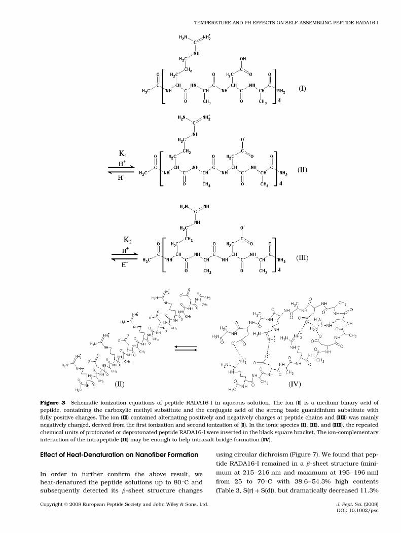
Figure 3 Schematic ionization equations of peptide RADA16-I in aqueous solution. The ion (I) is a medium binary acid ofpeptide, containing the carboxylic methyl substitute and the conjugate acid of the strong basic guanidinium substitute withfully positive charges. The ion (II) contained alternating positively and negatively charges at peptide chains and (III) was mainlynegatively charged, derived from the first ionization and second ionization of (I). In the ionic species (I), (II), and (III), the repeatedchemical units of protonated or deprotonated peptide RADA16-I were inserted in the black square bracket. The ion-complementaryinteraction of the intrapeptide (II) may be enough to help intrasalt bridge formation (IV).
Effect of Heat-Denaturation on Nanofiber Formation
In order to further confirm the above result, weheat-denatured the peptide solutions up to 80 °C andsubsequently detected its β-sheet structure changes
using circular dichroism (Figure 7). We found that pep-
tide RADA16-I remained in a β-sheet structure (mini-
mum at 215–216 nm and maximum at 195–196 nm)
from 25 to 70 °C with 38.6–54.3% high contents
(Table 3, S(r) + S(d)), but dramatically decreased 11.3%
Copyright 2008 European Peptide Society and John Wiley & Sons, Ltd. J. Pept. Sci. (2008)DOI: 10.1002/psc
YE ET AL.
Figure 4 The effect of pH on secondary structures of peptideRADA16-I (100 µM) at various pH values. All the spectra wererecorded at 25 °C after incubation overnight at 4 °C and peptideRADA16-I adopted typical β-sheet structure (minimum at217–218 nm, maximum at 195–206 nm) at pH 1.0–9.5. Thecolor patterns and pH values are shown in the frame.
β-sheet content and increased 14.6% unordered struc-ture content at 80 °C. Interestingly, the RADA 16-I,which lost some of its β-sheet at increasing temper-ature, was able to regain its β-sheet structure to asignificant level immediately back to 25 °C (46.7%) andafter three days incubation at 25 °C (data not shown).These data indicate that the thermal denaturationmight have only destroyed those ionic and hydrogenbonds responsible for secondary-structure interactionbut did not impair covalent interactions of the backbonemain chains. Thus, the β-sheet structure and some
good nanofibers can be regained after a slow renat-uration process (Figure 8(E) and (F)), although someaggregates still remained (Figure 8(B–E), arrow). TheAFM examination showed that heat-denatured peptideswere unable to form good nanofiber immediatelyafter heat-denaturation (Figure 8(B–D)), though it con-tained a good number of β-sheet structures (Figure 7,Table 3), and again confirmed that β-sheet structurewas essential, but not sufficient, for nanofiber self-assembly. Higher β-sheet contents or some other phys-ical or chemical complimentary factors are required fornanofiber formation.
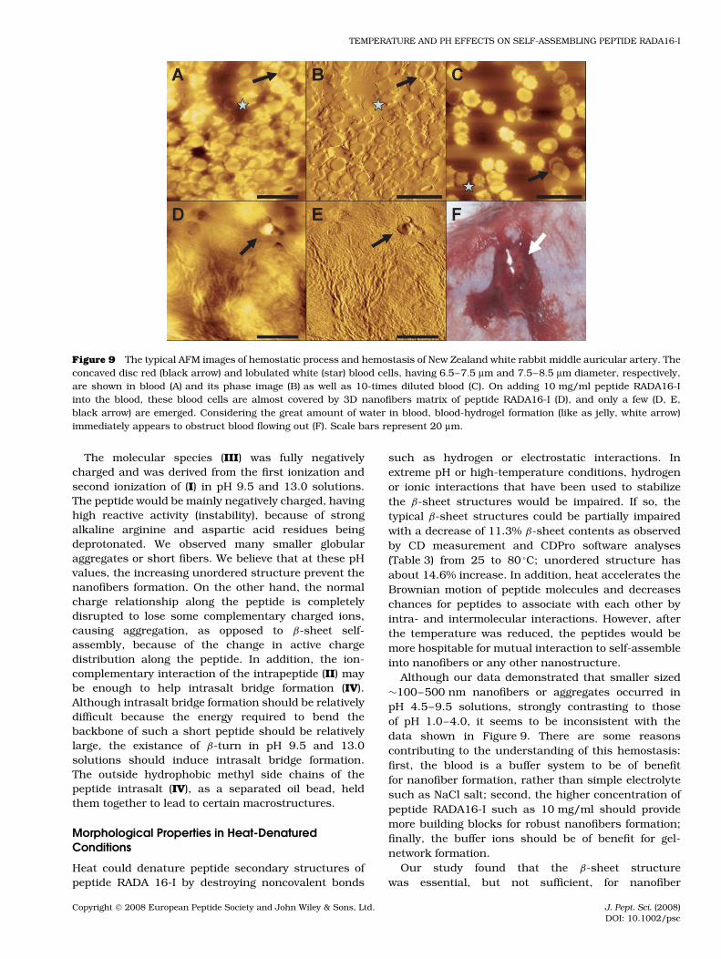
Peptide RADA16-I, under moderate pH and heat-denatured conditions, forms a very strong β-sheetstructure and self-assembles into long nanofibers and3D scaffolds. It is known that blood clotting dependson stable and insoluble fibrin fibers, which furthermechanically interact with blood cells for hemostasis[22,40,41]. We then tested if the RADA16-I fibers canmimic the naturally occurring process of blood clot-ting by examining heparin-treated blood (pH = 7.2–7.4)of New Zealand white rabbit ear artery in vitro treatedwith peptide RADA16-I (Figure 9). When 10 mg/ml pep-tide RADA16-I was added into the blood, the bloodcells such as red blood cells (Figure 9(A–C), at blackarrow) and white blood cells (Figure 9(A–C), with star)were almost covered by 3D nanofibers matrix of pep-tide RADA16-I (Figure 9(D)) with only a few emerged,as shown in Figure 9(D) and (E) with black arrow,indicating peptide matrix mechanically netting bloodcells. The practically occurring blood hydrogel displayeda jelly morphology and blood flowing out has been
Figure 5 The typical AFM morphological images of 100 µM peptide RADA16-I in pH = 1.0 (A–C) and 4.0 (D–F), large fieldimages (A), (D) and higher magnification of the images inside the black frame (A), (B), (D), and (E). The good 3D nanofiberscaffolds of self-assembling peptide were mediated with decreasing pH values by adding 0.1 M HCl and the typical nanofiber was17.5 ± 1.13 nm in width, 1.4 ± 0.36 nm in height, and 1108 ± 184 nm in length with 95% confidence. These data are consistentwith the previous report.
Copyright 2008 European Peptide Society and John Wiley & Sons, Ltd. J. Pept. Sci. (2008)DOI: 10.1002/psc
TEMPERATURE AND PH EFFECTS ON SELF-ASSEMBLING PEPTIDE RADA16-I
Figure 6 The typical AFM morphological images of 100 µM peptide RADA16-I at different pH values of about 4.5 (A), 7.0 (B),8.5 (C), 9.5 (D), (E), and 13.0 (F). The morphological changes of self-assembling peptide RADA16-I from nanofiber, filament (A),aggregation (B), (C), (E) to separated globular pieces (D) and (F) were mediated by increasing pH from 4.0 to 13.0 by adding 0.1 M
NaOH and 0.1 M HCl.
obstructed because of the formation of network betweenthe peptide-assembled fibers and the cells (white arrowin Figure 9(F)).
DISCUSSION
We examined the pH and temperature-inducedsecondary-structure transition of the self-assemblingpeptide RADA 16-I in solution and observed the good
Figure 7 Temperature effect on peptide RADA16-I (100 µM)structural transition in water (pH = 4.0). The peptideRADA16-I adopted a typical β-sheet structure (minimum at215–216 nm and maximum at 195–196 nm) with decreasingcontent before 80 °C. The characteristic signal of the β-sheetwas still detected, when the temperature was increased from25 to 80 °C. The random coil (minimum at 197 nm) wasobserved, when the denatured samples were shifted from 80to 25 °C. The color patterns and temperature are shown in theframe.
stability of the β-sheet structure at both extremeand moderate ranges of pH and temperature. Thepeptide lost its β-sheet content at the extreme con-ditions but held it together in moderate conditions.The morphological properties of peptide RADA 16-Iunderwent dramatic changes, ranging from the long,extensively interwoven 3D nanofibers, to shorter, lessinterwoven 3D nanofibers, then to small globular aggre-gated nanostructures. The hydrogen and ionic bondingamong inter- and intrapeptides and water are believedto play a critical role in maintaining the β-sheet struc-ture stability.
Morphological Properties and Ionization of PeptideRADA 16-I in Aqueous Solution
The amphiphilic RADA 16-I peptide is able to existin three different ionic states: acidic, zwitterionic andbasic. In water solution, the peptide had pK1 1.79, pI7.20 and pK2 12.58 and it contained different chargesat various pH values (shown in Table 1). Figure 3 showsthe molecular species (I), which is a medium binary acidof peptide. In pH 1.0 acidic environment, peptide RADA16-I was mainly positively charged because of arginineresidues and aspartic acid residues being protonated.It is possible that the backbone of the peptide chainRADA 16-I would be mutually repulsive and behave likea ‘rigid chain’, with a little decrease of the characteristicsignal of the β-sheet (Figure 4, pH = 1.0, and Table 2).However, the unordered structure and β-turn of peptideRADA16-I, having stable ‘rigid chain’, should be benefi-cial for nanofiber formation. On the other hand, nucle-ation or seeding processes would easily occur and end
Copyright 2008 European Peptide Society and John Wiley & Sons, Ltd. J. Pept. Sci. (2008)DOI: 10.1002/psc
YE ET AL.
Table 3 Estimated structure fractions of heat-denatured peptide RADA16-I
Temperature RMSD/NRMSD Secondary-structure fractions (%)a
(°C) H(r) H(d) S(r) S(d) Turn Unrd
25 0.113/0.054 1.3 3.0 39.2 15.1 20.7 20.737 0.228/0.045 20.4 1.3 35.1 13.4 14.9 14.950 0.185/0.039 18.2 1.1 34.9 13.4 14.8 17.760 0.143/0.038 13.8 2.5 33.8 14.3 15.8 19.970 0.160/0.080 6.7 4.4 26.3 12.3 21.8 28.580 0.151/0.124 0.0 1.6 28.7 14.3 20.1 35.3
Deviation(+/−)b 19.1/1.3 1.4/1.9 0.0/12.9 0.0/2.8 1.1/5.9 14.6/5.8Back to 25 0.114/0.059 0.1 1.0 31.7 15.0 22.9 29.2
a Including six types of secondary structures: H(r) is regular α-helix; H(d) is distorted α-helix; S(r) is regular β-strand;S(d) is distorted β-strand (a partial, but far from complete distortion of the regular β-strand because of the lack of somehydrogen bonds); Turn is β-turn structure; Unrd is unordered structure.b The maximum increase (+) and decrease (−) of secondary-structure fractions (%) compared with those at 25 °C (purewater, pH = 4.0).
Figure 8 The self-assembling peptide RADA16-I undergoes the morphological changes in response to temperature alternation.The typical AFM height images at 25 °C (A), 60 °C (B), 80 °C (C), back to 25 °C (D), and 3 days later (E), (F). The self-assemblingpeptide RADA16-I showed the morphological changes from good 3D nanofibers (A), to the aggregated and/or separated globularpieces (B), (C), and (D), to aggregations (E, arrow) and partly 3D nanofibers (E), (F).
up with peptide subunit association to form protofibrilsand further elongated filaments or nanofibers.
The molecular species (II) contained both posi-tive and negative charges in the peptide chain atpH 4.0. In near neutral pH, the peptide would fullybe both positively and negatively charged. It is gen-erally believed that ionic complementarity is mainlyresponsible for self-assembling peptides because ofboth electrostatic and hydrophobic interactions. AtpH = 7.0, the peptide is very close to its pI. Wewould expect a long nanofiber formation. However,little nanofiber could be ever detected (Figure 6(B)).
We think that the distorted β-strand [42], β-turn andunordered structure with total 66.4% high contentsof zwitterionic peptide, under simple electrolyte suchas NaCl salt condition, probably contribute to eitherintra- or interpeptide salt bridges formation. Theseattractions among peptides could prevent nanofibergrowth in one direction, or bend the peptides (Figure 3,IV) for globular aggregation, or initiate the so called‘impurities seeding’ process to produce small globularaggregates and fibers, though there exists a consider-able amount of regular β-strand (31.5%) structure atpH = 7.0–9.5.
Copyright 2008 European Peptide Society and John Wiley & Sons, Ltd. J. Pept. Sci. (2008)DOI: 10.1002/psc
TEMPERATURE AND PH EFFECTS ON SELF-ASSEMBLING PEPTIDE RADA16-I
Figure 9 The typical AFM images of hemostatic process and hemostasis of New Zealand white rabbit middle auricular artery. Theconcaved disc red (black arrow) and lobulated white (star) blood cells, having 6.5–7.5 µm and 7.5–8.5 µm diameter, respectively,are shown in blood (A) and its phase image (B) as well as 10-times diluted blood (C). On adding 10 mg/ml peptide RADA16-Iinto the blood, these blood cells are almost covered by 3D nanofibers matrix of peptide RADA16-I (D), and only a few (D, E,black arrow) are emerged. Considering the great amount of water in blood, blood-hydrogel formation (like as jelly, white arrow)immediately appears to obstruct blood flowing out (F). Scale bars represent 20 µm.
The molecular species (III) was fully negativelycharged and was derived from the first ionization andsecond ionization of (I) in pH 9.5 and 13.0 solutions.The peptide would be mainly negatively charged, havinghigh reactive activity (instability), because of strongalkaline arginine and aspartic acid residues beingdeprotonated. We observed many smaller globularaggregates or short fibers. We believe that at these pHvalues, the increasing unordered structure prevent thenanofibers formation. On the other hand, the normalcharge relationship along the peptide is completelydisrupted to lose some complementary charged ions,causing aggregation, as opposed to β-sheet self-assembly, because of the change in active chargedistribution along the peptide. In addition, the ion-complementary interaction of the intrapeptide (II) maybe enough to help intrasalt bridge formation (IV).Although intrasalt bridge formation should be relativelydifficult because the energy required to bend thebackbone of such a short peptide should be relativelylarge, the existance of β-turn in pH 9.5 and 13.0solutions should induce intrasalt bridge formation.The outside hydrophobic methyl side chains of thepeptide intrasalt (IV), as a separated oil bead, heldthem together to lead to certain macrostructures.
Morphological Properties in Heat-DenaturedConditions
Heat could denature peptide secondary structures ofpeptide RADA 16-I by destroying noncovalent bonds
such as hydrogen or electrostatic interactions. Inextreme pH or high-temperature conditions, hydrogenor ionic interactions that have been used to stabilizethe β-sheet structures would be impaired. If so, thetypical β-sheet structures could be partially impairedwith a decrease of 11.3% β-sheet contents as observedby CD measurement and CDPro software analyses(Table 3) from 25 to 80 °C; unordered structure hasabout 14.6% increase. In addition, heat accelerates theBrownian motion of peptide molecules and decreaseschances for peptides to associate with each other byintra- and intermolecular interactions. However, afterthe temperature was reduced, the peptides would bemore hospitable for mutual interaction to self-assembleinto nanofibers or any other nanostructure.
Although our data demonstrated that smaller sized∼100–500 nm nanofibers or aggregates occurred inpH 4.5–9.5 solutions, strongly contrasting to thoseof pH 1.0–4.0, it seems to be inconsistent with thedata shown in Figure 9. There are some reasonscontributing to the understanding of this hemostasis:first, the blood is a buffer system to be of benefitfor nanofiber formation, rather than simple electrolytesuch as NaCl salt; second, the higher concentration ofpeptide RADA16-I such as 10 mg/ml should providemore building blocks for robust nanofibers formation;finally, the buffer ions should be of benefit for gel-network formation.
Our study found that the β-sheet structurewas essential, but not sufficient, for nanofiber
Copyright 2008 European Peptide Society and John Wiley & Sons, Ltd. J. Pept. Sci. (2008)DOI: 10.1002/psc
YE ET AL.
self-assembly. Even so, there are still some other phys-ical or chemical factors such as peptide concentration,solvents, different ions as well as buffers to influenceself-assembling nanofiber formation. These problemswill stimulate us to further investigate them in subse-quent study.
AcknowledgementsWe thank Shuguang Zhang for his helpful sugges-tions and stimulating discussions. This research wasfinancially and technically supported by the ChineseNational ‘985 Project’ of Education Ministry of SichuanUniversity and the Analytical and Testing Center ofSichuan University, respectively, Chengdu, China.
REFERENCES
1. Zhang S. Fabrication of novel biomaterials through molecular self-assembly. Nat. Biotechnol. 2003; 21: 171–178.
2. Zhang S. Emerging biological materials through molecular self-assembly. Biotechnol. Adv. 2002; 20: 321–339.
3. Zhang S, Marini DM, Hwang W, Santoso S. Design of nanostruc-tured biological materials through self-assembly of peptide andproteins. Curr. Opin. Chem. Biol. 2002; 6: 865–871.
4. Gorman J. On-upping nature’s materials: striving for designersubstances that build themselves from individual molecules.Science 2000; 158: 364–367.
5. Schachner M. Nervous engineering. Nature 2000; 405: 747–748.6. Schwartz JJ, Zhang S. Peptide-mediated cellular delivery. Curr.
Opin. Mol. Ther. 2000; 2: 162–167.7. Holmes TC, Lacalle SD, Su X, Liu SS, Rich A, Zhang S. Extensive
neurite outgrowth and active synapse formation on self-assemblingpeptide scaffolds. Proc. Natl. Acad. Sci. U.S.A. 2000; 97:6728–6733.
8. Zhang S, Altman M. Peptide self-assembly in functional polymerscience and engineering. React. Funct. Polym. 1999; 41: 91–102.
9. Zhao X, Zhang S. Self-assembling nanopeptides become a new typeof biomaterial. Adv. Polym. Sci. 2006; 203: 145–170.
10. Zhao X, Zhang S. Molecular designer self-assembling peptides.Chem. Soc. Rev. 2006; 35: 1–7.
11. Zhao X, Zhang S. Fabrication of molecular materials using peptideconstruction motifs. Trends Biotechnol. 2004; 22: 470–476.
12. Zhao X, Nagai Y, Reeves PJ, Kiley P, Khorana HG, Zhang S.Designer short peptide surfactants stabilize G-protein coupledreceptor brovine rhodopsin. Proc. Natl. Acad. Sci. U.S.A. 2006; 103:17707–17712.
13. Zhang S, Holmes TC, Dipersio CM, Hynes RO, Su X, Rich A. Self-complementary oligopeptide matrices support mammalian cellattachment. Biomaterials 1995; 16: 1385–1393.
14. Leon EJ, Verma N, Zhang S, Lauffenburger DA, Kamm RD.Mechanical properties of a self-assembling oligopeptide matrix.J. Biomater. Sci. Polym. Ed. 1998; 9: 297–312.
15. Semino CE, Merok JR, Crane GG, Panagiotakos G, Zhang S.Functional differentiation of hepatocyte-like spheroid structuresfrom putative liver progenitor cells in three-dimensional peptidescaffolds. Differentiation 2003; 71: 262–270.
16. Semino CE, Kasahara J, Hayashi Y, Zhang S. Entrapment ofmigrating hippocampal neural cells in three-dimensional peptidenanofiber scaffold. Tissue Eng. 2004; 10: 643–655.
17. Bokharia MA, Akaya G, Zhang S, Birch MA. The enhancementof osteoblast growth and differentiation in vitro on a peptidehydrogel-polyHIPE polymer hybrid materials. Biomaterials 2005;26: 5198–5208.
18. Baig CK, Duhamel J, Fung SY, Bezaire J, Chen P. Self-assemblingpeptide as a potential carrier of hydrophobic compounds. J. Am.
Chem. Soc. 2004; 126: 7522–7532.19. Mira H, Vilar M, Esteve V, Martinell M, Kogan MJ, Giralt E,
Salom D, Mingarro I, Penarrubial L, Perez E. Ionic self-complementary induces amyloid-like fibril formation in an isolateddomain of a plant copper metallochaperone protein. BMC Struct.
Biol. 2004; 4: 7–21.20. Kisiday J, Jin M, Kurz B, Hung H, Semino C, Zhang S,
Grodzisky A. Self-assembling peptide hydrogel fosters chondrocyteextracellular matrix production and cell division: implications forcartilage tissue repair. Proc. Natl. Acad. Sci. U.S.A. 2002; 99:9996–10001.
21. Scheibel T, Parthasarathy R, Sawick G, Lin XM, Jaeger H.Conducting nanowires built by controlled self-assembly of amyloidfibers and selective metal deposition. Proc. Natl. Acad. Sci. U.S.A.
2003; 100: 4527–4532.22. Ellis-Behnke RG, Liang YX, Tay DK, Kau PWF, Schneider GE,
Zhang S, Wu W, So KF. Nano hemostat solution: immediatehemostasis at the nanoscale. Nanomedicine: Nanotechnology
Biology and Medicine 2006; 2(4): 207–215.23. Zhang S, Holmes T, Lockshin C, Rich A. Spontaneous assembly of
a self-complementary oligopeptide to form a stable macroscopicmembrane. Proc. Natl. Acad. Sci. U.S.A. 1993; 90: 3334–3338.
24. Lashuel HA, LaBrenz SR, Woo L, Serpell LC, Kelly JW. Protofila-ments, filaments, ribbons, and fibrils from peptidomimetic self-assembly: implication for amyloid fibrils formation and materialsscience. J. Am. Chem. Soc. 2000; 122: 5262–5277.
25. Aggeli A, Nyrkova IA, Bell M, Harding R, Carrick L, Mcleish TCB,et al. Hierarchical self-assembly of chiral rod-like molecules as amodel for peptide β-sheet tapes, ribbons, fibrils, and fibers. Proc.
Natl. Acad. Sci. U.S.A. 2001; 98: 11857–11862.26. Xiong H, Buckwalter BL, Shieh HM, Hecht MH. Periodicity of polar
and nonpolar amino acids is the major determinant of secondarystructure in self-assembling oligomeric peptides. Proc. Natl. Acad.
Sci. U.S.A. 1995; 92: 6349–6353.27. Caplan MR, Moore PN, Zhang S, Kamm RD, Lauffenburger DA.
Self-assembly of a β-sheet protein governed by relief of electrostaticrepulsion relative to van der Waals attraction. Biomacromolecules
2000; 1: 627–631.28. Yokoi H, Kinoshita T, Zhang S. Dynamic reassembly of peptide
RADA16 nanofiber scaffold. Proc. Natl. Acad. Sci. U.S.A. 2005; 102:8414–8419.
29. Zhang S, Lockshin C, Cook R, Rich A. Unusually stable β-sheetformation in an ionic self-complementary oligopeptide. Biopolymers
1994; 34: 663–672.30. Zhang S, Rich A. Direct conversion of an oligopeptide from a β-
sheet to an α-helix: a model for amyloid formation. Proc. Natl. Acad.
Sci. U.S.A. 1997; 94: 23–28.31. Altman M, Lee P, Rich A, Zhang S. Conformational behavior of ionic
self-complementary peptides. Protein Sci. 2000; 9: 1095–1105.32. Vale’ry C, Artzner F, Robert B, Gulick T, Keller G, Grabielle-
Madelmont C, Torres ML, Cherif-Cheikh R, Paternostrey M. Self-assembling process of a peptide in solution: from β-sheet filamentsto large embedded nanofibers. Biophys. J. 2004; 86: 2484–2501.
33. Hwang W, Zhang S, Kamm RD, Karplus M. Kinetic control of dimerstructure formation in amyloid fibrillogensis. Proc. Natl. Acad. Sci.
U.S.A. 2004; 101: 12916–12921.34. Hartgerink JD, Beniash E, Stupp SI. Peptide-amphiphile
nanofibers: a versatile scaffold for the preparation of self-assembling materials. Proc. Natl. Acad. Sci. U.S.A. 2002; 99:5133–5138.
35. Tjernberg LO, Tjernberg A, Bark N, Shi Y, Ruzsicxka BP, Bus Z,Thyberg J, Callaway DJE. Assembling amyloid fibrils from designedstructures containing a significant amyloid β-peptide fragment.Biochem. J. 2002; 366: 343–351.
36. Hong Y, Legge RL, Zhang S, Chen P. Effect of amino acid sequenceand pH on nanofiber formation of self-assembling peptides EAK16-II and EAK16-IV. Biomacromolecules 2003; 4: 1433–1442.
Copyright 2008 European Peptide Society and John Wiley & Sons, Ltd. J. Pept. Sci. (2008)DOI: 10.1002/psc
TEMPERATURE AND PH EFFECTS ON SELF-ASSEMBLING PEPTIDE RADA16-I
37. Fung SY, Keyes C, Duhamel J, Chen P. Concentration effect on theaggregation of a self-assembling oligopeptide. Biophys. J. 2003; 85:537–548.
38. Jun S, Hong Y, Imamura H, Ha BY, Bechhoefer J, Chen P. Self-assembly of the ionic peptide EAK16: the effect of chargedistributions on self-assembly. Biophys. J. 2004; 87: 1249–1259.
39. Pang GKH, Baba-Kishi KZ, Patel A. Topographic and phase-contrast imaging in atomic force microscopy. Ultramicroscopy 2000;81: 35–40.
40. Mankad PS, Codispoti M. The role of fibrin sealants in hemostasis.Am. J. Surg. 2001; 182: 21S–28S.
41. Jackson MR. Fibrin sealants in surgical practice: an overview. Am.
J. Surg. 2001; 182(Suppl. 2): 1S–7S.42. Viseu MI, Carvalho TI, Costa SMB. Conformational transitions in
β-Lactoglobulin induced by cationic amphiphiles: Equilibriumstudies. Biophys. J. 2004; 86: 2392–2402.
Copyright 2008 European Peptide Society and John Wiley & Sons, Ltd. J. Pept. Sci. (2008)DOI: 10.1002/psc