Tel2 Regulates the Stability of PI3K-Related Protein...
27
Tel2 Regulates the Stability of PI3K-Related Protein Kinases Hiroyuki Takai, 1 Richard C. Wang, 1,3 Kaori K. Takai, 1 Haijuan Yang, 2 and Titia de Lange 1, * 1 Laboratory for Cell Biology and Genetics, The Rockefeller University, 1230 York Avenue, New York, NY 10065, USA 2 Structural Biology Program, Sloan-Kettering Institute, Memorial Sloan-Kettering Cancer Center, New York, NY 10065, USA 3 Present address: Department of Dermatology, The University of Texas Southwestern Medical Center at Dallas, 5323 Harry Hines Boulevard, Dallas, TX 75390, USA. *Correspondence: [email protected] DOI 10.1016/j.cell.2007.10.052 SUMMARY We report an unexpected role for Tel2 in the expression of all mammalian phosphatidylino- sitol 3-kinase-related protein kinases (PIKKs). Although Tel2 was identified as a budding yeast gene required for the telomere length mainte- nance, we found no obvious telomeric function for mammalian Tel2. Targeted gene deletion showed that mouse Tel2 is essential in embry- onic development, embryonic stem (ES) cells, and embryonic fibroblasts. Conditional dele- tion of Tel2 from embryonic fibroblasts com- promised their response to IR and UV, dimin- ishing the activation of checkpoint kinases and their downstream effectors. The effects of Tel2 deletion correlated with significantly reduced protein levels for the PI3K-related kinases ataxia telangiectasia mutated (ATM), ATM and Rad3 related (ATR), DNA-dependent protein kinase catalytic subunit ataxia (DNA- PKcs). Tel2 deletion also elicited specific de- pletion of the mammalian target of rapamycin (mTOR), suppressor with morphological effect on genitalia 1 (SMG1), and transformation/tran- scription domain-associated protein (TRRAP), and curbed mTOR signaling, indicating that Tel2 affects all six mammalian PIKKs. While Tel2 deletion did not alter PIKK mRNA levels, in vivo pulse labeling experiments showed that Tel2 controls the stability of ATM and mTOR. Each of the PIKK family members asso- ciated with Tel2 in vivo and in vitro experiments indicated that Tel2 binds to part of the HEAT repeat segments of ATM and mTOR. These data identify Tel2 as a highly conserved regula- tor of PIKK stability. INTRODUCTION PI3K-related protein kinases are key signal transducers that inform eukaryotic cells on their nutrient supply and the status of their genome and its transcripts (Abraham, 2001; Bakkenist and Kastan, 2004; Guertin and Sabatini, 2005; Wullschleger et al., 2006). They phosphorylate target proteins on serine or threonine residues using a C-terminal region related to the catalytic domain of PI3 kinase (Lavin et al., 1995). The kinase domains of the PIKKs are flanked by conserved FAT and FATC domains (Bosotti et al., 2000), and their N termini carry long arrays of HEAT or Armadillo repeats (Andrade and Bork, 1995; Perry and Kleckner, 2003). Mammals have six PIKKs: taxia telangiectasia mutated (ATM), ATM and Rad3 related (ATR), DNA-dependent protein kinase catalytic subunit ataxia (DNA-PKcs), mammalian target of rapamycin, pre- viously FRAP (mTOR), suppressor with morphological effect on genitalia 1 (SMG1), and the catalytically inactive transformation/transcription domain-associated protein (TRRAP). The PIKKs interact with numerous proteins, but none of their binding partners or target proteins is held in common by all family members. The human PIKKs regulate a diverse set of signaling pathways that are relevant to human health and contribute to the suppression of tumorigenesis. ATM and ATR govern the response to genome damage by phosphorylating key substrates involved in DNA repair and cell-cycle control (Abraham, 2001; Kastan and Bartek, 2004; Shiloh, 2003). ATM is crucial for the response to DNA double-strand breaks (DSBs), whereas ATR is activated by replication stress and certain DNA repair intermediates. Mutations in ATM cause ataxia telangiectasia, while a mutation in ATR results in Seckel syndrome. DNA-PKcs promotes nonhomologous end joining and the diminished V(D)J recombination resulting from DNA-PKcs deficiency is re- sponsible for severe combined immunodeficiency (SCID) in mice. SMG1 is required for the nonsense-mediated decay (NMD) mRNA surveillance pathway (Conti and Izaurralde, 2005). TRRAP is involved in the regulation of 1248 Cell 131, 1248–1259, December 28, 2007 ª2007 Elsevier Inc.
-
Upload
duongxuyen -
Category
Documents
-
view
216 -
download
0
Transcript of Tel2 Regulates the Stability of PI3K-Related Protein...

Tel2 Regulates the Stabilityof PI3K-Related Protein KinasesHiroyuki Takai,1 Richard C. Wang,1,3 Kaori K. Takai,1 Haijuan Yang,2 and Titia de Lange1,*1Laboratory for Cell Biology and Genetics, The Rockefeller University, 1230 York Avenue, New York,
NY 10065, USA2Structural Biology Program, Sloan-Kettering Institute, Memorial Sloan-Kettering Cancer Center, New York,
NY 10065, USA3Present address: Department of Dermatology, The University of Texas Southwestern Medical Center at Dallas,
5323 Harry Hines Boulevard, Dallas, TX 75390, USA.
*Correspondence: [email protected]
DOI 10.1016/j.cell.2007.10.052
SUMMARY
We report an unexpected role for Tel2 in theexpression of all mammalian phosphatidylino-sitol 3-kinase-related protein kinases (PIKKs).Although Tel2 was identified as a budding yeastgene required for the telomere length mainte-nance, we found no obvious telomeric functionfor mammalian Tel2. Targeted gene deletionshowed that mouse Tel2 is essential in embry-onic development, embryonic stem (ES) cells,and embryonic fibroblasts. Conditional dele-tion of Tel2 from embryonic fibroblasts com-promised their response to IR and UV, dimin-ishing the activation of checkpoint kinasesand their downstream effectors. The effectsof Tel2 deletion correlated with significantlyreduced protein levels for the PI3K-relatedkinases ataxia telangiectasia mutated (ATM),ATM and Rad3 related (ATR), DNA-dependentprotein kinase catalytic subunit ataxia (DNA-PKcs). Tel2 deletion also elicited specific de-pletion of the mammalian target of rapamycin(mTOR), suppressor with morphological effecton genitalia 1 (SMG1), and transformation/tran-scription domain-associated protein (TRRAP),and curbed mTOR signaling, indicating thatTel2 affects all six mammalian PIKKs. WhileTel2 deletion did not alter PIKK mRNA levels,in vivo pulse labeling experiments showedthat Tel2 controls the stability of ATM andmTOR. Each of the PIKK family members asso-ciated with Tel2 in vivo and in vitro experimentsindicated that Tel2 binds to part of the HEATrepeat segments of ATM and mTOR. Thesedata identify Tel2 as a highly conserved regula-tor of PIKK stability.
1248 Cell 131, 1248–1259, December 28, 2007 ª2007 Elsevier
INTRODUCTION
PI3K-related protein kinases are key signal transducers
that inform eukaryotic cells on their nutrient supply and
the status of their genome and its transcripts (Abraham,
2001; Bakkenist and Kastan, 2004; Guertin and Sabatini,
2005; Wullschleger et al., 2006). They phosphorylate
target proteins on serine or threonine residues using
a C-terminal region related to the catalytic domain of PI3
kinase (Lavin et al., 1995). The kinase domains of the
PIKKs are flanked by conserved FAT and FATC domains
(Bosotti et al., 2000), and their N termini carry long arrays
of HEAT or Armadillo repeats (Andrade and Bork, 1995;
Perry and Kleckner, 2003). Mammals have six PIKKs: taxia
telangiectasia mutated (ATM), ATM and Rad3 related
(ATR), DNA-dependent protein kinase catalytic subunit
ataxia (DNA-PKcs), mammalian target of rapamycin, pre-
viously FRAP (mTOR), suppressor with morphological
effect on genitalia 1 (SMG1), and the catalytically inactive
transformation/transcription domain-associated protein
(TRRAP). The PIKKs interact with numerous proteins,
but none of their binding partners or target proteins is
held in common by all family members.
The human PIKKs regulate a diverse set of signaling
pathways that are relevant to human health and contribute
to the suppression of tumorigenesis. ATM and ATR govern
the response to genome damage by phosphorylating key
substrates involved in DNA repair and cell-cycle control
(Abraham, 2001; Kastan and Bartek, 2004; Shiloh, 2003).
ATM is crucial for the response to DNA double-strand
breaks (DSBs), whereas ATR is activated by replication
stress and certain DNA repair intermediates. Mutations
in ATM cause ataxia telangiectasia, while a mutation in
ATR results in Seckel syndrome. DNA-PKcs promotes
nonhomologous end joining and the diminished V(D)J
recombination resulting from DNA-PKcs deficiency is re-
sponsible for severe combined immunodeficiency (SCID)
in mice. SMG1 is required for the nonsense-mediated
decay (NMD) mRNA surveillance pathway (Conti and
Izaurralde, 2005). TRRAP is involved in the regulation of
Inc.

gene expression as a component of histone acetyltrans-
ferase complexes (Herceg and Wang, 2005). The sixth
mammalian PIKK, mTOR, integrates environmental cues,
mitogenic signals, and nutrient availability to control cell
growth (Hay and Sonenberg, 2004). The PI3K-AKT-
mTOR signaling pathway is frequently altered in human
cancer (Shaw and Cantley, 2006; Sabatini, 2006).
Here we report a link between all six mammalian
PIKKs and Tel2 (official gene symbol TELO2, also re-
ferred to as hCLK2), the mammalian ortholog of the bud-
ding yeast TEL2 gene. Along with TEL1, TEL2 was iden-
tified in the first screen for yeast mutants with altered
telomere length (Lustig and Petes, 1986). Cells that
harbor mutant alleles of tel1 or tel2 undergo telomere
attrition for �150 generations but ultimately maintain
their telomeres at a stable short length. Gene cloning re-
vealed that TEL1 is the budding yeast ortholog of ATM
(Greenwell et al., 1995; Morrow et al., 1995). TEL2 was
found to be an essential gene, but the TEL2 ORF lacked
obvious conserved motifs that might suggest a molecular
function (Runge and Zakian, 1996). The conservation of
TEL2 in Drosophila melanogaster suggested that its
function may not be limited to telomere maintenance,
since Drosophila and other dipterans lack the canonical
telomerase-based telomere system (Pimpinelli, 2005).
Hypomorphic alleles of the C. elegans TEL2 ortholog
(Clk-2/Rad-5) show pleiotropic phenotypes, including
an extended life span (Lakowski and Hekimi, 1996) and
hypersensitivity to DNA damage (Hartman and Herman,
1982) but no significant telomere-length changes. Re-
cent reports have suggested a role for human and fission
yeast Tel2 in the response to replication stress (Jiang
et al., 2003; Collis et al., 2007; Shikata et al., 2007).
We have used gene targeting in mouse cells to deter-
mine the function of Tel2 and show that it controls the
stability of all PIKKs proteins, thus potentially explaining
the pleiotropic nature of the phenotypes of Tel2 muta-
tions.
RESULTS
No Obvious Telomeric Functionfor Mammalian Tel2Guided by the work on budding yeast Tel2, we initially
explored the possibility that the human and mouse Tel2
orthologs might have telomere-related functions (Fig-
ure S1). Using Flag-tagged human Tel2 (Figures 1A and
1B), we asked whether Tel2 can localize to chromosome
ends. Indirect immunofluorescence (IF) showed that
ectopically expressed Tel2 localized in both the cyto-
plasm and nucleus (Figure S1C), in agreement with the
absence of canonical subcellular localization signals
from the Tel2 ORF. However, IF and ChIP analysis failed
to provide evidence for a telomeric localization of either
ectopically expressed Tel2 or the endogenous protein
(Figures S1C–S1E). In addition, shRNA knockdown of
Tel2 or overexpression of full-length Tel2 or truncated
Tel2 had no significant effect on telomere-length homeo-
Cell
stasis in human tumor cell lines or the shortening rate of
telomeres in primary human fibroblasts (data not shown).
Finally, cells with diminished Tel2 did not show accumu-
lation of 53BP1 or g-H2AX at their telomeres (data not
shown), which is a hallmark of telomere deprotection (Ta-
kai et al., 2003). Although these results did not exclude
a function for Tel2 at telomeres, they raised the possibil-
ity that Tel2 might have other functions in mammalian
cells.
Tel2 Is Essential for Embryonic Developmentand Cellular GrowthTo address the function of Tel2, we used targeted gene
deletion in the mouse. Mouse Tel2 mRNA appeared to
be ubiquitously expressed (Figure S1F) and encodes an
ORF starting in the second exon that predicts a 840 aa
protein with a molecular weight (MW) of 93 kDa. Our
targeting strategy deleted the third exon of the Tel2 gene
(Figures 1A and 1B), resulting in a shift in the reading frame
at amino acid (aa) 116 into a non-Tel2 ORF that terminates
52 amino acids downstream. Heterozygous Tel2DEL/+
mice were viable, fertile, and healthy, suggesting that
the truncated protein is either not expressed or has no
deleterious effects. Mouse embryonic fibroblasts (MEFs)
prepared from Tel2DEL/+ embryos grew normally and
showed the expected 2-fold reduction in Tel2 protein
levels (Figure 1C). Consistent with the lethality of Tel2
deficiency in yeast and C. elegans (Benard et al., 2001;
Runge and Zakian, 1996; Shikata et al., 2007), the Tel2
null mutation (DEL/DEL) caused embryonic lethality
(Figure 1D). Tel2 also appeared to be essential in ES cells
(Figure S2). When double-targeted Tel2TARGET/FLOX ES
cells carrying two conditional Tel2 alleles were treated
with Cre to induce Tel2 deficiency, the cells arrested (Fig-
ures S2A–S2D), and we were unable to isolate Tel2 null
clones from Tel2TARGET/FLOX ES cell cultures in which
Cre was expressed (Figure S2E).
To study the cellular consequence of Tel2 deficiency,
we prepared Tel2DEL/FLOX MEFs from which Tel2 could
be deleted by transduction of a self-deleting Cre retrovirus
(H&R Cre) (Silver and Livingston, 2001; Celli and de Lange,
2005) or by 4-hydroxytamoxifen (4-OHT) induction of
a Cre-ER fusion protein expressed from the GT(ROSA)26-
Sor locus (R26Cre-ER) (Feil et al., 1996; Badea et al.,
2003). Expression of Cre recombinase using either system
removed the floxed allele efficiently (Figures 2A and 4D
and see below). Tel2 protein became undetectable by
4 days after H&R Cre infection of the Tel2DEL/FLOX MEFs
(Figure 2A). Similarly, the Tel2 protein disappeared within
2 to 3 days after induction of R26Cre-ER with OHT (see
below, Figure 4E). Cells lacking the Tel2 gene initially
grew normally but arrested after 3 to 4 days with a 2N or
4N DNA content, reduced S and M phase index, and a flat-
tened senescence-like morphology (Figures 2B–2E, S3A,
and S3B and data not shown). This cell-cycle arrest phe-
notype was due to loss of Tel2 since ectopic expression of
either mouse or human Tel2 cDNA rescued the growth
defect (Figures 2C, 2D, S3A, and S3B).
131, 1248–1259, December 28, 2007 ª2007 Elsevier Inc. 1249

Figure 1. Conditional Deletion of Mouse
Tel2
(A) Schematic of the mouse Tel2 locus (WT,
Tel2+), the targeting vector (pTV), the conditional
allele (Tel2FLOX), and the null allele (Tel2DEL).
NdeI fragment sizes are indicated for each ge-
notype, and the probes p1 and p2 are shown.
f1, r1, and r2, primers for genomic PCR; N,
NdeI; S, ScaI; K, KpnI; A, ApaI.
(B) Genomic blots of ES clones. Genomic DNA
digested with NdeI and probed with p1. The tar-
geted ES clones were transiently transfected
with Cre to generate ES cells harboring the indi-
cated genotypes.
(C) Western blot of Tel2 from MEFs of the
indicated genotypes. *, nonspecific signal de-
tected by anti-Tel2 Ab #1039.
(D) Table of the genotypes found in the offspring
of heterozygous intercrosses of Tel2DEL/+ mice
at E13.5 and at weaning. The genotype was
ambiguous for two embryos (ND).
Tel2 Null Cells Show DiminishedDNA-Damage SignalingThe cell-cycle arrest of Tel2-deficient cells did not appear
to be due to a DNA-damage response since the nuclei did
not contain DNA-damage foci (data not shown) and the
phenotype was not affected by abrogation of the p53
pathway (Figure 2B). The arrest was also not accompa-
nied by induction of CDK inhibitors such as p21, p27,
and p16 (Figure 3A). There was also no evidence for upre-
gulation of p53, consistent with the lack of p21 induction
upon Tel2 deletion.
Interestingly, in cells lacking Tel2, the basal level of
phosphorylation of p53 on serine 18 was diminished, and
the steady-state levels of p53 and p21 were slightly but
reproducibly reduced (Figure 3A). As this reduction in basal
p53 activation could be explained if the Tel2-deficient cells
had a diminished response to intrinsic DNA damage, we
tested their ability to respond to exogenously induced gen-
otoxic stress. Whereas control cells showed the expected
IR-induced phosphorylation of Chk2 and a concomitant
increase in p53 serine 18 phosphorylation, Tel2-deficient
cells failed to show this response and also lacked the abil-
ity to upregulate p21 after IR (Figure 3B). These results sug-
gested that the slight reductions in p53 and p21 were due
to diminished Chk2 signaling in the Tel2 null cells. Interest-
1250 Cell 131, 1248–1259, December 28, 2007 ª2007 Elsevier
ingly, the cells were also defective in the induction of Chk1
phosphorylation after UV treatment (Figure 3C), suggest-
ing that Tel2-deficient cells might be generally compro-
mised in their ability to respond to DNA damage. Whereas
Tel2 deficiency affected the DNA-damage signaling path-
ways, IR-induced DNA damage did not appear to alter the
localization of human or mouse Tel2 (Figure S1G and data
not shown) nor did IR affect the migration of mouse Tel2
protein in SDS/PAGE (Figure 3B and data not shown).
Tel2 Affects PIKK Protein LevelsTo further address the effect of Tel2 on the DNA-damage
response, we evaluated the status of the ATM kinase in
Tel2-deficient cells. Immunoblots revealed that the dele-
tion of Tel2 resulted in a striking reduction in abundance
of ATM protein (Figure 4A). Quantification of the data indi-
cated that the residual ATM levels were less than 10% of
the controls (data not shown). The lowered abundance of
ATM protein was a specific consequence of Tel2 deletion,
since the effect was negated by expression of mouse Tel2
from a retroviral vector (Figure 4B). Whereas ATM levels
depended on Tel2, the converse was not the case:
Atm�/� MEFs had normal Tel2 levels (Figure 4C).
The diminished Chk1 phosphorylation in UV-treated
Tel2-deficient cells (Figure 3C) suggested that Tel2 might
Inc.

Figure 2. Tel2 Depletion Results in Cell-Cycle Arrest
(A) Immunoblot for mouse Tel2 protein from Tel2DEL/FLOX MEFs (top) and PCR analysis of genomic DNA isolated from Tel2DEL/FLOX (bottom) at the
indicated time points after introduction of Cre with the H&R retrovirus. *, nonspecific signal detected by the anti-Tel2 Ab #1039.
(B) Cell-cycle profile after deletion of Tel2 from p53+/+ or p53�/� MEFs. MEFs were infected with H&R Cre (or a mock treatment) and 5 (p53+/+) or
6 (p53�/�) days later incubated with BrdU for 1 hr, fixed, and analyzed by FACS for DNA (propidium iodide) and BrdU content.
(C) Graph of the growth curves of Tel2DEL/FLOX p53�/�MEFs after H&R Cre infection or mock infection (-Cre). Cells indicated as Tel2 were infected with
an hTel2 retrovirus and selected with puromycin before the H&R Cre infection.
(D) Immunoblots for Tel2 in the MEFs used in (C). *, nonspecific signal. control, nonspecific signals detected by the Tel2C antibody.
(E) Phase-contrast micrographs of Tel2DEL/FLOX p53�/� MEFs at day 9 after infection with H&R Cre. The dotted white line highlights a cell with
senescent morphology.
also affect the ATR kinase. Indeed, the steady-state level
of ATR was strongly diminished upon deletion of Tel2 us-
ing the R26Cre-ER system (Figures 4D and 4E). The effect
of Tel2 on ATM and ATR was specific since Rif1, a large
protein with HEAT repeats involved in the DNA-damage
response (Xu and Blackburn, 2004; Silverman et al.,
2004) was not affected by Tel2 deletion (Figures 4A and
S4A). Moreover, the levels of eight other proteins (Smc1,
Rent1, MCM2, NBS1, PTEN, PCNA, PARP, Raptor) were
not affected by Tel2 deletion (Figure S4A).
We next queried the other four mammalian PIKKs:
mTOR, DNA-PKcs, SMG1, and TRRAP. Each of these
PIKKs were affected showing significantly diminished
abundance in cells lacking Tel2 (Figure 4E). Quantitative
immunoblotting indicated that mTOR, DNA-PKcs, and
SMG1 were reduced to �20% of the control cells (data
not shown). The kinetics by which ATM, mTOR, and
DNA-PKcs dissipated was similar, whereas the response
was somewhat slower for ATR, SMG1, and TRRAP
(Figure 4E). As expected, the reduction in mTOR expres-
Cell
sion affected the signaling by this PIKK as evident from
the reduced phosphorylation of the mTOR target p70-
S6K on Thr389 (Figures 4F and 4G). Thus, Tel2 deletion
from MEFs had a specific effect on the abundance of all
mammalian PIKKs with functional consequences for at
least three of the PIKK signaling pathways.
We used several other approaches to confirm the effect
of Tel2 depletion on the steady-state level of PIKKs. First,
we used an shRNA-mediated knockdown system in
mouse ES cells that allowed reduction of Tel2 expression
through a doxycyclin-inducible shRNA (Figure 4H and I)
(Seibler et al., 2007). In these cells, the effect of Tel2 loss
on the expression of the ATM and mTOR kinases was con-
firmed. The ES clone shTel2#4 showed the strongest Tel2
knockdown and the greatest loss of ATM and mTOR
protein (Figure 4I and data not shown), suggesting that
the reduction in the levels of these two PIKKs correlated
with the efficiency of Tel2 depletion. Furthermore, shRNA-
mediated knockdown of human Tel2 in hTERT-immortal-
ized BJ fibroblasts resulted in lowered levels of the ATM
131, 1248–1259, December 28, 2007 ª2007 Elsevier Inc. 1251

Figure 3. Defective DNA-Damage Response in Cells Lacking Tel2
(A) Immunoblots of the indicated MEFs at day 4 and 6 after H&R Cre retrovirus infection.
(B) Immunoblots of time course of the indicated proteins from MEFs treated with IR. Cells were irradiated at day 5 after infection with H&R Cre.
*, nonspecific signals.
(C) Immunoblots of extracts prepared from UV and IR-treated MEFs. Cells were treated with or without 2 Gy of g-irradiation or 25 J/m2 of UV at day 4 of
Cre induction by OHT and cultured for 30 min or 1 hr, respectively.
kinase, indicating that Tel2 controls PIKK expression in
human cells as well (Figure 4J).
Finally, we determined whether the observed depletion
of the PIKK proteins was a secondary effect of changes in
the cell cycle progression of Tel2-deficient cells. One
observation arguing against this possibility is that in
MEFs the reduction in ATM levels was already evident at
day 3 after Tel2 deletion (Figure 4E), at which time the cul-
tures still proliferate at the same rate as the controls
(Figure 2C and data not shown). When Tel2-deficient cells
stop proliferating, they show diminished entry into
S phase and arrest with a 2N- and 4N-DNA content (Fig-
ure 2B). We therefore asked whether the lowered PIKK
levels could be explained from reduced progression
through S phase by examining the levels of ATM and
mTOR in quiescent cells. However, human fibroblasts
that were arrested in G0 through contact inhibition or
serum starvation showed no reduction in ATM and
mTOR protein levels (Figure S4B).
Tel2 Affects the Stability of ATM and mTORIn order to determine the mechanism whereby Tel2 con-
trols the expression of the PIKKs, we examined the effect
of Tel2 deficiency on PIKK mRNA levels. Northern blotting
revealed that Tel2 deletion did not significantly alter the
abundance of the mRNAs for ATM, DNA-PKcs, mTOR,
ATR, SMG1, or TRRAP over a period of up to 5 days after
deletion of Tel2 (Figure 5A). Additional analysis using other
probes to detect the mRNAs and semiquantitative
RT-PCR for ATM confirmed that the reduced PIKK levels
after Tel2 deletion were not due to lowered mRNA levels
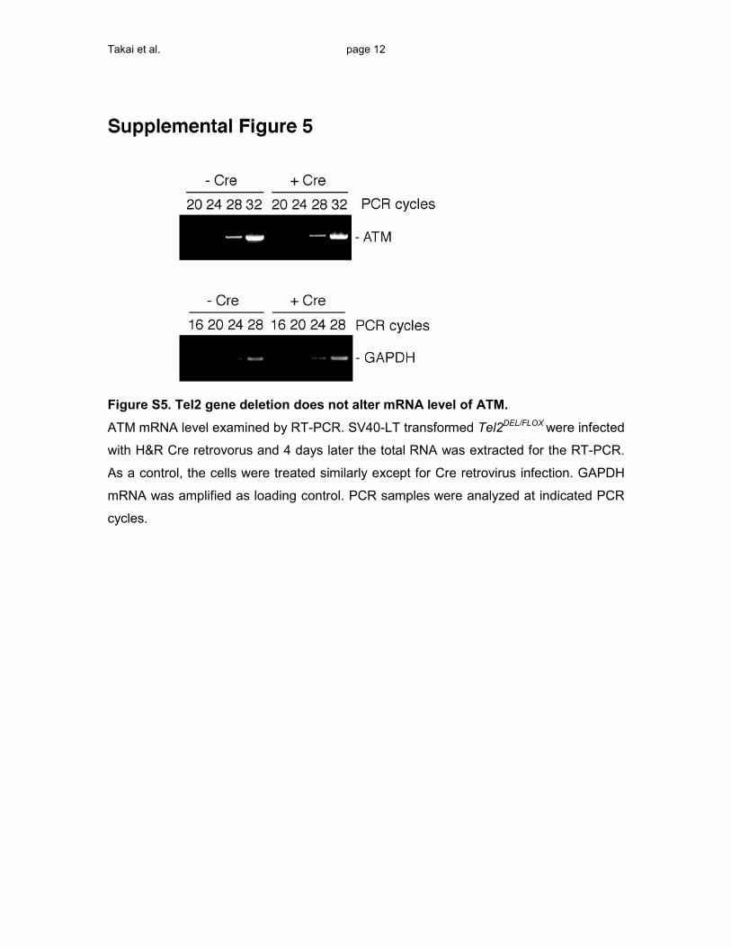
(Figure S5 and data not shown).
1252 Cell 131, 1248–1259, December 28, 2007 ª2007 Elsevier
To test whether Tel2 affected the synthesis or stability of
the ATM and mTOR kinases, cells were pulse labeled with35S-methionine and -cysteine and protein extracts were
harvested at various time points after a chase with unla-
beled amino acids (Figure 5B). The ATM and mTOR
kinases were isolated by immunoprecipitation, and their
abundance was quantified by autoradiography. Consis-
tent with the largely unaltered mRNA levels, both ATM
and mTOR were synthesized at close to normal rates.
Three days after Cre treatment, the incorporation of
labeled amino acids into ATM and mTOR was 70% and
90% of control cells, respectively. A 260 kDa control pro-
tein in the ATM IPs showed a similar reduction in synthesis
rate in Tel2-deficient cells (80% of the rate in wild-type
control cells). This minor decrease is most likely due to di-
minished protein synthesis associated with the depletion
of mTOR.
Tel2 had a profound effect on the stability of ATM and
mTOR. In Tel2-proficient control cells, the half-lives of
ATM and mTOR were approximately 8 and 18 hr, re-
spectively. In contrast, in the Tel2-depleted cells the
half-lives of ATM and mTOR were drastically reduced
to 1.5 and 2 hr, respectively (Figures 5C and 5D). As
a control, we determined the half-life of the 260 kDa
protein in the ATM IPs, which was 4 hr in both the con-
trol cells and Tel2-deficient cells. Together the results
suggested that the reduction of PIKK protein level was
primarily due to the destabilization of PIKKs in absence
of Tel2. Treatment of cells with the proteasome inhibi-
tors MG-132 and lactacystin did not increase the levels
of the PIKKs in Tel2-deficient cells (Figures S6A–S6C),
and we have not been able to detect ubiquitylated inter-
mediates. Future experiments will have to address
Inc.

Figure 4. Reduced PIKK Protein Levels upon Tel2 Deletion
(A) Tel2+/+ and Tel2DEL/FLOX MEFs were infected with H&R-Cre retrovirus and harvested after 4 days for immunoblot analysis of the indicated proteins.
*, nonspecific bands.
(B) Immunoblots of Tel2DEL/FLOX MEFs expressing mouse Tel2 treated with H&R Cre. The indicated proteins were analyzed at day 5 after Cre treat-
ment.
(C) Immunoblotting for Tel2 in Atm+/+ and Atm�/� MEFs.
(D) PCR for conversion of the FLOX allele to the DEL allele by R26Cre-ER induced by OHT; analysis at day 2 after OHT treatment. Cells were p53
deficient.
(E) Time course of PIKK expression in Tel2-depleted MEFs. Tel2DEL/FLOX MEFs were treated with 0.5 mM OHT for 6 hr to induce R26Cre-ER, and
lysates were prepared at the indicated times. Parallel control cultures were treated identically except for OHT treatment. Tel2 was detected with
a mouse polyclonal anti-Tel2 antibody.
(F) Verification of reagents used to query mTOR signaling. Immunoblot for the indicated protein phosphorylation in wild-type MEFs treated with or
without 20 nM rapamycin or 200 nM wortmannin. Cells were cultured in 0.5% serum medium for 16 hr. Rapamycin or wortmannin was added 30
min prior to serum stimulation.
(G) Tel2 deletion diminishes mTOR signaling. Immunoblot to determine the phosphorylation of mTOR targets (as in F) in MEFs of the indicated
genotypes at 3 days after with or without OHT treatment to induce Cre.
(H) Schematic of the inducible Tel2 shRNA Rosa26 cassette in ES cells. SA, splicing acceptor; pA, poly A sequence; H1, promoter; tetO, tet-operator;
CAGGS, promoter; tetR, tet repressor; Dox, doxycycline; *, ATG; open triangle, FRT sequence; closed triangle, FRT F3 sequence.
(I) Tel2 shRNA in ES cells diminishes ATM and mTOR. Tel2 shRNAs were induced with doxycycline in ES cells with the Rosa26 cassette shown in (F),
and cells were harvested after 4 days.
(J) Immunoblot for effect of Tel2 knockdown on ATM in human cells. Human BJ-hTERT fibroblasts were infected with the indicated shRNA retrovi-
ruses and subjected to puromycin selection for 3 days. *, nonspecific signal.
the degradation pathway of the PIKKs in Tel2-deficient
cells.
We considered the possibility that Tel2 might behave
like a (co)chaperone for the PIKKs and therefore asked
whether Tel2 deletion resulted in mislocalization and/or
Cell
aggregation of its client proteins. IF analysis did not reveal
an altered subcellular distribution of the residual ATM and
mTOR in Tel2-deficient cells, and there was no indication
of aberrant protein aggregation (Figure S7A). We also
tested whether the effect of Tel2 deletion was similar to
131, 1248–1259, December 28, 2007 ª2007 Elsevier Inc. 1253
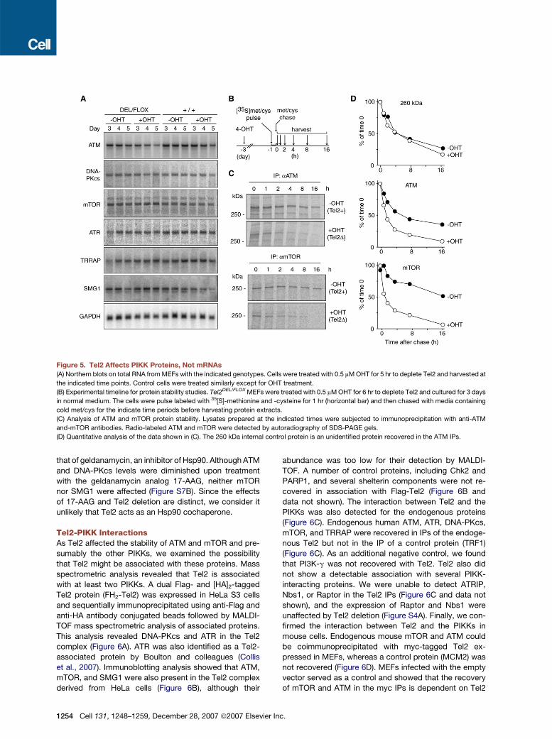
Figure 5. Tel2 Affects PIKK Proteins, Not mRNAs
(A) Northern blots on total RNA from MEFs with the indicated genotypes. Cells were treated with 0.5 mM OHT for 5 hr to deplete Tel2 and harvested at
the indicated time points. Control cells were treated similarly except for OHT treatment.
(B) Experimental timeline for protein stability studies. Tel2DEL/FLOX MEFs were treated with 0.5 mM OHT for 6 hr to deplete Tel2 and cultured for 3 days
in normal medium. The cells were pulse labeled with 35[S]-methionine and -cysteine for 1 hr (horizontal bar) and then chased with media containing
cold met/cys for the indicate time periods before harvesting protein extracts.
(C) Analysis of ATM and mTOR protein stability. Lysates prepared at the indicated times were subjected to immunoprecipitation with anti-ATM
and-mTOR antibodies. Radio-labeled ATM and mTOR were detected by autoradiography of SDS-PAGE gels.
(D) Quantitative analysis of the data shown in (C). The 260 kDa internal control protein is an unidentified protein recovered in the ATM IPs.
that of geldanamycin, an inhibitor of Hsp90. Although ATM
and DNA-PKcs levels were diminished upon treatment
with the geldanamycin analog 17-AAG, neither mTOR
nor SMG1 were affected (Figure S7B). Since the effects
of 17-AAG and Tel2 deletion are distinct, we consider it
unlikely that Tel2 acts as an Hsp90 cochaperone.
Tel2-PIKK InteractionsAs Tel2 affected the stability of ATM and mTOR and pre-
sumably the other PIKKs, we examined the possibility
that Tel2 might be associated with these proteins. Mass
spectrometric analysis revealed that Tel2 is associated
with at least two PIKKs. A dual Flag- and [HA]2-tagged
Tel2 protein (FH2-Tel2) was expressed in HeLa S3 cells
and sequentially immunoprecipitated using anti-Flag and
anti-HA antibody conjugated beads followed by MALDI-
TOF mass spectrometric analysis of associated proteins.
This analysis revealed DNA-PKcs and ATR in the Tel2
complex (Figure 6A). ATR was also identified as a Tel2-
associated protein by Boulton and colleagues (Collis
et al., 2007). Immunoblotting analysis showed that ATM,
mTOR, and SMG1 were also present in the Tel2 complex
derived from HeLa cells (Figure 6B), although their
1254 Cell 131, 1248–1259, December 28, 2007 ª2007 Elsevier
abundance was too low for their detection by MALDI-
TOF. A number of control proteins, including Chk2 and
PARP1, and several shelterin components were not re-
covered in association with Flag-Tel2 (Figure 6B and
data not shown). The interaction between Tel2 and the
PIKKs was also detected for the endogenous proteins
(Figure 6C). Endogenous human ATM, ATR, DNA-PKcs,
mTOR, and TRRAP were recovered in IPs of the endoge-
nous Tel2 but not in the IP of a control protein (TRF1)
(Figure 6C). As an additional negative control, we found
that PI3K-g was not recovered with Tel2. Tel2 also did
not show a detectable association with several PIKK-
interacting proteins. We were unable to detect ATRIP,
Nbs1, or Raptor in the Tel2 IPs (Figure 6C and data not
shown), and the expression of Raptor and Nbs1 were
unaffected by Tel2 deletion (Figure S4A). Finally, we con-
firmed the interaction between Tel2 and the PIKKs in
mouse cells. Endogenous mouse mTOR and ATM could
be coimmunoprecipitated with myc-tagged Tel2 ex-
pressed in MEFs, whereas a control protein (MCM2) was
not recovered (Figure 6D). MEFs infected with the empty
vector served as a control and showed that the recovery
of mTOR and ATM in the myc IPs is dependent on Tel2
Inc.

Figure 6. Tel2 Interacts with All Mammalian PIKKs and Binds HEAT-Repeat Segments of mTOR and ATM
(A) Silver-stained gel of a Tel2 complex isolated by FLAG/HA affinity purification of an N-terminally tagged FLAG-[HA]2-Tel2 construct expressed in
the human HeLa S3 clone. DNA-PKcs and ATR were identified based on 23 and 7 peptide sequences, respectively.
(B) Immunoblots showing specific association of Tel2 with all mammalian PIKKs. Tel2 was isolated as in (A) and queried for associated proteins by
immunoblotting.
(C) Association of endogenous Tel2 with PIKKs. Extract prepared form HeLa S3 cells was used for the immunoprecipitation with Tel2C, preimmune
serum, or TRF1 antibody. IPs were analyzed by immunoblotting for the indicated proteins.
(D) Immunoblot analysis of the association of mouse Tel2 with ATM and mTOR. N-terminal Myc-tagged hTel2 expressed in MEFs was affinity purified
using anti-Myc beads and queried for the indicated associated proteins.
(E) Tel2 binds to HEAT repeat and C-terminal regions of ATM. Equal amounts of purified bacterially expressed GST-ATM fusion proteins (top) and GST
alone were incubated with purified baculovirus-derived Tel2 or TRF1, bound to glutathione beads, and washed. Bound proteins were eluted and
visualized by immunoblotting using Tel2B (middle) or TRF1 371 antibodies (bottom). *, nonspecific peptide purified with GST fusion proteins.
(F) Tel2 binds to a HEAT-repeat segment of mTOR. Equal amounts of purified GST-mTOR fusion proteins (top) were treated as described in (E).
*, nonspecific peptide copurified with GST fusion proteins.
(G) Tel2-mediated stabilization conferred onto GFP by the HEAT-repeat fragment of mTOR. GFP or GFP fused to the indicated segments of mTOR
(see panel F) were expressed in Tel2DEL/FLOX MEFs, and Cre was induced with OHT. After 4 days, the levels of Tel2 and the GFP fusion proteins was
determined by immunoblotting as shown.
Cell 131, 1248–1259, December 28, 2007 ª2007 Elsevier Inc. 1255

expression. Collectively, these results indicate that Tel2
can interact with each of the six mammalian PIKKs.
Since our mass spectrometry analysis did not reveal
potential factors that might mediate the interaction of
Tel2 with the PIKKs, we set out to determine whether
Tel2 can bind PIKKs directly. Fragments of the ATM kinase
were fused to GST and used to pull down Tel2 produced in
insect cells (Figure 6E). TRF1 served as a negative control.
Tel2 bound to a fragment representing the middle of the
HEAT-repeat region (aa 830–1290) as well as to a fragment
from the C terminus of ATM representing the kinase
domain and the FATC region (aa 2680–3056). Tel2 also
bound to a portion of HEAT-repeat region of mTOR
(aa 400–806) but did not show a specific interaction with
the kinase domain or the FATC region of mTOR
(Figure 6F). The ability of Tel2 to bind mTOR directly was
further verified by examining the interaction of proteins
produced in insect cells (Figure S8). When tagged Tel2
and mTOR were coexpressed in insect cells, a complex
containing Tel2 and mTOR could be isolated using the
tags for either Tel2 or mTOR indicating a direct interaction.
Raptor did not appear to interfere with the binding of Tel2
to mTOR. These data suggest that Tel2 binds directly to
HEAT-repeat segments in the N terminus of mTOR and
ATM. The interaction appears specific since Tel2 did not
bind to six other HEAT-repeat fusion fragments tested
(Figures 6E and 6F), and Tel2 did not appear to
interact with PI3K-g, which also contains HEAT repeats
(Figure 6C). HEAT repeats occur in the N terminus of
each of the six mammalian PIKKs and are conserved
aspects of PIKKs in other eukaryotes, raising the possibil-
ity that Tel2 binds to each of the PIKKs through an interac-
tion with a subset of the HEAT repeats.
To determine whether the Tel2-binding fragment of
mTOR could confer Tel2-dependent stability onto a differ-
ent protein, GFP was fused to aa 400–806 of mTOR or to
aa 2–406, which served as a negative control. GFP and the
two GFP fusion proteins were introduced into Tel2DEL/FLOX
MEFs, and their abundance was determined by western
blotting before and after deletion of Tel2. The results
showed that while GFP itself and the fusion of GFP to
the HEAT repeats in aa 2–406 of mTOR were unaffected
by Tel2 deletion, the abudance of the GFP fusion contain-
ing the Tel2-binding site of mTOR was significantly dimin-
ished in Tel2-deficient cells. We note that while this
mTOR-GFP fusion protein was clearly affected by Tel2
status, the reduction in its expression level was less prom-
inent than the endogenous full-length mTOR. Therefore it
is possible that the stabilization of mTOR by Tel2 involves
regions outside aa 400–806.
DISCUSSION
PIKKs are eukaryotic signal transducers that govern path-
ways of major relevance to human disease. Our results
reveal that the six mammalian PIKKs (ATM, ATR,
DNA-PKcs, SMG1, mTOR, and TRRAP) share Tel2 as an
interacting partner and regulator. In the absence of Tel2,
1256 Cell 131, 1248–1259, December 28, 2007 ª2007 Elsevier I
the steady-state level of all PIKKs is drastically reduced
and the signaling pathways governed by ATM, ATR, and
mTOR are curtailed. Tel2 binds to a site in the HEAT-
repeat regions of ATM and mTOR and prevents their rapid
degradation, and we assume that binding of Tel2 to the
other PIKKs similarly affects their stability.
The role of Tel2 as a stabilizer of PIKKs could potentially
explain why Tel2 is essential in eukaryotes, including in
mice, as we show here for mouse embryos, MEFs, and
ES cells. Drastically lowered levels of one or more essen-
tial PIKKs can explain why Tel2 is essential in mouse cells
and embryos. Although mice lacking some PIKKs (e.g.,
ATM and DNA-PKcs) are viable, deficiency in mTOR,
ATR, and TRRAP results in early embryonic lethality and
curbs the growth of MEFs and/or ES cells (Brown and
Baltimore, 2000; Gangloff et al., 2004; Herceg et al.,
2001; Murakami et al., 2004). Similarly, the lethality of
Tel2 deletions in C. elegans and S. cerevisiae might be
due to loss of function of essential PIKKs.
The phenotypes associated with viable mutations in the
Tel2 orthologs of yeast and C. elegans can also be under-
stood in the context of effects on one or more of the
PIKKs. The telomere-length defect of the S. cerevisiae
tel2-1 mutant (S129N) could result from diminished activ-
ity of Tel1 and/or Mec1, which are required for telomere
maintenance (Ritchie et al., 1999). The defects in the S
phase checkpoint of C. elegans clk-2/rad-5 mutants
(C772Y for clk-2 and G135C for rad-5) could be due to
diminished expression of ATL-1, the ATR ortholog of C.
elegans. The replication checkpoint phenotype of reduced
Tel2 levels in S. pombe is also consistent with compro-
mised function of its ATR ortholog (Rad3) (Shikata et al.,
2007). Furthermore, the extended life span of the clk-2
mutant could be explained by partial inhibition of
LET-363, the mTOR ortholog of C. elegans (Vellai et al.,
2003). As this aging phenotype is not observed with the
rad-5 allele, the rad-5 G135C mutation in the C. elegans
TEL2 ortholog might affect ATL-1 and LET-363 in different
ways. Differential effects of the point mutations in TEL2
orthologs could also explain why the S. cerevisiae tel2-1
mutant shows no overt mec1 phenotype (e.g., sensitivity
to IR, MMS, or HU [Ahmed et al., 2001]).
The mechanism by which Tel2 stabilizes the PIKKs is
unknown. Tel2 could have a chaperone or cochaperone-
like role, facilitating correct folding of the large PIKK poly-
peptides. However, several observations argue against
this possibility. For instance, the localization of the PIKKs
and their rate of synthesis are not strongly affected by Tel2
status, and the effect of Tel2 loss does not resemble the
consequences of inhibition of the Hsp90 chaperone. It is
also not known which pathway is responsible for PIKK
degradation in Tel2-deficient cells. Although we antici-
pated that the proteasome pathway contributes to PIKK
degradation, proteasome inhibitors did not stabilize the
full-length PIKKs in Tel2 null cells and we did not detect
ubiquitylated forms of the PIKKs. One possibility is that,
rather than having a chaperone function, Tel2 protects
the PIKKs from cleavage by a specific protease. The
nc.
products of this initial cleavage could then be processed
by the proteasome. Such a scenario would explain why in-
hibition of the proteasome does not result in accumulation
of the full-length PIKKs in Tel2-deficient cells.
The finding that mammalian PIKKs and possibly other
eukaryotic PIKKs require Tel2 for their stability raises
questions about the functional significance of this depen-
dency. One possibility is that the universal Tel2-PIKK
interaction simply reflects the divergence of the PIKKs
from a common ancestor. If this primordial eukaryotic
PIKK was regulated through its interaction with Tel2, this
dependency may have persisted even if the original
Tel2-dependent control is no longer required in the con-
text of the newly diverged PIKKs. However, the existence
of a single protein required for the stability of all PIKKs
could also point to a common mechanism to govern
PIKK activity that has not yet been appreciated. For
instance, Tel2 could be a regulatory node that allows cells
to rapidly downmodulate all PIKKs in concert. The biolog-
ical utility of such a pathway is not obvious to us, espe-
cially given the diverse cellular roles of the PIKKs. Alterna-
tively, we imagine that specific and regulated disruption of
the Tel2-binding ability of individual PIKKs (e.g., by modi-
fication of the Tel2-interaction site) would allow cells to
rapidly downregulate specific signaling pathways. We
are currently testing a specific version of this model in
which Tel2 is proposed to dissociate from each PIKK
upon its activation. This mechanism would impose a short-
ened half-life on the activated PIKK while simultaneously
allowing cells to accumulate and maintain a pool of Tel2-
bound inactive forms of the same PIKK for later use. As
a result, the signaling activity would be of limited duration
once the original stimulus has dissipated, thus endowing
cells with an intrinsic mechanism by which they can exit
stress-response states. It will be interesting to determine
whether these regulatory options are used by mammals
and other eukaryotes and to explore the possibility that
manipulation of the Tel2-PIKK interaction might be bene-
ficial in the context of human diseases, including cancer.
EXPERIMENTAL PROCEDURES
Cell Culture
Primary MEFs were isolated from 13.5 day embryos and maintained in
DMEM supplemented with 15% fetal calf serum (FCS). MEFs trans-
formed by SV40-LT and TERT-BJ cells were cultured in medium
without pyruvate with 10% FCS. HeLa S3 cells were maintained in
Joklik’s medium (Sigma) with 10% FBS. Media were supplemented
4-OHT (Sigma) as indicated.
Tel2 Gene Targeting
A Bac carrying the mouse Tel2 gene was identified using the easy-
to-screen high-density filters (129/Sv, Release I, Incyte Genomics)
with a cDNA probe and a region containing exons 1 to 9 of Tel2 was
subcloned into vector pSL301 (Invitrogen). The targeting vector,
pTV-Tel2, designed to allow conditional deletion of exon 3, was con-
structed using the pGKneobpAlox2PGKDTA vector (a gift from T.
Jacks). The resulting pTV-Tel2 was linearized with KpnI, and gene tar-
geting of ES cells (E14 derived from 129P2/Ola) was performed using
standard techniques. Correctly targeted ES were transfected with Cre
Cell
recombinase (Taniguchi et al., 1998) and selected for 48 hr in 1 mg/ml
puromycin to obtain clones with either the deleted Tel2 locus (Tel2DEL/+)
or the conditional (Tel2FLOX/+) allele. ES clones were injected into
C57BL/6J blastocysts, and chimeric founders were crossed to
C57BL/6J females. PCR screening of Tel2 genomic locus was per-
formed to identify offspring carrying the modified alleles. Tel2FLOX/+
mice were crossed with p53�/� (Jacks et al., 1994) and R26Cre-ER
transgenic mice (B6;129-Gt(ROSA)26Sortm1(cre/Esr1)Nat/J, #004847,
Jackson Laboratories), (Badea et al., 2003).
Antibodies and Western Blot Analysis
Anti-mouse Tel2 antibody #1039 was affinity purified from rabbit serum
immunized with a KLH-conjugated mTel2 peptide (TGLKRYLGGTED
PVLPEEKEEFATC; aa 35–56). Anti-mouse Tel2 mouse polyclonal anti-
body was produced against GST-tagged mouse Tel2, and the serum
was absorbed using bacteria extract expressing GST-tag. Rabbit
antibody TelC was raised against human Tel2 produced in insect cells
and affinity purified. Rabbit anti-human Tel2 antibody #B was affinity
purified from rabbit serum immunized with KLH-conjugated hTel2
peptide (RSKTQRLSKGGPRQGPAGSPSRFC; aa 670–692). Other
antibodies are rabbit anti-mouse Rif1 antibody #1240 (a gift from S.
Buonomo), ATM MAT3 (a gift from M. Kastan), ATR N-19 (sc-1887,
Santa Cruz Biotechnology), DNA-PKcs Ab-4 (MS-423, Lab Vision),
mTOR (#2972, Cell Signaling Technology), SMG1 (A300-393A, Bethyl
Laboratories), TRRAP (RPH800, Serotec), AKT pS473 (#9271, Cell
Signaling Technology), ATRIP (MAB1579, R&D Systems), Chk1 (sc-
8408, Santa Cruz Biotechnology), Chk1 pS345 (#2348, Cell Signaling
Technology), Chk2 (611570, BD Biosciences), g-tubulin GTU-88
(T6557, Sigma), MCM2 (559542, BD PharMingen), PARP1 (NB100-
112, Novus), PI3-kinase p110g (#4254, Cell Signaling Technology),
p16 M-156 (sc-1207, Santa Cruz Biotechnology), p21 F-5 (sc-6246,
Santa Cruz Biotechnology), p27 (sc-528, Santa Cruz Biotechnology),
p53 AI25-13 (a gift from K. Helin [Pasini et al., 2004]), p53 phospho-
Serine 15 (#9284, Cell Signaling Technology), p70-S6K pT389
(#9234, Cell Signaling Technology), 53BP1 (a gift from T. Halazonetis),
and TRF1 (371).
For whole cell lysates, 2 3 106 cells/10 cm dish plated 24 hr prior to
harvesting were rinsed with cold PBS, and 500 ml of 23 Laemmli buffer
was added. Protein samples were separated by SDS-PAGE and
blotted to either nitrocellulose or PVDF (for ATM) membranes. The
blots were blocked in 5% nonfat powdered milk in PBS-T (0.1%
Tween-20 in PBS) for 30 min and incubated with primary antibodies
in 0.1% or 1% milk in PBS-T at room temperature for at least for
1 hr. For phosphospecific antibodies, blots were treated according
to the manufacturers protocol.
FACS Analysis
MEFs were pulse labeled with 10 mM BrdU (Sigma) for 60 min, har-
vested by trypsinization, fixed in 70% ethanol, and stored at �20�C.
Fixed cells were incubated with FITC-conjugated anti-BrdU antibodies
(BD PharMingen) according to the manufactur’s protocol and counter-
stained with a propidium iodide buffer containing 100 mg/ml RNase,
5 mg/ml propidium iodide (Sigma), and 0.5% (W/V) BSA in PBS. Flow
cytometry was performed with a FACScalibur (Becton Dickinson).
Data were analyzed with either CELL Quest software (Becton
Dickinson) or FlowJo (Tree Star).
Tel2 Knockdown by shRNA
Phoenix cells were used to produce shRNA expressing pSUPER.retro
(OligoEngine) retroviruses. Recipient BJ-hTERT cells were infected
four times every 12 hr and selected in puromycin. Target sequences
were as follows: luciferase, 50-CGT ACG CGG AAT ACT TCG A-30;
hTel2-sh2, 50-GGA ACC TGG TGG TGA AGA A-30; hTel2-sh5, 50-GGA
GAG TGC AGA TGC AGG A-30; hTel2-sh6, 50-AGA AGC TTC TGT TCT
TAC A-30. Inducible expression of Tel2 shRNA in mouse ES cells was
as described by Seibler and colleagues (Seibler et al., 2007).
131, 1248–1259, December 28, 2007 ª2007 Elsevier Inc. 1257

Proteomic Analysis and Immunoprecipitations
Isolation of the human Tel2 complex was performed by FLAG-HA
tandem affinity purification as described previously (Ye and de Lange,
2004). Eluted proteins were separated by SDS-PAGE (8%–16% gradi-
ent, Invitrogen) and the protein bands were excised and subjected to
trypsin digestion. The resulting peptides were extracted, and the
proteins were identified by mass spectrometry at the Rockefeller
University Proteomics Resource Center.
Tel2 immunoprecipitations from MEFs were performed using one
15 cm plate of MEFs expressing myc-tagged human full-length Tel2.
The cells were rinsed in cold PBS, scraped in 1 ml lysis buffer
(50 mM Tris-HCl pH 7.6, 150 mM NaCl, 1 mM EDTA, 1 mM PMSF,
1 mg/ml Aprotinin, 2 mg/ml Pepstatin, 1 mg/ml Leupeptin [Roche]),
and disrupted by sonication. Lysate was centrifuged at 16,000 g for
15 min at 4�C. The supernatant was used as the input lysate. Fifty ml
of anti-myc Agarose bead (A7470, Sigma) slurry (50% [v/v] in PBS,
blocked with 10% BSA in PBS) was added to 0.5–1 ml of lysate. Sam-
ples were nutated at 4�C for at least 2 hr. The beads were washed four
times with lysis buffer. After removal of buffer, the beads were sus-
pended in 50 ml 23 Laemmli buffer.
Interaction of endogenous Tel2 and PIKKs were analyzed as below.
1 3 108 HeLa S3 cells were collected, rinsed in cold PBS, and disrup-
ted by homogenization in 1 ml of lysis buffer (10 mM Tris-HCl pH 7.6,
150 mM KCl, 1.5 mM MgCl2, 5 mM b-mercaptoethanol,1 mM PMSF,
and 13 complete protease inhibitor [Roche]) on ice. The lysate was
cleared by centrifugation at 16,000 g for 15 min at 4�C, incubated
with protein G-Sepharose beads (GE Healthcare), centrifuded again.
The supernatant was used for the immunoprecipitation with Tel2C,
preimmune serum, or TRF1 antibody. The beads were washed four
times with lysis buffer. After removal of buffer, the beads were sus-
pended in 80 ml 23 Laemmli buffer.
Northern Blot Analysis and RT-PCR Analysis
Total cellular RNA was prepared using Trizol reagent (GIBCO) accord-
ing to the manufactur’s protocol, and northern blot analysis was
performed as described previously (Watanabe et al., 1995). Northern
probes were prepared by RT-PCR as described in the Supplemental
Data. The cDNA probes amplified by PCR were labeled with
[32P]dCTP by random priming. GAPDH mRNA served as an internal
control.
Metabolic Labeling and Immunoprecipitation
One day prior to labeling 1.5 3 106 cells were plated per 10 cm dish.
Cells were rinsed with methionine and cysteine (met/cys) free DMEM
(#21013-024, GIBCO) three times and incubated with 4 ml labeling
medium (met/cys-free DMEM, 10% dialyzed FBS, 2 mM L-glutamine)
containing 0.1 mCi/ml of a [35S]-met/cys mix (Expre35S 35S protein
labeling mix, #NEG772007MC, NEN) for 1 hr. The medium was
replaced with normal culture medium (15% FCS, 0.1 mM nonessential
amino acids, 100 units/ml of penicillin, 0.1 mg/ml streptomycin, and
2 mM L-glutamine in DMEM) supplemented with 15 mg/ml L-metionine
and L-cysteine-HCl (Sigma). IPs were performed as described (Ye and
de Lange, 2004) with ATM Ab MAT2 (a gift from Y. Shiloh) and mTOR
Ab #2972 (Cell Signaling Technology). Precipitates were lysed in 23
Laemmli buffer and separated by SDS-PAGE. The gels were
immersed in Amplify (NAMP100V, Amersham) and dried and exposed
to a PhosphoImager screen (GE Healthcare). The quantification of the
signal incorporated into ATM and mTOR proteins was done with the
Storm imaging system and the ImageQuant software (GE Healthcare).
In Vitro Binding Assays
ATM and mTOR cDNA fragments were amplified by PCR using human
ATM and rat mTOR cDNAs, respectively, and cloned into pGEX-4T-2.
GEX-4T-2 transformed BL21 cells were grown up in 1 l of 2xYT
medium, and 0.2 mM IPTG was added when the OD600 was�0.5. After
3 hr at 30�C, cells were harvested, resuspended in 16 ml of lysis buffer
(50 mM Tris [pH 8.0], 100 mM KCl, 1% Triton X-100, 2 mM DTT, 1 mg/ml
1258 Cell 131, 1248–1259, December 28, 2007 ª2007 Elsevier
lysozyme, 0.1 mM PMSF, and 13 Complete protease inhibitor mix
[Roche]) and sonicated on ice. The lysate was cleared by centrifuga-
tion at 40,000 g for 20 min at 4�C and incubated with 500 ml of equili-
brated glutathione beads for 2 hr at 4�C. Beads were washed three
times in PBS containing 2 mM DTT, 0.1 mM PMSF, 13 Complete pro-
tease inhibitor, and once in wash buffer 2 (50 mM Tris [pH 8.0], 100 mM
KCl, 10% glycerol, 2 mM DTT, 0.1 mM PMSF). Fusion proteins were
eluted in 500 ml of wash buffer 2 containing 20 mM glutathione
(reduced form). Three sequential elutions were collected and dialyzed
against PBS/10% glycerol. Five micrograms of GST-fusion protein or
GST alone were incubated with 2 mg of baculovirally expressed Tel2
or TRF1 in binding buffer (20 mM Tris [pH 8.0], 100 mM KCl, 1.5 mM
MgCl2, 10% glycerol, 5 mM b-mercaptoethanol, 1 mg/ml BSA, 13
Complete [-EDTA] protease inhibitor) at 4�C for 1 hr. Glutathione beads
(20 ml) were added and incubated for 1 hr at 4�C. Beads were collected
at 500 g, washed three times with binding buffer, and bound protein
was eluted in Laemmli buffer.
Supplemental Data
Supplemental Data include six figures, Supplemental Experimental
Procedures, and Supplemental References and can be found with
this article online at http://www.cell.com/cgi/content/full/131/7/1248/
DC1/.
ACKNOWLEDGMENTS
We gratefully acknowledge Jost Seibler and Holger Kissel (Artemice
Pharmaceuticals GmbH) for the inducible Tel2 knockdown in ES cells.
Devon White is thanked for outstanding mouse husbandry and the RU
Gene Targeting and Transgenic Facilities and Chinweng Yang for help
in generating gene targeted mice. We thank Tyler Jacks, Mike Kastan,
Yosef Shiloh, John Petrini, Noboru Motoyama, Noriko Oshiro, and
Kristian Helin for reagents and materials. Jill Donigian, Eros Lazzerini
Denchi, Dirk Hockemeyer, Sara B.C. Buonomo, and other member
of the de Lange laboratory are thanked for discussion and comments
on this manuscript. R.C.W. was supported by an NIH MSTP grant
(GM07739) to the Cornell/RU/MSK Tri-Institutional MD/PhD program.
This research was supported by grants from the Breast Cancer Re-
search Foundation and the NCI (CA076027) to T.d.L. H.T. and T.d.L.
planned the experiments and wrote the paper together. H.T. made
the figures and executed all experiments except for those in
Figure S1 (performed by RCW), Figure S8 (performed by HY), and Fig-
ures 6E and 6F (performed by K.K.T.).
Received: April 23, 2007
Revised: July 27, 2007
Accepted: October 30, 2007
Published: December 27, 2007
REFERENCES
Abraham, R.T. (2001). Cell cycle checkpoint signaling through the ATM
and ATR kinases. Genes Dev. 15, 2177–2196.
Ahmed, S., Alpi, A., Hengartner, M.O., and Gartner, A. (2001). C.
elegans RAD-5/CLK-2 defines a new DNA damage checkpoint pro-
tein. Curr. Biol. 11, 1934–1944.
Andrade, M.A., and Bork, P. (1995). HEAT repeats in the Huntington’s
disease protein. Nat. Genet. 11, 115–116.
Badea, T.C., Wang, Y., and Nathans, J. (2003). A noninvasive genetic/
pharmacologic strategy for visualizing cell morphology and clonal
relationships in the mouse. J. Neurosci. 23, 2314–2322.
Bakkenist, C.J., and Kastan, M.B. (2004). Initiating cellular stress
responses. Cell 118, 9–17.
Benard, C., McCright, B., Zhang, Y., Felkai, S., Lakowski, B., and
Hekimi, S. (2001). The C. elegans maternal-effect gene clk-2 is essen-
tial for embryonic development, encodes a protein homologous to
Inc.

yeast Tel2p and affects telomere length. Development 128,
4045–4055.
Bosotti, R., Isacchi, A., and Sonnhammer, E.L. (2000). FAT: a novel
domain in PIK-related kinases. Trends Biochem. Sci. 25, 225–227.
Brown, E.J., and Baltimore, D. (2000). ATR disruption leads to chromo-
somal fragmentation and early embryonic lethality. Genes Dev. 14,
397–402.
Celli, G., and de Lange, T. (2005). DNA processing not required for
ATM-mediated telomere damage response after TRF2 deletion. Nat.
Cell Biol. 7, 712–718.
Collis, S.J., Barber, L.J., Clark, A.J., Martin, J.S., Ward, J.D., and Boul-
ton, S.J. (2007). HCLK2 is essential for the mammalian S-phase check-
point and impacts on Chk1 stability. Nat. Cell Biol. 9, 391–401.
Conti, E., and Izaurralde, E. (2005). Nonsense-mediated mRNA decay:
molecular insights and mechanistic variations across species. Curr.
Opin. Cell Biol. 17, 316–325.
Feil, R., Brocard, J., Mascrez, B., LeMeur, M., Metzger, D., and
Chambon, P. (1996). Ligand-activated site-specific recombination in
mice. Proc. Natl. Acad. Sci. USA 93, 10887–10890.
Gangloff, Y.G., Mueller, M., Dann, S.G., Svoboda, P., Sticker, M.,
Spetz, J.F., Um, S.H., Brown, E.J., Cereghini, S., Thomas, G., and
Kozma, S.C. (2004). Disruption of the mouse mTOR gene leads to early
postimplantation lethality and prohibits embryonic stem cell develop-
ment. Mol. Cell. Biol. 24, 9508–9516.
Greenwell, P.W., Kronmal, S.L., Porter, S.E., Gassenhuber, J., Ober-
maier, B., and Petes, T.D. (1995). TEL1, a gene involved in controlling
telomere length in S. cerevisiae, is homologous to the human ataxia
telangiectasia gene. Cell 82, 823–829.
Guertin, D.A., and Sabatini, D.M. (2005). An expanding role for mTOR
in cancer. Trends Mol. Med. 11, 353–361.
Hartman, P.S., and Herman, R.K. (1982). Radiation-sensitive mutants
of Caenorhabditis elegans. Genetics 102, 159–178.
Hay, N., and Sonenberg, N. (2004). Upstream and downstream of
mTOR. Genes Dev. 18, 1926–1945.
Herceg, Z., Hulla, W., Gell, D., Cuenin, C., Lleonart, M., Jackson, S.,
and Wang, Z.Q. (2001). Disruption of Trrap causes early embryonic
lethality and defects in cell cycle progression. Nat. Genet. 29, 206–211.
Herceg, Z., and Wang, Z.Q. (2005). Rendez-vous at mitosis: TRRAPed
in the chromatin. Cell Cycle 4, 383–387.
Jacks, T., Remington, L., Williams, B.O., Schmitt, E.M., Halachmi, S.,
Bronson, R.T., and Weinberg, R.A. (1994). Tumor spectrum analysis in
p53-mutant mice. Curr. Biol. 4, 1–7.
Jiang, N., Benard, C.Y., Kebir, H., Shoubridge, E.A., and Hekimi, S.
(2003). Human CLK2 links cell cycle progression, apoptosis, and telo-
mere length regulation. J. Biol. Chem. 278, 21678–21684.
Kastan, M.B., and Bartek, J. (2004). Cell-cycle checkpoints and
cancer. Nature 432, 316–323.
Lakowski, B., and Hekimi, S. (1996). Determination of life-span in
Caenorhabditis elegans by four clock genes. Science 272, 1010–1013.
Lavin, M.F., Khanna, K.K., Beamish, H., Spring, K., Watters, D., and
Shiloh, Y. (1995). Relationship of the ataxia-telangiectasia protein
ATM to phosphoinositide 3-kinase. Trends Biochem. Sci. 20, 382–383.
Lustig, A.J., and Petes, T.D. (1986). Identification of yeast mutants with
altered telomere structure. Proc. Natl. Acad. Sci. USA 83, 1398–1402.
Morrow, D.M., Tagle, D.A., Shiloh, Y., Collins, F.S., and Hieter, P.
(1995). TEL1, an S. cerevisiae homolog of the human gene mutated
in ataxia telangiectasia, is functionally related to the yeast checkpoint
gene MEC1. Cell 82, 831–840.
Murakami, M., Ichisaka, T., Maeda, M., Oshiro, N., Hara, K.,
Edenhofer, F., Kiyama, H., Yonezawa, K., and Yamanaka, S. (2004).
Cell
mTOR is essential for growth and proliferation in early mouse embryos
and embryonic stem cells. Mol. Cell. Biol. 24, 6710–6718.
Pasini, D., Bracken, A.P., Jensen, M.R., Lazzerini Denchi, E., and Helin,
K. (2004). Suz12 is essential for mouse development and for EZH2
histone methyltransferase activity. EMBO J. 23, 4061–4071.
Perry, J., and Kleckner, N. (2003). The ATRs, ATMs, and TORs are
giant HEAT repeat proteins. Cell 112, 151–155.
Pimpinelli, S. (2005). Drosophila Telomeres. In Telomeres, T. de Lange,
V. Lundblad, and E. Blackburn, eds. (Cold Spring Harbor, New York:
Cold Spring Harbor Laboratory Press), pp. 433–464.
Ritchie, K.B., Mallory, J.C., and Petes, T.D. (1999). Interactions of
TLC1 (which encodes the RNA subunit of telomerase), TEL1, and
MEC1 in regulating telomere length in the yeast Saccharomyces cere-
visiae. Mol. Cell. Biol. 19, 6065–6075.
Runge, K.W., and Zakian, V.A. (1996). TEL2, an essential gene required
for telomere length regulation and telomere position effect in Saccha-
romyces cerevisiae. Mol. Cell. Biol. 16, 3094–3105.
Sabatini, D.M. (2006). mTOR and cancer: insights into a complex
relationship. Nat. Rev. Cancer 6, 729–734.
Seibler, J., Kleinridders, A., Kuter-Luks, B., Niehaves, S., Bruning, J.C.,
and Schwenk, F. (2007). Reversible gene knockdown in mice using
a tight, inducible shRNA expression system (. Nucleic Acids Res. 35,
e54.
Shaw, R.J., and Cantley, L.C. (2006). Ras, PI(3)K and mTOR signalling
controls tumour cell growth. Nature 441, 424–430.
Shikata, M., Ishikawa, F., and Kanoh, J. (2007). Tel2 is required for
activation of the mrc1-mediated replication checkpoint. J. Biol.
Chem. 282, 5346–5355.
Shiloh, Y. (2003). ATM and related protein kinases: safeguarding
genome integrity. Nat. Rev. Cancer 3, 155–168.
Silver, D.P., and Livingston, D.M. (2001). Self-excising retroviral
vectors encoding the Cre recombinase overcome Cre-mediated cellu-
lar toxicity. Mol. Cell 8, 233–243.
Silverman, J., Takai, H., Buonomo, S.B., Eisenhaber, F., and de Lange,
T. (2004). Human Rif1, ortholog of a yeast telomeric protein, is regu-
lated by ATM and 53BP1 and functions in the S-phase checkpoint.
Genes Dev. 18, 2108–2119.
Takai, H., Smogorzewska, A., and de Lange, T. (2003). DNA damage
foci at dysfunctional telomeres. Curr. Biol. 13, 1549–1556.
Taniguchi, M., Sanbo, M., Watanabe, S., Naruse, I., Mishina, M., and
Yagi, T. (1998). Efficient production of Cre-mediated site-directed
recombinants through the utilization of the puromycin resistance
gene, pac: a transient gene-integration marker for ES cells. Nucleic
Acids Res. 26, 679–680.
Vellai, T., Takacs-Vellai, K., Zhang, Y., Kovacs, A.L., Orosz, L., and
Muller, F. (2003). Genetics: influence of TOR kinase on lifespan in C.
elegans. Nature 426, 620.
Watanabe, K., Yamada, H., and Yamaguchi, Y. (1995). K-glypican:
a novel GPI-anchored heparan sulfate proteoglycan that is highly
expressed in developing brain and kidney. J. Cell Biol. 130,
1207–1218.
Wullschleger, S., Loewith, R., and Hall, M.N. (2006). TOR signaling in
growth and metabolism. Cell 124, 471–484.
Xu, L., and Blackburn, E.H. (2004). Human Rif1 protein binds aberrant
telomeres and aligns along anaphase midzone microtubules. J. Cell
Biol. 167, 819–830.
Ye, J.Z., and de Lange, T. (2004). TIN2 is a tankyrase 1 PARP modula-
tor in the TRF1 telomere length control complex. Nat. Genet. 36,
618–623.
131, 1248–1259, December 28, 2007 ª2007 Elsevier Inc. 1259