Cyclin D1 Expression Mediated by Phosphatidylinositol 3-Kinase through mTOR-p70S6K

1
UMEÅ UNIVERSITY MEDICAL DISSERTATIONS New Series No: 1117 ISSN: 0346-6612 ISBN: 978-91-7264-385-7
Edited by the Dean of the Faculty of Medicine
Studies of Phosphatidylinositol 3 Kinase (PI3K) Signaling Pathway in Mammalian
Ovarian Follicle Activation and Development
by
Singareddy Rajareddy
Department of Medical Biochemistry and Biophysics
Umeå University, Sweden Umeå, 2007

2
Copyright © 2007 by Singareddy Rajareddy
Printed in Sweden by VMC, KBC Umeå University
Umeå 2007
ISBN: 978-91-7264-385-7

3
TABLE OF CONTENTS ABBREVIATIONS………………………………………………………………………………..4 ABSTRACT.……………………………………………………………………………………… 5 PUBLICATION LIST…………………………………………………………………………….6 INTRODUCTION.………………………………………………………………………………..7 1. THE MAMMALIAN OVARY………………………………………………………………... 9 1.1 Follicle Formation…………………………………………………………………………… 10 1.2 Classification of Follicles……………………………………………………………………. 11 1.3 Follicular Activation and Early Development……………………………………………... 12 GDF9 and BMP15………………………………………………………………………….... 13 Smads………………………………………………………………………………………… 14 1.4 Regulation of Follicular Development by Follicle Stimulating Hormone (FSH)…………15 1.5 Ovulation and Luteinizing Hormone (LH)………………………………………………… 16 1.6 The Corpus Luteum (CL)……………………………………………………………………18 1.7 Follicular Atresia……………………………………………………………………………. 18 2. THE PHOSPHOTIDYLINOSITOL 3 KINASE (PI3K) PATHWAY…………………….. 20 2.1 Introduction………………………………………………………………………………….. 20 2.2 Akt……………………………………………………………………………………………. 20 2.3 Pten…………………………………………………………………………………………… 21 2.4 Foxo3a………………………………………………………………………………………... 23 2.5 GSK-3………………………………………………………………………………………… 24 2.6 Cyclin-Dependent Kinases (Cdks), Cyclins and p27……………………………………... 24 2.7 The role of p27 in the PI3K pathway………………………………………………………. 25 3. SUMMARY OF THE PRESENT STUDY………………………………………………….. 27 CONCLUSIONS………………………………………………………………………………… 31 ACKNOWLEDGEMENTS…………………………………………………………………….. 32 REFERENCES…………………………………………………………………………………...34 PAPERS

4
ABBREVIATIONS ADAMTS/1 a disintgrin and metalloproteinase with thrombosin like-motifs
BMP15 bone morphogenetic protein 15
Cdk cyclin-dependent kinase
CL corpus luteum
CNS central nervous system
CS cowden syndrome
ECM extracellular matrix
FSH follicle stimulating hormone
GDF9 growth differentiation factor 9
GSK-3 glycogen synthase kinase - 3
IRS-1 insulin receptor substrate – 1
KL kit ligand
LH luteinizing hormone
MMP matrix metallo proteinase
PA plasminogen activator
PARP poly – (ADP – ribose) – polymerase
PDE-3B phosphodiesterase-3B
PIP3 phosphatidylinositol-3,4,5-triphosphate
PI3K phosphatidylinositol 3 kinase
PR progesterone receptor
Pten phosphatase and tensin homolog deleted on chromosome ten
POF premature ovarian failure
SCF stem cell factor
TGFβ transforming growth factor – beta
5
ABSTRACT
The intra-oocyte signaling pathways that control oocyte growth and early follicular development
are largely unknown. The aim of this thesis was to investigate the regulation and functions of
phosphatidylinositol 3 kinase (PI3K) pathway in the oocyte, focusing in the roles of Foxo3a, p27,
and Pten (phosphatase and tensin homolog deleted on chromosome ten). The physiological
significance of Foxo3a in oocytes had been investigated by generating a transgenic mouse,
whereby constitutively active Foxo3a is maintained in oocytes using the oocyte-specific ZP3
(Zona pellucida) promoter. The expression of the constantly active “negative” molecule Foxo3a in
mouse oocytes was found to cause retardation of oocyte growth, resulting in a significant reduction
in oocyte volume in secondary follicles. The transgenic mice also showed arrested follicular
development and were infertile. In addition, when Foxo3a was overexpressed in oocytes of
primary follicles, oocyte growth and follicular development were retarded. One of the causes of
this phenotype may be the retained expression of the cyclin-dependent kinase (Cdk) inhibitor 1B
(Cdkn1b), commonly known as p27kip1 or p27, in the nuclei of oocytes. The role and related
mechanisms of p27 in controlling early follicular development and oocyte growth were then
investigated using wild-type and p27-deficient (p27-/-) mice, and we demonstrated that (i) p27
suppresses follicle endowment/formation and activation, (ii) p27 induces follicle atresia that occurs
prior to sexual maturity, and (iii) the overactivated follicles in p27-/- ovaries are depleted in early
adulthood, causing premature ovarian failure (POF). In this thesis, we also provide genetic
evidence that in mice with conditional deletion of Pten a major negative regulator of PI3K in
oocytes, the entire pool of primordial follicles becomes activated, and subsequently all activated
follicles are depleted in young adulthood, causing POF. Further mechanistic studies revealed that
loss of Pten in oocytes resulted in elevated Akt signaling, which led to upregulation of both
expression and activation of ribosomal protein S6 (rpS6) in oocytes. The results thus show that the
mammalian oocyte serves as the headquarters of programming of the occurrence of follicle
activation, and that the PI3K pathway of the oocyte governs follicle activation through control of
initiation of oocyte growth.

6
PUBLICATION LIST
I *Liu L., *Rajareddy S., *Reddy P., Jagarlamudi K., Du C., Shen Y., Guo Y., Boman K.,
Lundin E., Ottander U., Selstam G. and Liu K. Phosphorylation and inactivation of glycogen
synthase kinase-3 (GSK-3) by soluble kit ligand (KL) in mouse oocytes during early follicular
development (2007). Journal of Molecular Endocrinology 38(1-2):137-46. (* Equal contributions)
II *Liu L., *Rajareddy S., *Reddy P., Du C., Jagarlamudi K., Shen Y., Gunnarsson D., Selstam
G., Boman K. and Liu K. Infertility caused by retardation of follicular development in mice with
oocyte-specific expression of Foxo3a (2007). Development 134(1), 199-209. (* Equal
contributions)
III Rajareddy S., Reddy P., Du C., Liu L., Jagarlamudi K., Tang W.L., Shen Y., Berthet C., Peng.
S.L., Kaldis P. and Liu K. p27kip1 (Cdkn1b) controls ovarian development by suppressing follicle
endowment and activation, and promoting follicle atresia in mice (2007). Molecular
Endocrinology 21(9):2189-202
IV Reddy P., Rajareddy S., Liu L., Jagarlamudi K., Adhikari K., Shen Y., Hämäläinen T.,
Peng SL., Lan ZJ., Cooney AJ., Huhtaniemi I., and Liu K. Oocyte-specific deletion of Pten
causes activation of the entire primordial follicle pool (manuscript).

7
INTRODUCTION
The major function of the ovary is the differentiation and release of mature oocytes for fertilization
and for successful propagation of the species. In addition, the ovary produces steroid hormones
that allow the development of female secondary sexual characteristics and that also support
pregnancy (Mcgee and Hsueh, 2000; Vanderhyden, 2002).
In mammals, the ovary is a heterogeneous organ containing follicles and corpora lutea at various
stages of development. Each follicle contains an oocyte that is surrounded by granulosa cells. In
order to produce mature oocytes, follicles are activated from the pool of dormant primordial
follicles and they develop through primary and secondary stages before acquiring an antral cavity
(Mcgee and Hsueh, 2000). At the antral stage, most follicles undergo atretic degeneration, whereas
a few of them grow further and reach the preovulatory stage under the cyclic stimulation by
gonadotropin that occurs after puberty (Mcgee and Hsueh, 2000). In response to preovulatory
surges of gonadotropin during each reproductive cycle, the dominant graafian follicle ovulates to
release the mature oocyte for fertilization, whereas the remnant follicle undergoes luteinization to
become the corpus luteum (Elvin and Matzuk, 1998; Matzuk and Lamb, 2002; Matzuk et al., 2002;
Mcgee and Hsueh, 2000; Vanderhyden, 2002).
The communication between oocytes and the surrounding granulosa cells is largely dependent on
signaling between the receptor protein tyrosine kinase Kit and its only known ligand, Kit ligand
(KL), which is also referred to as stem cell factor (SCF) or steel factor. Kit is expressed on the
surface of mammalian oocytes at all stages of follicular development in postnatal ovaries of the
mouse, the rat and humans (Driancourt et al., 2000; Horie et al., 1991; Manova et al., 1990). The
growth of the ovarian follicle is dependent on KL-Kit signaling at a time when functional FSH
receptors are not yet expressed in the mouse ovary (Albertini and Barrett, 2002; Vanderhyden,
2002; Driancourt et al., 2000; Matzuk et al., 2002; Huang et al., 1993; Nilsson and Skinner, 2001;
Parrott and Skinner, 1999). KL-Kit-mediated signaling cascades inside mammalian oocytes,
however, are largely unknown. The rapid oocyte growth during follicle activation and early

8
development implies that this process may require certain common pathways that are involved in
cell proliferation and survival—such as the PI3K pathway—which can be activated by Kit in
somatic or cancer cells (Blume-Jensen and Hunter, 2001; Cantley, 2002; Stokoe, 2005).
PI3Ks themselves are known to be lipid kinases that phosphorylate the 3′-OH group of the inositol
ring of inositol phospholipids. The phosphatase Pten, which functions as a negative regulator of
PI3K, reverses this process (Cantley, 2002; Stokoe, 2005). And, proteins containing lipid-binding
domains, mainly, phosphatidylinositol-3,4,5-triphosphate (PIP3), recruit the serine/threonine
kinases 3′-phosphoinositide-dependent kinase-1 (PDK-1) and Akt from the cytoplasm to the cell
membrane (Blume-Jensen and Hunter, 2001; Cantley, 2002)
Akt is a key molecule of the PI3K pathway. It controls many downstream molecules of the PI3K
pathway after its activation. Upon activation, Akt regulates glycogen synthesis by phosphorylating
and inactivating glycogen synthase kinase-3 (GSK-3) (Cross et al., 1995). Among various Akt
substrates, the forkhead transcription factors of the Foxo family play a vital role in the PI3K
pathway. The Foxo factors are evolutionarily conserved, and three of the four members of this
family, Foxo1 (FKHR), Foxo3a (FKHRL1) and Foxo4 (AFX), are substrates of Akt (Accili and
Arden, 2004). Several in vitro studies have shown that Foxo factors have important roles in
mediating cell cycle arrest and apoptosis (Burgering and Medema, 2003; Accili and Arden, 2004).
Another important downstream molecule of the PI3K pathway is the Cdk inhibitor p27, which is a
negative regulator of the mammalian cell cycle and cell growth (Fero et al., 1996). By shuttling
between the cytoplasm and the nucleus of cells, the growth and cell cycle inhibitory function of
p27 is prevented or initiated, respectively. It has been demonstrated that in several types of cells
this process is under the control of Akt and Foxo3a. Akt can directly phosphorylate p27 at tyrosine
187 and trigger the shuttling of p27 from the nucleus to the cytoplasm—whereby its inhibitory
effect is abolished (Cunningham et al., 2004). Similarly, Foxo3a has also been shown to regulate
p27 expression, a process that is controlled by the upstream PI3K/Akt signaling (Chandramohan et
al., 2004).
9
1. THE MAMMALIAN OVARY
An ovary is an egg-producing reproductive organ in the female. It is often found in pairs as part of
the mammalian female reproductive system. The mammalian ovary consists of follicles at many
stages of development. Most of these follicles are primordial follicles and consist of a small oocyte
surrounded by single layer of granulosa cells, and exist in a quiescent state with the oocyte arrested
in prophase I of meiosis (Skinner, 2004). Studies in humans have shown that an ovary contains
around two million follicles at birth, but only a few hundred of these will reach ovulation whereas
most of them will instead go into the degenerative process called atresia (Kaipia and Hsueh, 1997).
In every estrus cycle, premature follicles are recruited into growth and development, mainly in
response to follicle stimulating hormone (FSH) which is secreted by the pituitary gland. The
developed follicles respond to the pituitary hormone luteinizing hormone (LH), which culminates
in rupture of the follicular wall and release of the oocyte (Hsueh et al., 1984; Richards, 1980;
Monniaux et al., 1997; Richards et al., 1987). The ovulated follicle will then develop into a corpus
luteum (CL). The CL produces progesterone, and prepares the uterus for embryo implantation. If
no successful implantation occurs, the CL stops producing progesterone (functional luteolysis) and
goes into structural regression (Niswender et al., 1994).
1.1. FOLLICLE FORMATION
Folliculogenesis involves orchestration of developmental programs in germ and somatic cells and
the interactions between them (Eppig, 1991). In prenatal mouse ovaries, female germ cells are
found in the form of clusters (syncytia), which are connected by intercellular bridges as a result of
incomplete cytokinesis (Pepling and Spradling, 1998). Further studies in mice and sheep have
revealed that during ovarian follicle formation, somatic cells invade the clusters of germ cells and
syncytial breakdown occurs just before primordial follicles are assembled (McNatty et al., 2000;
Pepling and Spradling, 2001), signifying that pregranulosa cells have an active role in the
formation of follicles (Epifano and Dean, 2002)

10
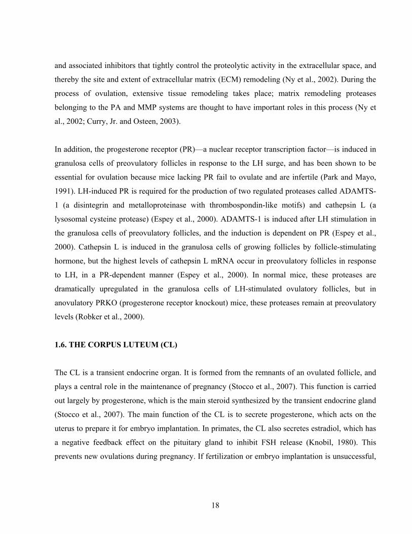
Fig. 1 Structure of a mammalian ovary (Obtained from Google Images).
By the time mice are a few day old, individual oocytes are enclosed in follicles with a few
flattened or squamous somatic cells called pregranulosa cells, and become arrested at the diplotene
stage of meiosis I. They are called primordial follicles (type 2 follicles in mice). Primordial
follicles are multicompartmental units that house single germ cells and a varying number of
somatic cells. These small follicles represent the fundamental developmental unit of the
mammalian ovary and, as such, serve the needs of the entire reproductive life span. In humans, the
ovarian endowment of primordial follicles is established during fetal life and folliculogenesis is a
very long process. Primordial follicles are first observed in the human ovary at approximately 18
weeks of gestation (Baker, 1963; Makabe et al., 2006). In the last few decades, there have been a
number of studies on follicle formation; however, the molecular mechanisms that control
primordial follicle assembly have not been thoroughly investigated.
The mammalian oocyte and somatic cell compartment are highly coordinated for the development
of ovarian follicles. There is a bidirectional communication between granulosa cells and oocytes
that occurs throughout follicular development, which is essential for successful development of
both follicular compartments (Eppig, 1991; Makabe et al., 2006). Communication between oocytes

11
and their associated somatic cells is established with primordial follicle formation. The oocyte
depends on its association with companion somatic granulosa cells to support its growth and
development; on the other hand, oocytes promote granulosa cell proliferation, differentiation, and
function (Eppig et al., 2002). This is a symbiotic process whereby the oocyte secretion factors
help in the proliferation of somatic cells for the formation of different types of follicles. Similarly,
factors produced by somatic cells aid the development and maturation of oocytes (Gilchrist et al.,
2004; Makabe et al., 2006).
Gene products expressed in oocytes have important roles in folliculogenesis. For example, FIGα, a
germ cell-specific basic helix-loop-helix (bHLH) factor that has been implicated in the coordinated
expression of the three zona genes (Zp1, Zp2, and Zp3) encoding the mouse egg coat (Liang et al.,
1997), is detected as early as embryonic day 13 (E13). Mouse lines lacking FIGα have been found
to have normal embryonic gonadogenesis, but primordial follicles were not formed at birth;
massive depletion of oocytes was observed, which resulted in shrunken ovaries and female sterility
(Soyal et al., 2000).
Nobox (newborn ovary homeobox gene), an oocyte-specific homeobox gene, is expressed in germ
cell cysts and in primordial and growing oocytes. Lack of Nobox accelerates loss of postnatal
oocytes and abolishes the transition from primordial to growing follicles (Rajkovic et al., 2004). In
some respects, female mice lacking Nobox is similar to nonsyndromic ovarian failure in women.
Thus, Nobox is critical for specifying an oocyte-restricted gene expression pattern that is essential
for postnatal follicle formation and development (Rajkovic et al., 2004). Sohlh1 encodes a basic
helix-loop-helix transcription factor with homologs in humans and other placental mammals
(Rajkovic et al., 2001). Disruption of Sohlh1 disturbs follicle formation in part by causing
downregulation of two genes that are known to disrupt folliculogenesis: Nobox and Figla (Pangas
et al., 2006). In addition, it has been shown that the transcription factor gene Lhx8 (Zhao et al.,
1999; Mori et al., 2004) is situated downstream of Sohlh1, and is critical for fertility (Pangas et al.,
2006). Adult Lhx8-/- ovaries lack germ cells and appear histologically identical to adult Sohlh1-/-
ovaries, thus demonstrating that Sohlh1 and Lhx8 are the two germ cell-specific genes that are

12
preferentially expressed during oogenesis in females and that are critical to early folliculogenesis
(Pangas et al., 2006).
1.2. CLASSIFICATION OF FOLLICLES
Accurate estimation of the number of ovarian follicles at various stages of development has been
an important indicator of the process of folliculogenesis, in relation to the endocrine signals and
paracrine/autocrine mechanisms that control the growth and maturation of oocytes and their
supporting follicular cells. Various classifications have been used to describe the different stages
of oocyte and follicle development. Some workers have used the shape of the granulosa cells and
the number of layers surrounding the oocyte as the main characteristic (Mandl and Zuckerman,
1950; Hadek, 1965). At first glance, the stages of follicular maturation appear to be
morphologically well-defined across species. In fact, a follicle from any mammalian model can
generally be categorized as being primordial, primary, or secondary—based on the presence and
number of cuboidal granulosa cell layers (Pedersen and Peters, 1968).
In mice, for example, the meiotically arrested oocytes are surrounded by somatic cells by the
second day after birth, forming primordial follicles that represent the first stage of folliculogenesis
and make up the reserve pool. Morphologically, a quiescent primordial follicle (type 2 follicle in
mice) in its largest cross section consists of a single small oocyte (< 20 µm in diameter)
surrounded by two to four squamous somatic cells, called pregranulosa cells, and a basement
membrane (Pedersen and Peters, 1968). Primary follicles have an oocyte surrounded by a single
layer of cuboidal granulosa cells. Secondary follicles are surrounded by more than one layer of
cuboidal granulosa cells, with no visible antrum (Myers et al., 2004). Also, early antral follicles
have emerging antral spaces whilst antral follicles possess a clearly defined antral space (Pedersen
and Peters, 1968; Knigge and Leathem, 1956). Preovulatory follicles are the largest of the different
types of follicle and have a defined cumulus granulosa cell layer (Myers et al., 2004).

13
1.3. FOLLICULAR ACTIVATION AND EARLY DEVELOPMENT
Initiation of follicle growth is defined as the transition of primordial follicles from the quiescent
phase to the growth phase (Braw-Tal, 2002). Follicular activation is believed to be a continuous
process that starts just after follicle formation, long before the onset of puberty (Mcgee and Hsueh,
2000). The follicular activation is broadly defined by three landmark events: (1) transition of the
granulosa cells surrounding the oocyte from a squamous to a cuboidal morphology, (2) slow
proliferation of granulosa cells, and (3) oocyte growth followed by rapid granulosa cell mitosis
(Braw-Tal, 2002).
The resting primordial follicles are thought to be important as the source of developing follicles
and oocytes (McGee and Hsueh, 2000; Liu et al., 2006). In women, folliculogenesis is a very long
process, requiring almost one year for a primordial follicle to grow and develop to the ovulatory
stage (Gougeon, 1996). In mice, on the other hand, an initial synchronous wave of follicular
recruitment occurs within a few days of birth. By 10–12 days of postnatal life, a cohort of
secondary-stage follicles develops, in which oocytes at midgrowth are surrounded by two or more
layers of granulosa cells (Matzuk et al., 2002).
One phenomenon during primordial follicle activation and early development that is worth noting
is the extremely rapid growth of the oocytes. In mice, for example, the oocytes grow aggressively
with an approximately 300-fold increase in volume during the 2- to 3-week growth phase (Liu et
al., 2006), which is also accompanied by a 300-fold increase in RNA content (Sternlicht and
Schultz, 1981; Liu et al., 2006). Oocytes complete most of their growth phase before the formation
of a follicular antrum, and the increase in oocyte diameter and volume during antral follicular
growth is relatively small (Liu et al., 2006). During the phase of oocyte growth, granulosa cells
proliferate from one layer of a few flattened pre-granulosa cells to three layers of cuboidal
granulosa cells by the time oocyte growth is almost complete (Liu et al., 2006).

14
The molecular mechanisms underlying follicular activation are not very well understood (Mcgee
and Hsueh, 2000). It is believed that unknown intra-ovarian factors stimulate some primordial
follicles to initiate growth while the rest of the follicles remain quiescent (Mcgee and Hsueh, 2000).
It is also possible that follicular activation is caused by release of primordial follicles from
inhibitory mechanisms that maintain them in a resting state (Mcgee and Hsueh, 2000). It is
common knowledge nowadays that bi-directional communication between oocytes and somatic
cells plays an important role in ovarian follicular development (Albertini et al., 2001; Elvin and
Matzuk, 1998; Eppig et al., 2002).
GDF9 and BMP15
Two important oocyte-derived TGF-β (transforming growth factor – beta) superfamily members,
namely GDF9 (growth differentiation factot 9) and BMP15 (bone morphogenetic protein 15), are
synthesized and secreted by oocytes of murine follicles and have been shown to be essential for
ovarian follicular growth (Juengel et al., 2004). Neither GDF9 nor BMP15 are thought to be
involved in the formation of follicles, as no disturbance of the primordial follicle population has
been found in GDF9 knockout mice and BMP15 knockout mice. Mice lacking a functional GDF9
gene are infertile, due to a block in follicular development beyond the primary one-layer follicle
stage (Dong et al., 1996). Oocyte growth and zona pellucida formation proceed normally, but
aberrant expression of mRNA encoding several proteins such as aromatase, activin-βB, follistatin,
and COX-2 has been observed; these were reduced compared to controls with an intact GDF9 gene
(Dong et al., 1996). In addition, levels of FSH and LH were found to be elevated and ovarian cysts
were often observed in GDF9 knockout mice (Dong et al., 1996). Female mice lacking a
functional BMP15 gene are fertile and follicular growth appears to be normal, with ovarian
morphology that is indistinguishable from that of wild-type littermates (Yan et al., 2001). However,
BMP15-deficient mice have been found to produce fewer litters of smaller litter size than their
heterozygous or wild-type counterparts, due to defects in ovulation (Yan et al., 2001; Galloway et
al., 2002). No apparent effect on ovulation rate or litter size was observed in mice heterozygous for
dormant copies of GDF9 or BMP15 alone (Juengel et al., 2004).
15
Mice heterozygous for inactive copies of both the BMP15 and GDF9 genes had smaller and less
frequent litters than control mice (Juengel et al., 2004). Double-mutant mice deficient in both
genes resemble GDF9-deficient mice and are infertile. On the other hand, BMP15-deficient mice
with one copy of GDF9 are even less productive than BMP15-deficient mice that have wild-type
levels of GDF9, a phenomenon that also implies a dose effect (Galloway et al., 2002; Yan et al.,
2001).
Smads
Smads have been shown to be indispensable for ovarian folliculogenesis (Pangas and Matzuk,
2004; Yi et al., 2001; Ying et al., 2001). The Smad family proteins were first identified in the fruit
fly Drosophila melanogaster in the mid-1990s by Sekelsky et al. (Sekelsky et al., 1995). Smads
function as intracellular transcription factors that mediate the signaling of the TGF-β superfamily,
which consists of more than 40 members including three isoforms of TGF-β, activins, inhibins,
GDFs and BMPs (Kaivo-Oja et al., 2006). All ligands in this protein family share common
sequence elements and structural motifs. They are multifunctional regulators of cell proliferation,
differentiation, migration, and apoptosis—promoting extracellular matrix production, tissue
homeostasis, and embryogenesis (Kaivo-Oja et al., 2006).
The role of the TGF-β superfamily has been studied extensively in ovarian organogenesis as well
as in folliculogenesis in animals (Kaivo-Oja et al., 2006). The growth of the follicle to the small
antral stage is considered gonadotropin-independent, and during these early phases folliculogenesis
appears to be driven by local autocrine and paracrine signals from the oocyte and the surrounding
somatic cells. A complex bidirectional communication between oocytes and granulosa cells, and
also between granulosa and thecal cells, drives the progression of follicular development through
its successive stages (Matzuk et al., 2002). Various TGF-β superfamily ligands expressed by the
different ovarian cell types are important in this interaction, and their expression is regulated in a
manner that is related to the stage of development (van den Hurk and Zhao, 2005). The developing
oocyte has been shown to express GDF-9, BMP-15, BMP-6, and TGF-β2 (Dube et al., 1998;
Laitinen et al., 1998; Schmid et al., 1994; Lyons et al., 1989; Gilchrist et al., 2003). Granulosa

16
cells produce activins, inhibins, TGF-βs, BMP-2, BMP-3, BMP-6, and also AMH at different
stages of folliculogenesis, while thecal cells have been reported to produce all the isoforms of
TGF-β, BMP-3b, BMP-4, and BMP-7 (Liao et al., 2004; Knight and Glister, 2003). TGF-βs,
activins and GDF9 signal through the Smad2/3 pathway whereas BMP-15, BMP-2, - 4, -6, and -7
utilize the Smad1/5/8 pathway (Kaivo-Oja et al., 2006).
1.4. REGULATION OF FOLLICULAR DEVELOPMENT BY FOLLICLE
STIMULATING HORMONE (FSH)
FSH is a glycoprotein, synthesized and secreted by gonadotropes in the anterior pituitary gland
(Burns and Matzuk, 2002). In the ovary, FSH stimulates the growth of immature follicles to
maturation and as the follicle grows it releases inhibin, which shuts off the production of FSH
(Burns and Matzuk, 2002). Inhibins (α:βA and α:βB heterodimers) and activins (βA:βA and βB:βB
homodimers, and βA:βB heterodimers) are members of the TGF-β superfamily, and are so-called
because of their respective abilities to suppress and enhance FSH production (Burns and Matzuk,
2002). These peptides are synthesized in granulosa cells of the ovary and sertoli cells of the testis,
and they are also found in other tissues where they have been implicated in diverse biological
processes (Burns and Matzuk, 2002). FSH acts by binding to its receptor, which is expressed on
granulosa cells (Ulloaaguirre et al., 1995; OShaughnessy et al., 1996). The FSH receptor plays an
essential role during follicular development; however, it becomes unnecessary once follicular cells
differentiate, and its expression subsequently declines after the LH surge during the process of
luteinization (Camp et al., 1991).
Studies on FSH knockout mice have revealed that loss of FSH signaling in male mice results in
smaller testis size, reduced spermatogenesis, impaired sperm motility, but the males are still fertile
(Dierich et al., 1998). In contrast, females without a functional FSH signaling pathway were
sterile, with thin uteri and hypoplastic ovaries due to a block in folliculogenesis prior to antrum
formation (Kumar et al., 1997). Similar results were obtained in studies of FSHRKO (follicle
stimulating receptor knockout mice). Follicles at all stages up to the preantral stage were observed
17
in the ovaries of these knockout mice, and there was no evidence of antral follicles or corpora lutea
(Abel et al., 2000). Moreover, it has been reported that loss of FSH signaling causes infertility in
women (Layman and McDonough, 2000).
1.5. OVULATION AND LUTEINIZING HORMONE (LH)
LH is a member of the glycoprotein hormone family that includes FSH and thyroid-stimulating
hormone (Pierce and Parsons, 1981; Bousfield and Ward, 1994). These hormones are heterodimers
consisting of a common α-subunit noncovalently linked to a hormone-specific β-subunit.
Ovulation is critically dependent on the stimulation of LH (Richards, 2001). In addition, in both
sexes LH stimulates secretion of sex steroids from the gonads. In the testis, LH binds to receptors
on Leydig cells and stimulates synthesis and secretion of testosterone. In the ovary, the theca cells
respond to LH stimulation by secretion of testosterone, which is converted to estrogen by adjacent
granulosa cells (Pierce and Parsons, 1981; Bousfield and Ward, 1994).
At the time of ovulation, the wall of the preovulatory follicle ruptures and the mature oocyte is
released into the oviduct. The process of ovulation is controlled by LH and FSH, which are
secreted in the anterior lobe of the pituitary gland (Chabbert et al., 1998). Once follicular selection
occurs, the follicle(s) destined for ovulation become(s) selectively vascularized, responding
preferentially to the release of LH and FSH (Espey and Lipner, 1965; Gougeon, 1996). However,
there is a significant mechanical hindrance that must be overcome in order for a successful
ovulation to occur.
The possibility that increased follicular pressure facilitates ovulation has been recognized for
centuries (Edwards, 1974). Although several studies have addressed this possibility, the issue has
still not been fully resolved. In early studies, no increased follicular pressure during ovulation
could be detected (Espey and Lipner, 1965). Two proteolytic systems that have been suggested to
play key roles in matrix remodeling are the plasminogen activator (PA) system and the matrix
metalloproteinase (MMP) system (Ny et al., 2002). These systems are comprised of both proteases
18
and associated inhibitors that tightly control the proteolytic activity in the extracellular space, and
thereby the site and extent of extracellular matrix (ECM) remodeling (Ny et al., 2002). During the
process of ovulation, extensive tissue remodeling takes place; matrix remodeling proteases
belonging to the PA and MMP systems are thought to have important roles in this process (Ny et
al., 2002; Curry, Jr. and Osteen, 2003).
In addition, the progesterone receptor (PR)—a nuclear receptor transcription factor—is induced in
granulosa cells of preovulatory follicles in response to the LH surge, and has been shown to be
essential for ovulation because mice lacking PR fail to ovulate and are infertile (Park and Mayo,
1991). LH-induced PR is required for the production of two regulated proteases called ADAMTS-
1 (a disintegrin and metalloproteinase with thrombospondin-like motifs) and cathepsin L (a
lysosomal cysteine protease) (Espey et al., 2000). ADAMTS-1 is induced after LH stimulation in
the granulosa cells of preovulatory follicles, and the induction is dependent on PR (Espey et al.,
2000). Cathepsin L is induced in the granulosa cells of growing follicles by follicle-stimulating
hormone, but the highest levels of cathepsin L mRNA occur in preovulatory follicles in response
to LH, in a PR-dependent manner (Espey et al., 2000). In normal mice, these proteases are
dramatically upregulated in the granulosa cells of LH-stimulated ovulatory follicles, but in
anovulatory PRKO (progesterone receptor knockout) mice, these proteases remain at preovulatory
levels (Robker et al., 2000).
1.6. THE CORPUS LUTEUM (CL)
The CL is a transient endocrine organ. It is formed from the remnants of an ovulated follicle, and
plays a central role in the maintenance of pregnancy (Stocco et al., 2007). This function is carried
out largely by progesterone, which is the main steroid synthesized by the transient endocrine gland
(Stocco et al., 2007). The main function of the CL is to secrete progesterone, which acts on the
uterus to prepare it for embryo implantation. In primates, the CL also secretes estradiol, which has
a negative feedback effect on the pituitary gland to inhibit FSH release (Knobil, 1980). This
prevents new ovulations during pregnancy. If fertilization or embryo implantation is unsuccessful,
19
the CL will lose its ability to produce progesterone (functional luteolysis) and will go into
structural regression, during which the luteal tissue is degraded (Niswender et al., 1994) and the
ovary enters a new estrus cycle (Michael et al., 1994).
There are several morphological features associated with luteolysis. The luteal cells accumulate
lipid droplets and become reduced in size. At a later stage, these cells undergo apoptosis and the
vascular network degenerates; an influx of macrophages into the regressing CL has been observed
(Gaytan et al., 1997; Paavola, 1979). These macrophages may participate in the destruction of
luteal tissue and phagocytosis of dead luteal cells (Gaytan et al., 1997; Paavola, 1979).
1.7. FOLLICULAR ATRESIA
In ovarian physiology, follicular atresia is a key phenomenon by which the ovary eliminates those
follicles that will not ovulate. The process of follicular selection results in the formation of one or
more dominant follicles that will ovulate, and in the concomitant atresia of multiple follicles that
will eventually die. Despite the widespread occurrence and fundamental importance of atresia, its
molecular mechanisms are still largely unknown. In humans, the ovarian endowment of primordial
follicles is established during fetal life (Hussein, 2005; Hsueh et al., 1994). Apoptotic cell death
depletes this endowment by at least two-thirds before birth (Hussein, 2005), executed with the help
of several players and pathways (Hussein, 2005). Several pro-survival and pro-apoptotic molecules
are involved in ovarian apoptosis, with a delicate balance existing between them and determining
the destiny of the follicular cells (Hussein, 2005).
The duration of fertility in a female is determined by the size of the primordial follicle pool formed
during fetal life, and by the rate of depletion of the follicle pool after birth (Erickson and
Shimasaki, 2001; Rodgers et al., 1995). In mice, a large proportion of follicles disappear from the
non-growing follicle pool during the first wave of follicle development (Bristol-Gould et al., 2006).
In the human ovary, follicles begin to form during the fourth month of fetal life (Baker, 1963).
Primordial follicles are first observed in human ovaries from approximately 18 weeks of gestation.

20
At the time of the first menstrual period, the ovaries of a woman contain some 250,000 follicles,
each of which has the potential to release an egg (Hussein, 2005; Hsueh et al., 1994). Over her
reproductive life, however, a woman will ovulate no more than 500 times, because her supply of
eggs is progressively eliminated through follicular atresia (Powell, 2004; Hsueh et al., 1994).
Apoptosis has a fundamental role in follicular atresia, and experimental studies have revealed that
Bax, which is expressed in both granulosa cells and oocytes, may be a molecule that is central to
ovarian cell death. It has been shown that young adult female Bax-/- mice possess three times more
primordial follicles in their ovarian reserve than their wild-type sisters, and this surfeit of follicles
is maintained in advanced chronological age, such that 20–22-month-old female Bax-/- mice
possess hundreds of follicles at all developmental stages and exhibit ovarian steroid-driven uterine
hypertrophy (Perez et al., 1999).
Caspases are known to be involved in mediating follicle atresia. For example, granulosa cells from
healthy follicles possess almost exclusively the inactive (unprocessed) form of caspase-3, whereas
granulosa cells from atretic follicles have increased concentrations of activated caspase-3. The
active processing of caspase-3 is associated with the cleavage of poly-(ADP-ribose)-polymerase
(PARP) and actin, and the formation of oligonucleosomes (Boone and Tsang, 1998). Loss of
caspase-3 function leads to defective atresia of maturing follicles in vivo. Furthermore, caspase-3
mutant female mice have been found to possess aberrant atretic follicles containing granulosa cells
that fail to be eliminated by apoptosis (Matikainen et al., 2001).
2. THE PHOSPHOTIDYLINOSITOL 3 KINASE (PI3K) PATHWAY 2.1. INTRODUCTION
The PI3K pathway is a “classic” signaling pathway consisting of various signaling molecules
including kinases, phosphatases, and transcription factors that establish cascades of intracellular
signaling to facilitate the fundamental regulation of cell proliferation, survival, migration, and

21
metabolism (Blume-Jensen and Hunter, 2001; Cantley, 2002; Stokoe, 2005). PI3Ks themselves are
lipid kinases that phosphorylate the 3'-OH group of the inositol ring of inositol phospholipids, and
a phosphatase—Pten—reverses this process and functions as a negative regulator of PI3K (Cantley,
2002; Simpson and Parsons, 2001; Stokoe, 2005). The PIP3 can then recruit proteins containing
lipid-binding domains (i.e. the FYVE and the pleckstrin-homology (PH) domains) from the
cytoplasm (Pawson and Nash, 2000), such as the serine/threonine kinases PDK-1 and Akt (also
known as protein kinase B, PKB) (Blume-Jensen and Hunter, 2001; Cantley, 2002).
2.2. Akt
The serine/threonine protein kinase Akt/PKB is the cellular homolog of the viral oncogene v-Akt
and is activated by various growth and survival factors (Song et al., 2005) . Many cell-surface
receptors induce the production of second messengers that activate PI3K (Song et al., 2005). There
are three closely related enzymatic isoforms, Akt1 (PKBα), Akt2 (PKBβ), and Akt3 (PKBγ)—
encoded by three different genes located on chromosomes 14q32, 19q13, and 1q43, respectively.
They are similar both in structure and size, and are thought to be activated by a common
mechanism (Okano et al., 2000).
The serine-threonine protein kinase Akt mediates many of the downstream effects of PI3K and
consequently plays a central role in both normal and pathological signaling by the PI3K pathway.
Activation of Akt is a multi-step process involving both membrane binding and phosphorylation.
Upon activation of PI3K and production of PIP3 and PIP2, Akt is recruited to the plasma
membrane where it binds to these phosphoinositides through its PH domain (Dudek et al., 1999).
In this way, Akt phosphorylates a variety of substrates involved in the regulation of key cellular
functions including cell growth and survival, glucose metabolism, and protein translation. These
targets include GSK3, IRS-1 (insulin receptor susbtrate-1) PDE-3B (phosphodiesterase-3B), BAD,
human caspase 9, Foxo and NF-kB transcription factors, mTOR, eNOS, Raf protein kinase,
BRCA1, and p21Cip1/WAF1 (Altiok et al., 1999; Datta et al., 1999; Galetic et al., 1999).

22
It has been demonstrated, however, that upon activation, Akt not only regulates glycogen synthesis
through phosphorylation and inactivation of GSK-3, but also regulates the cell cycle by preventing
GSK-3β-mediated phosphorylation and degradation of cyclin D1 (Diehl et al., 1998) and by
negatively regulating the Cdk inhibitor p27kip1 (Shin et al., 2005; Viglietto et al., 2002; Blume-
Jensen and Hunter, 2001).
For complete activation, Akt must be translocated to the plasma membrane where it is
phosphorylated at Thr308 and Ser473 (Bayascas and Alessi, 2005; Song et al., 2005). Akt activity
is negatively regulated by molecules that antagonize PI3K, such as the dual lipid and protein
phosphatase Pten (Cantley and Neel, 1999; Simpson and Parsons, 2001).
2.3. Pten (phosphatase and tensin homolog deleted on chromosome ten)
Pten negatively regulates the PI3K signaling pathway by dephosphorylating PIP3, the product of
PI3K (Cantley and Neel, 1999). The Pten gene also acts as a tumor suppressor gene. It is
frequently deleted or mutated not only in human glioblastoma and prostatic cancer, but also in a
wide range of advanced human malignancies such as endometrial, breast, lung, kidney, bladder,
testicular, and head and neck cancers, malignant melanoma, and lymphoma (Li et al., 1997; Steck
et al., 1997; Gronbaek et al., 1998; Teng et al., 1997; Tashiro et al., 1997; Okami et al., 1998).
Loss of Pten function—either in embryonic stem cells (ES cells) or in human cancer cell lines—
has been found to result in accumulation of PIP3 and activation of its downstream signaling
molecule, Akt/PKB. The activated PI3K/Akt pathway in turn stimulates progression of the cell
cycle, cell survival, and cell migration (Liliental et al., 2000; Stambolic et al., 1998).
Inactivation of Pten (on chromosome 19) in mouse models confirmed Pten to be a tumor
suppressor (Di Cristofano et al., 1998; Podsypanina et al., 1999; Lesche et al., 2002; Suzuki et al.,
1998). Pten+/- mice have been found to develop tumors in multiple organs including breast cancer,
partially resembling the spectrum of neoplasia observed in Cowden syndrome (CS) patients; Pten-/-
mice die during embryogenesis, before midgestation at E6.5–9.5 (Di Cristofano et al., 1998;
Podsypanina et al., 1999; Stambolic et al., 2000; Suzuki et al., 1998). Owing to the information

23
that the homozygous deletion of Pten in mice is an embryonic lethal trait, the Cre-loxP technology
has been introduced to study the function of the Pten gene in specific cells or organs. Pten is a
crucial negative regulator of breast tumorigenesis, and loss of Pten is associated with a poor
outcome in breast cancer (Depowski et al., 2001). Mammary-specific deletion of the Pten gene by
MMTV-Cre transgenic mice (Wagner et al., 2001) has been found to give precocious lobulo-
alveolar development, excessive ductal branching, delayed involution, and severely reduced
apoptosis in mammary tissue. Pten null mammary epithelial cells have been found to be
disregulated and hyperproliferative. In addition, mutant females developed mammary tumors early
in life (Li et al., 2002).
Targeted deletion of Pten in neural tissues by a nestin promoter-driven Cre transgene (Cre+/–) that
is activated in central nervous system (CNS) stem/progenitor cells at embry E9 or E10, resulted in
almost complete gene deletion in the CNS by midgestation (Bates et al., 1999; Fan et al., 2001).
Mice lacking Pten in the CNS had enlarged, histoarchitecturally abnormal brains that resulted from
increased cell proliferation, reduced cell death, and enlarged cell size (Groszer et al., 2001).
Continuous increase in brain weight was observed in Pten null mice from E14 to PD0, and the cell
numbers were double those of wild-type controls. The mutants died immediately after birth
(Groszer et al., 2001). Specific deletion of Pten in mouse brain by using Cre-mediated GFAP-Cre
transgenic mice (Zhuo et al., 2001) resulted in hyperphosphorylation of Akt, which is a signaling
molecule downstream of the Pten gene (Lesche et al., 2002). Inactivation of Pten in embryonic
fibroblasts has been found to result in elevated levels of PIP3. Consequently, Pten deficiency leads
to dosage-dependent increases in phosphorylation and activation of Akt. Activation of Akt
increases phosphorylation of BAD and promotes survival of Pten-/- cells (Sun et al., 1999).
Alteration of Pten has been strongly implicated in the development of prostate cancer. Pten
deletions and/or mutations have been found in 30% of primary prostate cancers (Dahia, 2000; Latil
and Lidereau, 1998) and in 63% of metastatic prostate tissue samples (Suzuki et al., 1998), making
mutations in the Pten gene among the most common genetic alterations reported in human prostate
cancers. Prostate-specific deletion of Pten in the ARR2Probasin-Cre transgenic line PB-Cre4, in

24
which the Cre recombinase is under the control of a modified rat prostate-specific probasin (PB)
promoter (Wu et al., 2001), resulted in elevated phosphorylation of Akt in Pten null prostate cells
compared to wild-type cells. Pten null, Akt-activated cells were also larger than their wild-type or
heterozygous controls (Wang et al., 2003), which is consistent with the role of Pten in controlling
cell size. Targeted deletion of Pten in murine liver resulted in increased fatty acid synthesis,
accompanied by hepatomegaly, fatty liver, and increased glycogen synthesis (Stiles et al., 2004).
2.4. Foxo3a
In mammals, Akt can phosphorylate three conserved residues of serine or threonine in Foxo1,
Foxo3a, and Foxo4, which then leads to their nuclear export and results in inhibition of their
apoptotic transcriptional activities (Accili and Arden, 2004; Arden and Biggs, III, 2002; Nakae et
al., 1999; Brunet et al., 1999). In the other words, after PI3K-induced activation of the
serine/threonine kinase Akt, Foxos are rapidly phosphorylated. Phosphorylated Foxos bind to 14-
3-3 chaperones, which sequester Foxo within the cytosol and prevent re-entry into the nucleus,
leading to functional inactivation. In the absence of environmental signals or growth factors, Foxo
members localize to the nucleus (Arden and Biggs, III, 2002).
Several in vitro studies have shown that Foxo factors have important roles in mediating cell cycle
arrest and apoptosis (Accili and Arden, 2004; Burgering and Medema, 2003; Tran et al., 2003). In
recent studies, it has been reported that Foxo proteins have essential roles in response to
physiological oxidative stress and thereby mediate quiescence and enhanced survival in the
hematopoietic stem cell compartment, a function that is required for the long-term regenerative
potential of the latter (Tothova et al., 2007). Although the three Foxos serve some discrete
functions, they most likely have significant redundancies, as they are broadly expressed during
embryonic development and in adult tissues (Furuyama et al., 2000). Among the Foxo family
members, Foxo3a is a transcription factor that mediates apoptosis and cell cycle arrest, and its
activity is manipulated by PI3K-Akt (Accili and Arden, 2004; Nakae et al., 1999; Arden and Biggs,
III, 2002; Brunet et al., 1999).

25
2.5. GSK-3
Two forms of GSK-3, designated GSK-3α and GSK-3β, have been identified, which are encoded
by two distinct genes (Welsh et al., 1996). GSK-3 was initially identified as a kinase that
phosphorylates and inactivates glycogen synthase (GS) and regulates glycogen synthesis in
response to insulin (Welsh et al., 1996). Later on, GSK-3 also became recognized as a critical
downstream element of the PI3K and the Wnt signaling pathways (Cross et al., 1995; Srivastava
and Pandey, 1998).
It has been reported that in GSK-3α and GSK-3β knock-in mice, the Akt phosphorylation sites—
serine 21 of GSK-3α and serine 9 of GSK-3β—were mutated to alanine, which led to resistance to
Akt inactivation in both molecules (McManus et al., 2005). The Knock-in mice were viable and
were not diabetic (McManus et al., 2005). And also, there was no defect in female reproduction
and follicular development, suggesting that mutations in the Akt phosphorylation sites of GSK-3α
and GSK-3β may not play an indispensable part in oocyte growth and follicular development in
mice.
2.6. Cyclin-dependent kinases (Cdks), cyclins and p27kip1
The Cdks are a family of serine/threonine protein kinases and are activated at specific points of the
cell cycle (Vermeulen et al., 2003). The progression of the cell cycle is catalyzed by Cdks which,
as the name suggests, are activated by a special class of proteins called cyclins (Gartel and
Radhakrishnan, 2005). Cdk protein levels remain stable during the cell cycle, in contrast to those
of their activating proteins, the cyclins. In mammals, the different cyclins are designated A, B, C,
and D, and so on. Cyclin protein levels rise and fall during the cell cycle, and in this way they
periodically activate Cdks (Evans et al., 1983).
p27, a member of the Cip/Kip family of Cdk inhibitors, is a negative regulator of cell-cycle
progression and is a tumor suppressor (Kaldis, 2007). p27 inhibits certain cyclin-Cdk complexes in
the cell nucleus and prevents proliferation of cells. It is a short-lived protein, and its degradation is

26
controlled by the ubiquitin-proteasome system (Pagano et al., 1995). From in vitro studies, it has
been shown that p27 is localized not only in the nucleus but also in the cytoplasm (Rodier et al.,
2001; Boehm et al., 2002). It is highly expressed in quiescent cells (at G0), where it binds and
inhibits cyclinE-Cdk2 activity (Hengst et al., 1994; Slingerland et al., 1994). p27 is located in the
nucleus in G0 and early in G1, and appears transiently in the cytoplasm at the G1/S transition. p27
also plays a role in mid-G1, in the assembly and nuclear import of D-type cyclin-Cdk complexes
(LaBaer et al., 1997). As cells enter the cell cycle and S phase, p27 levels decrease and the kinase
activity of Cdks increases. For example, p27 binds to Cdk2 (or to Cdk1, and to other Cdks) and
potently inhibits Cdk2 kinase activity.
The activities of Cdk complexes are thought to promote the transitions between the different
phases of the cell cycle by phosphorylating a plethora of substrates. Cdk activity is controlled by
several mechanisms, including cyclin binding, phosphorylation, dephosphorylation, localization,
transcriptional regulation, protein degradation, and binding to inhibitors (Sherr and Roberts, 1999).
Cdk2 (or Cdk1) phosphorylates p27 at threonine 187, which is then recognized by the SCFSkp2
ubiquitin ligase and degraded by the proteasome (Kaldis, 2007). Mice lacking p27 are viable but
are bigger than wild-type littermates, and develop pituitary tumors and in some instances retinal
dysplasia, thymic hyperplasia, female sterility, and hyperplasia of the adrenal gland (Fero et al.,
1996).
2.7. The role of p27 in the PI3K pathway
It is known that p27 is an negative regulator of the mammalian cell cycle and cell growth, and is
one of the important downstream molecules of the PI3K pathway (Fero et al., 1996). By shuttling
between the cytoplasm and the nucleus, the growth/cell cycle inhibitory function of p27 is
prevented or initiated in cells, respectively. It has been demonstrated that this process is under the
control of Akt and Foxo3a in several different types of cells. Akt can directly phosphorylate p27 at
tyrosine 187 and trigger the shuttling of p27 from the nucleus to the cytoplasm—whereby its
inhibitory effect is abolished (Cunningham et al., 2004; Shin et al., 2005; Viglietto et al., 2002).

27
However, some studies have revealed that Foxo3a regulates p27 expression, a process that is
controlled by the upstream PI3K/Akt signaling (Chandramohan et al., 2004; Dijkers et al., 2000).

28
3. SUMMARY OF THE PRESENT STUDY
PAPER-1
Phosphorylation and inactivation of glycogen synthase kinase-3 (GSK-3) by soluble kit
ligand (KL) in mouse oocytes during early follicular development
The KL-Kit downstream signaling pathways in mammalian oocytes are largely unknown. In this
study, the question of how GSK-3α and GSK-3β are regulated by soluble KL in cultured growing
mouse oocytes was investigated. Cultured oocytes were stimulated for various lengths of time
ranging from 2–30 min with recombinant soluble mouse KL. At 2 min, there were significantly
elevated levels of phosphorylated GSK-3α and GSK-3β (Fig. 3A (‘a’ and ‘b’, lane 2 vs. lane 1))—
indicating that GSK-3 is immediately regulated by KL—and the induced phosphorylation was
maintained until 30 min (Fig. 3A, lanes 2–5). To study the possible role of PI3K in the regulation
of GSK-3 by KL, the starved oocytes were pretreated with the PI3K-specific inhibitor LY294002
for 1 h before the KL stimulation, which resulted in complete abolishment of phosphorylation of
both GSK-3α and GSK-3β (Fig. 3A (‘a’ and ‘b’, lane 6)), indicating that both GSK-3α and GSK-
3β are phosphorylated through PI3K. The oocytes were also treated for longer times. As shown in
Fig. 4A (‘a’ and ‘b’, lanes 2–5 vs. lane 1), treatment of cultured 8-day-old oocytes with KL for 4–
16 h led to significantly elevated and sustained phosphorylation levels of both GSK-3α and GSK-
3β in the oocytes, indicating that both GSK-3α and GSK-3β were inactivated by longer-term KL
treatment. These results indicate that the KL-Kit signaling pathway is necessary to maintain GSK-
3 in an inactivated form in mouse oocytes.
PAPER II
Infertility caused by retardation of follicular development in mice with oocyte-specific
expression of Foxo3a
To determine whether intra-oocyte Foxo3a—another component of the PI3K-Akt signaling
pathway—influences follicular development and female fertility, a transgenic mouse model was

29
generated with constitutively active Foxo3a expressed in oocytes (Fig. 2). We found that the
fertility of Zp3-Foxo3a transgenic (Tg) mice was severely reduced—and our morphological
studies and follicle counting revealed that there was retarded follicular development in the Tg mice
(Fig. 3A –K). To determine the molecules downstream of Foxo3a in oocytes that control oocyte
growth and granulosa cell proliferation, we first measured mRNA levels of the oocyte secretion
factors GDF9 and BMP15 by quantitative real-time PCR at different developmental stages of the
ovary. In Tg ovaries at day 6, day 8, and days 15–17 we found that mRNA levels of Bmp15 were
dramatically reduced (Fig. 6A–C), whereas Gdf9 mRNA levels were normal at day 6 and day 8 in
Tg ovaries, but dramatically reduced in day 15 ovaries (Fig. 6D). Furthermore, we also found that
activation of the Smad pathway is suppressed in Tg ovaries (Fig. 6E, F, and H). These data
indicate that the downregulation of BMP15 in Zp3-Foxo3a Tg mice may result in suppressed
activation of the Smad pathway, leading to retardation of granulosa cell proliferation that is
responsible for the retarded follicle development in the transgenic mice.
PAPER III
p27kip1 (Cdkn1b) controls ovarian development by suppressing follicle endowment and
activation, and by promoting follicle atresia in mice
In this study, by using p27-deficient (p27-/-) mice we demonstrated that p27 suppresses follicle
endowment/formation and activation and induces the follicle atresia that occurs prior to sexual
maturity (Figs. 1-4). The overactivated follicles in p27-/- ovaries are depleted in early adulthood,
causing premature ovarian failure (POF). To determine the major cause of the accelerated rate of
follicular endowment in p27-/- mice, we measured the kinase activities associated with Cdks and
cyclins in newborn (PD0) p27+/+ and p27-/- ovaries—which is a developmental stage that precedes
follicle formation—and found that Cdk2 and cyclin A activities were elevated in newborn p27-/-
ovaries relative to those in PD0 p27+/+ ovaries (Fig. 5). These results suggest that the increased
kinase activity of the Cdk2-cyclin A complex may be one of the forces that drive the accelerated
follicle formation and endowment. Furthermore, in PD18 ovaries there was an overall increase in
kinase activities associated with Cdk2, Cdc2, cyclin A, and cyclin B1, but not with cyclin E1 (Fig.
7B), implying that the kinase activities of Cdk2/Cdc2-cyclin A/B1 complexes may be involved in

30
preventing the death of ovarian follicles in p27-/- ovaries prior to sexual maturity. Also, we found
that the caspase- dependent apoptotic pathway was dramatically downregulated in p27-/- ovaries
relative to that in p27+/+ ovaries (Fig. 7C). Our data therefore imply that the caspase-dependent
apoptosis pathway is suppressed in p27-/- ovaries, which leads to an elevated follicle survival rate.
Thus, p27 may induce follicle atresia prior to sexual maturity through activation of the caspase-
dependent apoptotic pathways.
PAPER IV
Oocyte-specific deletion of Pten causes activation of the entire primordial follicle pool
In this study, we investigated the role of Pten in mouse oocytes by conditionally deleting the Pten
gene with transgenic mice expressing GDF9 promoter-mediated improved Cre recombinase, which
is specifically expressed in oocytes of primordial and further developed follicles (Fig. 1A). We
found that the PtenloxP/loxP; GCre+ females become infertile in young adulthood (12–13 weeks)
(Fig. 2A). To investigate the follicular development in PtenloxP/loxP; GCre+ mice, we looked into
the morphology and follicular counts at different stages of development of the ovary, and found
that the entire pool of primordial follicles are activated, which leads to POF at young adulthood
(Figs. 2-3). In PtenloxP/loxP; GCre+ ovaries at PD23, no primordial follicles were observed (Fig.
2G). There were significantly higher levels of FSH, LH, and testosterone in the sera of PtenloxP/loxP;
GCre+ mice (Fig. 3J–L) than in the sera of control PtenloxP/loxP mice.
Further mechanistic studies revealed that loss of Pten in oocytes resulted in elevated Akt signaling,
which led to upregulation of both expression and activation of ribosomal protein S6 (rpS6) in
oocytes. The results thus show that the mammalian oocyte serves as the headquarters of
programming of the occurrence of follicle activation, and that the PI3K pathway of the oocyte
governs follicle activation through control of initiation of oocyte growth.

31
CONCLUSIONS
PAPER I
Recombinant soluble KL can lead to phosphorylation/inactivation of GSK-3α and GSK-3β in
cultured growing mouse oocytes, indicating that GSK-3 may participate in the regulation of
mammalian oocyte growth and early follicular development.
PAPER II
Intra-oocyte Foxo3a negatively controls oocyte growth, suppresses follicular development by
blocking paracrine and gap junctional communication between oocytes and somatic cells.
PAPER III
p27 is a suppressor of ovarian follicle endowment/formation and activation, and an enhancer of
ovarian follicle atresia. The deregulation—or malfunctioning—of the ovarian p27-mediated
cascade may lead to defects in follicular development, which may cause disturbed ovarian function
and pathological changes in the ovary.
PAPER IV
The mammalian oocyte serves as the headquarters for programming of the occurrence of follicle
activation, where the PI3K pathway governs follicle activation through control of initiation of
oocyte growth.

32
ACKNOWLEDGEMENTS
First of all, I would like to thank everybody (past and present) at the Department of Medical
Biochemistry and Biophysics, University of Umeå, for making these years interesting and
memorable.
Special thanks to:
My supervisor Kui Liu, for everything you have done for me both in science and in life in general.
Also, my vice-supervisor Tor Ny for encouraging and supporting me throughout my stay in the lab.
My group member, Pradeep Reddy, for supporting me through these years and for teaching me
many valuable skills that I will need in the future, and also for listening to my personal feelings.
Also, Lian Liu for helping me with your lab skills. Even though you have been with me for such a
short time, I have enjoyed your company a great deal. Krishna, Chun Du, and Deepak—for
helping me a lot in the lab with experiments and with the mouse work.
Malgorzata, for being a professional influence, Guo, Patrik, and Jinan for giving me valuable
suggestions! And also Rima, Nina and Björn for sharing and exchanging your culture, Pabba for
our fruitful discussions about science and life in general. Good luck with your projects. Tomasz
and Patrycja, for sharing your Polish culture—good luck in the future.
Ingrid and Lola, for being the essence of the Department. Your help during my stay has been
invaluable.
Our collaborators in other departments: Gunnar Selstam and those in his group.
The staff at BISAM, for helping with the mice.
My family and friends, for the support and encouragement you have given me.

33

34
Reference List
Abel, M. H., Wootton, A. N., Wilkins, V., Huhtaniemi, I., Knight, P. G., and Charlton, H. M. (2000). The effect of a null mutation in the follicle-stimulating hormone receptor gene on mouse reproduction. Endocrinology 141, 1795-1803.
Accili, D. and Arden, K. C. (2004). FoxOs at the crossroads of cellular metabolism, differentiation, and transformation. Cell 117, 421-426.
Albertini, D. F. and Barrett, S. L. (2002). Oocyte-somatic cell communication. Reproduction 49-54.
Albertini, D. F., Combelles, C. M. H., Benecchi, E., and Carabatsos, M. J. (2001). Cellular basis for paracrine regulation of ovarian follicle development. Reproduction 121, 647-653.
Altiok, S., Batt, D., Altiok, N., Papautsky, A., Downward, J., Roberts, T. M., and Avraham, H. (1999). Heregulin induces phosphorylation of BRCA1 through phosphatidylinositol 3-kinase/AKT in breast cancer cells. Journal of Biological Chemistry 274, 32274-32278.
Arden, K. C. and Biggs, W. H., III (2002). Regulation of the FoxO family of transcription factors by phosphatidylinositol-3 kinase-activated signaling. Arch. Biochem. Biophys. 403, 292-298.
BAKER, T. G. (1963). A Quantitative and Cytological Study of Germ Cells in Human Ovaries. Proc. R. Soc. Lond B Biol. Sci. 158, 417-433.
Bates, B., Rios, M., Trumpp, A., Chen, C., Fan, G., Bishop, J. M., and Jaenisch, R. (1999). Neurotrophin-3 is required for proper cerebellar development. Nat. Neurosci. 2, 115-117.
Bayascas, J. R. and Alessi, D. R. (2005). Regulation of Akt/PKB Ser473 phosphorylation. Mol. Cell 18, 143-145.
Blume-Jensen, P. and Hunter, T. (2001). Oncogenic kinase signalling. Nature 411, 355-365.
Boehm, M., Yoshimoto, T., Crook, M. F., Nallamshetty, S., True, A., Nabel, G J., and Nabel, E. G. (2002). A growth factor-dependent nuclear kinase phosphorylates p27(Kip1) and regulates cell cycle progression. Embo Journal 21, 3390-3401.
Boone, D. L. and Tsang, B. K. (1998). Caspase-3 in the rat ovary: Localization and possible role in follicular atresia and luteal regression. Biology of Reproduction 58, 1533-1539.
Bousfield, G. R. and Ward, D. N. (1994). Evidence for two folding domains in glycoprotein hormone alpha-subunits. Endocrinology 135, 624-635.
Braw-Tal, R. (2002). The initiation of follicle growth: the oocyte or the somatic cells? Mol. Cell Endocrinol. 187, 11-18.

35
Bristol-Gould, S. K., Kreeger, P. K., Selkirk, C. G., Kilen, S. M., Mayo, K. E., Shea, L. D., and Woodruff, T. K. (2006). Fate of the initial follicle pool: Empirical and mathematical evidence supporting its sufficiency for adult fertility. Developmental Biology 298, 149-154.
Brunet, A., Bonni, A., Zigmond, M. J., Lin, M. Z., Juo, P., Hu, L. S., Anderson, M. J., Arden, K. C., Blenis, J., and Greenberg, M. E. (1999). Akt promotes cell survival by phosphorylating and inhibiting a Forkhead transcription factor. Cell 96, 857-868.
Burgering, B. M. and Medema, R. H. (2003). Decisions on life and death: FOXO Forkhead transcription factors are in command when PKB/Akt is off duty. J. Leukoc. Biol. 73, 689-701.
Burns, K. H. and Matzuk, M. M. (2002). Minireview: Genetic models for the study of gonadotropin actions. Endocrinology 143, 2823-2835.
Camp, T. A., Rahal, J. O., and Mayo, K. E. (1991). Cellular-Localization and Hormonal-Regulation of Follicle-Stimulating-Hormone and Luteinizing-Hormone Receptor Messenger-Rnas in the Rat Ovary. Molecular Endocrinology 5, 1405-1417.
Cantley, L. C. (2002). The phosphoinositide 3-kinase pathway. Science 296, 1655-1657.
Cantley, L. C. and Neel, B. G. (1999). New insights into tumor suppression: PTEN suppresses tumor formation by restraining the phosphoinositide 3-kinase/AKT pathway. Proc. Natl. Acad. Sci. U. S A 96, 4240-4245.
Chabbert,B.N., Djakoure,C., Maitre,S.C., and Bouchard,P. (1998). Regulation of the human menstrual cycle. Front Neuroendocrinol. 19, 151-186.
Chandramohan, V., Jeay, S., Pianetti, S., and Sonenshein, G. E. (2004). Reciprocal control of Forkhead box O 3a and c-Myc via the phosphatidylinositol 3-kinase pathway coordinately regulates p27Kip1 levels. J. Immunol. 172, 5522-5527.
Cross, D. A. E., Alessi, D. R., Cohen, P., Andjelkovich, M., and Hemmings, B. A. (1995). Inhibition of Glycogen-Synthase Kinase-3 by Insulin-Mediated by Protein-Kinase-B. Nature 378, 785-789.
Cunningham, M. A., Zhu, Q., and Hammond, J. M. (2004). FoxO1a can alter cell cycle progression by regulating the nuclear localization of p27kip in granulosa cells. Mol. Endocrinol. 18, 1756-1767.
Curry, T. E., Jr. and Osteen, K. G. (2003). The matrix metalloproteinase system: changes, regulation, and impact throughout the ovarian and uterine reproductive cycle. Endocr. Rev. 24, 428-465.
Dahia, P. L. (2000). PTEN, a unique tumor suppressor gene. Endocr. Relat Cancer 7, 115-129.
36
Datta, S. R., Brunet, A., and Greenberg, M. E. (1999). Cellular survival: a play in three Akts. Genes & Development 13, 2905-2927.
Depowski, P. L., Rosenthal, S. I., and Ross, J. S. (2001). Loss of expression of the PTEN gene protein product is associated with poor outcome in breast cancer. Mod. Pathol. 14, 672-676.
Di Cristofano, A., Pesce, B., Cordon-Cardo, C., and Pandolfi, P. P. (1998). Pten is essential for embryonic development and tumour suppression. Nat. Genet. 19, 348-355.
Diehl, J. A., Cheng, M., Roussel, M. F., and Sherr, C. J. (1998). Glycogen synthase kinase-3beta regulates cyclin D1 proteolysis and subcellular localization. Genes Dev. 12, 3499-3511.
Dierich, A., Sairam, M. R., Monaco, L., Fimia, G. M., Gansmuller, A., LeMeur, M., and Sassone-Corsi, P. (1998). Impairing follicle-stimulating hormone (FSH) signaling in vivo: Targeted disruption of the FSH receptor leads to aberrant gametogenesis and hormonal imbalance. Proceedings of the National Academy of Sciences of the United States of America 95, 13612-13617.
Dijkers, P. F., Medema, R. H., Pals, C., Banerji, L., Thomas, N. S., Lam, E. W., Burgering, B. M., Raaijmakers, J. A., Lammers, J. W., Koenderman, L., and Coffer, P. J. (2000). Forkhead transcription factor FKHR-L1 modulates cytokine-dependent transcriptional regulation of p27(KIP1). Mol. Cell Biol. 20, 9138-9148.
Dong, J., Albertini, D. F., Nishimori, K., Kumar, T. R., Lu, N., and Matzuk, M. M. (1996). Growth differentiation factor-9 is required during early ovarian folliculogenesis. Nature 383, 531-535.
Driancourt, M. A., Reynaud, K., Cortvrindt, R., and Smitz,J . (2000). Roles of KIT and KIT LIGAND in ovarian function. Rev. Reprod. 5, 143-152.
Dube, J. L., Wang, P., Elvin, J., Lyons, K. M., Celeste, A. J., and Matzuk, M. M. (1998). The bone morphogenetic protein 15 gene is X-linked and expressed in oocytes. Molecular Endocrinology 12, 1809-1817.
Dudek, H., Greenberg, M. E., Franke, T. F., and Toker, A. (1999). Hot papers - Signal transduction - Regulation of neuronal survival by the serine-threonine protein kinase Akt by H. Dudek, S.R. Datta, T.F. Franke, M.J. Birnbaum, R. Yao, G.M. Cooper, R.A. Segal, D.R. Kaplan, M.E. Greenberg and Direct regulation of the Akt proto-oncogene product by phosphatidylinositol-3,4-bisphosphate by T.F. Franke, D.R. Kaplan, L.C. Cantley, A. Toker - Comments. Scientist 13, 15.
Edwards, R. G. (1974). Follicular fluid. J. Reprod. Fertil. 37, 189-219.
Elvin, J. A. and Matzuk, M. M. (1998). Mouse models of ovarian failure. Reviews of Reproduction 3, 183-195.
37
Epifano, O. and Dean, J. (2002). Genetic control of early folliculogenesis in mice. Trends in Endocrinology and Metabolism 13, 169-173.
Eppig, J. J. (1991). Intercommunication Between Mammalian Oocytes and Companion Somatic-Cells. Bioessays 13, 569-574.
Eppig, J. J., Wigglesworth, K., and Pendola, F. L. (2002). The mammalian oocyte orchestrates the rate of ovarian follicular development. Proceedings of the National Academy of Sciences of the United States of America 99, 2890-2894.
Erickson, G. F. and Shimasaki, S. (2001). The physiology of folliculogenesis: the role of novel growth factors. Fertility and Sterility 76, 943-949.
EspeyY, L. L. and Lipner, H. (1965). Enzyme-Induced Repture of Rabbit Graafian Follicle. Am. J. Physiol 208, 208-213.
Espey, L. L., Yoshioka, S., Russell, D. L., Robker, R. L., Fujii, S., and Richards, J. S. (2000). Ovarian expression of a disintegrin and metalloproteinase with thrombospondin motifs during ovulation in the gonadotropin-primed immature rat. Biology of Reproduction 62, 1090-1095.
Evans, T., Rosenthal, E. T., Youngblom, J., Distel, D., and Hunt, T. (1983). Cyclin: a protein specified by maternal mRNA in sea urchin eggs that is destroyed at each cleavage division. Cell 33, 389-396.
Fan, G., Beard, C., Chen, R. Z., Csankovszki, G., Sun, Y., Siniaia, M., Biniszkiewicz, D., Bates, B., Lee, P. P., Kuhn, R., Trumpp, A., Poon, C., Wilson, C. B., and Jaenisch, R. (2001). DNA hypomethylation perturbs the function and survival of CNS neurons in postnatal animals. J. Neurosci. 21, 788-797.
Fero, M. L., Rivkin, M., Tasch, M., Porter, P., Carow, C. E., Firpo, E., Polyak, K., Tsai, L. H., Broudy, V., Perlmutter, R. M., Kaushansky, K., and Roberts, J. M. (1996). A syndrome of multiorgan hyperplasia with features of gigantism, tumorigenesis, and female sterility in p27(Kip1)-deficient mice. Cell 85, 733-744.
Furuyama, T., Nakazawa, T., Nakano, I., and Mori, N. (2000). Identification of the differential distribution patterns of mRNAs and consensus binding sequences for mouse DAF-16 homologues. Biochem. J. 349, 629-634.
Galetic, I., Andjelkovic, M., Meier, R., Brodbeck, D., Park, J., and Hemmings,B. A. (1999). Mechanism of protein kinase B activation by insulin/insulin-like growth factor-1 revealed by specific inhibitors of phosphoinositide 3-kinase - Significance for diabetes and cancer. Pharmacology & Therapeutics 82, 409-425.
Galloway, S. M., Gregan, S. M., Wilson, T., McNatty, K. P., Juengel, J. L., Ritvos, O., and Davis, G. H. (2002). Bmp15 mutations and ovarian function. Mol. Cell Endocrinol. 191, 15-18.

38
Gartel, A. L. and Radhakrishnan, S. K. (2005). Lost in transcription: p21 repression, mechanisms, and consequences. Cancer Res. 65, 3980-3985.
Gaytan, F., Morales, C., Bellido, C., Aguilar, E., and Sanchez-Criado, J. E. (1997). Role of prolactin in the regulation of macrophages and in the proliferative activity of vascular cells in newly formed and regressing rat corpora lutea. Biol. Reprod. 57, 478-486.
Gilchrist, R. B., Morrissey, M. P., Ritter, L. J., and Armstrong, D. T. (2003). Comparison of oocyte factors and transforming growth factor-beta in the regulation of DNA synthesis in bovine granulosa cells. Molecular and Cellular Endocrinology 201, 87-95.
Gilchrist, R. B., Ritter, L. J., and Armstrong, D. T. (2004). Oocyte-somatic cell interactions during follicle development in mammals. Animal Reproduction Science 82-83, 431-446.
Gougeon, A. (1996). Regulation of ovarian follicular development in primates: facts and hypotheses. Endocr. Rev. 17, 121-155.
Gronbaek, K., Zeuthen, J., Guldberg, P., Ralfkiaer, E., and Hou-Jensen, K. (1998). Alterations of the MMAC1/PTEN gene in lymphoid malignancies. Blood 91, 4388-4390.
Groszer, M., Erickson, R., Scripture-Adams, D. D., Lesche, R., Trumpp, A., Zack, J. A., Kornblum, H. I., Liu, X., and Wu, H. (2001). Negative regulation of neural stem/progenitor cell proliferation by the Pten tumor suppressor gene in vivo. Science 294, 2186-2189.
Hadek, R. (1965). The structure of the mammalian egg. Int. Rev. Cytol. 18, 29-71.
Hengst, L., Dulic, V., Slingerland, J. M., Lees, E., and Reed, S. I. (1994). A Cell-Cycle-Regulated Inhibitor of Cyclin-Dependent Kinases. Proceedings of the National Academy of Sciences of the United States of America 91, 5291-5295.
Horie, K., Takakura, K., Taii, S., Narimoto, K., Noda, Y., Nishikawa, S., Nakayama, H., Fujita, J., and Mori, T. (1991). The Expression of C-Kit Protein During Oogenesis and Early Embryonic-Development. Biology of Reproduction 45, 547-552.
Hsueh, A. J., Adashi, E. Y., Jones, P. B., and Welsh, T. H., Jr. (1984). Hormonal regulation of the differentiation of cultured ovarian granulosa cells. Endocr. Rev. 5, 76-127.
Hsueh, A. J. W., Billig, H., and Tsafriri, A. (1994). Ovarian Follicle Atresia - A Hormonally Controlled Apoptotic Process. Endocrine Reviews 15, 707-724.
Huang, E. J., Manova, K., Packer, A. I., Sanchez, S., Bachvarova, R. F., and Besmer, P. (1993). The Murine Steel Panda Mutation Affects Kit Ligand Expression and Growth of Early Ovarian Follicles. Developmental Biology 157, 100-109.
Hussein, M. R. (2005). Apoptosis in the ovary: molecular mechanisms. Human Reproduction Update 11, 162-178.

39
Juengel, J. L., Bodensteiner, K. J., Heath, D. A., Hudson, N. L., Moeller, C. L., Smith, P., Galloway, S. M., Davis, G. H., Sawyer, H. R., and McNatty, K. P. (2004). Physiology of GDF9 and BMP15 signalling molecules. Anim Reprod. Sci. 82-83, 447-460.
Kaipia, A. and Hsueh, A. J. W. (1997). Regulation of ovarian follicle atresia. Annual Review of Physiology 59, 349-363.
Kaivo-Oja, N., Jeffery, L. A., Ritvos, O., and Mottershead, D. G. (2006). Smad signalling in the ovary. Reproductive Biology and Endocrinology 4.
Kaldis, P. (2007). Another piece of the p27Kip1 puzzle. Cell 128, 241-244.
Kniggre, K . M. and Leathem, J. H. (1956). Growth and atresia of follicles in the ovary of the hamster. Anat. Rec. 124, 679-707.
Knight, P. G. and Glister, C. (2003). Local roles of TGF-beta superfamily members in the control of ovarian follicle development. Animal Reproduction Science 78, 165-183.
Knobil, E. (1980). The neuroendocrine control of the menstrual cycle. Recent Prog. Horm. Res. 36, 53-88.
Kumar, T. R., Wang, Y., Lu, N., and Matzuk, M. M. (1997). Follicle stimulating hormone is required for ovarian follicle maturation but not male fertility. Nat. Genet. 15, 201-204.
LaBaer, J., Garrett, M. D., Stevenson, L. F., Slingerland, J. M., Sandhu, C., Chou, H. S., Fattaey, A., and Harlow, E. (1997). New functional activities for the p21 family of CDK inhibitors. Genes & Development 11, 847-862.
Laitinen, M., Vuojolainen, K., Jaatinen, R., Ketola, I., Aaltonen, J., Lehtonen, E., Heikinheimo, M., and Ritvos, O. (1998). A novel growth differentiation factor-9 (GDF-9) related factor is co-expressed with GDF-9 in mouse oocytes during folliculogenesis. Mechanisms of Development 78, 135-140.
Latil, A. and Lidereau, R. (1998). Genetic aspects of prostate cancer. Virchows Arch. 432, 389-406.
Layman, L. C. and McDonough, P. G. (2000). Mutations of follicle stimulating hormone-beta and its receptor in human and mouse: genotype/phenotype. Molecular and Cellular Endocrinology 161, 9-17.
Lesche, R., Groszer, M., Gao, J., Wang, Y., Messing, A., Sun, H., Liu, X., and Wu, H. (2002). Cre/loxP-mediated inactivation of the murine Pten tumor suppressor gene. Genesis 32, 148-149.
Li, G., Robinson, G. W., Lesche, R., Martinez-Diaz, H., Jiang, Z., Rozengurt, N., Wagner, K. U., Wu, D. C., Lane, T. F., Liu, X., Hennighausen, L., and Wu, H. (2002). Conditional loss of PTEN
40
leads to precocious development and neoplasia in the mammary gland. Development 129, 4159-4170.
Li, J., Yen, C., Liaw, D., Podsypanina, K., Bose, S., Wang, S. I., Puc, J., Miliaresis, C., Rodgers, L., McCombie, R., Bigner, S. H., Giovanella, B. C., Ittmann, M., Tycko, B., Hibshoosh, H., Wigler, M. H., and Parsons, R. (1997). PTEN, a putative protein tyrosine phosphatase gene mutated in human brain, breast, and prostate cancer. Science 275, 1943-1947.
Liang, L., Soyal, S. M., and Dean, J. (1997). FIGalpha, a germ cell specific transcription factor involved in the coordinate expression of the zona pellucida genes. Development 124, 4939-4947.
Liao, W. X., Moore, R. K., and Shimasaki, S. (2004). Functional and molecular characterization of naturally occurring mutations in the oocyte-secreted factors bone morphogenetic protein-15 and growth and differentiation factor-9. Journal of Biological Chemistry 279, 17391-17396.
Liliental, J., Moon, S. Y., Lesche, R., Mamillapalli, R., Li, D., Zheng, Y., Sun, H., and Wu, H. (2000). Genetic deletion of the Pten tumor suppressor gene promotes cell motility by activation of Rac1 and Cdc42 GTPases. Curr. Biol. 10, 401-404.
Liu, K., Rajareddy, S., Liu, L., Jagarlamudi, K., Boman, K., Selstam, G., and Reddy, P. (2006). Control of mammalian oocyte growth and early follicular development by the oocyte PI3 kinase pathway: new roles for an old timer. Dev. Biol. 299, 1-11.
Lyons, K. M., Pelton, R. W., and Hogan, B. L. M. (1989). Patterns of Expression of Murine Vgr-1 and Bmp-2A Rna Suggest That Transforming Growth Factor-Beta-Like Genes Coordinately Regulate Aspects of Embryonic-Development. Genes & Development 3, 1657-1668.
Makabe, S., Naguro, T., and Stallone, T. (2006). Oocyte-follicle cell interactions during ovarian follicle development, as seen by high resolution scanning and transmission electron microscopy in humans. Microsc. Res. Tech. 69, 436-449.
Mandl, A. M. and Zuckerman, S. (1950). The numbers of normal and atretic ova in the mature rat. J. Endocrinol. 6, 426-435.
Manova, K., Nocka, K., Besmer, P., and Bachvarova, R. F. (1990). Gonadal Expression of C-Kit Encoded at the W-Locus of the Mouse. Development 110, 1057-1069.
Matikainen, T., Perez, G. I., Zheng, T. S., Kluzak, T. R., Rueda, B. R., Flavell, R. A., and Tilly, J. L. (2001). Caspase-3 gene knockout defines cell lineage specificity for programmed cell death signaling in the ovary. Endocrinology 142, 2468-2480.
Matzuk, M. M., Burns, K. H., Viveiros, M. M., and Eppig, J. J. (2002). Intercellular communication in the mammalian ovary: Oocytes carry the conversation. Science 296, 2178-2180.

41
Matzuk, M. M. and Lamb, D. J. (2002). Genetic dissection of mammalian fertility pathways. Nature Cell Biology 4, S41-S49.
McGee, E. A. and Hsueh, A .J. (2000). Initial and cyclic recruitment of ovarian follicles. Endocr. Rev. 21, 200-214.
McManus, E. J., Sakamoto, K., Armit, L. J., Ronaldson, L., Shpiro, N., Marquez, R., and Alessi, D. R. (2005). Role that phosphorylation of GSK3 plays in insulin and Wnt signalling defined by knockin analysis. EMBO J. 24, 1571-1583.
McNatty, K. P., Fidler, A. E., Juengel, J. L., Quirke, L. D., Smith, P. R., Heath, D. A., Lundy, T., and O'Connell, A. (2000). Growth and paracrine factors regulating follicular formation and cellular function. Molecular and Cellular Endocrinology 163, 11-20.
Michael, A. E., Abayasekara, D. R., and Webley, G. E. (1994). Cellular mechanisms of luteolysis. Mol. Cell Endocrinol. 99, R1-R9.
Monniaux, D., Huet, C., Besnard, N., Clement, F., Bosc, M., Pisselet, C., Monget, P., and Mariana, J. C. (1997). Follicular growth and ovarian dynamics in mammals. J. Reprod. Fertil. Suppl 51, 3-23.
Mori, T., Yuxing, Z., Takaki, H., Takeuchi, M., Iseki, K., Hagino, S., Kitanaka, J., Takemura, M., Misawa, H., Ikawa, M., Okabe, M., and Wanaka, A. (2004). The LIM homeobox gene, L3/Lhx8, is necessary for proper development of basal forebrain cholinergic neurons. Eur. J. Neurosci. 19, 3129-3141.
Myers, M., Britt, K. L., Wreford, N. G., Ebling, F. J., and Kerr, J. B. (2004). Methods for quantifying follicular numbers within the mouse ovary. Reproduction 127, 569-580.
Nakae, J., Park, B. C., and Accili, D. (1999). Insulin stimulates phosphorylation of the forkhead transcription factor FKHR on serine 253 through a Wortmannin-sensitive pathway. J. Biol. Chem. 274, 15982-15985.
Nilsson, E. and Skinner, M. K. (2001). Cellular interactions that control primordial follicle development and folliculogenesis. Journal of the Society for Gynecologic Investigation 8, S17-S20.
Niswender, G. D., Juengel, J. L., McGuire, W. J., Belfiore, C. J., and Wiltbank, M. C. (1994). Luteal function: the estrous cycle and early pregnancy. Biol. Reprod. 50, 239-247.
Ny, T., Wahlberg, P., and Brandstrom, I. J. M. (2002). Matrix remodeling in the ovary: regulation and functional role of the plasminogen activator and matrix metalloproteinase systems. Molecular and Cellular Endocrinology 187, 29-38.

42
Okami, K., Wu, L., Riggins, G., Cairns, P., Goggins, M., Evron, E., Halachmi, N., Ahrendt, S. A., Reed, A. L., Hilgers, W., Kern, S. E., Koch, W. M., Sidransky, D., and Jen, J. (1998). Analysis of PTEN/MMAC1 alterations in aerodigestive tract tumors. Cancer Res. 58, 509-511.
Okano, J., Gaslightwala, I., Birnbaum, M. J., Rustgi, A. K., and Nakagawa, H. (2000). Akt/protein kinase B isoforms are differentially regulated by epidermal growth factor stimulation. Journal of Biological Chemistry 275, 30934-30942.
OShaughnessy, P. J., Dudley, K., and Rajapaksha, W. R. A. K. (1996). Expression of follicle stimulating hormone-receptor mRNA during gonadal development. Molecular and Cellular Endocrinology 125, 169-175.
Paavola, L. G. (1979). The corpus luteum of the guinea pig. IV. Fine structure of macrophages during pregnancy and postpartum luteolysis, and the phagocytosis of luteal cells. Am. J. Anat. 154, 337-364.
Pagano, M., Tam, S. W., Theodoras, A. M., Beer-Romero, P., Del Sal, G., Chau, V., Yew, P. R., Draetta, G. F., and Rolfe, M. (1995). Role of the ubiquitin-proteasome pathway in regulating abundance of the cyclin-dependent kinase inhibitor p27. Science 269, 682-685.
Pangas, S. A., Choi, Y., Ballow, D. J., Zhao, Y., Westphal, H., Matzuk, M. M., and Rajkovic, A. (2006). Oogenesis requires germ cell-specific transcriptional regulators Sohlh1 and Lhx8. Proc. Natl. Acad. Sci. U. S A 103, 8090-8095.
Pangas, S. A. and Matzuk, M. M. (2004). Genetic models for transforming growth factor beta superfamily signaling in ovarian follicle development. Molecular and Cellular Endocrinology 225, 83-91.
Park, O. K. and Mayo, K. E. (1991). Transient expression of progesterone receptor messenger RNA in ovarian granulosa cells after the preovulatory luteinizing hormone surge. Mol. Endocrinol. 5, 967-978.
Parrott, J. A. and Skinner, M. K. (1999). Kit-ligand/stem cell factor induces primordial follicle development and initiates folliculogenesis. Endocrinology 140, 4262-4271.
Pawson, T. and Nash, P. (2000). Protein-protein interactions define specificity in signal transduction. Genes Dev. 14, 1027-1047.
Pedersen, T. and Peters, H. (1968). Proposal for a classification of oocytes and follicles in the mouse ovary. J. Reprod. Fertil. 17, 555-557.
Pepling, M. E. and Spradling ,A. C. (1998). Female mouse germ cells form synchronously dividing cysts. Development 125, 3323-3328.

43
Pepling, M. E. and Spradling, A. C. (2001). Mouse ovarian germ cell cysts undergo programmed breakdown to form primordial follicles. Developmental Biology 234, 339-351.
Perez, G. I., Robles, R., Knudson, C. M., Flaws, J. A., Korsmeyer, S. J., and Tilly, J. L. (1999). Prolongation of ovarian lifespan into advanced chronological age by Bax-deficiency. Nature Genetics 21, 200-203.
Pierce, J. G. and Parsons, T. F. (1981). Glycoprotein hormones: structure and function. Annu. Rev. Biochem. 50, 465-495.
Podsypanina, K., Ellenson, L. H., Nemes, A., Gu, J., Tamura, M., Yamada, K. M., Cordon-Cardo, C., Catoretti, G., Fisher, P. E., and Parsons, R. (1999). Mutation of Pten/Mmac1 in mice causes neoplasia in multiple organ systems. Proc. Natl. Acad. Sci. U. S A 96, 1563-1568.
Powell, K. (2004). Age is no barrier. Nature 432, 40-42.
Rajkovic, A., Pangas, S. A., Ballow, D., Suzumori, N., and Matzuk, M. M. (2004). NOBOX deficiency disrupts early folliculogenesis and oocyte-specific gene expression. Science 305, 1157-1159.
Rajkovic, A., Yan,M. S. C., Klysik, M., and Matzuk, M. (2001). Discovery of germ cell-specific transcripts by expressed sequence tag database analysis. Fertil. Steril. 76, 550-554.
Richards, J. S. (1980). Maturation of ovarian follicles: actions and interactions of pituitary and ovarian hormones on follicular cell differentiation. Physiol Rev. 60, 51-89.
Richards, J. S. (2001). Perspective: the ovarian follicle--a perspective in 2001. Endocrinology 142, 2184-2193.
Richards, J. S., Jahnsen, T., Hedin, L., Lifka, J., Ratoosh, S., Durica, J. M., and Goldring, N. B. (1987). Ovarian follicular development: from physiology to molecular biology. Recent Prog. Horm. Res. 43, 231-276.
Robker, R. L., Russell, D. L., Espey, L. L., Lydon, J. P., O'Malley, B. W., and Richards, J. S. (2000). Progesterone-regulated genes in the ovulation process: ADAMTS-1 and cathepsin L proteases. Proceedings of the National Academy of Sciences of the United States of America 97, 4689-4694.
Rodgers, R. J., Lavranos, T. C., Rodgers, E. F., Young, F. M., and Vella, C. A. (1995). The Physiology of the Ovary - Maturation of Ovarian Granulosa-Cells and A Novel Role for Antioxidants in the Corpus-Luteum. Journal of Steroid Biochemistry and Molecular Biology 53, 241-246.

44
Rodier, G., Montagnoli, A., Di Marcotullio, L., Coulombe, P., Draetta, G. F., Pagano, M., and Meloche, S. (2001). p27 cytoplasmic localization is regulated by phosphorylation on Ser10 and is not a prerequisite for its proteolysis. EMBO J. 20, 6672-6682.
Schmid, P., Cox, D., Vanderputten, H., Mcmaster, G. K., and Bilbe, G. (1994). Expression of Tgf-Beta-S and Tgf-Beta Type-Ii Receptor Messenger-Rnas in Mouse Folliculogenesis - Stored Maternal Tgf-Beta-2 Message in Oocytes. Biochemical and Biophysical Research Communications 201, 649-656.
Sekelsky, J. J., Newfeld, S. J., Raftery, L. A., Chartoff, E. H., and Gelbart, W. M. (1995). Genetic-Characterization and Cloning of Mothers Against Dpp, A Gene Required for Decapentaplegic Function in Drosophila-Melanogaster. Genetics 139, 1347-1358.
Sherr, C. J. and Roberts, J. M. (1999). CDK inhibitors: positive and negative regulators of G1-phase progression. Genes Dev. 13, 1501-1512.
Shin, I., Rotty, J., Wu, F. Y., and Arteaga, C. L. (2005). Phosphorylation of p27Kip1 at Thr-157 interferes with its association with importin alpha during G1 and prevents nuclear re-entry. J. Biol. Chem. 280, 6055-6063.
Simpson, L. and Parsons, R. (2001). PTEN: life as a tumor suppressor. Exp. Cell Res. 264, 29-41.
Skinner, M. K. (2004). Regulation of ovarian primordial follicle assembly and development: Establishing female reproductive potential. Biology of Reproduction 89.
Slingerland, J. M., Hengst, L., Pan, C. H., Alexander, D., Stampfer, M. R., and Reed, S. I. (1994). A Novel Inhibitor of Cyclin-Cdk Activity Detected in Transforming Growth-Factor Beta-Arrested Epithelial-Cells. Molecular and Cellular Biology 14, 3683-3694.
Song, G., Ouyang, G., and Bao, S. (2005). The activation of Akt/PKB signaling pathway and cell survival. J. Cell Mol. Med. 9, 59-71.
Soyal, S. M., Amleh, A., and Dean, J. (2000). FIG alpha, a germ cell-specific transcription factor required for ovarian follicle formation. Development 127, 4645-4654.
Srivastava, A. K. and Pandey, S. K. (1998). Potential mechanism(s) involved in the regulation of glycogen synthesis by insulin. Molecular and Cellular Biochemistry 182, 135-141.
Stambolic, V., Suzuki, A., de la Pompa, J. L., Brothers, G. M., Mirtsos, C., Sasaki, T., Ruland, J., Penninger, J. M., Siderovski, D. P., and Mak, T. W. (1998). Negative regulation of PKB/Akt-dependent cell survival by the tumor suppressor PTEN. Cell 95, 29-39.
Stambolic, V., Tsao, M. S., Macpherson, D., Suzuki, A., Chapman, W. B., and Mak, T. W. (2000). High incidence of breast and endometrial neoplasia resembling human Cowden syndrome in pten+/- mice. Cancer Res. 60, 3605-3611.

45
Steck, P. A., Pershouse, M. A., Jasser, S. A., Yung, W. K., Lin, H., Ligon, A. H., Langford, L. A., Baumgard, M. L., Hattier, T., Davis, T., Frye, C., Hu, R., Swedlund, B., Teng, D. H., and Tavtigian, S. V. (1997). Identification of a candidate tumour suppressor gene, MMAC1, at chromosome 10q23.3 that is mutated in multiple advanced cancers. Nat. Genet. 15, 356-362.
Sternlicht, A. L. and Schultz, R. M. (1981). Biochemical studies of mammalian oogenesis: kinetics of accumulation of total and poly(A)-containing RNA during growth of the mouse oocyte. J. Exp. Zool. 215, 191-200.
Stiles, B., Wang, Y., Stahl, A., Bassilian, S., Lee, W. P., Kim, Y. J., Sherwin, R., Devaskar, S., Lesche, R., Magnuson, M. A., and Wu, H. (2004). Liver-specific deletion of negative regulator Pten results in fatty liver and insulin hypersensitivity [corrected]. Proc. Natl. Acad. Sci. U. S A 101, 2082-2087.
Stocco, C., Telleria, C., and Gibori, G. (2007). The molecular control of corpus luteum formation, function, and regression. Endocr. Rev. 28, 117-149.
Stokoe, D. (2005). The phosphoinositide 3-kinase pathway and cancer. Expert Rev. Mol. Med. 7, 1-22.
Sun, H., Lesche, R., Li, D. M., Liliental, J., Zhang, H., Gao, J., Gavrilova, N., Mueller, B., Liu, X., and Wu, H. (1999). PTEN modulates cell cycle progression and cell survival by regulating phosphatidylinositol 3,4,5,-trisphosphate and Akt/protein kinase B signaling pathway. Proc. Natl. Acad. Sci. U. S A 96, 6199-6204.
Suzuki, H., Freije, D., Nusskern, D. R., Okami, K., Cairns, P., Sidransky, D., Isaacs, W. B., and Bova, G. S. (1998). Interfocal heterogeneity of PTEN/MMAC1 gene alterations in multiple metastatic prostate cancer tissues. Cancer Res. 58, 204-209.
Tashiro, H., Blazes, M. S., Wu, R., Cho, K. R., Bose, S., Wang, S. I., Li, J., Parsons, R., and Ellenson, L. H. (1997). Mutations in PTEN are frequent in endometrial carcinoma but rare in other common gynecological malignancies. Cancer Res. 57, 3935-3940.
Teng, D. H., Hu, R., Lin, H., Davis, T., Iliev, D., Frye, C., Swedlund, B., Hansen, K. L., Vinson, V. L., Gumpper, K. L., Ellis, L., El Naggar, A., Frazier, M., Jasser, S., Langford, L. A., Lee, J., Mills, G. B., Pershouse, M. A., Pollack, R. E., Tornos, C., Troncoso, P., Yung, W. K., Fujii, G., Berson, A., Steck, P. A. (1997). MMAC1/PTEN mutations in primary tumor specimens and tumor cell lines. Cancer Res. 57, 5221-5225.
Tothova, Z., Kollipara, R., Huntly, B .J., Lee, B. H., Castrillon, D. H., Cullen, D. E., McDowell, E. P., Lazo-Kallanian, S., Williams, I. R., Sears, C., Armstrong, S. A., Passegue, E., DePinho, R. A., and Gilliland, D. G. (2007). FoxOs are critical mediators of hematopoietic stem cell resistance to physiologic oxidative stress. Cell 128, 325-339.

46
Tran, H., Brunet, A., Griffith, E. C., and Greenberg, M. E. (2003). The many forks in FOXO's road. Sci. STKE. 2003, RE5.
Ulloaaguirre, A., Midgley, A. R., Beitins, I. Z., and Padmanabhan, V. (1995). Follicle-Stimulating Isohormones - Characterization and Physiological Relevance. Endocrine Reviews 16, 765-787.
van den Hurk, R. and Zhao, H. (2005). Formation of mammalian oocytes and their growth, differentiation and maturation within ovarian follicles. Theriogenology 63, 1717-1751.
Vanderhyden, B. (2002). Molecular basis of ovarian development and function. Frontiers in Bioscience 7, D2006-D2022.
Vermeulen, K., Van Bockstaele, D. R., and Berneman, Z. N. (2003). The cell cycle: a review of regulation, deregulation and therapeutic targets in cancer. Cell Prolif. 36, 131-149.
Viglietto, G., Motti, M. L., Bruni, P., Melillo, R. M., D'Alessio, A., Califano, D., Vinci, F., Chiappetta, G., Tsichlis, P., Bellacosa, A., Fusco, A., and Santoro, M. (2002). Cytoplasmic relocalization and inhibition of the cyclin-dependent kinase inhibitor p27(Kip1) by PKB/Akt-mediated phosphorylation in breast cancer. Nat. Med. 8, 1136-1144.
Wagner, K. U., McAllister, K., Ward, T., Davis, B., Wiseman, R., and Hennighausen, L. (2001). Spatial and temporal expression of the Cre gene under the control of the MMTV-LTR in different lines of transgenic mice. Transgenic Res. 10, 545-553.
Wang, S., Gao, J., Lei, Q., Rozengurt, N., Pritchard, C., Jiao, J., Thomas, G. V., Li, G., Roy-Burman, P., Nelson, P. S., Liu, X., and Wu, H. (2003). Prostate-specific deletion of the murine Pten tumor suppressor gene leads to metastatic prostate cancer. Cancer Cell 4, 209-221.
Welsh, G. I., Wilson, C., and Proud, C. G. (1996). Gsk3: A Shaggy Frog Story. Trends in Cell Biology 6, 274-279.
Yan, C., Wang, P., DeMayo, J., DeMayo, F. J., Elvin, J. A., Carino, C., Prasad, S. V., Skinner, S. S., Dunbar, B. S., Dube, J. L., Celeste, A. J., and Matzuk, M. M. (2001). Synergistic roles of bone morphogenetic protein 15 and growth differentiation factor 9 in ovarian function. Mol. Endocrinol. 15, 854-866.
Yi, S. E., Lapolt, P. S., Yoon, B. S., Chen, J. Y. C., Lu, J. K. H., and Lyons, K. M. (2001). The type IBMP receptor BmprIB is essential for female reproductive function. Proceedings of the National Academy of Sciences of the United States of America 98, 7994-7999.
Ying, Y., Qi, X., and Zhao, G. Q. (2001). Induction of primordial germ cells from murine epiblasts by synergistic action of BMP4 and BMP8B signaling pathways. Proc. Natl. Acad. Sci. U. S A 98, 7858-7862.

47
Zhao, Y., Guo, Y. J., Tomac, A. C., Taylor, N. R., Grinberg, A., Lee, E. J., Huang, S., and Westphal, H. (1999). Isolated cleft palate in mice with a targeted mutation of the LIM homeobox gene lhx8. Proc. Natl. Acad. Sci. U. S A 96, 15002-15006.
Zhuo, L., Theis, M., Alvarez-Maya, I., Brenner, M., Willecke, K., and Messing, A. (2001). hGFAP-cre transgenic mice for manipulation of glial and neuronal function in vivo. Genesis 31, 85-94.