Tarrant County College District District Master Syllabus
22
1 SEIZURES & EPILEPSY Dr. Richard A. LeCouteur Professor of Neurology & Neurosurgery School of Veterinary Medicine University of California Davis CA 95616 USA Seizure disorders occur frequently in dogs and cats. Estimates of seizure incidence during a lifetime vary from 0.5% to 5.7% of all dogs, and from 0.5% to 1.0% of all cats. Discussion of seizure disorders of all types must precede consideration of the clinical management of epilepsy. Such a broad approach is necessary because dogs and cats with a seizure disorder frequently have similar histories and physical signs despite a wide variety of underlying causes of cerebral dysfunction (including epilepsy). Similarities in clinical histories of dogs or cats with a seizure disorder reflect the similar pathophysiological mechanisms that underlie seizure disorders of all types. Classification of Seizures An epileptic seizure is a definable event that may be measured, recorded, and classified. Classification of epileptic seizures is well established in people [Table 1]. Therapy for epilepsy of people is best directed toward the seizure type. A description of seizures learned from a history (or occasionally by means of videotape) is the single most important factor in providing a correct therapeutic approach in people. Table 1: Classification of Seizures. A classification system specifically for cats and dogs does not exist. Many types of epileptic seizure that occur in animals, however, are similar to those described for people, and attempts have been made to classify epileptic seizures of cats and dogs by using a system of classification designed for people. A classification system that is designed specifically for dogs and cats is needed to permit understanding and comparison of prospective studies of anticonvulsant efficacy in animals. The initial step in the classification of epileptic seizures of dogs and cats is to categorise the seizures as either partial (focal) or generalised episodes. I. Partial Seizures (Local, Focal) a. Simple Partial Seizures (consciousness not impaired) i. With motor signs ii. With somatosensory or special sensory signs iii. With autonomic signs iv. With behavioral signs b. Complex Partial Seizures (consciousness impaired) i. Beginning as a simple partial seizure & progressing to impairment of consciousness ii. With impairment of consciousness at onset c. Partial seizures evolving to secondary generalised seizures II. Primary Generalised Seizures (bilaterally symmetrical without local onset; consciousness may be impaired or lost) i. Absence seizures ii. Myoclonic seizures iii. Clonic seizures iv. Tonic seizures v. Tonic-clonic seizures vi. Atonic seizures III. Unclassified Epileptic Seizures (inadequate or incomplete data)
Transcript of Tarrant County College District District Master Syllabus

1
SEIZURES & EPILEPSY
Dr. Richard A. LeCouteur Professor of Neurology & Neurosurgery
School of Veterinary Medicine
University of California
Davis CA 95616 USA
Seizure disorders occur frequently in dogs and cats. Estimates of seizure incidence during a lifetime
vary from 0.5% to 5.7% of all dogs, and from 0.5% to 1.0% of all cats. Discussion of seizure disorders of all types must precede consideration of the clinical management of epilepsy. Such a
broad approach is necessary because dogs and cats with a seizure disorder frequently have similar
histories and physical signs despite a wide variety of underlying causes of cerebral dysfunction (including epilepsy). Similarities in clinical histories of dogs or cats with a seizure disorder reflect the
similar pathophysiological mechanisms that underlie seizure disorders of all types.
Classification of Seizures An epileptic seizure is a definable event that may be measured, recorded, and classified.
Classification of epileptic seizures is well established in people [Table 1]. Therapy for epilepsy of
people is best directed toward the seizure type. A description of seizures learned from a history (or occasionally by means of videotape) is the single most important factor in providing a correct
therapeutic approach in people.
Table 1: Classification of Seizures.
A classification system specifically for cats and dogs does not exist. Many types of epileptic seizure
that occur in animals, however, are similar to those described for people, and attempts have been made to classify epileptic seizures of cats and dogs by using a system of classification designed for
people. A classification system that is designed specifically for dogs and cats is needed to permit
understanding and comparison of prospective studies of anticonvulsant efficacy in animals. The
initial step in the classification of epileptic seizures of dogs and cats is to categorise the seizures as either partial (focal) or generalised episodes.
I. Partial Seizures (Local, Focal)
a. Simple Partial Seizures (consciousness not impaired)
i. With motor signs ii. With somatosensory or special sensory signs iii. With autonomic signs
iv. With behavioral signs b. Complex Partial Seizures (consciousness impaired)
i. Beginning as a simple partial seizure & progressing to impairment of
consciousness ii. With impairment of consciousness at onset
c. Partial seizures evolving to secondary generalised seizures
II. Primary Generalised Seizures (bilaterally symmetrical without local onset; consciousness may be impaired or lost)
i. Absence seizures
ii. Myoclonic seizures iii. Clonic seizures iv. Tonic seizures
v. Tonic-clonic seizures vi. Atonic seizures
III. Unclassified Epileptic Seizures (inadequate or incomplete data)
2
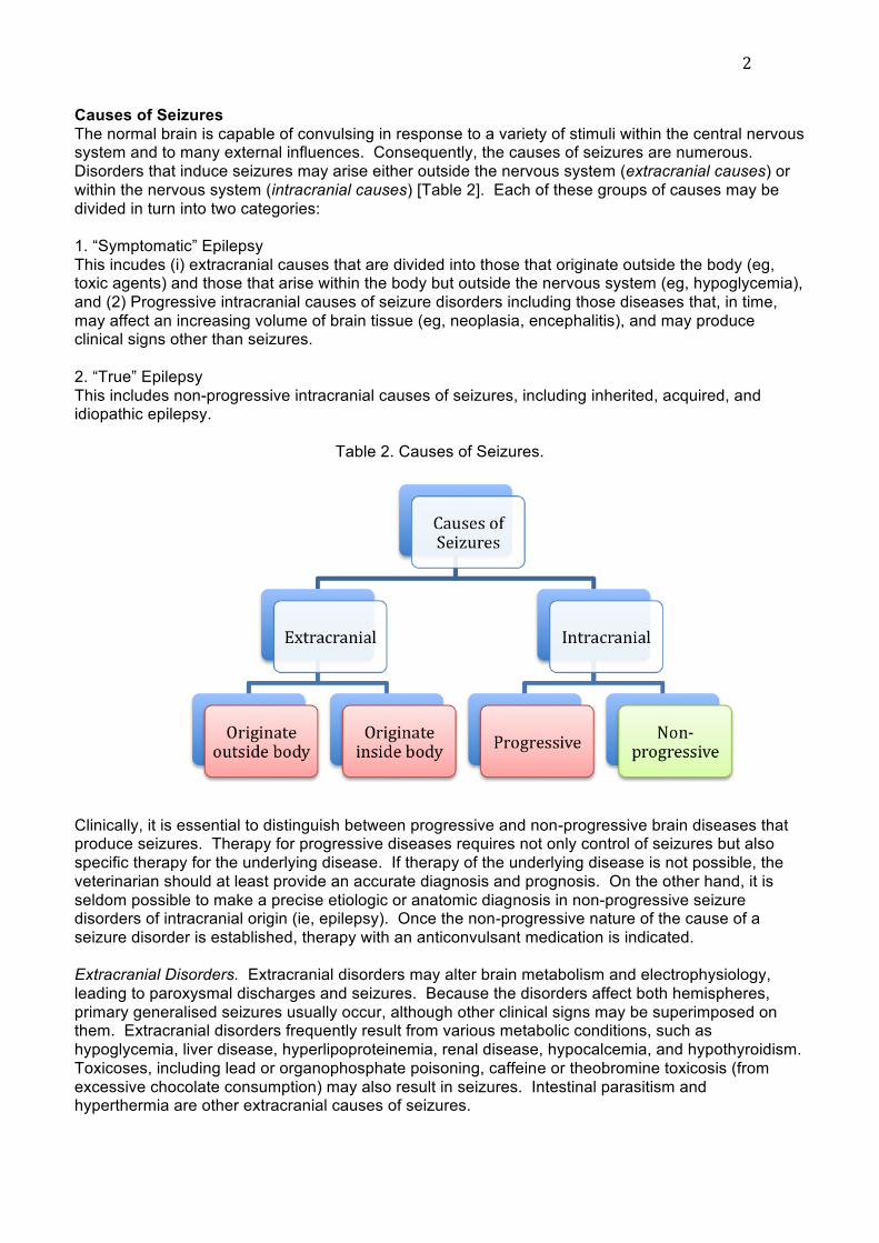
Causes of Seizures
The normal brain is capable of convulsing in response to a variety of stimuli within the central nervous system and to many external influences. Consequently, the causes of seizures are numerous.
Disorders that induce seizures may arise either outside the nervous system (extracranial causes) or
within the nervous system (intracranial causes) [Table 2]. Each of these groups of causes may be
divided in turn into two categories:
1. “Symptomatic” Epilepsy
This incudes (i) extracranial causes that are divided into those that originate outside the body (eg, toxic agents) and those that arise within the body but outside the nervous system (eg, hypoglycemia),
and (2) Progressive intracranial causes of seizure disorders including those diseases that, in time,
may affect an increasing volume of brain tissue (eg, neoplasia, encephalitis), and may produce clinical signs other than seizures.
2. “True” Epilepsy
This includes non-progressive intracranial causes of seizures, including inherited, acquired, and idiopathic epilepsy.
Table 2. Causes of Seizures.
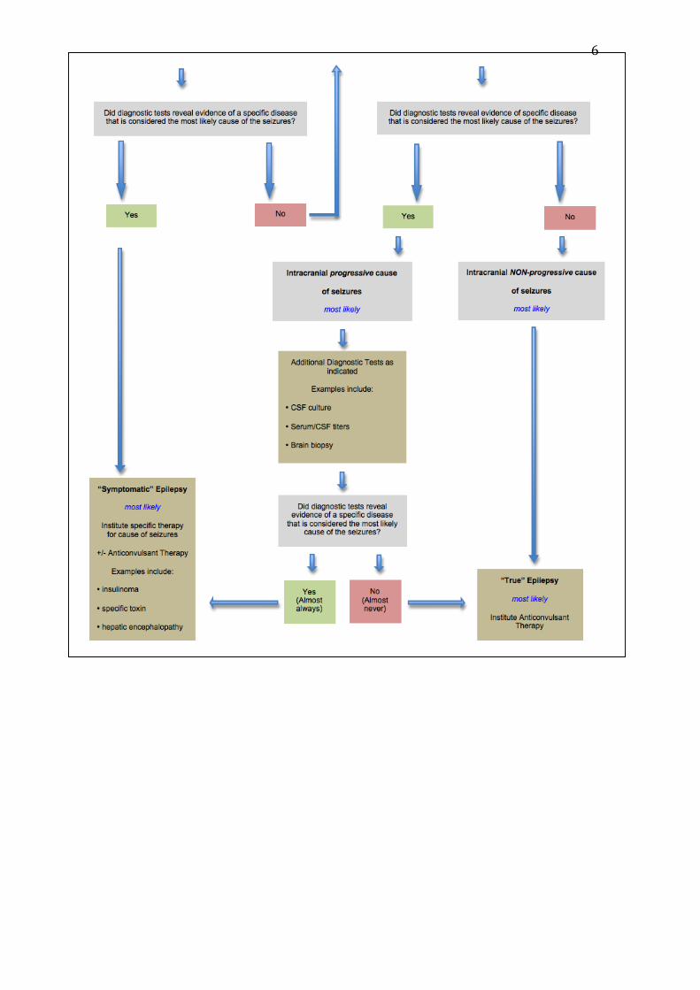
Clinically, it is essential to distinguish between progressive and non-progressive brain diseases that produce seizures. Therapy for progressive diseases requires not only control of seizures but also
specific therapy for the underlying disease. If therapy of the underlying disease is not possible, the
veterinarian should at least provide an accurate diagnosis and prognosis. On the other hand, it is
seldom possible to make a precise etiologic or anatomic diagnosis in non-progressive seizure disorders of intracranial origin (ie, epilepsy). Once the non-progressive nature of the cause of a
seizure disorder is established, therapy with an anticonvulsant medication is indicated.
Extracranial Disorders. Extracranial disorders may alter brain metabolism and electrophysiology,
leading to paroxysmal discharges and seizures. Because the disorders affect both hemispheres,
primary generalised seizures usually occur, although other clinical signs may be superimposed on them. Extracranial disorders frequently result from various metabolic conditions, such as
hypoglycemia, liver disease, hyperlipoproteinemia, renal disease, hypocalcemia, and hypothyroidism.
Toxicoses, including lead or organophosphate poisoning, caffeine or theobromine toxicosis (from
excessive chocolate consumption) may also result in seizures. Intestinal parasitism and hyperthermia are other extracranial causes of seizures.

3
Intracranial Disorders. Intracranial causes of seizures include malformations (eg, hydrocephalus),
inflammatory disorders (eg, canine distemper encephalitis), nutritional disorders (eg, thiamine deficiency), neoplasia, cranial trauma, degenerative conditions (eg, storage diseases), and cerebral
infarction. Affected animals typically present with progressive neurological disease. In some
animals, such as animals with a previous history of cranial trauma, morphological brain lesions may
have occurred long before the first seizures occur, and may be inactive but leave the brain in a seizure-prone state. In other animals, seizures may be an early sign of progressive brain disease,
such as cerebral neoplasia, and may be the sole clinical sign for a prolonged period.
With intracranial disease, secondary or primary generalised seizures occur in a wide variety of clinical manifestations, depending on the location and extent of the underlying lesion(s). The frequency of
seizures may vary considerably, and the association with rest and sleep seems to be less
pronounced than in idiopathic epilepsy (see later). Most intracranial diseases lead to other neurological or clinical signs during the interictal period, and these diseases may have a progressive
or non-progressive clinical course.
Epilepsy. The seizures seen in association with idiopathic epilepsy are caused by functional disorders of the brain in which both hemispheres are affected by paroxysmal neuronal discharges.
Epileptic seizures are generalised and symmetrical from the onset. Morphological lesions are not
observed in the cerebrum of animals with epilepsy, with the exception of animals with microdysgenesis (a condition where subtle alterations of embryo-foetal development, such as
increased neuron density, may result in a lowered seizure threshold). However, lesions such as
gliosis, atrophy, or laminar cortical necrosis, may occur secondary to severe seizures, clusters of seizures, or status epilepticus. These lesions may evolve into a secondary epileptic focus.
Idiopathic epilepsy of dogs or cats usually begins with a single seizure. The seizures most often
occur during or following a period of sleep or rest, and rarely occur during periods of activity. Seizure
frequency is variable (days to months between seizures), however the time between seizures usually decreases as the disorder becomes more chronic. Intervals between seizures may be uniform, or
highly variable. Clusters of seizures may occur over hours or days in some breeds of dog (eg,
German shepherd dogs, Saint Bernard dogs, Irish setters).
Diagnostic Approach
A comprehensive case history, complete physical and neurological examinations, and a minimum
data base consisting of results of hematological and serum chemistry analyses should be obtained for all animals suspected of having a seizure disorder, even if only one seizure has been observed.
On the basis of this information a list of differential diagnoses should be made. Further clinical
laboratory, toxicological, or radiographical procedures may be indicated after the results of these initial tests are known [Table 3] .
Differential Diagnosis Information obtained from the history, physical, and neurological examinations and the results of a
minimum data base may be used to form a list of differential diagnoses. Idiopathic epilepsy may
occur at any age, but it occurs most frequently in dogs or cats between 6 months and 5 years of age.
Using idiopathic epilepsy as a reference point, a list of differential diagnoses for seizure disorders may be formed for each of three age groups: <6 months of age, 6 months to 5 years of age, and >5
years of age. A list of the most common diseases other than epilepsy that may occur in association
with seizures in each of these age categories is included in Table 4.
Additional Diagnostic Tests
Selection of additional tests should be based on results of physical and neurological examinations and on the results of tests that comprise a minimum data base [Table 3]. The dog's or cat's age
should also be considered because certain disorders that may result in seizures are more frequently
associated with younger dogs and cats [Table 4].
An extracranial cause of seizures is most likely associated with an abnormal minimum data base
[Table 3]. Additional tests may be selected to investigate such extracranial causes. For example, in

4
animals with serum chemistry abnormalities that are consistent with liver disease (eg, low BUN,
elevated ALT and /or alkaline phosphatase, low glucose, low total protein, etc) further tests may be needed to assess liver function. Quantification of serum bile acids after a 12-hour fast, and 2 hours
postprandially, provides a reliable indicator of liver function.
After confirmation of an extracranial cause for seizures, specific therapy may be indicated. In certain instances, anticonvulsant medication may be instituted in addition to specific therapy of an
extracranial disorder to control seizure activity during such therapy. Depending on the nature of the
underlying extracranial disease, such anticonvulsant therapy may or may not be discontinued at some later date.
It should be remembered that in rare instances both an extracranial and an intracranial disorder may be present, and yet the extracranial disorder may not be related to the cause of the seizures. For
example, it is possible that a dog with seizures may have hepatic cirrhosis and a primary intracranial
neoplasm. For this reason, it is essential to monitor closely the response of an animal to therapy for
an extracranial cause of seizures. Should the seizures continue or worsen in the face of a response to therapy of the extracranial disease, then further diagnostic tests may be indicated.
An intracranial cause of seizures is most likely associated with normal results of a minimum data base [Table 3]. An intracranial cause should also be considered if the results of additional tests
completed to fully investigate abnormalities seen on a minimum data base prove to be normal.
Cerebrospinal fluid (CSF) analysis is essential for any dog or cat in which an intracranial cause for seizures is suspected. In addition to submission of CSF for cytological examination and protein
quantification, aerobic and anaerobic bacterial and/or mycotic culture and sensitivity testing may be
done, and titers for infectious agents may be completed (eg, cryptococcosis, canine distemper, etc).
Radiographs of the skull may be useful for detecting calvarial tumors, mineralised intracranial neoplasms, or fractures of the skull associated with head trauma.
Electroencephalography (EEG) is helpful in the diagnosis of congenital malformations such as hydrocephalus or lissencephaly. The EEG can also be useful to evaluate electrical events associated
with a seizure that may occur during recording and in the identification of paroxysmal electrical events
that occasionally occur interictally in the recordings of some epileptic animals. Advanced imaging
modalities, such as x-ray computed tomography or magnetic resonance imaging, may provide specific information regarding the location and extent of intracranial lesions such as neoplasms,
granulomas, infarcts, or hemorrhages.
Animals more than 6 months and less than 5 years of age that have a history of a seizure or of
recurrent seizures, that have normal physical and neurological examinations, and that have normal
results on a minimum data base most likely have a nonprogressive intracranial disorder. Ideally, additional diagnostic procedures should be done in such animals, however, consideration of costs
inolved and potential for morbidity and mortality associated with anaesthesia may result in a decision
to delay further tests pending assessment of response to anticonvulsant medication. If a response to
therapy is not seen, if seizure frequency or severity increase, or if additional clinical signs develop, then further diagnostic tests to investigate progressive intracranial causes of seizures should be
done.

5
Table 3: Diagnostic Approach for a Dog or Cat with Seizures
6

7
Table 4. Causes of Seizures
Anticonvulsant Therapy
Appropriate therapy for a seizure disorder depends on accurate determination of the cause of the seizures. Treatment with anticonvulsants is indicated for animals with idiopathic epilepsy. Seizures
resulting from a structural brain disorder (progressive intracranial disease) require additional therapy,
depending on the cause of the disease (eg, neoplasia or inflammation). Anticonvulsants usually are contraindicated in animals with extracranial causes of seizures, where therapy should be directed
towards the primary cause of the seizures (eg, hypoglycemia).
Objectives of Anticonvulsant Therapy. While the overall goal of anticonvulsant therapy is to eradicate
all seizure activity, this goal is rarely achieved. Most dogs and cats benefit from anticonvulsant
medication by reduction in frequency, severity, and duration of their seizures. A realistic goal is to
reduce seizure frequency to a point that is acceptable to an owner without intolerable or life-threatening adverse affects to the animal.
General Principles of Anticonvulsant Therapy. Prevention of seizures in cats or dogs with epilepsy is a pharmacological problem in clinical veterinary medicine. Surgical therapy for uncontrolled epilepsy
as applied in humans has not yet been reported for use in animals.
Prior to initiation of therapy for seizures induced by epilepsy, every reasonable effort must be made to rule out either extracranial or progressive intracranial causes for the seizures.
Decisions Regarding the Need for Anticonvulsant Therapy. Many factors must be considered prior to
the initiation of anticonvulsant therapy. The most important considerations are seizure frequency, seizure character, and owner factors.

8
Seizure Frequency. The seizures observed in epileptic animals occur with varying frequency, and
two general approaches exist regarding the institution of anticonvulsant therapy. Some authors state that therapy should not be started before the recurrent nature of the disease has been established.
This means that at least two seizures should have been observed. Otherwise animals may be
treated that would not have had additional seizures. However, there may be sound biological
reasons for beginning treatment after the first seizure. Experience in human epilepsy indicates that when this is done, seizure control may be more effective.
Character of Seizures. In certain instances, early and aggressive control of seizures is required. For example, in those animals where preictal and postictal phases are characterised by intolerable
changes in personality (eg, aggression) or in excretory behavior.
Owner Factors. In veterinary practice, the decision for or against anticonvulsant therapy ultimately
must be made by the owner of an epileptic dog or cat. This decision should be based on information
and advice provided by a veterinarian. An owner should be fully informed about the nature of the
disease and its treatment in terms that are easily understandable. The owner should have a realistic knowledge of the objectives of anticonvulsant therapy because frequently an owner will expect
successful therapy to be curative with complete elimination of seizures. An owner must appreciate
the need for regular administration of an anticonvulsant drug and also understand that an animal may require medication for the remainder of its life. Cost of medication and regular assessments by a
veterinarian should be discussed. Alterations in dosage without prior consultation must not occur.
Omission of a single dose may result in severe relapses and sometimes status epilepticus. Although seizure frequency and severity will be reduced in the majority of cats or dogs that receive
anticonvulsant medications, a proportion of animals (perhaps as high as 20-30%) may not be
controlled adequately despite intensive medical management. With high dosages of anticonvulsant
medications, the risk of drug-induced complications increases and must be weighed against the benefits of therapy. Once therapy has begun, a prescribed dosage schedule must be followed
exactly. An owner should have a detailed knowledge of expected undesirable effects of
anticonvulsant medications. Knowledge of these factors is essential for a high and intelligent degree of cooperation between an owner and veterinarian. Euthanasia of an animal should be considered
when an owner cannot commit to supervision and lifetime treatment of a dog or cat with severe
epilepsy.
General Recommendations for Anticonvulsant Therapy
In general, owners should be encouraged to begin anticonvulsant medication in epileptic dogs or cats
that are known to have had one or more seizures within an eight week period. Treatment is not routinely advised in animals with seizures that occur less frequently than once every eight weeks, as
owners of such animals often do not follow instructions diligently and may treat animals only
intermittently. Certain owners, however, are so distressed by seizures that occur in their pet that they are willing to medicate an animal daily despite a history of infrequent seizures.
In animals that have had only one seizure, institution of therapy may be delayed. Such a delay may
permit the seizure interval to become apparent, thereby providing a basis for a decision regarding the
need for therapy and also for an assessment of the efficacy of therapy once it is initiated.
Exceptions to these recommendations are made for animals that have seizures in clusters or
episodes of status epilepticus even though the interval between clusters may be greater than eight weeks. The seizure episodes have a tendency to become more frequent and severe in such animals
when control is not attempted.
When seizures have not occurred in a dog or cat for a period of 6-12 months, a cautious reduction of
dosage may be attempted. Such a reduction must be done slowly. In rare instances, a dog or cat
may remain free of seizures after withdrawal of drug therapy.
There are few alternatives to the use of pharmacological agents in the control of seizures.
Acupuncture, either as a sole therapy or in conjunction with conventional anticonvulsant therapy, has
9
been recommended by several authors. Results of these reports are encouraging, and it is likely that
acupuncture will be used with increasing frequency as an adjunctive therapy to conventional anticonvulsant therapy in dogs in the future.
Selection of an Anticonvulsant Medication
The efficacy of an anticonvulsant depends on its serum concentration, because this determines its concentration in the brain. Therapeutic success can be achieved only when serum concentrations of
a given anticonvulsant are consistently maintained within a therapeutic range. Therefore,
anticonvulsants that are eliminated slowly must be employed. The elimination half-lives of the various anticonvulsants differ considerably between different species. Few of the anticonvulsant
drugs used for the treatment of epilepsy in people are suitable for use in dogs and cats. This is
largely because of differences in pharmacokinetics of antiepileptic drugs in animals and in humans. Some drugs are metabolised so rapidly that it is not possible to reach consistently high serum
concentrations, even with very high doses. For many drugs, pharmacokinetic data and/or clinical
experience is lacking in cats, which usually metabolise anticonvulsants more slowly than dogs.

10
SEIZURES:
WHAT TO DO WHEN PHENOBARBITAL FAILS?
Dr. Richard A. LeCouteur
Professor of Neurology & Neurosurgery School of Veterinary Medicine
University of California
Davis CA 95616 USA
PHENOBARBITAL
Phenobarbital is a safe, effective, and inexpensive drug, suitable for long-term therapy
of seizures in dogs and cats. Phenobarbital may be considered a broad spectrum
anticonvulsant, in that it may be effective in many types of epileptic seizures observed
in cats and dogs. It is particularly effective in delaying the progressive intensification of seizure activity that may accompany epilepsy. Clinical reports indicate that
phenobarbital is effective in controlling seizures in 60% to 80% of epileptic dogs,
provided serum concentrations of the drug are maintained within the recommended therapeutic range (20 to 45 ug/ml).
Mechanism of action
The exact anticonvulsant mechanism of action of barbiturates is not completely understood, although it is known that phenobarbital both increases the seizure
threshold required for seizure discharge, and decreases the spread of discharge to
surrounding neurons. Unlike other barbiturates (e.g., pentobarbital), phenobarbital suppresses seizure activity at subhypnotic doses.
Four potential mechanisms of action are: 1) Increasing the activity of the inhibitory neurotransmitter gamma aminobutyric acid (GABA) in the central nervous system
(CNS) at the post-synaptic membrane, hence increasing neuronal inhibition (by means
of an allosteric effect, barbiturates increase the affinity of GABA for its own receptor,
and as a result the chloride ion channel is open for a longer time, resulting in greater chloride flux and enhanced neuronal inhibition); 2) barbiturates may also interact with
glutamate receptors to reduce neuronal excitotoxicity; 3) inhibition of voltage gaited
calcium channels; and 4) competitive binding of the picrotoxin site of the chloride channel.
For the chronic treatment of seizures, dosing should begin at 3-5 mg/kg/day per os divided BID or TID. In severely affected dogs with frequent, severe, or prolonged
seizures, it may be necessary to commence therapy at a higher initial dosage. In
some dogs or cats the starting dose may result in adverse effects. Should these
adverse effects not resolve after two weeks of therapy, a reduction in dose may be indicated. Steady state serum phenobarbital concentrations may be determined after
three weeks of therapy. If a patient’s seizures are adequately controlled, then serum
concentrations may be determined again after three to six months, or more frequently, should seizure activity resume at an unacceptable frequency. If a patient’s seizures
are not adequately controlled after three weeks of therapy, the dose of phenobarbital
may be increased by 25%, and serum concentrations should be determined again
after two weeks of therapy at this higher dose. This process should be continued until the patient’s seizures are controlled, or until a patient fails phenobarbital therapy (i.e.
becomes refractory). Dogs or cats may be considered refractory to phenobarbital
therapy should seizure activity or unacceptable effects persist after plasma concentrations reach 35 ug/ml.

11
Doses of phenobarbital as high as 18-20 mg/kg/day per os divided BID or TID rarely
may be necessary to achieve seizure control in some patients, due to variations in individual drug metabolism. Adverse effects are commonly observed at doses above
20 mg/kg/day.
Reductions in dosage should be made gradually as physical dependence to phenobarbital may develop, and withdrawal seizures may occur as serum levels
decline. Therapy should not be discontinued suddenly, except in animals that develop
fulminant liver dysfunction. Use of phenobarbital should be avoided in animals with hepatic dysfunction.
Phenobarbital may be administered intravenously for the treatment of toxic seizures or status epilepticus, however a lag time of 20-30 minutes may be observed prior to
control of seizures. Phenobarbital may be given as a loading dose of 12 to 24 mg/kg
IV to achieve therapeutic plasma concentrations immediately. Phenobarbital may also
be given as a bolus (2 - 4 mg/kg intravenously every 30 minutes) until a cumulative dose of 20 mg/kg has been given. The dose should be decreased proportionately if
the patient is currently receiving phenobarbital.
Pharmacokinetics
Phenobarbital is fairly rapidly absorbed after oral administration, although variation
exists between animals probably as a result of differences in tablet or capsule dissolution. Bioavailability is high (86-96%) and peak plasma levels are achieved at 4-
6 hours after administration. Although phenobarbital is widely distributed into tissues,
its lower lipid solubility means that it does not penetrate the CNS as rapidly as other
barbiturates. After intravenous injection, therapeutic concentrations are reached in the CNS in 15-20 minutes. Administration of the drug with food reduces absorption by
about 10%. Phenobarbital is primarily metabolised in the liver with only 25% excreted
unchanged by the kidneys. Alkalinization of the urine will enhance elimination of phenobarbital and its metabolites. Plasma protein binding is approximately 45%.
Reported elimination half-life of phenobarbital ranges from 30 to 90 hours in dogs and
from 3 to 83 hours in cats. The wide range of values is a result of several variables, including the use of multiple drug doses in different studies, and more importantly,
whether the drug was given as a single dose, a short course of several doses, or
administered for a length of time. In addition drugs capable of stimulating drug metabolising enzyme systems were included in some studies. In studies in cats, the
use of different populations of cats appeared to be a contributing factor in the reported
variability in elimination half-life.
In dogs, phenobarbital elimination half-life significantly decreases when the drug is
administered chronically (47.3 10.7 hours when administered for 90 days vs 88.7 19.6
hours in a single dose study). The degree of autoinduction of hepatic metabolism of the drug that occurs may be dose dependent. Phenobarbital increases its own rate of
metabolism, and therefore the drug concentration of phenobarbital may be expected to
decrease in patients receiving long-term therapy, and a dose increase should be anticipated in patients after three to six months. In contrast, a study in cats comparing
pharmacokinetics after single and multiple doses suggested that repeated
phenobarbital administration did not alter serum steady state concentrations, but that large differences in phenobarbital elimination rates may exist between different
populations of cats.
In dogs the elimination half-life of phenobarbital is similar after oral or intravenous administration. Steady state concentrations are achieved within 18 days of initiation of
therapy and therefore dosage adjustments should not be made until three weeks after

12
treatment has commenced, or been altered. The metabolism of phenobarbital is quite
variable in dogs and similar oral dosage regimes can result in up to a six fold difference in peak serum concentrations. Beagles are reported to metabolise
phenobarbital more quickly (32 hour elimination half-life) than cross-bred dogs.
Clinical observations suggest that phenobarbital elimination half-life may be shorter in
puppies.
Since there may be extreme variations between the oral dosage of phenobarbital
administered and the serum concentration of drug achieved, it is recommended that serum drug levels should be measured and used as a guide to alterations in drug
therapy. Therapeutic serum phenobarbital concentrations for dogs and cats are
estimated to be 15-45 &g/ml and 10-30 ug/ml respectively. Although monitoring of serum drug levels may be used as a guide to alterations in drug therapy, such
monitoring is not a substitute for clinical judgement. Expected therapeutic serum drug
levels are average values, and each animal will have an individual optimal value. In
dogs this therapeutic range has been demonstrated to be achievable with a dose of 5.5 - 11 mg/kg/day. Due to the longer elimination half-life of phenobarbital in cats, a
once daily dose of 2.5 mg/kg may be appropriate.
Although pharmacokinetic studies in both cats and dogs suggest that once daily
dosing should be adequate to maintain appropriate serum concentrations, twice or
even three times a day dosing is usually recommended to decrease the fluctuations in serum drug levels and minimise adverse effects. In measuring serum drug levels it
has been recommended that blood be collected at a similar time in the daily
administration cycle of phenobarbital. A blood sample should be collected within one
hour before the next scheduled drug administration time (so called trough levels). In a recent study, epileptic dogs receiving chronic phenobarbital therapy were shown to
have minimal variations in serum phenobarbital concentrations throughout the day.
Should other studies confirm these results, it may be acceptable to measure phenobarbital blood levels anytime during the day in chronically treated dogs.
In dogs that have an unacceptable level of seizure control, serum phenobarbital
concentrations should be maintained between 30 to 40 ug/ml for 1-2 months before the maximal effects of the drug may be fully assessed. After this time should
frequency and/or severity of the seizures remain unacceptable, additional or
alternative anticonvulsant medications should be considered. Serum separation tubes have been shown to falsely reduce drug concentrations, and should be avoided for
therapeutic drug monitoring.
Methylphenobarbital is available in several countries. It requires hepatic demethylation
for activation. Dosage recommendations are usually similar to those recommended
for phenobarbital. However, it is possible that serum concentrations of the active
constituent, phenobarbital, may not be as high at equivalent milligram doses of phenobarbital.
Adverse effects Adverse effects that may be observed initially with phenobarbital therapy include
sedation, ataxia, polydipsia, polyuria, polyphagia and weight gain. Polyuria is due to
an inhibitory effect on the release of antidiuretic hormone. Polyphagia is believed to be due to suppression of the satiety center in the hypothalamus. Most dogs develop a
tolerance to these effects after two to four weeks of therapy. Unless sedation is
extreme or persistent beyond four weeks, it should not be considered evidence of
toxicity and does not warrant an adjustment in the dose. Sedation at the initiation of therapy has been reported to be more severe in cats. Occasionally, a dog may appear
13
hyperactive during the initial phase of therapy, however this effect usually may be
overcome by increasing the dose.
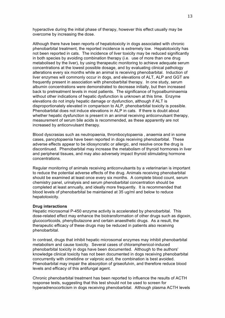
Although there have been reports of hepatotoxicity in dogs associated with chronic
phenobarbital treatment, the reported incidence is extremely low. Hepatotoxicity has
not been reported in cats. The incidence of liver toxicity may be reduced significantly in both species by avoiding combination therapy (i.e. use of more than one drug
metabolised by the liver), by using therapeutic monitoring to achieve adequate serum
concentrations at the lowest possible dosage, and by evaluating clinical pathology alterations every six months while an animal is receiving phenobarbital. Induction of
liver enzymes will commonly occur in dogs, and elevations of ALT, ALP and GGT are
frequently present in association with phenobarbital therapy. In one study, serum albumin concentrations were demonstrated to decrease initially, but then increased
back to pretreatment levels in most patients. The significance of hypoalbuminaemia
without other indications of hepatic dysfunction is unknown at this time. Enzyme
elevations do not imply hepatic damage or dysfunction, although if ALT is disproportionately elevated in comparison to ALP, phenobarbital toxicity is possible.
Phenobarbital does not induce elevations in ALP in cats. If there is doubt about
whether hepatic dysfunction is present in an animal receiving anticonvulsant therapy, measurement of serum bile acids is recommended, as these apparently are not
increased by anticonvulsant therapy.
Blood dyscrasias such as neutropaenia, thrombocytopaenia , anaemia and in some
cases, pancytopaenia have been reported in dogs receiving phenobarbital. These
adverse effects appear to be idiosyncratic or allergic, and resolve once the drug is
discontinued. Phenobarbital may increase the metabolism of thyroid hormones in liver and peripheral tissues, and may also adversely impact thyroid stimulating hormone
concentrations.
Regular monitoring of animals receiving anticonvulsants by a veterinarian is important
to reduce the potential adverse effects of the drug. Animals receiving phenobarbital
should be examined at least once every six months. A complete blood count, serum
chemistry panel, urinalysis and serum phenobarbital concentration should be completed at least annually, and ideally more frequently. It is recommended that
blood levels of phenobarbital be maintained at 35 ug/ml and below to reduce
hepatotoxicity.
Drug interactions
Hepatic microsomal P-450 enzyme activity is accelerated by phenobarbital. This dose-related effect may enhance the biotransformation of other drugs such as digoxin,
glucocorticoids, phenylbutazone and certain anaesthetic drugs. As a result, the
therapeutic efficacy of these drugs may be reduced in patients also receiving
phenobarbital.
In contrast, drugs that inhibit hepatic microsomal enzymes may inhibit phenobarbital
metabolism and cause toxicity. Several cases of chloramphenicol-induced phenobarbital toxicity in dogs have been documented. Although to the authors'
knowledge clinical toxicity has not been documented in dogs receiving phenobarbital
concurrently with cimetidine or valproic acid, the combination is best avoided. Phenobarbital may impair the absorption of griseofulvin, and therefore reduce blood
levels and efficacy of this antifungal agent.
Chronic phenobarbital treatment has been reported to influence the results of ACTH response tests, suggesting that this test should not be used to screen for
hyperadrenocorticism in dogs receiving phenobarbital. Although plasma ACTH levels

14
are reported to be unchanged, not all studies have confirmed this observation.
Similarly, dexamethasone suppression tests may also be influenced in some, but not all, dogs chronically treated with phenobarbital.
Total and free serum thyroxine concentrations have been reported to be significantly
reduced in dogs receiving phenobarbital.
BROMIDE
Bromide is a halide element, discovered in 1826 by Balard, and first reported for use
as an anticonvulsant in people by Sir Charles Locock in 1857. Until 1912 when
phenobarbital was introduced, bromide was the only effective anticonvulsant available. There are reports of the use of bromide in dogs as early as 1907, but it was not until
1986 that interest in its use clinically to control seizures was reported. Until recently,
bromide was used only as an adjunct to phenobarbital therapy for the management of
dogs with refractory epilepsy. More recently, its use as a sole anticonvulsant therapy in epileptic dogs has been recommended by some neurologists, particularly in dogs
with hepatic dysfunction. Although bromide is an effective anticonvulsant, it is not
approved for use for medical purposes in any species.
Dosage and formulations
Bromide is available as a potassium or a sodium salt. Bromide may be formulated as a 200 mg/ml or 250 mg/ml solution of reagent grade potassium bromide in syrup or
distilled water. Potassium bromide is available in tablet form in some countries.
Alternatively, the powder may be placed in gelatin capsules. In solution in distilled
water, bromide has been found to be stable for more than one year at room temperature or refrigerated, and in glass, plastic, clear or brown containers.
Information concerning stability of bromide in solution in syrup is not available,
however current recommendations include keeping the solution refrigerated, and discarding it after three months.
Bromide may also be administered as the sodium salt to dogs that dislike the taste of
potassium or cannot tolerate it (e.g., due to hypoadrenocortism). Sodium bromide should not be used in dogs with congestive heart failure, hypertension or liver disease.
The dose of sodium bromide should be reduced by 15% to account for the higher
bromide content per gram (KBr = 67% bromide, NaBr = 78% bromide).
The primary indication for bromide has been in combination with phenobarbital in
refractory epileptics or in patients that have developed liver disease. Bromide is also the drug best suited for use by noncompliant owners because of its long half-life.
Bromide may also be used as the sole drug in patients whose seizure history is limited
to mild seizure activity.
Because reaching a steady state may require two to three months, a loading dose is
recommended to achieve therapeutic concentrations more rapidly. The maintenance
dose is designed to maintain concentrations achieved after loading. Alternatively the maintenance dose may be given without a loading dose to allow more gradual
accommodation to effective serum concentrations.
The appropriate dose of bromide depends on concurrent anticonvulsants, diet, and
renal function. The maintenance dose of potassium bromide as a first line
anticonvulsant is 20-40 mg/kg/day PO once daily or in divided doses in both dogs and
cats. Steady state concentrations are not achieved for two to three months in dogs. Steady state concentrations in dogs following administration of 30 mg/kg per day are
about 0.8 to 1.2 mg/ml. When used in conjunction with other anticonvulsants, the

15
recommended dose is 22-30 mg/kg PO once daily or divided. The author’s protocol
for a loading dose of potassium bromide is 400-600 mg/kg divided into four doses, given over 24 hours.
Distribution of bromide is to the extracellular space. Bromide is eliminated slowly
(perhaps due to marked reabsorption) in the kidney. Its rate of elimination can change with salt administration. Increased dietary salt will increase the rate of elimination of
bromide, and decreased dietary salt will result in the opposite effect.
The elimination of bromide in cats appears to be faster than that in dogs, with a mean
half-life of approximately six weeks. Following administration of 30 mg/kg orally for
eight weeks, mean bromide concentrations were 1.2 mg/ml at steady state. Cats developed no adverse effects to bromide administration in one report.
Serum bromide concentrations may be determined one month after a maintenance
dose is instituted, regardless of whether a loading dose was given. This approach will enable modification prior to steady state and prior to therapeutic failure or signs of
adverse reactions. Recommended target ranges are controversial and depend on
whether phenobarbital is being given concurrently. Most laboratories use 1-3 mg/ml regardless of whether bromide is being given as a sole agent or in combination with
phenobarbital.
Mechanism of action
The exact mechanisms of anticonvulsant action of bromide are incompletely
understood. The action appears to involve chloride ion channels. These channels are
an important part of the inhibitory neuronal network of the CNS. Their function is modulated by GABA, the most important inhibitory neurotransmitter in the mammalian
CNS. Increased chloride ion flow as a result of activation of GABA receptors by
barbiturates, or benzodiazepines results in increased neuronal inhibition and increases the threshold for seizures. Bromide appears to cross neuronal chloride channels more
readily than chloride because it has a smaller hydrated diameter. It is therefore
believed that bromide, by competing with chloride ions, hyperpolarises post-synaptic
neuronal membranes and facilitates the action of inhibitory neurotransmitters. Barbiturates may act synergistically with bromide to raise the seizure threshold by
enhancing chloride conductance via GABA-ergic activity.
Pharmacokinetics
The pharmacokinetics of bromide have not been well established. Bromide is
absorbed from the small intestine with peak absorption achieved 1.5 hours after oral administration. It is not protein bound or metabolised and does not undergo hepatic
metabolism. Therefore bromide does not affect hepatic enzymes and is a useful
anticonvulsant for dogs with hepatic disease. Elimination of bromide from the body is
via the kidneys.
Although bromide replaces chloride throughout the body, the sum of these two halides
remains constant. Lower concentrations of bromide are found in the brain than would be expected based on the chloride concentration, as it appears there is a barrier to the
free passage of bromide. However, the concentration of bromide in the brain of dogs
is higher than that found in humans. In humans after bromide treatment the CSF to serum ratio is 31% whereas in dogs the ratio is 87%. As a result, seizure control may
be achieved in dogs at a lower serum bromide concentration than in humans, and the
potential for toxicity is much reduced.
The half-life of bromide is longer in dogs than in humans (24.9 vs 12 days). Two to
three weeks of therapy are required before serum bromide levels enter the therapeutic

16
range, and steady state levels are achieved in four months. Higher serum
concentrations are required if dogs are treated with bromide alone as compared to dogs treated with bromide and phenobarbital. The therapeutic range for potassium
bromide when administered with phenobarbital is 0.8-2.4 mg/ml and 0.8 - 3.0 mg/ml
when it is used alone.
Adverse effects
Clinical signs of bromide toxicity appear to be dose dependent, and include
polyphagia, vomiting, anorexia, constipation, pruritis, muscle pain, sedation, and pelvic limb weakness. Ataxia and sedation have been reported to be the major dose limiting
side effects in dogs. Other infrequently reported adverse effects include pancreatitis,
increased attention seeking, aggression, coprophagia and hyperactivity. High dose bromide therapy has been associated with thyroid disfunction in people and rats.
Bromide toxicity has been reported in a dog with renal insufficiency, which resulted in
decreased clearance of bromide and therefore higher plasma levels. Reduced
efficacy of the drug has been reported in a dog fed a high chloride diet. Toxic concentrations of bromide may be reached rapidly when chloride intake is decreased.
Bromide readily crosses the placenta is people, and there are a few reports of neonatal bromism. Due the lack of information in dogs, bromide should be avoided in
breeding animals.
Drug interactions
Because bromide is not protein bound and not metabolized, it does not interact with
many drugs. However, chloride containing foods, food supplements, fluids, drugs, and
loop diuretics may enhance bromide elimination and lower serum bromide concentrations. Bromide is a product of halothane metabolism in dogs, and therefore
small increases in serum bromide may be apparent after halothane anaesthesia. In
people, bromide administration has be reported to be associated with increased visualisation of cerebral vessels with non-contrast computed tomography.
Pseudohyperchloraemia occurs with bromide administration as bromide ions interfere
with colourimetric and automated ion-specific electrolyte analysers used to measure chloride. Treatment of bromide toxicity includes chloride loading (intravenous sodium
chloride) to increase bromide elimination.
Table 1: Recommended Anticonvulsant Drug Dosages
DRUG RECOMMENDED DOSES
Phenobarbital Dogs: 2 - 5 mg/kg divided BID (up to 18 - 20 mg/kg BID if
necessary)
Cats: 0.5 - 1.5 mg/kg divided BID
Potassium bromide Dogs and Cats: 20 - 40 mg/kg PO SID or divided BID
Dogs: Loading dose 400-600 mg/kg divided into four doses, given
over 24 hours.
Sodium Bromide Dogs: 20-40 mg/kg PO SID, reduce by 15%
Dogs: 1200-1500 mg/kg over 24 hours as a continuous rate
infusion of 3% NaBR in D5W.

17
Phenobarbital Resistant Epilepsy Using multiple anti-convulsant drugs has several potential disadvantages, including the
increased cost, the need to monitor and to interpret serum concentrations of multiple drugs,
potential drug interactions, and more complicated dosing schedules. Indications for such
'polytherapy' include failure of diligent monotherapy and the treatment of cluster seizures. Before polytherapy is started, all reasonable options for monotherapy should be tried. If the
initial drug is ineffective, a second drug should be added. If the dog responds, the veterinarian
should attempt to withdraw the first drug gradually and continue with polytherapy if this is unsuccessful.
Factors to consider when choosing an add-on medication include 1) mechanism of action, with preference given to drugs with a differing mechanism, 2) side effects, 3) the potential for
drug interactions among antiepileptic drugs, 4) the required frequency of administration, which
in turn may influence compliance, and 5) the cost.
Potassium Bromide (KBr)
Potassium bromide has no known hepatic toxicity and all the adverse effects of KBr are
completely reversible once the drug is discontinued. Potassium bromide is excreted unchanged in the urine and is not metabolised by the liver. There have been no liver enzyme
alterations or thyroid axis effects after administration of this drug. KBr controls approximately
70-80% of the epileptic dogs it is used to treat and is often effective in dogs that fail phenobarbital therapy. When high dose KBr and low dose PB are used together,
approximately 95% of epileptic dogs can be controlled.
Where do you turn when an epileptic dog responds poorly to PB and/or KBr? Management of refractory epilepsy presents a considerable challenge to veterinary
practitioners and pet owners. Appropriate therapy and monitoring should be used to optimise
treatment results. Ongoing patient follow-up is critical. With the approval of several new antiepileptic drugs for the management of human epilepsy over the last two decades, the
treatment options available for dogs and cats have increased. Although information on the use
of these novel antiepileptic drugs in animals is lacking, studies and clinical experience
available to date suggest that they have the potential to improve seizure control and minimise adverse effects. Continued evaluation of these drugs in veterinary patients should provide
additional details on their use in managing canine epilepsy. Aspects of these new
antiepileptic drugs have been summarised by Munana.1
Newer anticonvulsant drugs to consider include:
Felbamate Gabapentin
Pregabalin
Zonisamide
Levetiracetam
Efficacy of new anticonvulsant drugs
When recommending a novel treatment, it is helpful if you can provide information on the expected effectiveness of the treatment, especially when the treatment involves a
considerable investment of time or money on the pet owner's part. Unfortunately, extensive
data of this nature are not available for the use of the new antiepileptic drugs in canine or feline epilepsy. Rather, information on drug use has been obtained by administering the new
antiepileptic drug in an open-label fashion: Dogs with poorly controlled seizures were given a
new antiepileptic drug, and seizure frequency during administration of the new drug was
compared with seizure frequency before the new drug was initiated. Because the studies were not placebo-controlled, the information obtained regarding drug efficacy may be inaccurate.
This has been called the placebo effect.

18
From the information available to date, a veterinarian can recommend a trial with a new antiepileptic drug, but, no representation should be made with respect to efficacy. As
experience with these drugs grows in veterinary medicine and more research is performed on
their applicability to dogs and cats, additional information will become available to further
guide recommendations on their use.
Factors to consider when choosing an add-on medication include:
1. Mechanism of action, with preference given to drugs with a differing mechanism,
2. Adverse effects,
3. Potential for drug interactions among antiepileptic drugs, 4. Required frequency of administration, which in turn may influence compliance,
and,
5. Cost.
New antiepileptic drugs approved for use in people that may be considered for extra-
label use in dogs are summarised in Table 1.1
Levetiracetam Levetiracetam is one of the more recently approved human antiepileptic drugs. Points
of interest regarding levetiracetam are as follows:
1. levetiracetam has a unique mechanism of action, which is a potential
advantage when the drug is used in combination with other antiepileptic drugs, 2. Levetiracetam has minimal hepatic metabolism in dogs, with more than 80% of
the drug excreted in the urine, 3. The half-life in dogs is three to four hours, which necessitates frequent
administration,
4. The recommended oral dose is 20 mg/kg every eight hours.
A recent study2 evaluated levetiracetam as an add-on medication in dogs with
idiopathic epilepsy that was refractory to phenobarbital and potassium bromide. Nine of 14 dogs responded, and the only adverse effect was sedation (observed in one
dog).
The main factor limiting the use of levetiracetam in dogs has been its expense,
although a generic form of this drug recently has become available.
Levetiracetam also is available in a parenteral formulation. Levetiracetam may prove
useful in treating cluster seizures and status epilepticus in dogs, with the option of
administering the drug intramuscularly if venous access cannot be obtained. The disposition of levetiracetam has been evaluated in six dogs after intravenous and
intramuscular administration.3 A dose of 20 mg/kg IV resulted in desirable serum
concentrations in a short period; with intramuscular administration, peak
concentrations were reached in 40 minutes.
Felbamate
Felbamate was the first of the newer antiepileptic drugs to be approved in the United
States for epilepsy in people. Side effects of aplastic anemia and hepatotoxicosis
have since been reported in people, so its use has declined.
Points of interest regarding felbamate are as follows:
1. The half-life of felbamate in dogs is five to eight hours,

19
2. The recommended oral dosage is 15 to 60 mg/kg every eight hours. 3. Treatment should be initiated at the low end of the dosage range, and the
dosage should be increased as needed to control seizures. 4. Felbamate serum concentrations are not routinely monitored, as is the case for
many of the newer antiepileptic drugs. Rather, the drug is used to effect, and
dosage adjustments are based on seizure frequency and side effects, 5. Felbamate is mostly excreted in the urine in dogs, although some hepatic
metabolism occurs, increasing the potential for drug interactions when
phenobarbital and felbamate are administered concurrently.
Felbamate is used infrequently in veterinary patients because of the potential for
adverse effects and drug interactions, and because of the expense. It is
recommended that complete blood counts and liver enzyme activities be measured
every two or three months during treatment to assess for adverse effects.
Felbamate was evaluated as a sole antiepileptic drug in six dogs with partial onset
seizures.4 Dosages ranged from 60 to 220 mg/kg/day, and all dogs showed reduced
seizure frequency. Two dogs developed blood dyscrasias, characterised by
thrombocytopenia, lymphopenia, and leukopenia, which resolved after the drug was discontinued. One dog developed keratoconjunctivitis sicca, although it was not
determined that this problem was drug-related.
The use of felbamate as an add-on therapy has been reported in 16 dogs refractory to
phenobarbital and potassium bromide.5 Twelve dogs had improved seizure control, but
four of these dogs developed signs of liver dysfunction. Other side effects anecdotally reported in dogs receiving felbamate in combination with phenobarbital include
sedation, nausea, and vomiting.6
Gabapentin Gabapentin was approved in the United States for use in people as an antiepileptic
drug shortly after felbamate. Since its introduction, gabapentin has also been
approved to treat neuropathic pain. In people, gabapentin is eliminated entirely by the
kidneys.
1. In dogs gabapentin undergoes partial hepatic metabolism, 2. The elimination half-life in dogs is two to four hours, requiring frequent drug
administration to achieve steady-state concentrations,
3. The recommended oral dosage is 10 to 20 mg/kg every six to eight hours.
Two studies have examined the use of gabapentin in refractory canine epilepsy. The
first study involved 11 dogs administered gabapentin (10 mg/kg t.i.d.) in addition to phenobarbital and potassium bromide.7 A positive response to gabapentin, defined as
≥ 50% reduction in seizure frequency, was reported in six of the 11 dogs. The second
study evaluated 17 dogs with refractory seizures that were administered gabapentin at a dose of 35 to 50 mg/kg/day divided twice or three times daily, also in conjunction
with phenobarbital and potassium bromide (16 dogs) or phenobarbital alone (1 dog).8
This study found no significant decrease in the number of seizures over the study
period for the entire population of dogs. However, seizures resolved in three dogs while they were receiving the medication. The adverse effects reported in both studies
were sedation and ataxia.
The availability of a generic form of gabapentin has made it affordable relative to other
antiepileptic drugs used in veterinary medicine.

20
A liquid formulation (50 mg/ml) of gabapentin exists, which facilitates the
administration of lower doses to smaller patients. However, the liquid product contains 300 mg xylitol/ml, so it has the potential of causing adverse effects associated with the
ingestion of this sugar alcohol.9
Pregabalin Pregabalin is a newer-generation drug in the same class as gabapentin. Limited
information exists on its use in dogs for the treatment for epilepsy. Based on
pharmacokinetic data in normal dogs, a dose of 2 to 4 mg/kg orally every eight hours
has been recommended and was administered to six dogs with idiopathic epilepsy refractory to treatment with phenobarbital, potassium bromide, or both drugs.10 One
dog was considered a drug failure, and four of the five remaining dogs had a mean
seizure reduction of 59.3%. Five dogs exhibited side effects (sedation and ataxia)
attributed to pregabalin.
Zonisamide Zonisamide is a sulfonamide-derived antiepileptic drug introduced in the United States
in 2000.
Points of interest are as follows: 1. The half-life of zonisamide in dogs is 15 to 20 hours, which is relatively long
when compared with the other new antiepileptic drugs,
2. Zonisamide requires only twice-daily administration 3. Most of the drug is excreted unchanged by the kidneys, although some hepatic
metabolism occurs,
4. The dosage in dogs is 5 to 10 mg/kg given orally every 12 hours. The high end of the dose range is recommended when the drug is used in combination with
phenobarbital since phenobarbital appears to facilitate zonisamide clearance.11
For this reason, it may also be helpful to measure serum zonisamide
concentrations in dogs being treated concurrently with Phenobarbital,
5. A therapeutic range of 10 to 40 µg/ml has been suggested,12 which is similar to
the therapeutic range in people, 6. Zonisamide is a known teratogen in dogs, so its use should be avoided in
pregnant animals.
A generic form of zonisamide is available, which has reduced the cost considerably
since its introduction.
Two recent reports on zonisamide as add-on therapy in dogs with refractory epilepsy
demonstrated a favorable response in seven of 12, and nine of 11 dogs,
respectively.13,14 Reported side effects included sedation, ataxia, and loss of appetite.
Efficacy of new antiepileptic drugs – The placebo effect15 Extensive data are not available regarding the effectiveness of treatment of the new
antiepileptic drugs in canine epilepsy. Rather, the information on drug use described above was obtained by administering the new antiepileptic drug in an open-label
fashion: Dogs with poorly controlled seizures were given a new antiepileptic drug, and
seizure frequency during administration of the new drug was compared with seizure
frequency before the new drug was initiated. Because the studies were not placebo-
controlled, the information obtained regarding drug efficacy may be inaccurate.
A reduction in seizure frequency during placebo administration has been documented
in people with epilepsy,15 and, a similar phenomenon may occur in dogs. One likely
cause for this placebo effect is regression to the mean, which is a statistical term used

21
to describe the fluctuations that occur in biological variables over time and take the
form of a sine wave around the mean. Epilepsy is a waxing and waning disorder, and fluctuations in seizure frequency are common over the course of the disease. Clients
are most likely to seek a change in therapy for their pets when seizures are poorly
controlled. Over time, improvement in the seizure frequency is probable, regardless of
the treatment administered. However, this improvement may be erroneously attributed to a recently instituted change in therapy. Open-label studies cannot account for this
bias, and, consequently, the efficacy reported in such studies may be falsely elevated.
Thus, from the information available to date, a veterinarian may recommend a trial with
a new antiepileptic drug, but should not make any representation with respect to
efficacy. As experience with these drugs grows in veterinary medicine and more research is performed on their applicability to dogs, additional information will become
available to further guide recommendations on their use.
Conclusion The management of refractory seizures can pose a considerable challenge to
practitioners and pet owners. Ongoing patient follow-up is critical, and directed,
appropriate therapy and monitoring should be used to optimize treatment results. With
the approval of several new antiepileptic drugs for the management of human epilepsy over the last two decades, the treatment options available for our canine patients have
also increased. Although our knowledge is still somewhat limited on these novel
antiepileptic drugs, studies and clinical experience available to date suggest that they have the potential to improve seizure control and minimize adverse effects in affected
dogs. Continued evaluation of these drugs in veterinary patients should provide
additional details on how to use these therapies most effectively in managing canine
epilepsy.
References 1. Munana KR. Newer options for medically managing refractory canine epilepsy.
http://veterinarymedicine.dvm360.com/vetmed/ArticleStandard/Article/detail/608398 2. Volk HA, Matiasek LA, Reliu-Pascual AL, et al. The efficacy and tolerability of
levetiracetam in pharmacoresistant epileptic dogs. Vet J 2008;176(3):310-319.
3. Patterson EE, Goel V, Cloyd JC, et al. Intramuscular, intravenous and oral levetiracetam in dogs: safety and pharmacokinetics. J Vet Pharmacol Ther
2008;31(3):253-258. 4. Ruehlmann D, Podell M, March P. Treatment of partial seizures and seizure-like
activity with felbamate in six dogs. J Small Anim Pract 2001;42(8):403-408. 5. Boothe DM. Anticonvulsant therapy in small animals. Vet Clin North Am Small Anim
Pract 1998;28(2):411-448. 6. Dayrell Hart B, Tiches D, Vite C, et al. Efficacy and safety of felbamate as an
anticonvulsant in dogs with refractory seizures (abst). J Vet Intern Med 1996;10:174
7. Platt SR, Adams V, Garosi LS, et al. Treatment with gabapentin of 11 dogs with
refractory idiopathic epilepsy. Vet Rec 2006;159(26):881-884. 8. Govendir M, Perkins M, Malik R. Improving seizure control in dogs with refractory
epilepsy using gabapentin as an adjunctive agent. Aust Vet J 2005;83(10):602-608. 9. Dunayer EK, Gwaltney-Brant SM. Acute hepatic failure and coagulopathy
associated with xylitol ingestion in eight dogs. J Am Vet Med Assoc 2006;229(7):1113-
1117. 10. Dewey CW, Cerda-Gonzalez S, Levine JM, et al. Pregabalin therapy for refractory
idiopathic epilepsy in dogs (abst). J Vet Intern Med 2008;22:765.
11. Orito K, Saito M, Fukunaga K, et al. Pharmacokinetics of zonisamide and drug
interaction with phenobarbital in dogs. J Vet Pharmacol Ther 2008;31(3):259-264.
22
12. Dewey CW, Guiliano R, Boothe DM, et al. Zonisamide therapy for refractory
idiopathic epilepsy in dogs. J Am Anim Hosp Assoc 2004;40(4):285-291. 13. von Klopmann T, Rambeck B, Tipold A. Prospective study of zonisamide therapy
for refractory idiopathic epilepsy in dogs. J Small Anim Pract 2007;48(3):134-138. 14. Burneo JG, Montori VM, Faught E. Magnitude of the placebo effect in randomised
trials of antiepileptic agents. Epilepsy Behav 2002;3(6):532-534. 15. Munana KR, Zhang D, Patterson EE. Placebo effect in canine epilepsy trials. J Vet
Intern Med 2010; 24:166-170.
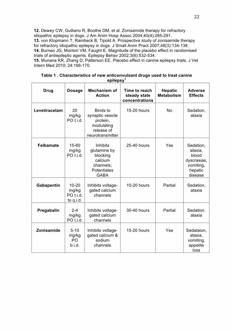
Table 1 . Characteristics of new anticonvulsant drugs used to treat canine
epilepsy1
Drug
Dosage
Mechanism of Action
Time to reach steady state
concentrations
Hepatic Metabolism
Adverse Effects
Levetiracetam
20
mg/kg
PO t.i.d.
Binds to
synaptic vesicle
protein,
modulating release of
neurotransmitter
15-20 hours
No
Sedation,
ataxia
Felbamate
15-60
mg/kg
PO t.i.d.
Inhibits
glutamine by
blocking
calcium channels;
Potentiates
GABA
25-40 hours
Yes
Sedation,
ataxia,
blood
dyscrasias, vomiting,
hepatic
disease
Gabapentin
10-20
mg/kg
PO t.i.d. to q.i.d.
Inhibits voltage-
gated calcium
channels
10-20 hours
Partial
Sedation,
ataxia
Pregabalin
2-4 mg/kg
PO t.i.d.
Inhibits voltage-gated calcium
channels
30-40 hours
Partial
Sedation. ataxia
Zonisamide
5-10 mg/kg
PO
b.i.d.
Inhibits voltage-gated calcium &
sodium
channels
15-20 hours
Yes
Sedataion, ataxia,
vomiting,
appetite loss