
Targeting BCAA Catabolism to Treat Obesity …...Targeting BCAA Catabolism to Treat...
17
Targeting BCAA Catabolism to Treat Obesity-Associated Insulin Resistance Meiyi Zhou, 1 Jing Shao, 1 Cheng-Yang Wu, 2 Le Shu, 3 Weibing Dong, 1 Yunxia Liu, 1 Mengping Chen, 1 R. Max Wynn, 2 Jiqiu Wang, 4 Ji Wang, 1 Wen-Jun Gui, 2 Xiangbing Qi, 5 Aldons J. Lusis, 6 Zhaoping Li, 7 Weiqing Wang, 4 Guang Ning, 4 Xia Yang, 3 David T. Chuang, 2 Yibin Wang, 8 and Haipeng Sun 1,8 Diabetes 2019;68:1730–1746 | https://doi.org/10.2337/db18-0927 Recent studies implicate a strong association between elevated plasma branched-chain amino acids (BCAAs) and insulin resistance (IR). However, a causal relationship and whether interrupted BCAA homeostasis can serve as a therapeutic target for diabetes remain to be established experimentally. In this study, unbiased integrative pathway analyses identified a unique genetic link between obesity- associated IR and BCAA catabolic gene expression at the pathway level in human and mouse populations. In genet- ically obese (ob/ob) mice, rate-limiting branched-chain a-keto acid (BCKA) dehydrogenase deficiency (i.e., BCAA and BCKA accumulation), a metabolic feature, accompa- nied the systemic suppression of BCAA catabolic genes. Restoring BCAA catabolic flux with a pharmacological inhibitor of BCKA dehydrogenase kinase (BCKDK) ( a sup- pressor of BCKA dehydrogenase) reduced the abundance of BCAA and BCKA and markedly attenuated IR in ob/ob mice. Similar outcomes were achieved by reducing protein (and thus BCAA) intake, whereas increasing BCAA intake did the opposite; this corroborates the pathogenic roles of BCAAs and BCKAs in IR in ob/ob mice. Like BCAAs, BCKAs also suppressed insulin signaling via activation of mammalian target of rapamycin complex 1. Finally, the small-molecule BCKDK inhibitor significantly attenu- ated IR in high-fat diet–induced obese mice. Collectively, these data demonstrate a pivotal causal role of a BCAA catabolic defect and elevated abundance of BCAAs and BCKAs in obesity-associated IR and provide proof-of- concept evidence for the therapeutic validity of manipu- lating BCAA metabolism for treating diabetes. The three branched-chain amino acids (BCAAs) are leucine, isoleucine, and valine. A strong association between ele- vated plasma BCAAs and insulin resistance (IR) has been repeatedly observed in human and rodent models (1–9). Moreover, longitudinal studies suggest that a high plasma BCAA level is predictive of the future onset of diabetes (10–13). Circulating BCAAs are also a significant prognos- tic marker associated with outcomes of diabetes interven- tions (4,10,14–16). On the other hand, elevated plasma branched-chain a-keto acids (BCKAs), the products of BCAA transamination, are also associated with IR and are potentially better biomarkers for diabetes (8,9). These observations strongly suggest a causal role of disrupted BCAA homeostasis in IR, which remains to be established experimentally. BCAAs are essential amino acids derived from protein- containing foods. The BCAA catabolic pathway, consisting of more than 40 enzymes in mitochondria, plays a pivotal role in maintaining BCAA homeostasis in mammals (2). 1 Department of Pathophysiology, Key Laboratory of Cell Differentiation and Apoptosis of the Chinese Ministry of Education, Hongqiao International Institute of Medicine, Shanghai Tongren Hospital/Faculty of Basic Medicine, Shanghai Jiao Tong University School of Medicine, Shanghai, China 2 Department of Biochemistry, University of Texas Southwestern Medical Center, Dallas, TX 3 Department of Integrative Biology and Physiology, University of California at Los Angeles, Los Angeles, CA 4 Department of Endocrinology and Metabolism, Ruijin Hospital, Shanghai Jiao Tong University School of Medicine, Shanghai, China 5 Chemistry Center, National Institute of Biological Science, Beijing, China 6 Departments of Medicine, Microbiology, and Human Genetics, University of California at Los Angeles, Los Angeles, CA 7 Department of Clinical Nutrition, University of California at Los Angeles, Los Angeles, CA 8 Departments of Anesthesiology, Medicine, and Physiology, University of California at Los Angeles, Los Angeles, CA Corresponding author: Haipeng Sun, [email protected] Received 26 August 2018 and accepted 29 May 2019 This article contains Supplementary Data online at http://diabetes .diabetesjournals.org/lookup/suppl/doi:10.2337/db18-0927/-/DC1. M.Z., J.S., C.-Y.W., and L.S. contributed equally to this work. © 2019 by the American Diabetes Association. Readers may use this article as long as the work is properly cited, the use is educational and not for profit, and the work is not altered. More information is available at http://www.diabetesjournals .org/content/license. 1730 Diabetes Volume 68, September 2019 METABOLISM
Transcript of Targeting BCAA Catabolism to Treat Obesity …...Targeting BCAA Catabolism to Treat...
Targeting BCAA Catabolism to Treat Obesity-AssociatedInsulin ResistanceMeiyi Zhou,1 Jing Shao,1 Cheng-Yang Wu,2 Le Shu,3 Weibing Dong,1 Yunxia Liu,1 Mengping Chen,1
R. Max Wynn,2 Jiqiu Wang,4 Ji Wang,1 Wen-Jun Gui,2 Xiangbing Qi,5 Aldons J. Lusis,6 Zhaoping Li,7
Weiqing Wang,4 Guang Ning,4 Xia Yang,3 David T. Chuang,2 Yibin Wang,8 and Haipeng Sun1,8
Diabetes 2019;68:1730–1746 | https://doi.org/10.2337/db18-0927
Recent studies implicate a strong association betweenelevated plasma branched-chain amino acids (BCAAs)and insulin resistance (IR). However, a causal relationshipand whether interrupted BCAA homeostasis can serve asa therapeutic target for diabetes remain to be establishedexperimentally. In this study, unbiased integrative pathwayanalyses identified a unique genetic link between obesity-associated IR and BCAA catabolic gene expression at thepathway level in human and mouse populations. In genet-ically obese (ob/ob) mice, rate-limiting branched-chaina-keto acid (BCKA) dehydrogenase deficiency (i.e., BCAAand BCKA accumulation), a metabolic feature, accompa-nied the systemic suppression of BCAA catabolic genes.Restoring BCAA catabolic flux with a pharmacologicalinhibitor of BCKA dehydrogenase kinase (BCKDK) ( a sup-pressor of BCKA dehydrogenase) reduced the abundanceof BCAA and BCKA and markedly attenuated IR in ob/obmice. Similar outcomeswere achieved by reducing protein(and thus BCAA) intake, whereas increasing BCAA intakedid the opposite; this corroborates the pathogenic roles ofBCAAs and BCKAs in IR in ob/ob mice. Like BCAAs,BCKAs also suppressed insulin signaling via activationof mammalian target of rapamycin complex 1. Finally,the small-molecule BCKDK inhibitor significantly attenu-ated IR in high-fat diet–induced obese mice. Collectively,these data demonstrate a pivotal causal role of a BCAA
catabolic defect and elevated abundance of BCAAs andBCKAs in obesity-associated IR and provide proof-of-concept evidence for the therapeutic validity of manipu-lating BCAA metabolism for treating diabetes.
The three branched-chain amino acids (BCAAs) are leucine,isoleucine, and valine. A strong association between ele-vated plasma BCAAs and insulin resistance (IR) has beenrepeatedly observed in human and rodent models (1–9).Moreover, longitudinal studies suggest that a high plasmaBCAA level is predictive of the future onset of diabetes(10–13). Circulating BCAAs are also a significant prognos-tic marker associated with outcomes of diabetes interven-tions (4,10,14–16). On the other hand, elevated plasmabranched-chain a-keto acids (BCKAs), the products ofBCAA transamination, are also associated with IR andare potentially better biomarkers for diabetes (8,9). Theseobservations strongly suggest a causal role of disruptedBCAA homeostasis in IR, which remains to be establishedexperimentally.
BCAAs are essential amino acids derived from protein-containing foods. The BCAA catabolic pathway, consistingof more than 40 enzymes in mitochondria, plays a pivotalrole in maintaining BCAA homeostasis in mammals (2).
1Department of Pathophysiology, Key Laboratory of Cell Differentiation andApoptosis of the Chinese Ministry of Education, Hongqiao International Instituteof Medicine, Shanghai Tongren Hospital/Faculty of Basic Medicine, Shanghai JiaoTong University School of Medicine, Shanghai, China2Department of Biochemistry, University of Texas SouthwesternMedical Center, Dallas, TX3Department of Integrative Biology and Physiology, University of California at LosAngeles, Los Angeles, CA4Department of Endocrinology and Metabolism, Ruijin Hospital, Shanghai JiaoTong University School of Medicine, Shanghai, China5Chemistry Center, National Institute of Biological Science, Beijing, China6Departments of Medicine, Microbiology, and Human Genetics, University ofCalifornia at Los Angeles, Los Angeles, CA7Department of Clinical Nutrition, University of California at Los Angeles, LosAngeles, CA
8Departments of Anesthesiology, Medicine, and Physiology, University ofCalifornia at Los Angeles, Los Angeles, CA
Corresponding author: Haipeng Sun, [email protected]
Received 26 August 2018 and accepted 29 May 2019
This article contains Supplementary Data online at http://diabetes.diabetesjournals.org/lookup/suppl/doi:10.2337/db18-0927/-/DC1.
M.Z., J.S., C.-Y.W., and L.S. contributed equally to this work.
© 2019 by the American Diabetes Association. Readers may use this article aslong as the work is properly cited, the use is educational and not for profit, and thework is not altered. More information is available at http://www.diabetesjournals.org/content/license.
1730 Diabetes Volume 68, September 2019
METABOLISM

BCAA catabolism is initiated by branched-chain amino-transferase (BCAT), which facilitates a reversibletransamination reaction generating BCKAs includinga-ketoisocaproic acid (from leucine), a-keto-b-methylvalericacid (from isoleucine), and a-ketoisovaleric acid (fromvaline). Mitochondrial BCAT (BCAT2) is expressed ubiq-uitously and plays a main role in peripheral BCAA catab-olism (2,17). The subsequent irreversible decarboxylationof BCKAs by BCKA dehydrogenase (BCKD) is the rate-limiting step in BCAA catabolism, giving rise to CoAmoieties that feed into the citric acid cycle after severalreactions. In addition to substrate-dependent allostericmodulation, BCKD activity is also regulated by posttrans-lational modifications. Phosphorylation of the BCKD E1asubunit by BCKD kinase (BCKDK) inhibits BCKD, whereasdephosphorylation by protein phosphatase 2Cm (PP2Cm)activates BCKD. Loss-of-function mutations in BCAA cat-abolic genes encoding BCKD subunits and PP2Cm lead toBCKD deficiency, BCAA and BCKA accumulation, andmaple syrup urine disease (18–21).
Given the critical role of the BCAA catabolic pathway inmaintaining BCAA homeostasis and the strong associationbetween diabetes and elevated BCAAs and BCKAs, theBCAA catabolic pathway may play an important role inIR pathogenesis. Individual genes (BCKDHA and PP2Cm)of the BCAA catabolic pathway have been genetically linkedwith IR (22–25). In this study, integrated genomic analysesrevealed a unique association between BCAA catabolic geneexpression at the pathway level and obesity-associated IRin human and mouse populations. We then examined thecausal role of suppressed BCAA catabolism in IR in bothob/ob and high fat–induced obese mice and the therapeuticpotential of targeting BCAA catabolism to treat type 2diabetes.
RESEARCH DESIGN AND METHODS
Human Genome-Wide Association Studies for Insulin-Related TraitsPublicly available human genome-wide association study(GWAS) data sets for fasting glucose, fasting insulin,and IR (unadjusted and adjusted for BMI) were retrievedfrom large meta-analysis consortia including MAGIC (theMeta-Analyses of Glucose and Insulin-related Traits Con-sortium) (26) and the GENESIS (Genetics of Insulin Sen-sitivity) Consortium (27). After retrieving summary-levelstatistics for all single nucleotide polymorphisms (SNPs),we removed SNPs with a weak association (,80%). Theremaining SNPs with high linkage disequilibrium (r2. 0.5)were filtered by using a previously described method (28).Linkage disequilibrium data for European ancestry wasobtained from HapMap3 (29) and the 1000 GenomesProject (30). Comprehensive lists of human cis–expressionquantitative trait loci (eQTL) from human adipose, liver,and muscle tissues were accessible from our Mergeomicsweb server (31). Cis-eQTL were defined as eQTL in whichthe associated SNP and gene pairs are within 1 MB of eachother. Details of the eQTL data sets used in the study are
listed in Supplementary Table 1. A total of 1,690 coex-pression modules were constructed from adipose, liver,and muscle tissue samples generated from multiple humanand mouse studies (Supplementary Table 1) by using theWGCNA package in R software (32).
Amino Acid Pathways and Other Canonical PathwaysWe retrieved BCAA catabolism pathways and 11 pathwaysfor amino acids that are not BCAAs from the KyotoEncyclopedia of Genes and Genomes (KEGG) Database(33). PPM1K and BCKDK were manually added into theKEGG BCAA pathway, which was further categorized intogroups of genes specific to the degradation of leucine,valine, and isoleucine. For non-BCAA pathways, we alsoremoved genes overlapping with BCAA.We used the aminoacid pathways, along with other pathways from the KEGGDatabase, to annotate all 1,690 coexpression modules;we tested the enrichment of pathway genes using theFisher exact test. Statistical significance was indicatedby Bonferroni-corrected P , 0.05, fold enrichment .5,and .2 overlapping genes.
Integrative Genomics Analysis Using the MergeomicsPipelineWe used the Marker Set Enrichment Analysis (MSEA)library in Mergeomics to determine the association ofcoexpression networks with human insulin traits byleveraging human GWAS and eQTLs (34) (Fig. 1A). Spe-cifically, coexpression modules were first mapped to adi-pose, liver, and muscle eQTLs in order to derive thecorresponding representative expression SNP sets. TheP values indicating the association of the expressionSNPs with disease were then extracted from the filteredsummary-level statistics as described above. We assessedthe significance of enrichment for SNPs indicating mod-erate to strong risk for a trait within each module usinga x2 like statistic followed by multiple-testing correction inorder to estimate false discovery rate (FDR) (34). Moduleswith an FDR ,5% and P , 0.05 were considered to besignificantly or suggestively associated with the respectivetrait.
To test whether BCAA genes play key regulatory roles inthe trait-associated BCAA modules, we used the weightedKey Driver Analysis library in Mergeomics to identify keydrivers of the significant modules. Key drivers of a givenmodule were defined as genes whose neighboring sub-network exhibited significant enrichment (FDR ,1%) formember genes in that module. Adipose Bayesian networksused in key driver analysis were constructed through theuse of a previously developed method (35,36).
Gene-Trait Correlation in the Hybrid Mouse DiversityPanelPearson correlations of the expression of BCAA genesin liver, adipose, and muscle tissues with fasting glu-cose, fasting insulin, and HOMA-IR measurementswere retrieved from the Hybrid Mouse Diversity Panel
diabetes.diabetesjournals.org Zhou and Associates 1731

(HMDP) (https://systems.genetics.ucla.edu/data/hmdp),which comprises ;100 strains of mice fed a high-fat diet(HFD) (37,38).We assessed clinical traits using 8–12mice perstrain, whereas we profiled gene expression using 3 mice perstrain per tissue.
AnimalsMale ob/ob mice or male wild-type C57BL/6 mice werepurchased from The Jackson Laboratory or SLAC Laboratory
Animals Co. Ltd, Shanghai, China. Animals from TheJackson Laboratory were used for our investigation of 3,6-dichlorobenzo[b]thiophene-2-carboxylic acid (BT2), and ani-mals from SLAC were used for other studies. All mice werehoused at 22°C under a 12-h light/12-h dark cycle, with freeaccess to water and standard chow. An isocaloric normalprotein diet (NPD) (20% protein; TD.91352) and a low-protein diet (LPD) (6% protein; TD.90016) (SupplementaryTable 5) were purchased from Envigo Teklad Diet. A normal
Figure 1—Integrative genomic analyses associate the BCAA catabolic pathway with IR-related traits in human populations. A: Theintegrative genomics workflow we used to investigate the association of BCAAs with IR-related traits in humans. Specifically, human GWASwere integrated with eQTLs and coexpression networks matched by tissue, and then analyzed using the Mergeomics pipeline in order toidentify coexpression modules that showed significant genetic association with IR-related clinical traits. Coexpression modules withsignificant over-representation of BCAA genes among the module genes were then annotated as BCAA modules. B: Number of BCAAmodules with significant trait association (FDR ,5% or P , 0.05, as assessed by using MSEA in the Mergeomics pipeline). Numbers in thebars indicate the fold enrichment of BCAA modules among all significant coexpression modules for the corresponding trait. Statisticalsignificance of enrichment of BCAA modules among all significant modules was determined by using the Fisher exact test. Details ofenrichment test are in Supplementary Table 2. C: Comparison of tissue origin distribution of BCAA modules and all coexpression modulessignificantly associated with fasting insulin and IR (BMI unadjusted) at FDR ,5% and P , 0.05. Significance of differences in the meancorrelation strength between gene categories was calculated by using the Student t test. *P, 0.05; **P, 0.01; ***P, 0.001; ****P, 0.0001.
1732 Targeting BCAA Catabolism to Treat IR Diabetes Volume 68, September 2019

BCAA diet (A11072001) and a low-BCAA diet (60% lessBCAAs than the normal BCAA diet; A12030802) (Supplemen-tary Table 6) were purchased from OpenSource Diets. Tissuesamples from white adipose tissue (epididymal fat), skeletalmuscle (soleus/gastrocnemius), and liver were quickly har-vested and frozen in liquid nitrogen andmaintained at280°Cuntil processed. All animal procedures were carried out inaccordance with the guidelines and protocols approved by theCommittee for Humane Treatment of Animals at ShanghaiJiao Tong University School of Medicine, the University ofTexas SouthwesternMedical Center Institutional Animal Careand Use Committee, or the University of California at LosAngeles Institutional Animal Care and Use Committee.
Western Blotting AnalysisAntisera against the BCKD E1a subunit were a gift fromDr. Yoshiharu Shimomura (Nagoya Institute of Technology,Nagoya, Japan). We purchased other antibodies againstpBCKD E1a (Novus Biologicals); BCKDE2 (Thermo FisherScientific); and phospho-Akt (Thr308), Akt, phospho-p70S6K (Thr389), total p70S6K, tubulin, GAPDH, andb-actin (Cell Signaling Technology). Densitometry wasperformed with ImageJ software.
RNA Isolation and Quantitative RT-PCRTotal RNA was extracted from the various tissues by usingthe TRIzol Reagent (Invitrogen), according to the manu-facturer’s instructions. Total RNA (2 mg) was reversetranscribed by using random primers and Maloney murineleukemia virus (Promega). Each cDNA sample was ana-lyzed in triplicate with the Applied Biosystems Prism7900HT Real-Time PCR System using Absolute SYBRGreen (Applied Biosystems). The primer sequences arelisted in Supplementary Table 4.
Metabolomic AnalysisThe global metabolomic analysis was carried out by Metab-olon, Inc. (Durham, NC), as described previously (39). TheWelch two-sample t test was used to test whether twounknown means were different from two independentpopulations. The wild-type mice were fed the NPD (20%protein by weight) and ob/ob mice were fed the NPD orLPD (6% protein by weight) for 4 weeks beginning at10 weeks of age. For the BCAA group (LPD + BCAA),drinking water was supplemented with BCAA (3 mg/mL)beginning after 2 weeks of eating the LPD; this supple-mentation lasted 2 weeks.
Determination of BCAA and BCKA ConcentrationsWithMass SpectrometryPlasma was precleared of protein by using ethanol. Thesupernatant was lyophilized and resuspended. Standardsand samples were diluted in butanolic HCL, heated to 60°Cfor 20 min, and then dried in a speed vacuum. The sampleswere resuspended in distilled H2O and acetonitrile (50:50)containing 0.1% formic acid, and analyzed by liquidchromatography–tandem mass spectrometry in order to
measure BCAA concentrations. To determine BCKA con-centrations, plasma, a solution of [13C] a-ketoisovalericacid, and o-phenylenediamine were mixed and incubatedat 80°C for 20 min. Na2SO4 and ethyl acetate were addedto the cooled solutions, which were then centrifuged. Theupper organic phase was re-extracted, pooled with thefirst, and dried under mild heat (40°C). The samples wereresuspended in MeOH and 5 mmol/L NH4 acetate (50:50)and analyzed by liquid chromatography–tandem massspectrometry.
Glucose and Insulin Tolerance TestsMice were deprived of food for 6 h. For the insulin tolerancetest, mice were injected intraperitoneally with insulin (0.75units/kg body weight; Sigma). For the glucose tolerance test,mice were injected intraperitoneally with D-glucose (1.5 g/kgbody weight; Sigma). Blood glucose concentrations weremeasured in tail blood by using a portable glucometer(Johnson & Johnson) at the times indicated afterinjection in figures. Plasma insulin was measured withMILLIPLEX Multiplex Immunoassay Kits (MMHMAG-44K-14; Merck Millipore, Darmstadt, Germany). To allowus to examine in vivo insulin signaling, mice were deprivedof food for 6 h and injected intraperitoneally with insulin(2 units/kg body weight) for 10 min.
Mouse Treatment With the BCKDK Inhibitor BT2The animals were fed an HFD (60% kcal from fat) (catalogno. D12492; Research Diets) for;12 weeks starting at theage of ;6 weeks in order to induce obesity in wild-typemice (diet-induced obesity [DIO]). For ob/ob mice, thetreatment started at 8 weeks of age. Obese mice (DIOmice or ob/ob mice) were randomized into two groups andreceived either the vehicle or BT2 treatment. BT2 wasdissolved in DMSO and diluted in 5% DMSO, 10% Cre-mophor EL, and 85% 0.1 mol/L sodium bicarbonate (pH9.0) for delivery. Mice received via oral gavage daily dosesof BT2 (40 mg/kg/day) or an equal volume of vehicle for8–10 weeks. BCKD activity was analyzed as describedpreviously (40).
StatisticsUnless otherwise specified, statistical analyses were per-formed with the two-sided Student t test (two groups) andone-way ANOVA (more than two groups) or two-wayANOVA (tolerance tests), followed by a Tukey post hoctest, where appropriate, using GraphPad Prism software.Data were calculated as the mean 6 SD unless otherwiseindicated. A P value ,0.05 was considered to be statisti-cally significant.
RESULTS
BCAA Catabolic Pathway Is Genetically AssociatedWith Obesity-Associated IR in Human PopulationsTo explore the connection between specific metabolicpathways and IR in an unbiased manner, we leveragedexisting genetics and functional genomics data sets from
diabetes.diabetesjournals.org Zhou and Associates 1733
human and mouse populations using an integrative frame-work (Fig. 1A).
For the human-focused analysis, we mainly used GWASof IR from the GENESIS Consortium (27) and glycemictraits from MAGIC (26) to capture genetic associationsignals. Instead of focusing on individual genetic signalsin individual genes, we focused on the aggregate behaviorof groups of functionally related genes by integrating theGWAS data sets with tissue-specific eQTLs and coexpres-sion network modules containing sets of coregulated genesin IR-relevant tissues—including adipose tissue, liver, andmuscle (Fig. 1A)—using the MSEA library in the Mergeo-mics pipeline (34). Among all significant coexpressionmodules for fasting insulin and IR traits at either anFDR ,5% (correcting for multiple testing) or P , 0.05(uncorrected), we observed significant over-representationof modules annotated with the BCAA catabolic pathway,among other well-established IR-related processes such asglucose metabolism, oxidative phosphorylation, and in-flammation (Fig. 1B and Supplementary Table 2). Forinstance, 57 of a total of 1,690 modules were found tobe significantly associated with IR. Among these, sevenmodules were enriched for BCAA catabolic genes, repre-senting a 4.2-fold enrichment for BCAA-related modules(enrichment P = 1.8e24) (Fig. 1B and SupplementaryTable 2). In contrast, the BCAA catabolic pathway wasnot among coexpression modules associated with fastingglucose or BMI-adjusted IR, suggesting an adiposity-dependent association between the BCAA catabolic path-way and IR (Fig. 1B). No association was observed formetabolic pathways for amino acids that are not BCAAs(Fig. 1B). Details of the BCAA catabolic genes, correspond-ing eQTLs, and P values for trait associations for themodules are provided in Supplementary Table 3.
After matching the tissue origin of coexpression mod-ules and eQTLs, we found that the BCAA modules asso-ciated with IR were primarily from the adipose tissue, notskeletal muscle or liver (Fig. 1C). In addition, we used theweighted Key Driver Analysis library in the Mergeomicspipeline to pinpoint potential key drivers (28,41–43),revealing BCAA catabolic genes as key drivers for allBCAA modules. These results support BCAA metabolicgenes as playing a pivotal regulatory role in theIR-associated BCAA modules (Supplementary Fig. 1). To-gether, these unbiased genomics analyses from humanpopulations reveal a unique and strong association be-tween obesity-associated IR and the BCAA catabolicpathway.
BCAA Catabolic Gene Expression at the Pathway LevelIs Correlated With IR in the HMDPTo connect the human genomic analysis results to mousemodels in which IR is typically studied, we evaluated theassociation of the expression levels of BCAA catabolicgenes in adipose, liver, and muscle with IR-relevant traitsin ;100 genetically divergent strains of mice fed withan HFD in the HMDP (37,38) (Supplementary Fig. 2).
Consistent with the findings from the aforementionedhuman studies, the expression levels of BCAA metabolicgenes in the adipose tissue exhibited a strong negativecorrelation with fasting insulin level and IR (HOMA-IR),but not fasting glucose level, in the HMDP cohort (Fig. 2Aand B). The expression levels of a smaller set of hepaticBCAA catabolic genes showed either a positive or a negativeassociation with fasting glucose, fasting insulin, and IRtraits (Fig. 2B). The strength of the correlation in liver wasweaker than that in the adipose tissue (Fig. 2B) and wasnot significantly different from the genes in amino acid(non-BCAA) pathways (Fig. 2A). The muscle tissue, on theother hand, did not exhibit specific correlations betweenBCAA catabolic gene expression and glycemic traits (Fig.2A and B). These data from the HMDPmouse cohort studyfurther support the association between IR and BCAAcatabolic gene expression observed in the human popula-tion, substantiating a common genetic feature related tothe BCAA catabolic pathway in diabetes.
BCKD Deficiency and BCAA/BCKA AccumulationCharacterize BCAA Catabolic Defect in Obese ob/obMiceWe next investigated the relationship between the BCAAcatabolic pathway and obesity-associated IR in a well-established experimental obese/diabetic animal model,leptin-deficient (ob/ob) mice (Supplementary Fig. 3). Ex-amination of gene expression revealed systemic down-regulation of the BCAA catabolic pathway in whiteadipose tissue and liver, but not in skeletal muscle, inob/ob mice (Fig. 3A). These expression patterns corre-sponded to the genetic association between IR and theBCAA catabolic pathway (particularly the negative corre-lation in adipose tissue) identified by the genomic analysesin human and mouse populations (Figs. 1 and 2).
We performed a targeted metabolomics analysis tocharacterize BCAA catabolism in ob/ob mice by examiningthe abundance of BCAAs and their metabolites in plasmaand various tissues (Fig. 3B). Fasting plasma levels ofBCAAs were unexpectedly not higher in obese mice (Fig.3C). Mass spectrometry also detected similar specificmeasurements of plasma BCAA levels in food-deprivedwild-type and ob/ob mice (Supplementary Fig. 4). BCAAabundances were, however, higher in white adipose tissue,lower in liver, and unchanged in skeletal muscle in food-deprived obese mice relative to lean mice (Fig. 3C). Thefasting plasma BCKA concentrations were significantlyhigher, in accordance with previous reports (8,9). Signif-icant elevation of BCKAs in white adipose tissue andtrends of elevation in liver and skeletal muscle werealso detected in food-deprived ob/ob mice (Fig. 3C). Onthe other hand, the concentrations of BCKD metaboliteswere significantly lower in plasma, liver, white adiposetissue, and probably skeletal muscle of food-deprived ob/obmice (Fig. 3C).
The elevation of plasma BCAA levels has been viewed asa metabolic hallmark of IR in obesity (2,4–9). The absolute
1734 Targeting BCAA Catabolism to Treat IR Diabetes Volume 68, September 2019
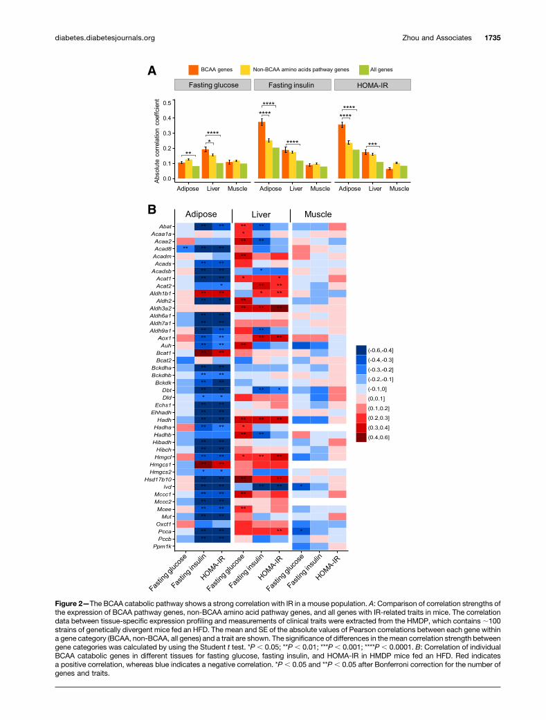
Figure 2—The BCAA catabolic pathway shows a strong correlation with IR in a mouse population. A: Comparison of correlation strengths ofthe expression of BCAA pathway genes, non-BCAA amino acid pathway genes, and all genes with IR-related traits in mice. The correlationdata between tissue-specific expression profiling and measurements of clinical traits were extracted from the HMDP, which contains;100strains of genetically divergent mice fed an HFD. The mean and SE of the absolute values of Pearson correlations between each gene withina gene category (BCAA, non-BCAA, all genes) and a trait are shown. The significance of differences in themean correlation strength betweengene categories was calculated by using the Student t test. *P , 0.05; **P , 0.01; ***P , 0.001; ****P , 0.0001. B: Correlation of individualBCAA catabolic genes in different tissues for fasting glucose, fasting insulin, and HOMA-IR in HMDP mice fed an HFD. Red indicatesa positive correlation, whereas blue indicates a negative correlation. *P , 0.05 and **P , 0.05 after Bonferroni correction for the number ofgenes and traits.
diabetes.diabetesjournals.org Zhou and Associates 1735
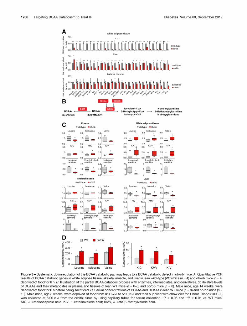
Figure 3—Systematic downregulation of the BCAA catabolic pathway leads to a BCAA catabolic defect in ob/obmice. A: Quantitative PCRresults of BCAA catabolic genes in white adipose tissue, skeletal muscle, and liver in lean wild-type (WT) mice (n = 4) and ob/obmice (n = 4)deprived of food for 6 h. B: Illustration of the partial BCAA catabolic process with enzymes, intermediates, and derivatives. C: Relative levelsof BCAAs and their metabolites in plasma and tissues of lean WT mice (n = 6–8) and ob/ob mice (n = 8). Male mice, age 14 weeks, weredeprived of food for 6 h before being sacrificed. D: Serum concentrations of BCAAs and BCKAs in lean WT mice (n = 8) and ob/obmice (n =10). Male mice, age 8 weeks, were deprived of food from 8:00 A.M. to 5:00 P.M. and then supplied with chow diet for 1 hour. Blood (100 mL)was collected at 6:00 P.M. from the orbital sinus by using capillary tubes for serum collection. *P , 0.05 and **P , 0.01 vs. WT mice.ΚΙC, a-ketoisocaproic acid; KIV, a-ketoisovaleric acid; KMV, a-keto-b-methylvaleric acid.
1736 Targeting BCAA Catabolism to Treat IR Diabetes Volume 68, September 2019

increases are usually moderate, however, and they becomesmaller or even disappear under fasting conditions(6,44,45). Our data show that, in fed animals, plasmaBCAA levels are significantly higher in ob/ob obese micethan in lean mice (Fig. 3D), and the plasma BCKA levelswere even higher in ob/ob obese mice (Fig. 3C and D). Themagnitudes of BCKA elevations were more pronouncedthan those of BCAAs in different settings, reflecting BCKDdeficiency.
Therefore, although BCAA catabolic genes are sup-pressed at the pathway level in various tissues, systemicBCAA catabolism in obese ob/ob mice is characterized bya deficiency of the rate-limiting enzyme BCKD, accompa-nied by reduced catabolic flux and by BCAA/BCKA accu-mulation. Diminished BCKD protein expression andincreased BCKD E1a phosphorylation further supportBCKD deficiency in obese ob/ob mice (SupplementaryFig. 5), in agreement with previous studies (8,9,45–49).
A BCAA Catabolic Defect Contributes to thePathogenesis of IR in ob/ob MiceBCKDK phosphorylates the E1a subunit of BCKD andinhibits its activity (2). BT2 is a specific and potent in-hibitor of BCKDK (39,40). We next assessed whether theBCAA catabolic defect played a causal role in obesity-associated IR by using BT2 to restore BCAA catabolismin obese mice. BT2 administration significantly enhancedBCKD activities in various tissues of ob/ob mice, includingliver, skeletal muscle, and white adipose tissue (Fig. 4A).BT2 treatment significantly reduced plasma BCAA levels inthe obese ob/ob mice, but the magnitudes of reductions inplasma BCKA concentrations were far more pronounced(Fig. 4B). Importantly, BT2-treated mice displayed signif-icantly improved glucose tolerance and insulin sensitivity(Fig. 4C and D), accompanied by the attenuation of hyper-insulinemia (Fig. 4E). BT2 treatment reduced the phos-phorylation of BCKD E1a in liver and induced theexpression of BCKD E1a and BCKDK protein in skeletalmuscle (Supplementary Fig. 6). BT2 also increased theexpression of numerous distal genes in the BCAA catabolicpathway in skeletal muscle and adipose tissue (Supple-mentary Fig. 7). Among the 17 amino acids that are notBCAAs, tryptophan was the only one to show a reducedconcentration in plasma in the BT2-treated mice (Supple-mentary Fig. 8); this reduction is potentially due to in-creased tryptophan transport into tissues when BCAAlevels are reduced (21). BT2 treatment did not affectbody weight or food intake in the ob/ob mice (Fig. 4Fand G). These results strongly support a causal role for theBCAA catabolic defect in the onset of IR in obese ob/obmice.
Elevated BCAAs and BCKAs Are Pathogenic Factorsfor IR in ob/ob MiceThe BCAA catabolic defect results in BCAA/BCKA accu-mulation and impaired BCAA catabolic flux. BCAA canenhance mammalian target of rapamycin complex
1 (mTORC1) activity, and the catabolic flux may affectglucose and lipid oxidation, both of which have beenimplicated as possible contributors to IR (2,50). Weshowed that BT2 treatment reduced BCAA levels whileenhancing catabolic flux. To assess further which of thesetwo changes contributes to the improved insulin sensitiv-ity in ob/ob mice, we examined the influences of variousdietary manipulations that alter BCAA abundance and thecatabolic flux in different ways. We used an isocaloric LPD(6% protein by weight), instead of an NPD (20% protein byweight), to reduce BCAA intake and consequently the loadto the catabolic flux. The LPD significantly reduced theplasma levels of BCAAs and BCKAs (Fig. 5A) and markedlyimproved systemic glucose tolerance (Fig. 5B). In contrast,increasing dietary intake of BCAAs by administering themin water increased plasma levels of BCAAs/BCKAs (Fig. 5A)and significantly exacerbated the systemic glucose toler-ance of ob/obmice being fed the LPD (Fig. 5B). The insulintolerance test showed that the LPD dramatically improvedsystemic insulin sensitivity in ob/ob mice, whereas BCAAsupplementation significantly impaired insulin sensitivityin mice fed the LPD (Fig. 5C). The hyperinsulinemia inob/ob mice was significantly attenuated by the LPD butaggravated by BCAA supplementation (Fig. 5D). Fastingplasma glucose levels were not affected by the LPD orBCAA supplementation (Fig. 5B). The body weight of ob/obmice was slightly reduced by the LPD, whereas BCAAsupplementation did not affect food intake or body weight(Fig. 5E and Supplementary Fig. 9). The LPD significantlyreduced plasma levels of multiple amino acids; BCAAs werethe only ones reversed by BCAA supplementation (Sup-plementary Fig. 10). These results show that the BT2treatment and reducing BCAA intake both attenuate IRthrough a smaller abundance of BCAAs and BCKAs. On theother hand, BT2 treatment enhanced the BCAA catabolicflux, whereas reducing BCAA intake probably decreasedthe flux. In addition, increasing BCAA intake promoted IRwith more BCAAs/BCKAs and higher catabolic flux. Col-lectively, all these data are consistent with the notion thatmore BCAAs/BCKAs, rather than the status of catabolicflux, plays a key role in the onset of IR in obese ob/obmice.
Both BCAAs and BCKAs Can Impair Insulin SignalingWe next examined insulin signaling in food-deprived micein the aforementioned experimental settings. Comparedwith the response (indicated by the fold induction ofpAKT-Thr308) in ob/obmice fed the NPD, insulin signalingin ob/ob mice fed the LPD was significantly higher inskeletal muscle (7.6 vs. 1.34 fold), liver (2.55 vs. 1.31fold), and white adipose tissue (2.61 vs. 1.37 fold), in-dicating enhanced insulin sensitivity (Fig. 6A). In contrast,BCAA supplementation impaired insulin response in liver,white adipose tissue, and, to a much lesser extent, skeletalmuscle in ob/ob mice fed the LPD (Fig. 6B). Similar to theLPD, BT2 treatment significantly enhanced AKT signalingin response to insulin, indicated by the higher fold in-duction of pAKT-Thr308 in various tissues (Fig. 6C). The
diabetes.diabetesjournals.org Zhou and Associates 1737

Figure 4—The BCKDK inhibitor restores BCAA catabolism and attenuates IR in ob/obmice. ob/obmice were treated with the vehicle (Ctrl) orBT2 (at 10 weeks [A,B, and E] or 4–6weeks [C andD]) by oral gavage (n = 6mice in each group). We analyzed BCKD activity in various tissues(A), fasting plasma levels of BCAAs (B, left) and BCKAs (B, right), glucose tolerance test results (C ), insulin tolerance test results (D), fastingplasma insulin level (E), bodyweight (F ), and food intake (G). *P, 0.05; **P, 0.01; ***P, 0.005; ****P, 0.0001 vs. Ctrl.ΚΙC, a-ketoisocaproicacid; KIV, a-ketoisovaleric acid; KMV, a-keto-b-methylvaleric acid; WAT, white adipose tissue.
1738 Targeting BCAA Catabolism to Treat IR Diabetes Volume 68, September 2019

enhanced response to insulin in the LPD and BT2 groupswas primarily attributable to the lower basal AKT activity,consistent with the lower fasting insulin levels in the samegroups (Figs. 4E and 5D).
BCAA-enhanced activation of mTORC1 has been impli-cated as an important mechanism underlying the impairmentof insulin signaling (1). The LPD and BT2 treatmentreduced the basal or insulin-stimulated mTORC1 activity(as indicated by p70S6K phosphorylation on Thr389), orboth, in all three tissues, consistent with an enhancedresponse to insulin (Fig. 6A and C) and reduced BCAAlevels (Figs. 4B and 5A). On the other hand, BCAA sup-plementation increased the amount of BCAAs in ob/obmice fed the LPD (Fig. 5A) and enhanced the insulin-stimulated mTORC1 activity in skeletal muscle and probablyadipose tissue (Fig. 6B). Of note, BCAA supplementationimpaired insulin signaling without enhancing mTORC1activity in liver in ob/ob mice fed the LPD.
As the abundances of BCKAs and BCAAs changed con-cordantly (Figs. 4B and 5A), we also examined the directinfluence of BCKAs on insulin signaling in cultured cells.Interestingly, similar to BCAAs, BCKAs impaired insulinsignaling while increasing insulin-stimulated mTORC1 ac-tivity (Fig. 6D). Further examination showed that BCKAssuppressed basal AKT phosphorylation while augmentingmTORC1 activity in the absence of BCAAs in culturemedium (Fig. 6E). The mTORC1 inhibitor rapamycin abol-ished BCKA-repressed AKT phosphorylation, indicating anmTORC1-dependent action (Fig. 6F). These data suggestthat BCKAs, in addition to BCAAs, may also contribute toimpaired insulin signaling in obese ob/ob mice.
The Pharmacological Inhibitor of BCKDK Attenuates IRin DIO MiceGiven the importance of the BCAA catabolic defect in thepathogenesis of obesity-associated IR, enhancing BCKDactivity through small-molecule treatment may representa new pharmacological approach to treating diabetes. Weassessed the effect of the BCKDK inhibitor BT2 on insulinsensitivity in wild-type mice with DIO, a model frequentlyused to mimic the metabolic impairments of obesity inhumans. As expected, BT2 treatment augmented BCKDactivities in various tissues of DIO mice and reducedplasma BCAA and BCKA levels (Fig. 7A and B). Moreimportantly, BT2 treatment improved glucose tolerance,insulin sensitivity, and hyperinsulinemia in DIO mice (Fig.7C–E) without affecting body weight (Fig. 7F). Theseresults demonstrate that restoring BCAA catabolic activitywith a small-molecule BCKDK inhibitor is effective toimprove insulin sensitivity in DIO mice. This in vivoproof-of-concept demonstration validates targeting theBCAA catabolic defect as a potential therapeutic approachto attenuating obesity-associated IR and diabetes.
DISCUSSION
This study examines the causal role of disrupted BCAAhomeostasis in obesity-associated IR and the therapeuticpotential of targeting the elevated amounts of BCAAs andBCKAs. The unbiased integrative genomic analyses reveala specific and robust association between the BCAA cat-abolic pathway and obesity-related IR in both human and
Figure 5—Altering BCAA intake affects IR in ob/obmice. ob/obmicewere fed an NPD (20% protein by weight) or an LPD (6% protein byweight) for 4 weeks beginning at 10 weeks of age. For the BCAAgroup (LPD + BCAA), supplementation of BCAAs in drinking water(3 mg/mL) was started after mice had eaten the LPD for 2 weeks andlasted 2 weeks. We analyzed fasting plasma levels of BCAAs andmetabolites (n = 8 mice; *P , 0.05 vs. NPD; &P , 0.05 vs. LPD) (A),glucose tolerance test results (n 5 6–13 mice; **P , 0.01, LPD vs.NPD; &P , 0.05, LPD vs. LPD 1 BCAA) (B), insulin tolerance testresults (n 5 6 mice in each group; *P , 0.05, ***P , 0.001, LPD vs.NPD; &P , 0.05, LPD vs. LPD 1 BCAA) (C), fasting plasma insulinlevels (n = 8–12mice; *P, 0.05; &P, 0.05) (D), and body weight (n =14 mice in each group; **P , 0.01).
diabetes.diabetesjournals.org Zhou and Associates 1739

Figure 6—BCAAs and BCKAs contribute to impaired insulin signaling in ob/obmice. A–C: Representative immunoblots for specific proteins,created by using tissue lysates from skeletal muscle, liver, and white adipose tissue of mice without or with insulin (Ins) injection for 10 minfollowing 6 h of food deprivation. ob/ob mice were fed an NPD (20% protein by weight) or an LPD (6% protein by weight) for 4 weeksbeginning at 10 weeks of age (A). BCAA supplementation (LPD + BCAA or LPD/amino acids) in drinking water (3 mg/mL) was started aftermice had consumed the LPD for 2 weeks and lasted 2 weeks (B). ob/ob mice were treated with the vehicle (Ctrl) or BT2 by oral gavage for5 weeks (C). The graphs below the immunoblots in A–C present densitometric values of the bands. D–F: Representative immunoblots forspecific proteins, created by using cell lysates. 3T3-L1 cells were treated with FBS- and BCAA-free DMEM for 1–2 h before BCAA (500mmol/L), BCKA (500 mmol/L), or rapamycin (100 nmol/L) treatment for 1 h (D and F ) or various times (E ), followed by insulin treatment (10nmol/L) for 1 h (D). *P , 0.05; **P , 0.01; ***P , 0.005; ****P , 0.0001.
1740 Targeting BCAA Catabolism to Treat IR Diabetes Volume 68, September 2019

mouse populations. In genetically obese mice, characteristicBCKD deficiency and BCAA/BCKA buildup accompany thesuppression of BCAA catabolic genes at the pathway level.Correcting this BCAA catabolic defect abolishes BCAA/BCKA elevation and attenuates IR in obese ob/ob mice,supporting a causal role for the BCAA catabolic defect in IRonset in obese mice. Furthermore, reducing protein (andthus BCAA) intake effectively reduces BCAA/BCKA abun-dances and improves insulin sensitivity, whereas BCAA
supplementation increases BCAA/BCKA abundances andpromotes IR in ob/ob mice. These results together demon-strate the pathogenic role of elevated BCAAs/BCKAs in IR inobese mice. Similar to BCAAs, BCKAs also can impair insulinsignaling. Finally, directly targeting the BCAA catabolicdefect with a small-molecule BCKDK inhibitor reducesBCAA/BCKA abundances and attenuates IR in DIO mice.These findings suggest that the BCAA catabolic defect andelevated BCAAs/BCKAs play causal roles in the onset of
Figure 7—The BCKDK inhibitor enhances BCAA catabolism and attenuates IR in DIO mice. DIO mice were treated with the vehicle (Ctrl) orBT2 by oral gavage for 8 weeks (n = 7 mice in each group). We analyzed BCKD activity in various tissues (A), fasting plasma levels of BCAAs(B, left) and BCKAs (B, right), glucose tolerance test results (C ), insulin tolerance test results (D), fasting plasma insulin level (E), and bodyweight (F ). *P, 0.05, **P, 0.01, ***P, 0.005, ****P, 0.0001, all vs. Ctrl.ΚΙC, a-ketoisocaproic acid; KIV,a-ketoisovaleric acid; KMV,a-keto-b-methylvaleric acid.
diabetes.diabetesjournals.org Zhou and Associates 1741

obesity-associated IR and provide a valid target for dietaryor pharmacological interventions to treat diabetes.
Several studies have established genetic links betweenindividual BCAA catabolic genes (BCKHA or PP2Cm) anddysfunctional glucose metabolism (9,22–25). Instead ofindividual genes, the comprehensive genomic analyses inthis study identified a genetic link between IR and theentire BCAA catabolic pathway. The significant and uniquegenetic association is based on information about bothgenetics and gene expression. The latter is of particularinterest because nutritional status in obesity profoundlyaffects BCAA catabolic gene expression. Numerous studieshave shown aberrant BCAA catabolic gene expression inanimals and humans that are obese and have diabetes(4,8,9,45–49). Interestingly, a study of monozygotic twinsdiscordant for BMI, which aimed to profile obesity-alteredbiological pathways in identical genetic backgrounds,revealed significant downregulation of the BCAA adiposecatabolic pathway in obese cotwins, which correlatedclosely with IR and hyperinsulinemia (46). Thus, BCAAcatabolism can be impaired by obesity status and linkedwith IR. In the bioinformatics study, we performed bothBMI-adjusted and BMI-unadjusted analyses to look forIR-associated pathways that are independent of BMI(based on BMI-adjusted analysis) and that can be depen-dent on or independent of BMI (based on unadjustedanalysis). That the genetic association between theBCAA catabolic pathway and IR is mainly observed inthe unadjusted analysis and disappeared in the BMI-adjusted analysis strongly supports an obesity-dependentrelationship. Our study, together with other data, indicatesthat BCAA catabolism is suppressed in obesity, which con-tributes to IR.
The special link between the adipose BCAA catabolicpathway and IR is intriguing. Studies in humans androdents have indicated that an impaired adipose BCAAcatabolic pathway contributes to the rise of plasma BCAAs/BCKAs and is associated with IR in obesity (4,8,9,45–49).In this study, the genetic association between the BCAAcatabolic pathway and obesity-associated IR is mainlyobserved when gene expression in adipose tissue is con-sidered in the genomics analyses. In ob/ob mice, adiposetissue shows the most dramatic loss of BCAA catabolicgenes and the most significant accumulation of intratissueBCAAs and BCKAs, when compared with the liver andskeletal muscle. Therefore, BCAA catabolic activity inadipose tissue seems to play a unique role in regulatingsystemic BCAA homeostasis and IR in obesity. However, itremains unclear how adipose tissue contributes signifi-cantly to systemic BCAA catabolism despite its relativelylow BCKD activity. It is possible that BCAA cataboliccapacity of tissue is determined not only by BCKD activitybut also by other factors such as BCAA/BCKA uptake fromblood by tissues. On the other hand, as a main contributorto obesity-associated IR, an adipose BCAA catabolic defectmay exert its effect locally or systemically. The localaccumulation of BCAA/BCKA may trigger functional
remodeling in adipose tissue. As adipose tissue playsa critical role in systemic lipid and glycemic metabolism,changes in adipose physiology may in turn contribute tolocal and systemic IR in obese mice. It is also possible,however, that an adipose BCAA catabolic defect functionsas a main contributor to the systemic BCAA/BCKA accu-mulation that in turn promotes IR in various tissues. Ineither case, BT2 can enhance BCAA catabolism in heart,liver, kidney, and other tissues, relieving the systemic andadipose BCAA catabolic defect, irrespective of the site, andthus improving systemic insulin sensitivity; thus, BT2should be considered for therapy.
Hepatic BCAA metabolism demonstrates weak and in-consistent correlations with IR. Directional discrepanciesof correlations are observed between liver BCAA catabolicgenes and fasting insulin level and IR traits. Some BCAAcatabolic genes in liver are positively correlated withfasting glucose levels. The molecular details and functionalimplications for this discrepancy are unclear. We speculatethat some positive correlations between BCAA genes inliver and glycemic traits in the HMDP study are likelya reactive rather than upstream causal process in thedevelopment of hyperglycemia or IR. Indeed, it is notuncommon to observe directional discrepancies in gene-trait correlations in various tissues and experimental set-tings because of feedback regulations and systems-levelinteractions between tissues. Meanwhile, the key enzymesdetermining the BCAA metabolic pattern are either neg-atively (DBT) or not significantly (BCAT2, BCKHDA,BCKDHB, BCKDK) associated with glycemic traits. Inaddition, our human genetics analysis does not supportan enrichment of genetic signals in liver BCAA modules forany of the glycemic traits examined. Therefore, although itis possible that the BCAA catabolic pathway in liver alsoplays a role in glycemic regulation, the genetic evidence isnot as strong as that for a role in adipose tissues andrequires further investigation.
A high level of BCAAs in blood has been viewed asa metabolic hallmark of IR and diabetes in numerousstudies (2,4–9). Thus, it was unexpected that the fastingplasma BCAA levels were not elevated in ob/ob mice.Plasma BCAA abundances were higher in fed obeseob/ob mice, suggesting that they are strongly affected byboth the dietary intake and degradation in tissues. Sim-ilarly, it has been shown that BCAA levels are significantlyincreased in fed obese mice, although the differencesdramatically decline or even disappear when the miceare deprived of food (45). On the other hand, the eleva-tions of plasma BCKAs are significant in both food-deprived and fed obese ob/ob mice, in agreement withprevious studies indicating higher plasma BCKA as a betterbiomarker for diabetes than BCAA (8,9). In addition,BCKAs are catabolites generated by tissues. ElevatedBCKA levels in blood reflect a BCKD deficiency in tissuesof obese mice.
BCAAs can trigger different and even opposite meta-bolic effects in mammals, depending on their catabolic and
1742 Targeting BCAA Catabolism to Treat IR Diabetes Volume 68, September 2019
anabolic states (51). Thus, a causal relationship betweendisrupted BCAA metabolism and obesity-associated IR canbe addressed only in animals that are already obese. Pre-vious studies examining the effects of BCAA intake onmetabolism in obese animals have fallen short of establish-ing this causal relationship. In obese New Zealand mice,reducing protein intake significantly improved glucosehomeostasis, whereas BCAA repletion did not reverse it(52). In a mouse model of preexisting DIO, reducingdietary BCAAs improved glucoregulatory control whilereducing body weight (53). In this study, however, thecausal role of BCAAs remains unclear, because its impacton glucose regulation could be attributed to changes inbody weight rather than BCAAs. In fatty Zucker rats, BCAArestriction showed no impact on systemic glucose toler-ance but improved skeletal muscle insulin sensitivity (7).Similarly, we found that a low-BCAA diet did not affectsystemic insulin sensitivity in ob/ob mice (SupplementaryFig. 11). However, reducing protein intake significantlyimproved systemic glucose tolerance in ob/ob mice. Re-ducing protein intake reduced the uptake of all amino acidsand the plasma levels of eight amino acids, includingBCAAs, phenylalanine, tryptophan, glutamine, tyrosine,and proline (Supplementary Fig. 10). Increasing BCAAintake in mice fed the LPD restored plasma BCAA levelsand significantly impaired systemic insulin sensitivity,specifying the causal role of BCAAs. The impacts ofnon-BCAA amino acids affected by the LPD remain tobe tested, whereas phenylalanine and tyrosine have beenassociated with IR (1). BCAA supplementation did notreduce glucose tolerance to the level of that in the NPDgroup, suggesting that the systemic insulin-sensitizingeffect of BCAA restriction in obese ob/ob mice may needassistance from other amino acids. Importantly, BCAAsupplementation did not change the body weight ofob/ob mice fed the LPD. Compared with the total energyintake (;94% from carbohydrate and fat in the LPD), thecaloric intake from BCAAs is likely very small. Thus, thecaloric contribution from BCAA oxidation is unlikely toaccount for IR pathogenesis. Together with results show-ing that BT2 attenuated IR while enhancing BCAA oxida-tion without affecting food intake and body weight in bothobese ob/ob and DIOmice (Figs. 4 and 7), these data clearlydemonstrate the causal role of a BCAA catabolic defect inobesity-associated IR. On the other hand, although theeffects of BT2 on ob/ob mice are similar to those recentlyreported in a genetic obese rat model (54) (Fig. 4), thebeneficial impacts of BT2 in DIO mice (Fig. 7) add to theclinical relevance of our findings, as the DIO rodentsremain the most clinically relevant model by mimickingthe common cause and metabolic impairments of obesityin humans.
It has been suggested that, because of reduced catab-olism by adipose tissue in obese mice, BCAA catabolism isenhanced in liver and skeletal muscle, which “clogs” lipidand glucose oxidation and promotes IR (50). A more recentstudy showed that obese and insulin-resistant mice shunt
BCAA oxidation from the liver and fat toward muscle (55).In this study, BT2 reduced BCAA/BCKA abundances andimproved insulin sensitivity while enhancing BCAA cata-bolic activities in various tissues including liver and skele-tal muscle in both obese ob/ob and DIO mice. On the otherhand, the LPD attenuated IR and BCAA/BCKA abundanceswhile probably reducing BCAA catabolism through lower-ing BCAA load to the catabolic pathway in ob/ob mice.Furthermore, increasing BCAA intake promoted IR whileelevating BCAA/BCKA abundances and probably the cat-abolic flux. Therefore, IR is consistently attenuated byreducing BCAA/BCKA abundances, rather than alteringthe status of catabolic flux, in obese mice. One recentindependent study reported similar beneficial effects ofBT2 on insulin sensitivity in genetically obese rats (54).Other reports have suggested that reducing BCAA intakeimproves insulin sensitivity via various mechanisms in-cluding reducing body weight or increasing energy expen-diture (7,52,53). The common feature of these studies isthe reduced abundances of BCAAs (and probably ofBCKAs), corroborating the pathogenic roles of elevatedBCAAs and BCKAs in obesity-associated IR.
BCAA-stimulated mTORC1 activation has been widelyimplicated in reducing insulin signaling (2,7,56–58). Ourdata show that BCKAs are also capable of suppressinginsulin signaling. Thus, in addition to BCAAs per se, othercomponents of the BCAA catabolic pathway may also playimportant roles in obesity-associated IR. Furthermore, ourdata suggest that mTORC1 probably mediates the sup-pression of AKT phosphorylation by BCAAs and BCKAs.However, disconnections have been observed betweenenhanced mTORC1 and impaired insulin signaling. Forexample, BCAA supplementation impaired insulin signal-ing without enhancing mTORC1 activity in liver in ob/obmice fed the LPD. The tissue-specific impacts of manipu-lations of insulin signaling and mTORC1 activity by BCAAalso remain puzzling. A recent study showed that BCAAsbecoming replete while dietary protein is reduced restoresmTORC1 signaling but does not induce IR in obese NewZealand mice (52). Thus, mTORC1 activation cannot fullyexplain the effects of modulating BCAA catabolism oninsulin sensitivity, and additional mTORC1-independentmechanisms warrant further investigation.
The roles of BCAA catabolic flux and the distal steps ofBCAA catabolism in obesity-associated IR remain to befurther investigated. In addition to the “clogging” model,recent studies have indicated that elevated BCAA oxidationin skeletal muscle in obese db/db mice may lead to theproduction of 3-hydroxyisobutyrate from valine and pro-mote IR (55,57). In contrast, another report suggested thatdefective BCAA metabolism in muscle contributes to im-paired lipid metabolism and thus the development ofobesity-associated IR (59). BT2 treatment enhancesBCAA catabolism in muscle in healthy mice (55), obeserats (54), and obese mice (Figs. 4 and 7), while attenuatingIR in obese animals, supporting the detrimental effects ofimpaired BCAA oxidation in muscle of obese animals.
diabetes.diabetesjournals.org Zhou and Associates 1743

Interestingly, BT2 treatment increased BCKD activity(Figs. 4A and 7A) without reducing its phosphorylationin skeletal muscle of obese ob/obmice (Supplementary Fig.6) and obese rats (54). But BT2 increases the expression ofBCKD subunits (Supplementary Fig. 6) and multiple distalgenes in the BCAA catabolic pathway (Supplementary Fig.7), which may contribute to enhanced BCAA catabolic flux.Quantitative measurements of BCAA catabolic flux atvarious steps in tissues of obese animals upon BT2 treat-ment or dietary manipulation will shed light on the rolesof BCAA catabolic flux and the distal steps in obesity-associated IR.
Tissue-specific patterns of BCAA catabolism andresponses to BT2 treatment have been observed in variousexperimental settings. In the bioinformatics analyses, theBCAA catabolic pathway in skeletal muscle showed a muchweaker association with IR than that in adipose tissue andliver. BCAA catabolic gene expression and phosphorylationpatterns vary dramatically in various tissues of ob/obmice.BCAA abundances are elevated in adipose tissue, un-changed in skeletal muscle, and reduced in liver in ob/obmice. Furthermore, BT2 treatment and dietary approachesalter insulin signaling differently in liver, skeletal muscle,and adipose tissue. Interestingly, BT2 treatment affectsBCKD activity, BCKD phosphorylation, BCKD subunits,and distal gene expression in a tissue-specific pattern. Tomake it even more complicated, one recent study showedthat acute BT2 treatment in healthy mice activates BCAAoxidation specifically in muscle even though BCKD phos-phorylation is significantly reduced in liver, skeletal mus-cle, and heart (55). These intriguing patterns of BCAAcatabolism and the impacts of BT2 may be attributed tothe tissue-specific expression of BCAA catabolic enzymesand the pharmacokinetics of BT2. For example, althoughBCKDK expression is high in liver, skeletal muscle, andadipose tissue, BCKD phosphatase (PP2Cm) expression islower in skeletal muscle and adipose tissue than in theliver. The strong dephosphorylation of BCKD by BT2 inliver might be the result of both BCKDK inhibition andPP2Cm activation. Nevertheless, despite these tissue-specific patterns of BCAA catabolism and BT2 effects,systemic BCAA catabolism is defective in obese mice butcan be relieved by BT2 treatment, irrespective of the site,in order to improve systemic insulin sensitivity. Mean-while, more mechanistic details remain to be determined.
BT2 is a potent allosteric inhibitor of BCKDK. BCKDKand pyruvate dehydrogenase kinases (PDKs) share similarstructures. PDKs phosphorylate and inhibit the pyruvatedehydrogenase (PDH) complex, a key player in glucosemetabolism. However, the allosteric ligand-binding pocketin BCKDK (volume 412Å3) is two- to fourfold larger thanits counterparts in PDKs (90Å3–213Å3) (SupplementaryFig. 12). The larger allosteric ligand-binding volume inBCKDK allows for the occupancy of natural inhibitors likeBCKAs or the artificial inhibitor BT2. By comparison, theallosteric sites in PDKs can accommodate up to 3-carbona-keto acids such as pyruvate and its small-molecule
inhibitor dichloroacetate (Supplementary Fig. 12). Thestructural constraints that preclude BT2 binding toPDKs are reflected by the absence of PDK inhibition and PDHactivation by BT2 (data not shown). Therefore, the metabolicphenotypes resulting from BT2 treatment are unlikely tobe related to PDK-regulated PDH activity; rather, they areexerted via the reprogramming of BCAA catabolism.
In summary, this study provides a compelling mecha-nistic basis for the strong association between elevatedplasma BCAAs and the onset of IR in humans, demonstrat-ing the causal role of a BCAA catabolic defect and thepathogenic role of elevated BCAAs/BCKAs in obesity-associated IR. The influences of BCAA intake on insulinsensitivity indicate the potential impact of dietary proteinon IR and diabetes in obese people, consistent with a recentreport showing that protein restriction improves glycemiccontrol in overweight and mildly obese humans (60).BCKDK and its inhibitor represent new pharmacologicaltargets and an approach to attenuating obesity-associatedIR. Finally, the genetic link between IR and the BCAAcatabolic pathway in human populations highlights theclinical significance of BCAA catabolism as a potential ther-apeutic target in treating obesity-associated IR and diabetes.
Funding. This work was supported by the Ministry of Science and Technologyof China (grant nos. 2012BAI02B05 and 2013YQ030923), the National Interna-tional Science Cooperation Foundation (grant no. 2015DFA30560), the NationalNatural Science Foundation of China (grant nos. NSFC81570717 and 81522011),the National Institutes of Health (grant nos. HL108186, HL103205, HL098954, andDK62306), the National Heart, Lung, and Blood Institute (grant no. HL080111), theLaubisch Fund (to the University of California Los Angeles), the Welch Foundation(grant no. I-1286), and the Science and Technology Commission of ShanghaiMunicipality (grant nos. 13ZR1423300 and 16JC1404400). L.S. is supported bya China Scholarship Council scholarship, a UCLA Eureka and Hyde scholarship,and a Burroughs Wellcome Fund fellowship. X.Y. is supported by the NationalInstitutes of Health/National Institute of Diabetes and Digestive and KidneyDiseases (grant no. R01DK104363), the American Heart Association (grant no.13SDG17290032), an American Heart Association Cardiovascular Genome-Phenome Study Pathway Grant, and a Leducq Foundation Transatlantic Networksof Excellence Grant.Duality of Interest. Y.W. and H.S. participated in an advisory board forRamino Bio Ltd. No other potential conflicts of interest relevant to this article werereported.Author Contributions. M.Z., J.S., C.-Y.W., L.S., W.D., Y.L., M.C., R.M.W.,Jiq.W., Ji W., and W.-J.G. performed the research. M.Z., J.S., C.-Y.W., L.S., W.D.,Y.L., M.C., R.M.W., Jiq.W., Ji W., W.-J.G., X.Q., A.J.L., Z.L., W.W., G.N., X.Y.,D.T.C., Y.W., and H.S. prepared the manuscript. M.Z., C.-Y.W., L.S., W.D., Y.W.,and H.S. analyzed the data. X.Q. performed the chemical design and synthesis.A.J.L., Z.L., W.W., G.N., X.Y., D.T.C., and Y.W. designed the overall study andanalyzed the data. H.S. designed the research. H.S. is the guarantor of this workand, as such, had full access to all the data in the study and takes responsibilityfor the integrity of the data and the accuracy of the data analysis.
References1. Newgard CB, An J, Bain JR, et al. A branched-chain amino acid-relatedmetabolic signature that differentiates obese and lean humans and contributes toinsulin resistance. Cell Metab 2009;9:311–3262. Lynch CJ, Adams SH. Branched-chain amino acids in metabolic signallingand insulin resistance. Nat Rev Endocrinol 2014;10:723–736
1744 Targeting BCAA Catabolism to Treat IR Diabetes Volume 68, September 2019
3. Felig P, Marliss E, Cahill GFJ Jr. Plasma amino acid levels and insulinsecretion in obesity. N Engl J Med 1969;281:811–8164. Lu J, Xie G, Jia W, Jia W. Insulin resistance and the metabolism of branched-chain amino acids. Front Med 2013;7:53–595. Lu Y, Wang Y, Ong C-N, et al. Metabolic signatures and risk of type 2 diabetesin a Chinese population: an untargeted metabolomics study using both LC-MS andGC-MS. Diabetologia 2016;59:2349–23596. Chen T, Ni Y, Ma X, et al. Branched-chain and aromatic amino acid profilesand diabetes risk in Chinese populations. Sci Rep 2016;6:205947. White PJ, Lapworth AL, An J, et al. Branched-chain amino acid restriction inZucker-fatty rats improves muscle insulin sensitivity by enhancing efficiency offatty acid oxidation and acyl-glycine export. Mol Metab 2016;5:538–5518. She P, Olson KC, Kadota Y, et al. Leucine and protein metabolism in obeseZucker rats. PLoS One 2013;8:e594439. Menni C, Fauman E, Erte I, et al. Biomarkers for type 2 diabetes and impairedfasting glucose using a nontargeted metabolomics approach. Diabetes 2013;62:4270–427610. Wang TJ, Larson MG, Vasan RS, et al. Metabolite profiles and the risk ofdeveloping diabetes. Nat Med 2011;17:448–45311. Yu D, Moore SC, Matthews CE, et al. Plasma metabolomic profiles in as-sociation with type 2 diabetes risk and prevalence in Chinese adults. Metabolomics2016;12. pii: 312. Würtz P, Soininen P, Kangas AJ, et al. Branched-chain and aromatic amino acidsare predictors of insulin resistance in young adults. Diabetes Care 2013;36:648–65513. Floegel A, Stefan N, Yu Z, et al. Identification of serummetabolites associatedwith risk of type 2 diabetes using a targeted metabolomic approach. Diabetes2013;62:639–64814. Melnik BC. Leucine signaling in the pathogenesis of type 2 diabetes andobesity. World J Diabetes 2012;3:38–5315. Shah SH, Crosslin DR, Haynes CS, et al. Branched-chain amino acid levelsare associated with improvement in insulin resistance with weight loss. Dia-betologia 2012;55:321–33016. Laferrère B, Reilly D, Arias S, et al. Differential metabolic impact of gastricbypass surgery versus dietary intervention in obese diabetic subjects despiteidentical weight loss. Sci Transl Med 2011;3:80re217. Hutson SM, Sweatt AJ, Lanoue KF. Branched-chain [corrected] amino acidmetabolism: implications for establishing safe intakes. J Nutr 2005;135(Suppl.):1557S–1564S18. Lu G, Sun H, She P, et al. Protein phosphatase 2Cm is a critical regulator ofbranched-chain amino acid catabolism in mice and cultured cells. J Clin Invest2009;119:1678–168719. Burrage LC, Nagamani SC, Campeau PM, Lee BH. Branched-chain aminoacid metabolism: from rare Mendelian diseases to more common disorders. HumMol Genet 2014;23:R1–R820. Oyarzabal A, Martínez-Pardo M, Merinero B, et al. A novel regulatory defect inthe branched-chain a-keto acid dehydrogenase complex due to a mutation in thePPM1K gene causes a mild variant phenotype of maple syrup urine disease. HumMutat 2013;34:355–36221. Zinnanti WJ, Lazovic J, Griffin K, et al. Dual mechanism of brain injury andnovel treatment strategy in maple syrup urine disease. Brain 2009;132:903–91822. Tiffin N, Adie E, Turner F, et al. Computational disease gene identification:a concert of methods prioritizes type 2 diabetes and obesity candidate genes.Nucleic Acids Res 2006;34:3067–308123. Taneera J, Lang S, Sharma A, et al. A systems genetics approach identifies genesand pathways for type 2 diabetes in human islets. Cell Metab 2012;16:122–13424. Xu M, Qi Q, Liang J, et al. Genetic determinant for amino acid metabolites andchanges in body weight and insulin resistance in response to weight-loss diets: thePreventing Overweight Using Novel Dietary Strategies (POUNDS LOST) trial.Circulation 2013;127:1283–128925. Goni L, Qi L, Cuervo M, et al. Effect of the interaction between diet com-position and the PPM1K genetic variant on insulin resistance and b cell functionmarkers during weight loss: results from the Nutrient Gene Interactions in Human
Obesity: implications for dietary guidelines (NUGENOB) randomized trial. Am J ClinNutr 2017;106:902–90826. Dupuis J, Langenberg C, Prokopenko I, et al.; DIAGRAM Consortium; GIANTConsortium; Global BPgen Consortium; Anders Hamsten on behalf of ProcardisConsortium; MAGIC investigators. New genetic loci implicated in fasting glucosehomeostasis and their impact on type 2 diabetes risk. Nat Genet 2010;42:105–11627. Knowles JW, Xie W, Zhang Z, et al.; RISC (Relationship between InsulinSensitivity and Cardiovascular Disease) Consortium; EUGENE2 (EuropeanNetwork on Functional Genomics of Type 2 Diabetes) Study; GUARDIAN(Genetics UndeRlying DIAbetes in HispaNics) Consortium; SAPPHIRe (StanfordAsian and Pacific Program for Hypertension and Insulin Resistance) Study.Identification and validation of N-acetyltransferase 2 as an insulin sensitivitygene [published correction appears in J Clin Invest 2016;126:403]. J Clin Invest2015;125:1739–175128. Mäkinen VP, Civelek M, Meng Q, et al.; Coronary Artery Disease Genome-Wide Replication and Meta-Analysis (CARDIoGRAM) Consortium. Integrative ge-nomics reveals novel molecular pathways and gene networks for coronary arterydisease. PLoS Genet 2014;10:e100450229. International HapMap Consortium. The International HapMap Project. Nature2003;426:789–79630. 1000 Genomes Project Consortium; Abecasis GR, Auton A, Brooks LD, et al.An integrated map of genetic variation from 1,092 human genomes. Nature 2012;491:56–6531. Arneson D, Bhattacharya A, Shu L, Mäkinen VP, Yang X. Mergeomics: a webserver for identifying pathological pathways, networks, and key regulators viamultidimensional data integration. BMC Genomics 2016;17:72232. Langfelder P, Horvath S. WGCNA: an R package for weighted correlationnetwork analysis. BMC Bioinformatics 2008;9:55933. Kanehisa M, Goto S. KEGG: Kyoto encyclopedia of genes and genomes.Nucleic Acids Res 2000;28:27–3034. Shu L, Zhao Y, Kurt Z, et al. Mergeomics: multidimensional data integration toidentify pathogenic perturbations to biological systems. BMC Genomics 2016;17:87435. Zhu J, Wiener MC, Zhang C, et al. Increasing the power to detect causalassociations by combining genotypic and expression data in segregating pop-ulations. PLoS Comput Biol 2007;3:e6936. Zhu J, Zhang B, Smith EN, et al. Integrating large-scale functional genomicdata to dissect the complexity of yeast regulatory networks. Nat Genet 2008;40:854–86137. Bennett BJ, Farber CR, Orozco L, et al. A high-resolution association mappingpanel for the dissection of complex traits in mice. Genome Res 2010;20:281–29038. Parks BW, Nam E, Org E, et al. Genetic control of obesity and gut microbiotacomposition in response to high-fat, high-sucrose diet in mice. Cell Metab 2013;17:141–15239. Sun H, Olson KC, Gao C, et al. Catabolic defect of branched-chain aminoacids promotes heart failure. Circulation 2016;133:2038–204940. Tso S-C, Gui W-J, Wu C-Y, et al. Benzothiophene carboxylate derivatives asnovel allosteric inhibitors of branched-chain a-ketoacid dehydrogenase kinase. JBiol Chem 2014;289:20583–2059341. Yang X, Zhang B, Molony C, et al. Systematic genetic and genomic analysis ofcytochrome P450 enzyme activities in human liver. Genome Res 2010;20:1020–103642. Wang IM, Zhang B, Yang X, et al. Systems analysis of eleven rodent diseasemodels reveals an inflammatome signature and key drivers. Mol Syst Biol 2012;8:59443. Meng Q, Ying Z, Noble E, et al. Systems nutrigenomics reveals brain genenetworks linking metabolic and brain disorders. EBioMedicine 2016;7:157–16644. Tai ES, Tan MLS, Stevens RD, et al. Insulin resistance is associated witha metabolic profile of altered protein metabolism in Chinese and Asian-Indian men.Diabetologia 2010;53:757–76745. She P, Van Horn C, Reid T, Hutson SM, Cooney RN, Lynch CJ. Obesity-relatedelevations in plasma leucine are associated with alterations in enzymes involved in
diabetes.diabetesjournals.org Zhou and Associates 1745

branched-chain amino acid metabolism. Am J Physiol Endocrinol Metab 2007;293:E1552–E156346. Pietiläinen KH, Naukkarinen J, Rissanen A, et al. Global transcript profiles offat in monozygotic twins discordant for BMI: pathways behind acquired obesity.PLoS Med 2008;5:e5147. Lackey DE, Lynch CJ, Olson KC, et al. Regulation of adipose branched-chainamino acid catabolism enzyme expression and cross-adipose amino acid flux inhuman obesity. Am J Physiol Endocrinol Metab 2013;304:E1175–E118748. Herman MA, She P, Peroni OD, Lynch CJ, Kahn BB. Adipose tissue branchedchain amino acid (BCAA) metabolism modulates circulating BCAA levels. J BiolChem 2010;285:11348–1135649. Zimmerman HA, Olson KC, Chen G, Lynch CJ. Adipose transplant for inbornerrors of branched chain amino acid metabolism in mice. Mol Genet Metab 2013;109:345–35350. Newgard CB. Interplay between lipids and branched-chain amino acids indevelopment of insulin resistance. Cell Metab 2012;15:606–61451. Bifari F, Nisoli E. Branched-chain amino acids differently modulate catabolicand anabolic states in mammals: a pharmacological point of view. Br J Pharmacol2017;174:1366–137752. Maida A, Chan JSK, Sjøberg KA, et al. Repletion of branched chain aminoacids reverses mTORC1 signaling but not improved metabolism during dietaryprotein dilution. Mol Metab 2017;6:873–881
53. Cummings NE, Williams EM, Kasza I, et al. Restoration of metabolic health bydecreased consumption of branched-chain amino acids. J Physiol 2018;596:623–64554. White PJ, McGarrah RW, Grimsrud PA, et al. The BCKDH kinase andphosphatase integrate BCAA and lipid metabolism via regulation of ATP-citratelyase. Cell Metab 2018;27:1281–1293.e755. Neinast MD, Jang C, Hui S, et al. Quantitative analysis of the whole-bodymetabolic fate of branched-chain amino acids. Cell Metab 2019;29:417–429.e456. Xiao F, Huang Z, Li H, et al. Leucine deprivation increases hepatic insulinsensitivity via GCN2/mTOR/S6K1 and AMPK pathways. Diabetes 2011;60:746–75657. Jang C, Oh SF, Wada S, et al. A branched-chain amino acid metabolite drivesvascular fatty acid transport and causes insulin resistance. Nat Med 2016;22:421–42658. Xiao F, Yu J, Guo Y, et al. Effects of individual branched-chain amino acidsdeprivation on insulin sensitivity and glucose metabolism in mice. Metabolism2014;63:841–85059. Lerin C, Goldfine AB, Boes T, et al. Defects in muscle branched-chain aminoacid oxidation contribute to impaired lipid metabolism. Mol Metab 2016;5:926–93660. Fontana L, Cummings NE, Arriola Apelo SI, et al. Decreased consumption ofbranched-chain amino acids improves metabolic health. Cell Rep 2016;16:520–530
1746 Targeting BCAA Catabolism to Treat IR Diabetes Volume 68, September 2019