Targeted next-generation-sequencing for reliable detection ...
9
Zurich Open Repository and Archive University of Zurich Main Library Strickhofstrasse 39 CH-8057 Zurich www.zora.uzh.ch Year: 2018 Targeted next-generation-sequencing for reliable detection of targetable rearrangements in lung adenocarcinoma-a single center retrospective study Velizheva, Nadezda P ; Rechsteiner, Markus P ; Valtcheva, Nadejda ; Freiberger, Sandra N ; Wong, Christine E ; Vrugt, Bart ; Zhong, Qing ; Wagner, Ulrich ; Moch, Holger ; Hillinger, Sven ; Schmitt-Opitz, Isabelle ; Soltermann, Alex ; Wild, Peter J ; Tischler, Verena Abstract: Oncogenic rearrangements leading to targetable gene fusions are well-established cancer driver events in lung adenocarcinoma. Accurate and reliable detection of these gene fusions is crucial to select the appropriate targeted therapy for each patient. We compared the targeted next-generation-sequencing Oncomine Focus Assay (OFA; Thermo Fisher Scientifc) with conventional ALK FISH and anti-Alk im- munohistochemistry in a cohort of 52 lung adenocarcinomas (10 ALK rearranged, 18 non-ALK rearranged, and 24 untested cases). We found a sensitivity and specifcity of 100% for detection of ALK rearrange- ments using the OFA panel. In addition, targeted next generation sequencing allowed us to analyze a set of 23 driver genes in a single assay. Besides EML4-ALK (11/52 cases), we detected EZR-ROS1 (1/52 cases), KIF5B-RET (1/52 cases) and MET-MET (4/52 cases) fusions. All EML4-ALK, EZR-ROS1 and KIF5B-RET fusions were confrmed by multiplexed targeted next generation sequencing assay (Oncomine Solid Tumor Fusion Transcript Kit, Thermo Fisher Scientifc). All cases with EML4-ALK rearrangement were confrmed by Alk immunohistochemistry and all but one by ALK FISH. In our experience, tar- geted next-generation sequencing is a reliable and timesaving tool for multiplexed detection of targetable rearrangements. Therefore, targeted next-generation sequencing represents an effcient alternative to time-consuming single target assays currently used in molecular pathology. DOI: https://doi.org/10.1016/j.prp.2018.02.001 Posted at the Zurich Open Repository and Archive, University of Zurich ZORA URL: https://doi.org/10.5167/uzh-153165 Journal Article Published Version The following work is licensed under a Creative Commons: Attribution 4.0 International (CC BY 4.0) License. Originally published at: Velizheva, Nadezda P; Rechsteiner, Markus P; Valtcheva, Nadejda; Freiberger, Sandra N; Wong, Chris- tine E; Vrugt, Bart; Zhong, Qing; Wagner, Ulrich; Moch, Holger; Hillinger, Sven; Schmitt-Opitz, Isabelle; Soltermann, Alex; Wild, Peter J; Tischler, Verena (2018). Targeted next-generation-sequencing for reli- able detection of targetable rearrangements in lung adenocarcinoma-a single center retrospective study. Pathology, Research and Practice, 214(4):572-578.
Transcript of Targeted next-generation-sequencing for reliable detection ...

Zurich Open Repository andArchiveUniversity of ZurichMain LibraryStrickhofstrasse 39CH-8057 Zurichwww.zora.uzh.ch
Year: 2018
Targeted next-generation-sequencing for reliable detection of targetablerearrangements in lung adenocarcinoma-a single center retrospective study
Velizheva, Nadezda P ; Rechsteiner, Markus P ; Valtcheva, Nadejda ; Freiberger, Sandra N ; Wong,Christine E ; Vrugt, Bart ; Zhong, Qing ; Wagner, Ulrich ; Moch, Holger ; Hillinger, Sven ;
Schmitt-Opitz, Isabelle ; Soltermann, Alex ; Wild, Peter J ; Tischler, Verena
Abstract: Oncogenic rearrangements leading to targetable gene fusions are well-established cancer driverevents in lung adenocarcinoma. Accurate and reliable detection of these gene fusions is crucial to selectthe appropriate targeted therapy for each patient. We compared the targeted next-generation-sequencingOncomine Focus Assay (OFA; Thermo Fisher Scientific) with conventional ALK FISH and anti-Alk im-munohistochemistry in a cohort of 52 lung adenocarcinomas (10 ALK rearranged, 18 non-ALK rearranged,and 24 untested cases). We found a sensitivity and specificity of 100% for detection of ALK rearrange-ments using the OFA panel. In addition, targeted next generation sequencing allowed us to analyze aset of 23 driver genes in a single assay. Besides EML4-ALK (11/52 cases), we detected EZR-ROS1 (1/52cases), KIF5B-RET (1/52 cases) and MET-MET (4/52 cases) fusions. All EML4-ALK, EZR-ROS1 andKIF5B-RET fusions were confirmed by multiplexed targeted next generation sequencing assay (OncomineSolid Tumor Fusion Transcript Kit, Thermo Fisher Scientific). All cases with EML4-ALK rearrangementwere confirmed by Alk immunohistochemistry and all but one by ALK FISH. In our experience, tar-geted next-generation sequencing is a reliable and timesaving tool for multiplexed detection of targetablerearrangements. Therefore, targeted next-generation sequencing represents an efficient alternative totime-consuming single target assays currently used in molecular pathology.
DOI: https://doi.org/10.1016/j.prp.2018.02.001
Posted at the Zurich Open Repository and Archive, University of ZurichZORA URL: https://doi.org/10.5167/uzh-153165Journal ArticlePublished Version
The following work is licensed under a Creative Commons: Attribution 4.0 International (CC BY 4.0)License.
Originally published at:Velizheva, Nadezda P; Rechsteiner, Markus P; Valtcheva, Nadejda; Freiberger, Sandra N; Wong, Chris-tine E; Vrugt, Bart; Zhong, Qing; Wagner, Ulrich; Moch, Holger; Hillinger, Sven; Schmitt-Opitz, Isabelle;Soltermann, Alex; Wild, Peter J; Tischler, Verena (2018). Targeted next-generation-sequencing for reli-able detection of targetable rearrangements in lung adenocarcinoma-a single center retrospective study.Pathology, Research and Practice, 214(4):572-578.

DOI: https://doi.org/10.1016/j.prp.2018.02.001
2
Contents lists available at ScienceDirect
Pathology - Research and Practice
journal homepage: www.elsevier.com/locate/prp
Targeted next-generation-sequencing for reliable detection of targetable
rearrangements in lung adenocarcinoma—a single center retrospective
study
Nadezda P. Velizhevaa,1, Markus P. Rechsteinera,1, Nadejda Valtchevaa,1, Sandra N. Freibergera,
Christine E. Wonga, Bart Vrugta, Qing Zhonga, Ulrich Wagnera, Holger Mocha, Sven Hillingerb,
Isabelle Schmitt-Opitzb, Alex Soltermanna, Peter J. Wilda,c, 1, Verena Tischlera,⁎,1
a Institute of Pathology and Molecular Pathology, University Hospital Zurich, Zurich, SwitzerlandbDepartment of Thoracic Surgery, University Hospital Zurich, Zurich, Switzerlandc Dr. Senckenberg Institute of Pathology, University Hospital Frankfurt, Frankfurt am Main, Germany
A R T I C L E I N F O
Keywords:
Next generation sequencing
Lung adenocarcinoma
Gene fusion
ALK gene
A B S T R A C T
Oncogenic rearrangements leading to targetable gene fusions are well-established cancer driver events in lung
adenocarcinoma. Accurate and reliable detection of these gene fusions is crucial to select the appropriate tar-
geted therapy for each patient. We compared the targeted next-generation-sequencing Oncomine Focus Assay
(OFA; Thermo Fisher Scientific) with conventional ALK FISH and anti-Alk immunohistochemistry in a cohort of
52 lung adenocarcinomas (10 ALK rearranged, 18 non-ALK rearranged, and 24 untested cases). We found a
sensitivity and specificity of 100% for detection of ALK rearrangements using the OFA panel. In addition, tar-
geted next generation sequencing allowed us to analyze a set of 23 driver genes in a single assay. Besides EML4-
ALK (11/52 cases), we detected EZR-ROS1 (1/52 cases), KIF5B-RET (1/52 cases) and MET-MET (4/52 cases)
fusions. All EML4-ALK, EZR-ROS1 and KIF5B-RET fusions were confirmed by multiplexed targeted next gen-
eration sequencing assay (Oncomine Solid Tumor Fusion Transcript Kit, Thermo Fisher Scientific). All cases with
EML4-ALK rearrangement were confirmed by Alk immunohistochemistry and all but one by ALK FISH. In our
experience, targeted next-generation sequencing is a reliable and timesaving tool for multiplexed detection of
targetable rearrangements. Therefore, targeted next-generation sequencing represents an efficient alternative to
time-consuming single target assays currently used in molecular pathology.
1. Introduction
Since its discovery in 2007, oncogenic EML4-ALK rearrangements
have been intensively studied in lung cancer biology and therapy [1–4].
Meanwhile, first line Alk kinase inhibitor therapy with crizotinib is the
current standard of care in ALK rearranged lung adenocarcinoma
(LUAD) with increased progression-free survival compared with con-
ventional chemotherapy [5]. Therefore, a reliable and accurate detec-
tion of such ALK rearrangements is essential for the molecular pa-
thology workflow. Approximately 3–7% of LUADs harbor ALK
rearrangements in Caucasian populations. Histological morphology of
ALK rearranged LUAD is typically solid with few foci of signet ring cells
[3]. Other cancer driver fusion genes in LUAD are ROS1 and RET [6–8].
The resulting chimeric proteins also are therapeutic targets [9–11].
ROS1 rearrangements are found in approximately 2% of LUADs and
RET rearrangements in 1%, respectively. MET splice site mutations re-
sulting in exon 14 skipping and activation of the c-Met pathway occur
in approximately 4% of LUADs [12,13]. Patients with these mutations
were shown to respond to MET inhibition [14]. Multiplexed assays like
targeted next-generation-sequencing (NGS) approaches allow the ana-
lysis of large set of genomic alterations compared with single target
assays like fluorescence in-situ hybridization (FISH) and im-
munohistochemistry (IHC). We have previously demonstrated the fea-
sibility and reliable application of DNA- and RNA- based targeted se-
quencing in a cohort of small tissue samples and cytological specimens
[15]. The aim of the present study was to investigate the performance
of a RNA-based targeted NGS assay for detection of targetable fusion
genes and compare the results with corresponding FISH and IHC assays.
https://doi.org/10.1016/j.prp.2018.02.001
Received 25 October 2017; Received in revised form 7 February 2018; Accepted 8 February 2018
⁎ Corresponding author at: University Hospital Zurich, Schmelzbergstrasse 12, 8091 Zurich, Switzerland.
1 These authors contributed equally to this work.
E-mail address: [email protected] (V. Tischler).
Pathology - Research and Practice 214 (2018) 572–578
0344-0338/ © 2018 The Authors. Published by Elsevier GmbH. This is an open access article under the CC BY license (http://creativecommons.org/licenses/BY/4.0/).
T

2. Materials and methods
2.1. Patient samples and cell lines
We tested a cohort of advanced lung adenocarcinomas (LUADs)
(n=52) in this retrospective validation study (Table 1). Formalin-fixed
paraffin-embedded (FFPE) LUAD tissue blocks were collected from our
archives between 2003 and 2008. All samples were processed according
to National Comprehensive Cancer Network (NCCN) and Swiss Society
of Pathology (SSPath) guidelines. Tumor cell content was assessed by
board-certified pathologists on a multi-headed microscope (VT, AS,
BV). Only unambiguous LUAD samples with tumor cell content≥ 60%
were included in the study cohort. Among included LUAD cases, 10
samples were ALK rearranged, 18 cases were non-ALK rearranged as
detected by fluorescence in-situ hybridization (FISH). The remaining 24
cases had not been tested before (Table 1). The study was approved by
the Cantonal Ethics Committee of Zurich (StV-No. 2009-0029 and KEK-
ZH-No. 2014-0604).
H3122 (EML4-ALK rearranged) and HCC-44 (no EML4-ALK re-
arrangement) were grown in RPMI 1640 medium (Thermo Fisher
Scientific, Carlsbad, CA) with 10% FBS at 37 °C in humidified atmo-
sphere with 5% CO2 to 70% confluency. The cells were harvested after
rinsing with phosphate buffered saline using 0.25% trypsine (Thermo
Fisher Scientific, Carlsbad, CA). Cells were washed in RPMI medium,
pelleted and formalin fixated. For cell blocks, protein glycerol
(Morphisto GmbH, Frankfurt) clotting followed by routine histological
processing was performed.
2.2. RNA extraction
We extracted RNA from three tissue cores (diameter 0.6mm) pun-
ched from the formalin-fixed paraffin-embedded (FFPE) tissue blocks or
from FFPE sections of the cell blocks from cell lines. Normal tissue was
not analyzed. Tumor tissue cylinders were deparaffinized with 1000 μl
xylene and washed twice in 800 μl ethanol. After drying at 37 °C the
samples were digested with proteinase K at 56 °C overnight. To avoid
genomic DNA contamination, samples were treated with DNase1 for
15min at room temperature (RT). RNA extraction was performed ap-
plying the Maxwell 16 LEV RNA FFPE Purification Kit (Promega). RNA
was quantified with Qubit 2.0 using the RNA HS Assay Kit (Thermo
Fisher Scientific). We assessed RNA quality with the Agilent 2100
Bioanalyzer (Agilent Technologies, Basel, Switzerland).
2.3. Targeted NGS
Targeted RNA-based NGS of LUADs has already been performed on
a cohort of small biopsies and cytology smears at the Institute of
Pathology and Molecular Pathology [15]. Targeted RNA-based NGS was
conducted with the Oncomine Focus Assay (OFA) panel (Thermo Fisher
Scientific, Carlsbad, CA) which is designed to detect gene fusions in-
volving 23 fusion drivers (ABL1, ALK, AKT3, AXL, BRAF, EGFR, ERBB2,
ERG, ETV1, ETV4, ETV5, FGFR1, FGFR2, FGFR3, MET, NTRK1, NTRK2,
NTRK3, PDGFRA, PPARG, RAF1, RET, ROS1).
We validated the OFA results with the Oncomine Solid Tumor
Fusion Transcript Kit (Thermo Fisher Scientific), another targeted NGS
assay to detect relevant gene fusions (ALK, ROS1, RET, NTRK). All
libraries were prepared using 10 ng of starting RNA. RNA was first re-
verse transcribed with the Invitrogen SuperScript VILO cDNA Synthesis
Kit (Thermo Fisher Scientific). The resulting cDNA was used as input for
the targeted amplification. After targeted amplification with the cor-
responding panel, all libraries were labeled with Ion Xpress™ Barcode
Adapters (Thermo Fisher Scientific). Libraries were quantified and
mixed according to manufacturer’s recommendations. The Ion Hi-Q
Chef Kit and the Ion Chef System were used for template preparation
and enrichment. Enriched libraries were then loaded on the Ion 318
Select Chip and sequenced on the Ion PGM System (Thermo Fisher
Scientific) using the Ion PGM Hi-Q Sequencing Kit (Thermo Fisher
Scientific). The detection of a rearrangement in a sample was judged as
true positive following the manufacturer’s recommendations (Thermo
Fisher Scientific). Statistics and sequencing data analysis were per-
formed as described [15]. Visualization of detected fusion events was
made using Integrative Genomics Viewer (IGV; Broad Institute, Cam-
bridge, MA) demonstrating the alignment of sequenced reads to the
reads of known fusion breakpoints and the reference human genome
hg19.
2.4. Fluorescence in-situ hybridization (FISH)
For testing of ALK, ROS1, and RET rearrangements, the Abbott
Molecular/Vysis LSI ALK Break Apart Rearrangement Probe (Abbott
Molecular, Baar, Switzerland), ZytoLight SPEC ROS1 Dual Color Break
Apart Probe (Zytovision GmbH, Bremerhaven, Germany), and ZytoLight
SPEC RET Dual Break Apart Probe (Zytovision GmbH, Bremerhaven,
Germany) were applied. FISH testing was performed on whole sections
of LUAD specimens. For each particular case, a board certified pathol-
ogist analyzed 100 tumor nuclei. A sample was called true positive if
≥15% of tumor nuclei showed split signals according to the manu-
facturer’s evaluation guidelines (Abbott Molecular, Des Plaines, IL). A
second pathologist independently counted borderline cases.
2.5. Immunohistochemistry (IHC)
Alk and Ros1 IHC was performed on 0.6mm tissue cylinders as
previously described [16,17]. For Alk IHC, the mouse anti-human ALK
monoclonal antibody was applied (clone 5A4, Leica Biosystems). Ros1
IHC was conducted using a rabbit anti-human ROS1 monoclonal anti-
body (clone D4D6, Cell Signaling Technology). For c-Met IHC a
monoclonal rabbit anti-human Met antibody was used (clone SP44,
Spring Biosciences). All buffers, including pretreatment CC1 standard
incubation buffer, secondary antibody (UltraMap anti-Rabbit HRP) and
detection system Discovery ChromoMap DAB, were purchased from
Roche Ventana (Tucson, AZ). Immunostainings were performed on the
automated immunostainer DiscoveryUltra (Roche Ventana).
3. Results
3.1. RNA metrics
We examined 52 LUAD cases, of which 10 had ALK rearrangement
confirmed by FISH and immunohistochemistry (Table 1). Eighteen
cases were confirmed non-ALK rearranged samples by FISH and im-
munohistochemistry (Table 1). The remaining 24 cases had an un-
known ALK status (Table 1). The extracted RNA of the three FFPE tissue
cores per case showed an average RNA concentration of 48 ng/μl
(median 42.2 ng/μl, standard deviation ± 29.5 ng/μl, range
11.2–152 ng/μl) and a low RNA integrity number (RIN) of 1.1–2.6 (13
cases RIN not available, Table 2). The fragment length>150 bp ranged
from 22 to 90% (13 cases not available, Table 2). All RNA samples were
processed using two assays, the Oncomine Focus Assay (OFA) and the
Oncomine Solid Tumor Fusion Transcript Kit, respectively. For the OFA
panel, the total mapped fusion panel reads ranged from
25,705–269,517 (Table 2). Amplification of the 5 control genes was
Table 1
LUAD samples included in the study cohort (n= 52).
ALK status Sample No.
Positive, n= 10 1–10
Negative, n= 18 11–20, 32, 36, 42, 43, 45, 46, 51, 52
Unknown, n= 24 21–31, 33–35, 37–41, 44, 47–50
ALK, anaplastic lymphoma kinase.
N.P. Velizheva et al. Pathology - Research and Practice 214 (2018) 572–578
573

successful in all cases. The number of total mapped fusion panel reads
(TMFPR) was not correlated with the RIN (correlation coefficient 0.108,
p-value 0.5139) or with the percentage of fragments> 150 bp (corre-
lation coefficient 0.268, p-value 0.09).
3.2. Detected gene fusions and performance of NGS based assays
EML4-ALK fusion gene variant 1 with EML4 exon 13 being fused to
ALK exon 20 was found in 3/11 (27%) cases and variants 3a/b with
EML4 exon 6 being fused to ALK exon 20 were detected in 8/11 (72%)
cases (Table 2). Variant 2 with EML4 exon 20 being fused to ALK exon
20 was not detected in our cohort. Other fusion genes than EML4-ALK,
EZR-ROS1, KIF5B-RET and MET-MET were not detected in our LUAD
cohort. We detected one additional EML4-ALK fusion, one EZR-ROS1
fusion and a single KIF5B-RET fusion in our LUAD cohort (Fig. 1).
Another EZR-ROS1 fusion (case 49) was detected at a very low read
number. However, Ros1 IHC was negative, so that the genomic event
could not be confirmed on expression level. The Oncomine Solid Tumor
Fusion Transcript Kit confirmed all EML4-ALK, EZR-ROS1 and KIF5B-
RET events (Fig. 1). In addition, 4 cases of MET-MET fusion, resulting in
MET exon 14 skipping were detected by the OFA and confirmed by c-
Met overexpression (Fig. 1). One of the MET-MET fusion genes co-oc-
curred with an EZR-ROS1 fusion (case 49, Fig. 1). For determination of
the sensitivity of RNA-based targeted sequencing we compared dif-
ferent ratios of the EML4-ALK rearranged cell line H3122 on a back-
ground of the non-EML4-ALK rearranged cell line HCC-44 (Fig. 2). The
Table 2
Metrics of extracted RNA and NGS parameters of the Oncomine Focus Assay panel.
Sample No. Quantification Quality NGS metrics
Qubit (ng/μl) RIN RNA fragments> 150 base pairs (%) TMFPR Gene (Exons) Read Counts
1 27 2.2 67 194460 EML4(6) – ALK(20) 527
2 29.8 2.5 46 106905 EML4(6) – ALK(20) 518
3 152 NA NA 38816 EML4(6) – ALK(20) 632
4 11.2 2.5 51 199487 EML4(6) – ALK(20) 2277
5 11.3 2.4 59 194261 EML4(6) – ALK(20) 370
6 12.4 2.5 38 137627 EML4(13) – ALK(20) 3108
7 40 NA NA 49725 EML4(6) – ALK(20) 574
8 18.3 NA NA 68458 EML4(13) – ALK(20) 3294
9 26 NA NA 42774 EML4(6) – ALK(20) 1790
10 20.6 NA NA 27414 EML4(6) – ALK(20) 533
11 14.7 2.3 67 179102 EZR(10) – ROS1(34) 8981
12 44.2 2.4 24 148025 no fusion –
13 42.6 1.1 22 151081 no fusion –
14 49.8 NA NA 41152 no fusion –
15 24.4 NA NA 63798 no fusion –
16 52.2 NA NA 196200 no fusion –
17 35.4 NA NA 230033 no fusion –
18 47 NA NA 131974 no fusion –
19 35.4 2.3 60 269517 no fusion –
20 34.8 2.5 47 99363 no fusion –
21 64.4 2.4 68 114466 no fusion –
22 76.6 2.3 73 186135 no fusion –
23 66.4 2.4 49 84206 no fusion –
24 30.6 2.5 46 135882 no fusion –
25 23.2 2.5 42 38390 no fusion –
26 37.2 2.4 52 150894 no fusion –
27 22.2 2.5 46 172565 no fusion –
28 21 2.5 43 192538 no fusion –
29 40.6 2.5 40 114472 no fusion –
30 63 2.3 63 253671 EML4(13) – ALK(20) 2626
31 87.2 2.3 64 183584 no fusion –
32 50.8 2.4 51 234307 no fusion –
33 19.6 1.6 76 35200 no fusion –
34 55.8 2.4 57 215167 no fusion –
35 83.6 2.3 58 196873 no fusion –
36 74.8 2.4 57 237336 KIF5B(15) – RET(12) 14257
37 50.8 2.4 65 169419 no fusion –
38 73 2.4 54 131217 no fusion –
39 61.6 2.3 65 183111 no fusion –
40 130 2 74 114982 no fusion –
41 26.6 2.2 38 25705 no fusion –
42 26.4 2.4 52 188899 no fusion –
43 50.4 2.4 62 206721 no fusion –
44 50.8 2.5 43 169713 no fusion –
45 25.6 2.6 37 102602 no fusion –
46 80 2.4 50 119869 no fusion –
47 56.4 2.2 58 206807 no fusion –
48 100 2.1 69 175943 no fusion –
49 30.8 2.3 90 181785 EZR(10) – ROS1(34) 76
50 41.8 NA NA 95416 no fusion –
51 100 NA NA 143052 no fusion –
52 43.6 NA NA 59579 no fusion –
Legend: RIN, RNA integrity number; NGS, next-generation sequencing; TMFPR, total mapped fusion panel reads; NA, not assessed; EML4-ALK, echinoderm microtubule-associate protein-
like 4-anaplastic lymphoma kinase; EZR-ROS1, ezrin gene-proto-oncogene tyrosine-protein kinase 1; KIF5B-RET, the kinesin family 5 B gene-ret proto-oncogene.
N.P. Velizheva et al. Pathology - Research and Practice 214 (2018) 572–578
574

lowest detectable ratio was 0.01 with 159 gene specific reads (Table 4).
3.3. Comparison of targeted NGS with FISH and IHC
All 10 known ALK fusions and 18 non-rearranged cases were de-
tected by OFA and Oncomine Solid Tumor Fusion Transcript Kit assay
(sensitivity 100%, specificity 100%, positive predictive value 100%,
negative predictive value 100%, Table 3). In addition, we found one
EML4-ALK rearranged case (ID 30) within the set of the 24 previously
non-tested cases (Fig. 3A). Interestingly, retrospective Alk im-
munohistochemistry of the same case was positive whereas ALK FISH
analysis by an investigator blinded for the NGS results revealed 14% of
nuclei with break-apart signals, slightly below the threshold of 15%
break-apart signals required to confirm ALK rearranged LUAD. Re-
analysis by an independent second pathologist confirmed the borderline
nature of the case.
To confirm ROS1 rearrangement detected by OFA and the
Oncomine Solid Tumor Fusion Transcript Kit assay all ROS1 positive
cases were further referred to FISH and IHC. Case no. 11 was ROS1
positive when tested by FISH and IHC (Fig. 3B). The second case (ID 49)
was negative for ROS1 IHC. Unfortunately, due to lack of tissue re-
sources, ROS1 FISH analysis could not be performed. We therefore
classified this result as false positive (Table 3). One case with KIF5B-
RET fusion identified by both assays, the OFA and the Oncomine Solid
Tumor Fusion Transcript Kit assay (Fig. 3C), showed a break-apart
signal in a retrospective RET FISH.
All four samples with MET exon 14 skipping stained positive with c-
Met IHC. In addition, we found c-Met overexpression in 27 of the re-
maining cases, indicating additional other mechanisms resulting in c-
Met overexpression. All MET(13)-MET(15) fusion cases had high
numbers ( > 200) of specific MET reads and sufficient total mapped
fusion panel reads.
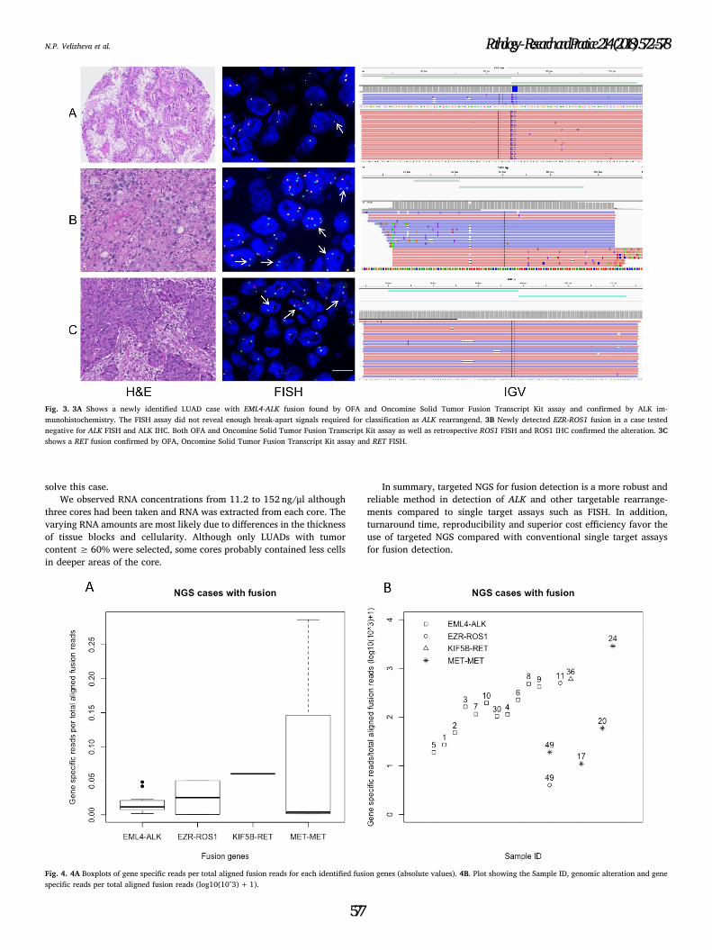
3.4. Variation of fusion read counts
We observed gene specific read counts ranging from 76 to 38946 for
all detected gene fusions. The ratio of gene specific read counts and
total mapped fusion panel reads ranged from 0.0004–0.2866. Fig. 4A
shows boxplots of read counts for all detected fusion genes. Sample no.
49 had low read counts of 76 for ROS1. EZR-ROS1 rearrangement was
confirmed with Oncomine Solid Tumor Fusion Transcript Kit assay.
However, Ros1 IHC was repeatedly negative, indicating that the low
number of fusion transcripts did not result in a detectable amount of
endogenous Ros1 protein with standard Ros1 IHC protocol. Fig. 4 B
summarizes the ratios of gene specific reads per total aligned fusion
reads.
4. Discussion
We studied the performance of a RNA-based targeted sequencing
Fig. 1. Genetic rearrangements in study cohort of LUADs. Overview of NGS results compared to FISH and IHC. LUAD lung adenocarcinoma; NGS, next-generation-sequencing; FISH,
fluorescence in-situ hybridization; IHC, immunohistochemistry; EML4-ALK, echinoderm microtubule-associate protein-like 4 − anaplastic lymphoma kinase; EZR-ROS1, ezrin gene −
proto-oncogene tyrosine-protein kinase 1; KIF5B-RET, the kinesin family 5 B gene-ret proto-oncogene.
N.P. Velizheva et al. Pathology - Research and Practice 214 (2018) 572–578
575

approach versus the gold standard FISH analysis for detection of ALK
fusions in a retrospective cohort of LUADs. All previously known ALK
fusions could be detected by targeted NGS. In addition, we found an
additional EZR-ROS1 fusion in one of the ALK non-rearranged cases and
another potential EZR-ROS1 fusion in the untested cohort. We also
found one additional EML4-ALK fusion in the untested cohort.
Our results are concordant with findings by other groups. Pfarr et al.
also reported a sensitivity and specificity of 100% of a targeted mas-
sively parallel sequencing approach for the detection of known
targetable gene fusions [18]. Paasinen-Sohns et al. compared in their
study the overall performance of DNA and RNA-based OFA panels and
could confirm all detected EML4-ALK, EZR-ROS1 and MET-MET fusions
with IHC in their cohort [19]. Another study investigating FISH, IHC
and NGS regarding EML4-ALK rearrangements found 4 tumors positive
by FISH and 8 positive by IHC in a cohort of 51 LUAD patients [20]. Of
these, only 3 cases were positive by both FISH and IHC [20]. Moreover,
4 of 5 IHC positive and FISH negative patient tumors could be con-
firmed being EML4-ALK rearranged by NGS [20]. Two of these patients
with EML4-ALK rearranged tumors confirmed by NGS received crizo-
tinib treatment with durable progression-free survival suggesting that
especially for borderline cases NGS can tip the scales [20]. A further
study was focused at solving discordant ALK testing results determined
by IHC and FISH with NGS [21]. NGS testing revealed three ALK FISH
positive Alk IHC negative cases as ALK non-rearranged [21]. Interest-
ingly, Alk immunohistochemistry with ALK1 clone was positive in 12
ALK FISH negative cases but turned out to be false positive when Alk
IHC was repeated with clone D5F3 which we also used for our study
[21].
The recommended cut-offs for FISH analyses remain controversial
and might explain the false positive and negative results when com-
paring FISH with other assays such as NGS. Seventy four per cent of
patients with advanced ALK rearranged LUAD as determined by FISH
responded to first line crizotinib treatment [5]. The median progression
free survival was 10.9 months [5]. False negative test results, however,
make it impossible to identify patients who might benefit from targeted
therapy. False positive results will exclude the patient from further
molecular testings in hierarchical single target assays. In addition, false
positive results lead to false hope and assignment to wrong therapeutic
strategies. The ALK reads per total aligned fusion reads of our NGS
positive ALK FISH negative case no. 30 match the median of all de-
tected EML4-ALK cases (0.01) so that a low ALK read ratio reflecting a
very low transcript number can be ruled out. NGS results for this par-
ticular case also fit to the observation that Alk IHC was positive. To
further rule out a lack of sensitivity we performed a dilution series of
ALK rearranged cell line RNA on a non-ALK rearranged RNA back-
ground. Ratios of 1, 0.1, 0.05, and 0.01 could be detected with RNA-
based targeted sequencing. This means that even 1% of EML4-ALK re-
arranged RNA or 1% of EML4-ALK rearranged cells can be detected on a
background without EML4-ALK rearrangement. This also explains the
discrepancy of the case being negative according to EML4-ALK FISH
evaluation criteria but positive by Alk IHC and RNA-based targeted
sequencing. Case 30 could harbor a heterogeneous EML4-ALK re-
arrangement so that the rearrangement detection can be difficult by
FISH. In addition, the ALK FISH scoring criteria could fail for hetero-
geneous cases aggravated by the fact that tissue sections are used where
nuclei are typically clipped.
Case 49 with potential detection of EZR-ROS1 rearrangement but
negative Ros1 IHC could be most likely false positive. However, since
we did not perform a dilution series of RNA of a ROS1rearranged cell
line and tissue for FISH testing was not available, we cannot completely
Fig. 2. Immunohistochemical staining for Alk, upper tile H3122, lower tile HCC-44.
Table 4
NGS metrics of different ratios of EML4-ALK to non-EML4-ALK rearrangement containing RNA.
Ratio RNA H3122/HCC-44 NGS metrics
TMFPR Gene (Exons) Read Counts Ratio Read Counts/TMFPR
1 236702 EML4(6) – ALK(20) 42452 0.179347872
0.1 238830 EML4(6) – ALK(20) 3067 0.01284177
0.05 197984 EML4(6) – ALK(20) 1999 0.010096775
0.01 155527 EML4(6) – ALK(20) 159 0.001022331
0.005 270342 EML4(6) – ALK(20) 0 0
0.001 234415 EML4(6) – ALK(20) 0 0
0.0005 214911 EML4(6) – ALK(20) 0 0
0 167480 EML4(6) – ALK(20) 0 0
Table 3
Accuracy of NGS-based targeted RNA-sequencing in detection of ALK and ROS1 re-
arrangements.
ALK rearrangements
Condition positive Condition negative
Test positive 11 (TP) 0 (FP)
Test negative 0 (FN) 18 (TN)
Sensitivity 100% Specificity 100%
ROS1 rearrangements
Condition positive Condition negative
Test positive 1 (TP) 1 (FP)
Test negative 0 (FN) 51 (TN)
Sensitivity 100% Specificity 98%
ALK, anaplastic lymphoma kinase; TP, true positive; FP, false positive; FN, false negative;
TN, true negative; ROS1, proto-oncogene tyrosine-protein kinase 1.
N.P. Velizheva et al. Pathology - Research and Practice 214 (2018) 572–578
576
solve this case.
We observed RNA concentrations from 11.2 to 152 ng/μl although
three cores had been taken and RNA was extracted from each core. The
varying RNA amounts are most likely due to differences in the thickness
of tissue blocks and cellularity. Although only LUADs with tumor
content ≥ 60% were selected, some cores probably contained less cells
in deeper areas of the core.
In summary, targeted NGS for fusion detection is a more robust and
reliable method in detection of ALK and other targetable rearrange-
ments compared to single target assays such as FISH. In addition,
turnaround time, reproducibility and superior cost efficiency favor the
use of targeted NGS compared with conventional single target assays
for fusion detection.
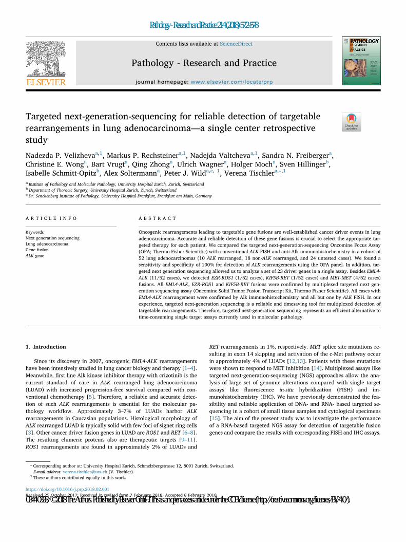
Fig. 3. 3A Shows a newly identified LUAD case with EML4-ALK fusion found by OFA and Oncomine Solid Tumor Fusion Transcript Kit assay and confirmed by ALK im-
munohistochemistry. The FISH assay did not reveal enough break-apart signals required for classification as ALK rearrangend. 3B Newly detected EZR-ROS1 fusion in a case tested
negative for ALK FISH and ALK IHC. Both OFA and Oncomine Solid Tumor Fusion Transcript Kit assay as well as retrospective ROS1 FISH and ROS1 IHC confirmed the alteration. 3C
shows a RET fusion confirmed by OFA, Oncomine Solid Tumor Fusion Transcript Kit assay and RET FISH.
Fig. 4. 4A Boxplots of gene specific reads per total aligned fusion reads for each identified fusion genes (absolute values). 4B. Plot showing the Sample ID, genomic alteration and gene
specific reads per total aligned fusion reads (log10(10^3)+ 1).
N.P. Velizheva et al. Pathology - Research and Practice 214 (2018) 572–578
577

5. Conclusion
Detection of targetable fusion genes is crucial for appropriate clin-
ical action. We experienced the RNA-based OFA panel as a reliable and
accurate tool in comparison with standard single target assays like FISH
and IHC. Especially borderline cases can benefit from the added value
of NGS testing.
Conflict of interest
The authors declare that no conflict of interest exists.
Acknowledgements
We thank Susanne Dettwiler, Fabiola Prutek and Peter Schraml from
the Tissue Biobank at USZ and Ursula Rommerscheid-Fuss, Department
of Pathology, University Hospital Cologne, Germany, for their excellent
technical support. Cell lines were a kind gift of Prof. Dr. Roman
Thomas, Cologne, Germany. This project was funded in part by
Innovation Pool Grants provided by the University Hospital Zurich to
V.T. and P.J.W., respectively. V. T. is the recipient of a joint ERS/EMBO
Long-Term Research fellowship n° LTRF 2014-2951 and a Swiss Cancer
League postdoctoral research fellowship (BIL KFS-3402-02-2014).
References
[1] M. Soda, Y.L. Choi, M. Enomoto, S. Takada, Y. Yamashita, S. Ishikawa, S. Fujiwara,
H. Watanabe, K. Kurashina, H. Hatanaka, M. Bando, S. Ohno, Y. Ishikawa,
H. Aburatani, T. Niki, Y. Sohara, Y. Sugiyama, H. Mano, Identification of the
transforming EML4?ALK fusion gene in non-small-cell lung cancer, Nature 448
(2007) 561–566, http://dx.doi.org/10.1038/nature05945.
[2] Y.L. Choi, K. Takeuchi, M. Soda, K. Inamura, Y. Togashi, S. Hatano, M. Enomoto,
T. Hamada, H. Haruta, H. Watanabe, K. Kurashina, H. Hatanaka, T. Ueno,
S. Takada, Y. Yamashita, Y. Sugiyama, Y. Ishikawa, H. Mano, Identification of novel
isoforms of the EML4-ALK transforming gene in non-small cell lung cancer, Cancer
Res. 68 (2008) 4971–4976, http://dx.doi.org/10.1158/0008-5472.CAN-07-6158.
[3] T. Sasaki, S.J. Rodig, L.R. Chirieac, P.A. Jänne, The biology and treatment of EML4-
ALK non-small cell lung cancer, Eur. J. Cancer 46 (2010) 1773–1780, http://dx.doi.
org/10.1016/j.ejca.2010.04.002.
[4] A.T. Shaw, B.Y. Yeap, M. Mino-Kenudson, S.R. Digumarthy, D.B. Costa, R.S. Heist,
B. Solomon, H. Stubbs, S. Admane, U. McDermott, J. Settleman, S. Kobayashi,
E.J. Mark, S.J. Rodig, L.R. Chirieac, E.L. Kwak, T.J. Lynch, A.J. Iafrate, Clinical
features and outcome of patients with non-small-cell lung cancer who harbor EML4-
ALK, J. Clin. Oncol. 27 (2009) 4247–4253, http://dx.doi.org/10.1200/JCO.2009.
22.6993.
[5] B.J. Solomon, T. Mok, D.-W. Kim, Y.-L. Wu, K. Nakagawa, T. Mekhail, E. Felip,
F. Cappuzzo, J. Paolini, T. Usari, S. Iyer, A. Reisman, K.D. Wilner, J. Tursi,
F. Blackhall, First-line crizotinib versus chemotherapy in ALK–positive lung cancer,
N. Engl. J. Med. 371 (2014) 2167–2177, http://dx.doi.org/10.1056/
NEJMoa1408440.
[6] K. Rikova, A. Guo, Q. Zeng, A. Possemato, J. Yu, H. Haack, J. Nardone, K. Lee,
C. Reeves, Y. Li, Y. Hu, Z. Tan, M. Stokes, L. Sullivan, J. Mitchell, R. Wetzel,
J. MacNeill, J.M. Ren, J. Yuan, C.E. Bakalarski, J. Villen, J.M. Kornhauser, B. Smith,
D. Li, X. Zhou, S.P. Gygi, T.L. Gu, R.D. Polakiewicz, J. Rush, M.J. Comb, Global
survey of phosphotyrosine signaling identifies oncogenic kinases in lung cancer,
Cell 131 (2007) 1190–1203, http://dx.doi.org/10.1016/j.cell.2007.11.025.
[7] K. Takeuchi, M. Soda, Y. Togashi, R. Suzuki, S. Sakata, S. Hatano, R. Asaka,
W. Hamanaka, H. Ninomiya, H. Uehara, Y. Lim Choi, Y. Satoh, S. Okumura,
K. Nakagawa, H. Mano, Y. Ishikawa, RET, ROS1 and ALK fusions in lung cancer,
Nat. Med. 18 (2012) 378–381, http://dx.doi.org/10.1038/nm.2658.
[8] T. Kohno, H. Ichikawa, Y. Totoki, K. Yasuda, M. Hiramoto, T. Nammo, H. Sakamoto,
K. Tsuta, K. Furuta, Y. Shimada, R. Iwakawa, H. Ogiwara, T. Oike, M. Enari,
A.J. Schetter, H. Okayama, A. Haugen, V. Skaug, S. Chiku, I. Yamanaka, Y. Arai,
S. Watanabe, I. Sekine, S. Ogawa, C.C. Harris, H. Tsuda, T. Yoshida, J. Yokota,
T. Shibata, KIF5B-RET fusions in lung adenocarcinoma, Nat. Med. 18 (2012)
375–377, http://dx.doi.org/10.1038/nm.2644.
[9] K. Bergethon, A.T. Shaw, S.H.I. Ou, R. Katayama, C.M. Lovly, N.T. McDonald,
P.P. Massion, C. Siwak-Tapp, A. Gonzalez, R. Fang, E.J. Mark, J.M. Batten, H. Chen,
K.D. Wilner, E.L. Kwak, J.W. Clark, D.P. Carbone, H. Ji, J.A. Engelman, M. Mino-
Kenudson, W. Pao, A.J. Iafrate, ROS1 rearrangements define a unique molecular
class of lung cancers, J. Clin. Oncol. 30 (2012) 863–870, http://dx.doi.org/10.
1200/JCO.2011.35.6345.
[10] O. Gautschi, J. Milia, T. Filleron, J. Wolf, D.P. Carbone, D. Owen, R. Camidge,
V. Narayanan, R.C. Doebele, B. Besse, J. Remon-Masip, P.A. Janne, M.M. Awad,
N. Peled, C.C. Byoung, D.D. Karp, M. Van Den Heuvel, H.A. Wakelee, J.W. Neal,
T.S.K. Mok, J.C.H. Yang, S.H.I. Ou, G. Pall, P. Froesch, G. Zalcman, D.R. Gandara,
J.W. Riess, V. Velcheti, K. Zeidler, J. Diebold, M. Früh, S. Michels, I. Monnet,
S. Popat, R. Rosell, N. Karachaliou, S.I. Rothschild, J.Y. Shih, A. Warth, T. Muley,
F. Cabillic, J. Mazières, A. Drilon, Targeting RET in patients with RET-rearranged
lung cancers: results from the global, multicenter RET registry, J. Clin. Oncol. 35
(2017) 1403–1410, http://dx.doi.org/10.1200/JCO.2016.70.9352.
[11] A. Drilon, L. Wang, A. Hasanovic, Y. Suehara, D. Lipson, P. Stephens, J. Ross,
V. Miller, M. Ginsberg, M.F. Zakowski, M.G. Kris, M. Ladanyi, N. Rizvi, Response to
cabozantinib in patients with RET fusion-positive lung adenocarcinomas, Cancer
Discov. 3 (2013) 630–635, http://dx.doi.org/10.1158/2159-8290.CD-13-0035.
[12] R. Salgia, MET in lung cancer: biomarker selection based on scientific rationale,
Mol. Cancer Ther. 16 (2017) 555–565, http://dx.doi.org/10.1158/1535-7163.
MCT-16-0472.
[13] A.B. Cortot, Z. Kherrouche, C. Descarpentries, M. Wislez, S. Baldacci, A. Furlan,
D. Tulasne, Exon 14 deleted MET receptor as a new biomarker and target in cancers,
J. Natl. Cancer Inst. 109 (2017), http://dx.doi.org/10.1093/jnci/djw262.
[14] P.K. Paik, A. Drilon, P.D. Fan, H. Yu, N. Rekhtman, M.S. Ginsberg, L. Borsu,
N. Schultz, M.F. Berger, C.M. Rudin, M. Ladanyi, Response to MET inhibitors in
patients with stage IV lung adenocarcinomas harboring met mutations causing exon
14 skipping, Cancer Discov. 5 (2015) 842–850, http://dx.doi.org/10.1158/2159-
8290. CD-14-1467.
[15] N.P. Velizheva, M.P. Rechsteiner, C.E. Wong, Q. Zhong, M. Rössle, B. Bode,
H. Moch, A. Soltermann, P.J. Wild, V. Tischler, Cytology smears as excellent
starting material for next-generation sequencing-based molecular testing of patients
with adenocarcinoma of the lung, Cancer Cytopathol. 125 (2017) 30–40, http://dx.
doi.org/10.1002/cncy.21771.
[16] F.H. Blackhall, S. Peters, L. Bubendorf, U. Dafni, K.M. Kerr, H. Hager,
A. Soltermann, K.J. O’Byrne, C. Dooms, A. Sejda, J. Hernández-Losa, A. Marchetti,
S. Savic, Q. Tan, E. Thunnissen, E.J.M. Speel, R. Cheney, D. Nonaka, J. De Jong,
M. Martorell, I. Letovanec, R. Rosell, R.A. Stahel, Prevalence and clinical outcomes
for patients with ALK-positive resected stage I to III adenocarcinoma: results from
the European Thoracic Oncology Platform Lungscape project, J. Clin. Oncol. 32
(2014) 2780–2787, http://dx.doi.org/10.1200/JCO.2013.54.5921.
[17] T.-M. Rogers, P.A. Russell, G. Wright, Z. Wainer, J.-M. Pang, L.A. Henricksen,
S. Singh, S. Stanislaw, J. Grille, E. Roberts, B. Solomon, S.B. Fox, Comparison of
methods in the detection of ALK and ROS1 rearrangements in lung cancer, J.
Thorac. Oncol. 10 (2015) 611–618, http://dx.doi.org/10.1097/JTO.
0000000000000465.
[18] N. Pfarr, A. Stenzinger, R. Penzel, A. Warth, H. Dienemann, P. Schirmacher,
W. Weichert, V. Endris, High-throughput diagnostic profiling of clinically action-
able gene fusions in lung cancer, Genes Chromosom. Cancer 55 (2016) 30–44.
[19] A. Paasinen-Sohns, V.H. Koelzer, A. Frank, J. Schafroth, A. Gisler, M. Sachs,
A. Graber, S.I. Rothschild, A. Wicki, G. Cathomas, K.D. Mertz, Single-center ex-
perience with a targeted next generation sequencing assay for assessment of re-
levant somatic alterations in solid tumors, Neoplasia (United States) 19 (2017)
196–206, http://dx.doi.org/10.1016/j.neo.2017.01.003.
[20] M. Pekar-Zlotin, F.R. Hirsch, L. Soussan-Gutman, M. Ilouze, A. Dvir, T. Boyle,
M. Wynes, V.A. Miller, D. Lipson, G.A. Palmer, S.M. Ali, S. Dekel, R. Brenner,
P.A. Bunn, N. Peled, Fluorescence in situ hybridization, immunohistochemistry, and
next-generation sequencing for detection of EML4-ALK rearrangement in lung
cancer, Oncologist 20 (2015) 316–322, http://dx.doi.org/10.1634/theoncologist.
2014-0389.
[21] J.S. Jang, X. Wang, P.T. Vedell, J. Wen, J. Zhang, D.W. Ellison, J.M. Evans,
S.H. Johnson, P. Yang, W.R. Sukov, A.M. Oliveir, G. Vasmatzis, Z. Sun, J. Jen,
E.S. Yi, Custom gene capture and next-generation sequencing to resolve discordant
ALK status by FISH and IHC in lung adenocarcinoma, J. Thorac. Oncol. 11 (2016)
1891–1900, http://dx.doi.org/10.1016/j.jtho.2016.06.001.
N.P. Velizheva et al. Pathology - Research and Practice 214 (2018) 572–578
578