Tailoring adhesion and wetting properties of cellulose fibers and model surfaces509273/... · ·...
41
Tailoring adhesion and wetting properties of cellulose fibers and model surfaces Emil Gustafsson Licentiate thesis School of Chemical Science and Engineering Department of Fibre and Polymer Technology Wallenberg Wood Science Center and Fibre Technology KTH Royal Institute of Technology Stockholm, Sweden 2012 AKADEMISK AVHANDLING som med tillstånd av Kungliga Tekniska Högskolan i Stockholm framlägges till offentlig granskning för avläggande av teknologie licentiatexamen 19 mars 2012, kl. 14.00 i K1, Teknikringen 56, KTH, Stockholm Avhandlingen försvaras på engelska.
Transcript of Tailoring adhesion and wetting properties of cellulose fibers and model surfaces509273/... · ·...
Tailoring adhesion and wetting properties of cellulose
fibers and model surfaces
Emil Gustafsson
Licentiate thesis
School of Chemical Science and Engineering
Department of Fibre and Polymer Technology
Wallenberg Wood Science Center and Fibre Technology
KTH Royal Institute of Technology
Stockholm, Sweden 2012
AKADEMISK AVHANDLING
som med tillstånd av Kungliga Tekniska Högskolan i Stockholm
framlägges till offentlig granskning för avläggande av teknologie licentiatexamen
19 mars 2012, kl. 14.00 i K1, Teknikringen 56, KTH, Stockholm
Avhandlingen försvaras på engelska.
Tailoring adhesion and wetting properties of cellulose fibers and model
surfaces
Emil Gustafsson
Thesis for the degree of Licentiate of Technology in Fibre and Polymer Science
KTH Royal Institute of Technology
School of Chemical Science and Engineering
Department of Fibre and Polymer Technology
Wallenberg Wood Science Center
SE-10044 Stockholm, Sweden
ISBN 978-91-7501-268-1
TRITA-CHE Report 2012:8
ISSN 1654-1081
Copyright © Emil Gustafsson, 2012
Printed by: Universitetsservice US-AB, Stockholm 2012

Abstract
The layer-by-layer (LbL) technique was used to modify the surface of cellulose fibers by
consecutive adsorption of poly(allylamine hydrochloride) (PAH) and poly(acrylic acid)
(PAA) followed by a final adsorbed layer of anionic paraffin wax colloids. Paper hand sheets
made from the modified fibers were found to be highly hydrophobic with a contact angle of
150°. In addition to the significantly increased hydrophobicity, the sheets showed improved
mechanical properties, such as a higher tensile strength. Heat treatment of the prepared sheets
further enhanced both the mechanical properties and the hydrophobicity. These results
demonstrate the flexibility and robustness of the LbL technique, which allows us to combine
the known adhesive effect of PAH/PAA LbL films with the functionality of wax
nanoparticles, creating a stronger and highly hydrophobic paper.
It was further observed that LbL modified sheets without wax also displayed increased
hydrophobicity when heat treated. The mechanism was studied through model experiments
where LbL films of PAH/PAA were assembled on flat non-porous model cellulose surfaces.
Contact angle measurements showed the same trend due to heat treatment of the model films,
although, the absolute value of the contact angles were smaller. Analysis using the highly
interfacial sensitive vibrational sum frequency spectroscopy technique showed an enrichment
of CH3 groups (from the polymer chain ends) at the solid/air interface. These results indicate
that during the heat treatment, a reorientation of polymer chains occurs to minimize the
surface energy of the LbL film.
In the second part of this work, the adhesive interactions between the main constituents of
wood fibers were studied using high-resolution measuring techniques and well-defined model
films of cellulose, hemicellulose and lignin. Successful surface modification of
polydimethylsiloxane (PDMS) caps, needed in the Johnson-Kendall-Roberts (JKR) measuring
methodology, by LbL deposition of nanofibrillated cellulose (NFC) and poly(ethylene imine)
(PEI) allowed for the first known all-wood biopolymer JKR measurements of the adhesion
between cellulose/cellulose, cellulose/lignin and the cellulose/glucomannan surfaces. The
work of adhesion on loading and the adhesion hysteresis were similar for all three systems,
suggesting that adhesion between the different wood biopolymers does not differ greatly.

Sammanfattning
Layer-by-layer (LbL) tekniken har använts för att ytmodifiera cellulosafibrer genom växelvis
adsorption av poly(allylamin hydroklorid) (PAH) and poly(akrylsyra) (PAA) följt av
adsorption av ett lager med anjoniska vaxkolloider. Pappersark tillverkades av de modifierade
fibrerna och dessa ark fanns vara hydrofoba, med en kontaktvinkel på 150°. I tillägg till den
signifikant förbättrade hydrofobiteten uppvisade arken även förbättrade mekaniska
egenskaper. Värmebehandling av arken förbättrade både styrkan och hydrofobiteten.
Resultaten demonstrerar robustheten och flexibiliteten hos LbL-tekniken genom att visa att
styrkeförbättrande polyelektrolyter kan kombineras med vaxpartiklar för att skapa starka
papper som samtidigt är hydrofoba.
Vidare observerades det att LbL modifierade ark även utan adsorberat vax blev hydrofoba när
de värmebehandlades. Mekanismen bakom detta fenomen studerades vidare genom att
cellulosamodellytor modifierades med PAH/PAA för att sedan värmebehandlas.
Kontakvinkelmätningar på dessa modellytor visade att ytorna blev mer hydrofoba om än inte i
samma utsträckning som de värmda arken. Mekanismen undersöktes vidare med hjälp av
vibrational sum frequency spectroscopy (VSFS) och resultaten visade att värmebehandling
ger en ökad andel CH3-grupper på ytan och även signalen för polymerens ryggrad ökade i
intensitet. Resultaten indikerar att värmebehandlingen leder till omorientering av
polymerkedjorna i LbL-filmen för att minimera ytenergin.
I den andra delen av det här arbetet studerades adhesionsarbetet mellan väldefinierade
modellytor av de huvudsakliga vedpolymererna. PDMS-halvsfärer ytmodifierades genom
LbL-deponering av nanofibrillerad cellulosa (NFC) och polyetylenimin (PEI) för att skapa en
halvsfärisk modellyta av cellulosa. Detta möjliggjorde de första adhesionsmätningarna mellan
cellulosa/cellulosa, cellulosa/glukomannan och cellulosa/lignin med Johnson-Kendall-Roberts
(JKR) metoden. Adhesionsarbetet och hysteresen för mätningarna skiljde sig inte signifikant
för de olika systemen, vilket föreslår att adhesionen mellan de olika vedpolymererna inte
skiljer sig markant.

List of publications
I Treatment of cellulose fibres with polyelectrolytes and wax colloids to create
tailored highly hydrophobic fibrous networks
E. Gustafsson, P.A. Larsson and L. Wågberg
Submitted
II Vibrational sum frequency spectroscopy on polyelectrolyte multilayers –
modeling of hydrophobic fibres
E. Gustafsson, J. Hedberg, P.A Larsson, L. Wågberg and C.M. Johnson
Submitted
III Direct adhesive measurements between wood biopolymer model surfaces
E. Gustafsson, E. Johansson, L. Wågberg and T. Pettersson
Submitted
The contribution of the author of the thesis to the appended papers are:
I All experiments on model surfaces, 50% of the writing
II All sample preparation, AFM, contact angle determinations, major part of the writing
III 50% of the experiments, 50% of the writing


Contents 1 Objectives ....................................................................................................................................... 1
2 Introduction ................................................................................................................................... 2
2.1 Adhesion .................................................................................................................................. 2
2.2 Paper and fiber networks ......................................................................................................... 2
2.2.1 Adhesion in paper and fiber networks ............................................................................. 2
2.2.2 Water interactions in cellulose fiber materials ................................................................ 3
2.3 Tailoring of surfaces by the layer-by-layer (LbL) technique .................................................. 4
2.4 Wood biopolymer model systems for evaluating adhesion and wetting ................................. 5
3 Experimental .................................................................................................................................. 8
3.1 Materials .................................................................................................................................. 8
3.1.1 Substrates ......................................................................................................................... 8
3.1.2 Chemicals ........................................................................................................................ 8
3.2 Methods ................................................................................................................................... 9
3.2.1 LbL formation ................................................................................................................. 9
3.2.2 Handsheet formation and sheet evaluation .................................................................... 10
3.2.3 Wood biopolymer model surfaces ................................................................................. 10
3.2.4 AFM imaging and scratch height analysis .................................................................... 10
3.2.5 JKR adhesion measurements ......................................................................................... 10
3.2.6 Contact angle determinations ........................................................................................ 11
3.2.7 Vibrational Sum Frequency Spectroscopy (VSFS) ....................................................... 12
4 Results and discussion ................................................................................................................. 13
4.1 Improved paper strength and hydrophobicity by LbL and wax adsorption ........................... 13
4.1.1 Polyelectrolyte- and wax adsorption on fibers .............................................................. 13
4.1.2 Sheet formation and mechanical characterization ......................................................... 14
4.1.3 Wetting properties of the sheets .................................................................................... 15
4.1.4 Heat-induced hydrophobation of PAH/PAA films without wax addition ..................... 16
4.2 Dry adhesion between cellulose, lignin, and glucomannan model surfaces .......................... 21
4.2.1 Model surface characterization ...................................................................................... 21
4.2.2 Adhesion measurements ................................................................................................ 23
5 Conclusions .................................................................................................................................. 26
6 Acknowledgements ...................................................................................................................... 27
7 List of abbreviations .................................................................................................................... 28
8 References .................................................................................................................................... 29
Appendices: Paper I-III


1
1 Objectives The overall objective of the present work has been to identify new routes to molecularly tailor
the wetting and adhesive properties of cellulose fibers. This overall objective was divided into
three different studies where both cellulose fibers and model surfaces of cellulose, lignin and
hemicelluloses were used.
The purpose of papers I and II was to create a highly hydrophobic paper without any
deterioration in the mechanical properties of the paper by using the layer-by-layer (LbL)
technique with a combination of polyelectrolytes and nanoparticles of wax.
In paper III, the objective was to develop a suitable cellulose model surface deposited on
elastic PDMS in order use the Johnson-Kendall-Roberts (JKR) approach to examine the work
of adhesion between well-defined model surfaces of wood biopolymers. The purpose of the
study was to seek a fundamental understanding of interactions both within the fiber wall and
in the fiber-fiber joints in paper.

2
2 Introduction
2.1 Adhesion Adhesion science is the science describing how surfaces adhere. Covering all length scales,
from the assembly of construction materials to cell adhesion and the interaction of pigments
down to the nanoscale interaction between molecules, adhesion phenomena are highly
relevant and a great challenge in engineering, chemistry and biology. The thermodynamic
work of adhesion upon separation of two surfaces theoretically depends solely on the
interfacial energy of the two surfaces and can be predicted by the Dupré equation [1]. The
adhesion measured in practice however usually is a non-equilibrium irreversible process in
which phenomena like surface roughness, plastic deformation, and molecular reorientation
give rise to adhesion hysteresis and real adhesion energies which are orders of magnitude
greater than the thermodynamic work of adhesion [1]. It is challenging to measure and
decouple these contributions, and if done properly it can provide an important tool towards
the tailoring of adhesive interactions and the improvement of material properties.
2.2 Paper and fiber networks
2.2.1 Adhesion in paper and fiber networks
Paper is a three dimensional network of fibers, the overall strength of which depends on the
strength of the individual fibers, the fiber length, the number of fiber-fiber joints per volume
and the strength of the fiber-fiber joints. Depending on the type of paper, the different factors
vary in relative importance. It has been suggested by a theoretical analysis that the maximum
tensile strength of a paper ultimately depends on the strength of the individual fibers [2], but it
has also been suggested that the adhesive strength of the individual fiber-fiber joints is always
the limiting factor for the maximum strength [3]. Without a doubt, it can be stated however
that adhesion plays a very important role when it comes to papermaking and that the
understanding and tailoring thereof can have large implications for paper applications. The
adhesion between fibers and matrix material is also of crucial importance for the performance
of fiber-reinforced biocomposites, a material branch that has gained a lot of interest due to the
environmental driving force for new, renewable materials.
The dry adhesion between two fibers, i.e. the strength of the fiber-fiber joint, depends on the
contact area between the fibers and on the molecular interactions in the contact zone.
Traditionally, the adhesion between cellulose fibers is improved by the mechanical treatment
of the fibers, so-called “beating”, in order to make the fibers more flexible and to create
fibrillar fines [4], both of which contribute to increasing the contact area in the fiber-fiber
joint. There are drawbacks of the method though; including impaired dewatering on the paper
machine, densification of the paper and high energy consumption.
An alternative to beating is the use of chemicals, usually water-soluble cationic
polyelectrolytes, which improve the joint strength without densification. It is well established
that the addition of polyelectrolytes increases the molecular contact area and/or the number of
fiber-fiber joints [5]. Cationic starch is the most commonly used dry strength additive,
primarily due to its low cost, but synthetic polymers such as polyethylenimine (PEI) are also

3
used [6]. Recently it has been demonstrated that the sequential deposition of oppositely
charged polyelectrolytes by the so-called layer-by-layer (LbL) assembly technique can further
improve paper strength [7]. LbL assembly also allows for the incorporation of charged
nanoparticles, which opens up further possibilities of tailoring fiber and fiber-fiber joint
properties. This approach is used in this work and will be further outlined in later sections of
this thesis.
2.2.2 Water interactions in cellulose fiber materials
One of the major drawbacks when comparing paper to e.g. plastics is its sensitivity to both
liquid water as and moist air. Commonly used paper grades are protected from liquid water by
hydrophobation, or sizing. Traditionally this is done by treating the fibers with rosin sizes and
alum (acidic sizing), or by adsorbing Alkyl Ketene Dimer (AKD) wax or alkenylsuccinic
anhydride waxes (alkaline sizing) prior to sheet forming to make the paper hydrophobic [8].
With AKD sizing, it is possible to achieve advancing contact angles of water of
approximately 110° [9]. To reach higher degrees of hydrophobicity however, not only are
other methods and/or chemicals needed, but also the introduction of a structured surface in the
micrometer range [10, 11].
The wetting of a fibrous material such as paper is complex since it has a chemically and
physically heterogeneous surface. The problem is further complicated by the simultaneous
spreading of the liquid on the surface and absorption of the liquid into the pores of the
material.
When a drop of liquid is placed on a surface, the drop appears to adopt a
constant shape with an angle towards the surface which is called the contact angle, θ. The
equilibrium contact angle is considered to be a characteristic of the particular liquid-solid
interaction and serves as an indication of the wettability of the surface by the liquid. This
equilibrium situation for a smooth, homogeneous, impermeable and non-deformable surface
is described by the Young equation:
(1)
where θ is the contact angle of the liquid at the solid-liquid-vapor boundary γsv is the surface
tension of the solid, γlv is the surface tension of the liquid and γsl is the interfacial tension
between the solid and the liquid.
However, most real surfaces do not have the ideal characteristics which allow the Young
equation to be used in its original form. The main reasons, which are certainly the case for
wood fibers and paper, are surface roughness and heterogeneity. The experimentally observed
contact angle is called the apparent contact angle and if inserted into Young’s equation the
result will only be approximate. Furthermore, on very thin films the test liquid can diffuse
through the film and the measured contact angle may thus be influenced by the interfacial
properties of the underlying substrate.
The contact between a liquid and a rough surface can adopt two states; either the liquid is in
complete contact with the solid surface or the drop rests on a composite surface of solid and

4
air. The relation between the apparent contact angle on a rough surface (θ*), the roughness
coefficient (r) and the intrinsic contact angle (θ) has been described by the Wenzel equation:
(2)
and by the Cassie-Baxter equation:
(3)
where is the surface area of the liquid in contact with the solid divided by the projected
area, and is the surface area of liquid in contact with the trapped air divided by the
projected surface area.
These equations suggest that there are two basic ways of improving the hydrophobicity of a
surface; (a) by lowering the surface energy, and b) increasing the surface roughness, i.e.
increasing the surface area, on a meso- and microscopic scale [10-13]. In addition to the effect
of microscopic roughness, the hydrophobicity of a surface can be further increased by the
introduction of additional roughness on the nanometer scale [14].
2.3 Tailoring of surfaces by the layer-by-layer (LbL) technique The layer-by-layer (LbL) technique for modifying the surface of charged solid substrates by
the consecutive adsorption of polyelectrolytes or charged nanoparticles was introduced as a
general method by Decher in the 1990s [15]. The technique, by which polyelectrolyte
multilayers (PEM) are formed, has since developed rapidly as a robust, flexible and
environment friendly surface engineering technique and notable applications include contact
lens coatings [16], sensor technology [17], antibacterial fibers [18] and hollow capsules for
the controlled release of drugs [19].
The principle is that when a polyelectrolyte is adsorbed to the surface it recharges the surface,
after which the surface is rinsed to wash away unadsorbed or loosely adsorbed
polyelectrolytes, followed by the adsorption of an oppositely charged polyelectrolyte and yet
another rinsing step. This procedure is repeated until the desired thickness is reached. The
adsorption is entropy-driven by the counter ions that are released from the surface during the
adsorption resulting in an increase in entropy. LbL deposition can be performed by spin-
coating, spraying or most commonly by dipping [16]. The properties of the LbL film and the
thickness of the adsorbed layers can be tuned by varying one or several parameters during the
LbL process. If a weak polyelectrolyte, i.e. where the degree of dissociation of the polymer
chain is dependent on pH, is used, the polyelectrolyte conformation can be tuned by varying
the pH. In addition to the charge of the polyelectrolytes, the salt concentration, type of salt,
molecular weight and temperature influence the properties of the PEM [16].
Wågberg successfully applied the LbL technique to engineer the surface properties of
cellulose fibers in order to improve paper strength [7] and since then the relationships
between adsorption, film properties and paper strength have been examined [20, 21]. Even
though tensile tests on paper provide only indirect information about the adhesive strength of
the fiber-fiber joints, the results obtained strongly indicate that the LbL technique improved
the fiber-fiber joint-strength. The influence of PAH/PAA multilayers on the adhesion in the
5
fiber joint have also been evaluated using fiber crosses, the results indicating an increased
joint strength [5]. The adhesion between PEMs under wet conditions, typical of those when
fiber-fiber joints are formed, has also been studied using colloid probe atomic force
microscopy (AFM). The influences of molecular weight, number of layers and polyelectrolyte
in the outermost layer were examined [22]. Although these measurements give information
about the adhesion in the wet state they do not provide any information about the effect of the
drying of the fiber joint.
Lately, several research groups have started to utilize the potential of the surface charge and
nano-dimensions of nanocellulose (NFC,CNC) and have started to include fibrils and
nanocrystals as a component in LbL assembly, to produce coatings [23-25], and freestanding
films [26] and this technique has been applied in this work as cellulose model surfaces for
interaction studies.
LbL assembly has also been used to prepare hydrophobic coatings. Glinel [27] showed that
charged wax particles could be incorporated as a barrier that protects the polyelectrolytes
from liquid water. Zhai et.al [28] used another route where LbL assembly was used to
combine polyelectrolytes and silica nanoparticles to create the porous and rough template
needed to create a superhydrophobic surface. Contact angles greater than 90° have also been
reported for water on cellulose fibers modified by LbL assembly of polyallylamine (PAH) and
polyacrylic acid (PAA), provided the cationic polyelectrolyte was in the external layer [29].
The contact angle of water on films assembled from these polyelectrolytes on flat substrates
has meanwhile been reported to be 40-50° [30, 31]. The structural or surface chemistry reason
for the observed discrepancy has yet to be resolved. Vibrational sum frequency spectroscopy
(VSFS) is an interfacial sensitive spectroscopic method that can provide information about the
molecular composition at the solid/air interface of a material. In the current work vibrational
sum frequency spectroscopy (VSFS) has been used to study the molecular composition at the
solid/air interface of LbL films on glass substrates, to study the connection between interfacial
chemistry and wettability of the films.
2.4 Wood biopolymer model systems for evaluating adhesion and wetting There are several high resolution methods available for the direct measurement of adhesive
forces between solid substrates, including the surface force apparatus (SFA) [32], colloid
probe AFM [33] and a variety of devices for adhesion measurements based on the Johnson-
Kendall-Roberts (JKR) theory [34-36] In this work, a JKR device has been used for
fundamental studies of the interaction between wood biopolymers. Adhesion measurement
according to the standard JKR approach used in this work does not provide any information
about the influence of the drying process on the joint strength. A JKR based technique for
performing such measurements is however being developed in our laboratory.
A major challenge when it comes to the measurement of adhesion in real engineering
situations, including papermaking and the design of biocomposites, is that they involve
heterogeneous surfaces that are both chemically and structurally ill-defined. Such surfaces are
ill-suited for studies using the above mentioned techniques, since all the methods, in order to
permit conclusions relating to molecular-scale mechanisms, require substrates that are smooth

6
and chemically well-defined. A common approach is to use neat or gently surface modified
silica or mica, which have a low surface roughness and permit controlled surface
modifications, as model surfaces. In this work, sufficiently smooth and chemically
homogeneous, wood biopolymer model films were prepared and characterized and ultimately
used for direct adhesive measurements using the JKR-technique.
Various procedures to prepare more advanced model surfaces representing the three main
wood components, cellulose, hemicellulose and lignin, are described in the literature [24, 37].
Earlier work on cellulose model surfaces has been based mostly on the spin-coating of
regenerated cellulose dissolved in mixtures of N-methylmorpholine-N-oxide (NMMO) and
dimethylsulfoxide (DMSO) [38] or lithium chloride/ dimethyldiacetamine (LiCl/DMAc) [39].
Spin-coated surfaces have also been prepared from suspensions of cellulose nanocrystals
(CNC) [40] and nanofibrillated cellulose [41]. CNC and NFC have also been used in LbL
assembly together with cationic polyelectrolytes to form model surfaces [23, 42]. The
morphology and crystallinity vary depending on the starting material and preparation
technique [24]. Cellulose model films have also been prepared by LbL assembly and such
films terminated with a layer of NFC were the main cellulose model surfaces used for
adhesion measurements in the present work. NFC consists of native cellulose I and also
contains both crystalline and paracrystalline regions, and this makes it a good cellulose model
surface [24]. The fibrilar structure obtained when assembling the films also resembles that of
the cellulose fiber.
The interaction between cellulose surfaces in water has been studied using SFA [43] and
colloidal probe AFM [44-47] and two comparative studies have demonstrated that the
preparation technique used, which determines the surface charge and roughness, was crucial
for the cellulose interactions [47, 48]. The interactions between surfaces coated with LbL
films containing NFC [24] and CNC [49] in water have been investigated.
Lignin model surfaces were first prepared in the early 1970s [50] and since then surfaces have
been prepared using various lignin sources, with different routes for the isolation of the lignin
and by different surface preparation techniques. Norgren [37] recently prepared smooth,
stable lignin model surfaces by the spin-coating of kraft lignin dissolved in an aqueous
alkaline solution. These surfaces were later used for interaction studies between lignin and
cellulose in water at various salt concentrations [46] and for wetting studies, where it was
shown that the lignin was partially wetted by water. Spin-coated surfaces have recently also
been produced from milled wood lignin [51]. The lignin model surfaces in the present work
were prepared from a commercial kraft lignin, Indulin AT.
Hemicelluloses occur in different amounts in wood and, depending on the wood species
different hemicelluloses are dominating. In softwood, hemicelluloses from the
galactoglucomannan (GGM) family are predominant [52]. There are two different GGMs, one
galactose-rich type that is soluble in water and one galactose-poor type called glucomannan
(GM) that is insoluble in water. The model surfaces of hemicellulose examined in the present
thesis were made from glucomannan. In the literature, surface interaction studies involving

7
hemicellulose model surfaces are rare, but one recent study examined the interaction between
xyloglucan grafted onto gold and a cellulose sphere using colloid probe AFM [53].
In addition to providing information about the interactions in the fiber-fiber joints, the wood
biopolymer model surfaces described above can be used to study the fundamental interactions
within the fibers. The fibers now found in trees and annual plants are the result of an
evolutionary process that has created an intricate chemistry and advanced nano- and
mesostructures to optimize the fiber properties, for which both fiber structure and chemistry
are crucial. The interactions between the wood biopolymers have a profound effect on the
mechanical properties of the fibers. However, we possess limited fundamental understanding
of the importance of the adhesive interactions between the various chemical constituents of
the fiber wall, and this motivates adhesion studies using well-defined model surfaces
representing these wood components.

8
3 Experimental This chapter presents the key experimental details of this work. For further details the reader
is referred to papers I-III and the references therein.
3.1 Materials
3.1.1 Substrates
The fibers used throughout this work were an unbeaten, once-dried, virgin softwood kraft
pulp (SCA, Östrand Mill, Sweden) bleached according to a (OO)Q(OP)(ZQ)(PO)-sequence.
Before use, the pulp was washed and the carboxyl groups of the fibers were converted to their
sodium form according to an earlier described procedure [54].
Dissolving grade pulp from Domsjö Fabriker (Örnsköldsvik, Sweden) was used for the
preparation of regenerated cellulose II model surfaces. The pulp was extracted with acetone
before use.
Polished silicon wafers with a natural oxide layer of 1.4-1.7 nm, were purchased from MEMC
Electronic Materials SpA (Novara, Italy). The surfaces were cleaned thoroughly with milli-Q
water and ethanol, blown dry with nitrogen and finally plasma-cleaned for 2 min at 30 W
(PDC-002 plasma cleaner; Harrick Scientific Inc., Pleasantville, USA). Microscope glass
slides were cleaned in a similar manner.
PDMS (Dow Sylgard 182 silicone elastomer kit, base and curing agent) was purchased from
Dow Corning (Midland, USA). Droplets of degassed 10:1 mixture of base and catalyst were
placed onto hydrophobated glass to form semi-spherical PDMS caps with a typical radius of
1.1 mm. The size of each individual PDMS cap used in the experimental series was measured.
The caps were cured at 100 °C for 1 h and were thereafter extracted in heptane for 12 h in a
Soxhlet equipment in order to remove unreacted monomer. Flat slabs of PDMS were also
prepared in a glass mold, supported by a silicon wafer using the same proportions of base and
catalyst.
3.1.2 Chemicals
Poly(allylamine hydrochloride) (PAH; Mw = 15000 g mol–1
) and poly(acrylic acid) (PAA;
Mw = 7000 mol–1
) were purchased from Sigma-Aldrich (Munich, Germany). Poly(ethylene
imine) (PEI; Mw = 60000 g mol–1
, 53% aqueous solution) was obtained from Acros Organics
(Geel, Belgium). Anionic paraffin wax, Ultralube E-340, was purchased from Keim-Additec
Surface Gmbg (Kirchberg, Germany). The wax colloids were of commercial grade with a
melting temperature of 56–58 °C according to information provided by the supplier. The
polyelectrolytes and wax were used without further purification.
Anionic nanofibrillated cellulose (NFC) was prepared at Innventia AB (Stockholm, Sweden)
according to a method described earlier [23, 55]. In brief, the NFC was prepared from a
commercial sulfite softwood dissolving pulp (Domsjö Dissolving Plus, Domsjö Fabriker,
Örnsköldsvik, Sweden) using high-pressure homogenization similar to a previously described
method [56], but with carboxymethylation [57] pretreatment of the fibers. The carboxylated
nanofibrils had a typical diameter of 5-15 nm and a length of up to 1 µm [23].
9
Commercial kraft lignin, Indulin AT, was obtained from MeadWestvaco (Richmond, USA)
and was purified according to a procedure previously described by Norgren et al. [37]. To
remove carbohydrates, the lignin was dissolved in a dioxane:water mixture (9:1) and stirred
for 2 h at room temperature. The solution was then centrifuged and the residue, containing
undissolved carbohydrates, was removed. The dioxane was thereafter evaporated in a
rotavapor and the lignin was freeze-dried. To remove extractives, the freeze-dried lignin was
subjected to pentane extraction for 8 h. The number average molecular weight of Indulin AT
has previously been determined to be Mn = 1062 mol-1
with a polydispersity index of 5.18
using size exclusion chromatography [58].
Glucomannan from Norway spruce (Picea abies) wood holocellulose was extracted with
NaOH/H3BO4 and precipitated with Fehling reagent [59]. The precipitate was washed with
deionized water, dissolved in 1 M HCl, and further precipitated and washed with ethanol. The
purified glucomannan was composed of galactose, glucose, and mannose in a <0.1:1:3.5 ratio
and the degree of polymerization, DP, as determined using size exclusion chromatography,
was 150.
Methyl iodide was obtained from Acros Organics (Geel, Belgium) and glycerol from Sigma
Aldrich (Munich, Germany). Both chemicals were of analytical grade and were used as
received.
All other chemicals (hydrochloric acid, sodium hydroxide, sodium chloride and sodium
bicarbonate) were of analytical grade.
All solutions were prepared in mill-Q purified water with a resistivity of 18.2 MΩ cm
(Synergy 185, Millipore, Bellerica, USA)
3.2 Methods
3.2.1 LbL formation
The bleached fibers were consecutively treated with PAH and PAA according to a method
described earlier [7]. The adsorption was performed in a 4 g/L suspension with a background
salt concentration of 0.01 M NaCl. Amounts of 30 mg/g fiber were added in each adsorption
step. In brief, the cationic PAH was added to the fiber suspension and allowed to adsorb for
20 min. The water with the excess polyelectrolyte was filtered off and the fiber pad was rinsed
with water. The pad was then re-suspended and the PAA was added and allowed to adsorb for
20 min, followed by filtering and washing. The procedure was repeated until 2.5 bilayers of
polyelectrolyte (PAH/PAA2.5) had been added. PAH and PAA were adsorbed at pH 7.5 and
3.5 respectively to optimize the adsorption [20].
LbL films were formed on cellulose model surface substrates by hand-dipping when there
were only a few layers or by using a dipping robot (StratoSequence VI, nanoStrata Inc.,
Tallahassee, USA) according to the same pH strategy and electrolyte concentrations as for the
deposition on fibers. The adsorption time for each polyelectrolyte was 10 min followed by
3x2 min rinsing in Milli-Q water. The films were dried with nitrogen gas following the last
deposition step.
10
3.2.2 Handsheet formation and sheet evaluation
Sheets with an average grammage of 100 g/m2 were prepared using tap water with the aid of a
Rapid Köthen handsheet former (Paper Testing Instruments, Pettenbach, Austria). The sheets
were dried at 93 °C under a reduced pressure of 95 kPa for 15 min. Some of the PEM-treated
sheets were cut in half, and one half of each sheet was further heat-treated for 30 min at
160°C to induce cross-links in the multilayer structure and thus to increase the tensile strength
of the sheets [5, 60], and allowing both a spreading of the adsorbed wax on the surface and a
fusing with the PEM [27].
Sheets were conditioned at 23 °C and 50% RH, after which the sheet grammage (mass per
unit area) was determined and dry tensile testing was performed according to the ISO 1924-3
Standard. The paper strength is presented as the tensile index, which is the maximum tensile
force at paper breakage per unit width and unit grammage of the paper. Sheet thickness and
density were evaluated by measuring the structural thickness according to a method
developed at STFI (now Innventia AB) [61].
3.2.3 Wood biopolymer model surfaces
Cellulose I model surfaces were prepared on silicon oxide and PDMS by LbL assembly of
PEI and NFC. An initial layer of PAH was adsorbed followed by 1.5 bilayer of NFC/PEI. The
adsorption time for PAH and PEI was 10 min, NFC was allowed to adsorb for 20 min. Three
rinsing steps, each of two minutes, were included between the adsorption steps.
A KW-4A spin coater (Chemat Technology, Northridge, USA) was used to prepare model
surfaces of NFC in paper I and of lignin and glucomannan in paper III. Lignin and
glucomannan were dissolved in NH3 at concentrations 1 wt% and 2 wt% respectively for 24 h
and thereafter filtered through 0.45-µm polyethersulfone membrane syringe filters. The
solutions were thereafter spin-coated onto cleaned silicon oxide substrates at 2000 rpm for 60
s. Cellulose model surfaces were prepared by spin coating from a 0.1 wt% solution of
carboxymethylated NFC onto cleaned silicon oxide surfaces at 1500 rpm for 15 s followed by
3500 rpm for 30 s.
3.2.4 AFM imaging and scratch height analysis
The surface morphology of the flat model surfaces and LbL-modified films was imaged using
a Nanoscope IIIa AFM (Veeco Instruments, now Bruker AXS, Santa Barbara, USA) with a
type E piezoelectric scanner. Images were acquired in tapping mode in air using RTESP Si
cantilevers (Bruker Probes, Camarillo, USA) with a nominal resonance frequency of 300 kHz
and a typical spring constant of 40 N m–1
. In paper III, AFM was also used to determine the
film thickness by means of digital image analysis of scratched surfaces using the AFM
software (Nanoscope version 6.13, Veeco Instruments). The thickness was calculated from
the difference between the height of the film and the height of the underlying silicon oxide
surface.
3.2.5 JKR adhesion measurements
The adhesion measurement protocol used in this work, where adhesion is measured using
surface functionalized soft PDMS hemispherical caps, is based on the JKR contact mechanics
theory [62] and was developed in the early 1990s by Chaudhury and Whitesides [35]. In a
11
typical experiment, an elastic hemispherical PDMS cap is pressed stepwise towards a flat
surface placed on a balance. Since the cap is transparent, the contact area between the surfaces
can be monitored using a light microscope connected to a CCD camera. The cap is pressed in
finite steps against the flat surface and at each step the system is allowed to equilibrate for 10
min. The load and the contact area are recorded at the end of the equilibrating period. The
surfaces are loaded until a trigger value of 0.25 g is recorded, at which point the surfaces are
unloaded again in the same stepwise manner until the cap and the flat surface are completely
separated.
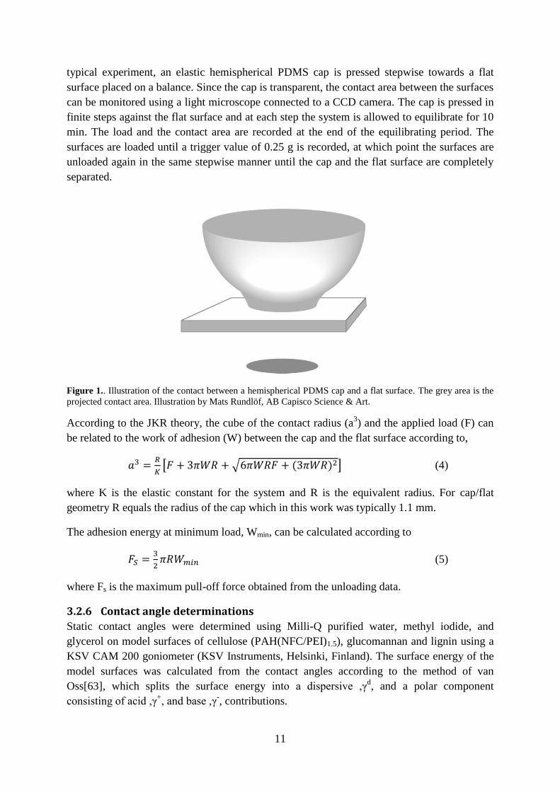
Figure 1.. Illustration of the contact between a hemispherical PDMS cap and a flat surface. The grey area is the
projected contact area. Illustration by Mats Rundlöf, AB Capisco Science & Art.
According to the JKR theory, the cube of the contact radius (a3) and the applied load (F) can
be related to the work of adhesion (W) between the cap and the flat surface according to,
[ √ ] (4)
where K is the elastic constant for the system and R is the equivalent radius. For cap/flat
geometry R equals the radius of the cap which in this work was typically 1.1 mm.
The adhesion energy at minimum load, Wmin, can be calculated according to
(5)
where Fs is the maximum pull-off force obtained from the unloading data.
3.2.6 Contact angle determinations
Static contact angles were determined using Milli-Q purified water, methyl iodide, and
glycerol on model surfaces of cellulose (PAH(NFC/PEI)1.5), glucomannan and lignin using a
KSV CAM 200 goniometer (KSV Instruments, Helsinki, Finland). The surface energy of the
model surfaces was calculated from the contact angles according to the method of van
Oss[63], which splits the surface energy into a dispersive ,γd, and a polar component
consisting of acid ,γ+, and base ,γ
-, contributions.

12
√
√
√
(6)
where γl is the surface tension of the liquid and γs is the surface energy of the sample.
The method requires a minimum of three test liquids, of which one must be water, since the
equation contains three unknowns. In the present work water, methylene iodide, and glycerol
were used as test liquids, and the surface tensions, including dispersive, acid, and base
components, were taken from Della Volpe [64].
The contact angle for water was also used to determine the effect of surface modifications and
heat treatment of surface modified paper sheets and model surfaces.
3.2.7 Vibrational Sum Frequency Spectroscopy (VSFS)
The VSFS spectrometer has been described in detail elsewhere [65]. In brief, an Ekspla
(Vilnius, Lithuania) Nd:YAG picosecond laser with an output wavelength of 1064 nm was
used to pump an optical parametric generator/optical parametric amplifier (OPG/OPA) from
Laservision (Bellevue, USA). A tuneable infrared beam and a visible beam (532 nm) were
generated in the OPG/OPA. The output energy from the OPG/OPA in the CH region (2750–
3100 cm-1
) was approximately 300 µJ. The scanning speed for the infrared beam was 1 cm-1
/s.
A Spectrogon filter was used at the output of the OPG to remove residual reflections of
unwanted frequencies.

13
4 Results and discussion
4.1 Improved paper strength and hydrophobicity by LbL and wax
adsorption
4.1.1 Polyelectrolyte- and wax adsorption on fibers
The LbL assembly technique was used to modify fibers in 4 g fiber/L suspension. PAH and
PAA were added at a concentration of 30 mg/g fiber and adsorbed at pH 7.5 and 3.5
respectively with a background electrolyte concentration of 10 mM. These conditions have
previously been found to maximize the thickness of the LbL film on the fibers [20]. A layer of
anionic paraffin wax particles was adsorbed following the deposition of the final PAH layer.
The addition of three different wax concentrations, 3, 15 and 30 mg/g fiber, was evaluated.
The particle size distribution of the anionic paraffin wax colloids used was characterized in
the pH range between 7.5 – 9.5, with and without the addition of 0.01 M NaCl, by dynamic
light scattering. Figure 2 shows a well-defined size distribution and importantly it can also be
noted that no particle agglomeration was induced by changes in pH or salt concentration.
Furthermore Mütek titrations showed that the charge density was constant within the pH
interval 7.5-8.5 and that the presence of salt had only a small influence on the charge density.
Wax particles were adsorbed onto the fibers at pH 8.5.
Figure 2. Wax particle size distribution in emulsions of pH 7.5, 8.5 and 9.5 without added NaCl and with an
added background NaCl concentration of 0.01 M. The bars indicate 95% confidence limits.
The adsorption of PAH, PAA and wax onto the fibers was determined via titration of non-
adsorbed polyelectrolytes from each consecutive step and by extraction of the non-adsorbed
wax in the filtrates. As shown in figure 3, the polyelectrolyte adsorption increased for each
adsorbed layer and after five layers a total amount of about 75 mg/g fiber had been adsorbed.
The wax adsorption was 0.4, 8.1 and 18.6 mg/g for addition of 3, 15 and 30 mg/g fiber added
respectively.
0 102
103
104
0
2
4
6
8
10
12
14
16 pH 7.5 - no salt
pH 8.5 - no salt
pH 9.5 - no salt
pH 7.5 - 0.01 M NaCl
pH 8.5 - 0.01 M NaCl
pH 9.5 - 0.01 M NaCl
Tota
l volu
me f
raction (
%)
Diameter (nm)

14
Figure 3. Adsorption analysis via titration of non-adsorbed polyelectrolytes and extraction of adsorbed wax.
95% confidence limits indicate the uncertainty of the titrations.
4.1.2 Sheet formation and mechanical characterization
Handsheets were formed from the surface-modified fibers with the aid of a Rapid Köthen
sheet former. After sheet preparation, half the sheets were heat treated for 30 min at 160°C to
induce cross-links in the multilayer and thus to increase the strength of the sheets [5, 60] and
to allow the adsorbed wax to spread and fuse into the LbL film [27].
LbL formation of (PAH/PAA)2.5 on the fibers significantly improved the strength of the hand-
sheets, i.e. both tensile strength and strain at break increased (Figure 4). This is in agreement
with earlier investigations of the LbL treatment of fibers [7, 20, 66, 67]. Adsorption of wax
however led to a reduction in the improved tensile strength as well as in the strain at break.
Figure 5 also shows that the modifications had very little effect on the tensile stiffness and
density of the sheets.
Figure 4. Tensile strength index and strain at break of sheets with five polyelectrolyte layers followed by the
addition of wax particles (in mg/g fiber) before sheet forming. Filled symbols indicate samples heat-treated for
30 min at 160 °C, and open symbols indicate samples without any extra heat-treatment. The bars indicate 95%
confidence limits.
Layer 1
(PAH)
Layer 2
(PAA)
Layer 3
(PAH)
Layer 4
(PAA)
Layer 5
(PAH)
Wax
0
4
8
12
16
20
24
28 3 mg/g
15 mg/g
30 mg/g
Adsorp
tion p
er
layer
(mg/g
)
Control PEM PEM +
3 mg/g
PEM +
15 mg/g
PEM +
30 mg/g
-10
0
10
20
30
40
50
60
Tensile
str
ength
index (
kN
m/k
g)
0
2
4
6
8
10
Str
ain
at
bre
ak (
%)

15
Figure 5. Tensile stiffness index and density of sheets with five polyelectrolyte layers followed by the addition
of wax particles (in mg/g fiber) before sheet forming. Filled symbols indicate samples heat-treated for 30 min at
160 °C, and open symbols indicate samples without any extra heat-treatment. The bars indicate 95% confidence
limits.
The increase in strength observed for the LbL-modified sheets is interpreted as being a result
of an increase in the molecular contact area in the fiber-fiber joints, combined with a greater
molecular adhesion in the contact zone due to the intermixing of polymers at the interface [5,
22]. The adsorption of wax colloids partly removed the positive effect of the LbL
modification; probably due to the formation of a thin wax layer on top of the PAH/PAA film.
This layer has a low surface energy with poor interaction across the interface and may block
the migration of molecules from the LbL film across the interface [22]. The heat curing of the
sheets led to a further improvement in the paper strength, for both the wax-treated fibers and
the LbL-treated fibers without wax addition (Figure 4). This heat-induced improvement in the
mechanical properties has previously been reported for PAH/PAA modified fibers by
Eriksson [20] and is assigned to a crosslinking reaction within the PEM by amide linkages
between the amino groups on the PAH and the carboxyl groups of the PAA, as outlined by
Harris [60]. Furthermore an increase in temperature increases the mobility of the polymer
chains in the multilayer and this could presumably enhance the intermixing and thus result in
a stronger adhesion between the PAH/PAA-modified fibers. Since the adsorption of wax
particles led to a deterioration in the improved mechanical properties of the PAH/PAA LbL
film, the paper strength in this combined polyelectrolyte/wax colloid system depended on the
amount of wax added i.e. the more wax the lower the degree of interpenetration between the
polymer layers. However the net effect of the LbL film and the highest wax addition was a
sheet with tensile properties similar to those sheets made from the non-treated fibers.
4.1.3 Wetting properties of the sheets
The purpose of the incorporation of wax colloids was to produce a hydrophobic paper and the
wetting properties of the sheets were therefore quantified by means of static contact angle
determinations. From the contact angles presented in Table 1, the success of the approach is
obvious, since the adsorption of wax colloid completely changed the wetting behavior of the
resulting sheet. For the sheets with no extra heat treatment, wax adsorption changes the paper
from being highly hydrophilic, rapidly absorbing the water drop, to being highly hydrophobic,
Control PEM PEM +
3 mg/g
PEM +
15 mg/g
PEM +
30 mg/g
0.0
2.4
2.8
3.2
3.6
4.0
4.4
4.8
Tensile
stiff
ness index (
MN
m/k
g)
0
580
600
620
640
660
680
700
Density (
kg/m
3)
16
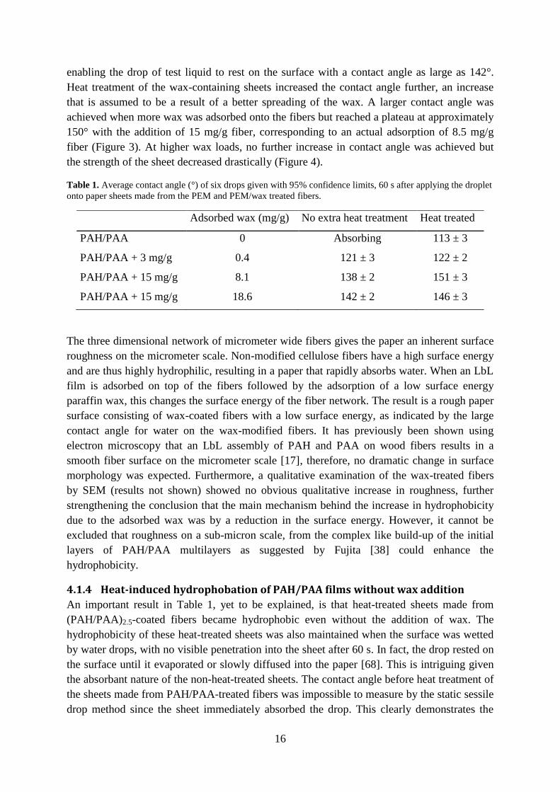
enabling the drop of test liquid to rest on the surface with a contact angle as large as 142°.
Heat treatment of the wax-containing sheets increased the contact angle further, an increase
that is assumed to be a result of a better spreading of the wax. A larger contact angle was
achieved when more wax was adsorbed onto the fibers but reached a plateau at approximately
150° with the addition of 15 mg/g fiber, corresponding to an actual adsorption of 8.5 mg/g
fiber (Figure 3). At higher wax loads, no further increase in contact angle was achieved but
the strength of the sheet decreased drastically (Figure 4).
Table 1. Average contact angle (°) of six drops given with 95% confidence limits, 60 s after applying the droplet
onto paper sheets made from the PEM and PEM/wax treated fibers.
Adsorbed wax (mg/g) No extra heat treatment Heat treated
PAH/PAA 0 Absorbing 113 ± 3
PAH/PAA + 3 mg/g 0.4 121 ± 3 122 ± 2
PAH/PAA + 15 mg/g 8.1 138 ± 2 151 ± 3
PAH/PAA + 15 mg/g 18.6 142 ± 2 146 ± 3
The three dimensional network of micrometer wide fibers gives the paper an inherent surface
roughness on the micrometer scale. Non-modified cellulose fibers have a high surface energy
and are thus highly hydrophilic, resulting in a paper that rapidly absorbs water. When an LbL
film is adsorbed on top of the fibers followed by the adsorption of a low surface energy
paraffin wax, this changes the surface energy of the fiber network. The result is a rough paper
surface consisting of wax-coated fibers with a low surface energy, as indicated by the large
contact angle for water on the wax-modified fibers. It has previously been shown using
electron microscopy that an LbL assembly of PAH and PAA on wood fibers results in a
smooth fiber surface on the micrometer scale [17], therefore, no dramatic change in surface
morphology was expected. Furthermore, a qualitative examination of the wax-treated fibers
by SEM (results not shown) showed no obvious qualitative increase in roughness, further
strengthening the conclusion that the main mechanism behind the increase in hydrophobicity
due to the adsorbed wax was by a reduction in the surface energy. However, it cannot be
excluded that roughness on a sub-micron scale, from the complex like build-up of the initial
layers of PAH/PAA multilayers as suggested by Fujita [38] could enhance the
hydrophobicity.
4.1.4 Heat-induced hydrophobation of PAH/PAA films without wax addition
An important result in Table 1, yet to be explained, is that heat-treated sheets made from
(PAH/PAA)2.5-coated fibers became hydrophobic even without the addition of wax. The
hydrophobicity of these heat-treated sheets was also maintained when the surface was wetted
by water drops, with no visible penetration into the sheet after 60 s. In fact, the drop rested on
the surface until it evaporated or slowly diffused into the paper [68]. This is intriguing given
the absorbant nature of the non-heat-treated sheets. The contact angle before heat treatment of
the sheets made from PAH/PAA-treated fibers was impossible to measure by the static sessile
drop method since the sheet immediately absorbed the drop. This clearly demonstrates the

17
hydrophilicity of the sheets not exposed to the additional heat treatment. It should be noted,
however, that by single fiber measurements using a Cahn balance, Lingström [29] also
showed contact angles larger than 90° also for non-heat-treated PAH/PAA-treated fiber,
indicating the hydrophobic potential of PAH/PAA LbL films on non-smooth substrates.
To explore the mechanisms of the increase in hydrophobicity upon heat-treatment,
(PAH/PAA)2.5 were assembled on smooth model surfaces. Silicon oxide surfaces were used as
well as two different spin-coated NFC and regenerated cellulose model surfaces. For
spectroscopic evaluation of the molecular composition at the solid/air interface, films were
also assembled on transparent microscope glass slides. The PAH/PAA films were assembled
by dipping the model substrates into polyelectrolyte solutions at 120 mg/L with 10 mM NaCl
background electrolyte. A total of 2.5 bilayers were adsorbed, similar to the LbL formation on
fibers. Half the surfaces were then heat-treated for 30 min at 160°C. The results of the contact
angle determinations, presented in Table 2, show that the (PAH/PAA)2.5 film was hydrophilic
on all three substrates with a contact angle of about 40°. These values are in reasonable
agreement with the 47° previously measured by Choi [31] for a PAH-terminated LbL film of
PAH/PAA assembled on a silicon oxide substrate at pH 7.5/3.5. This contact angle is however
higher than that measured on cellulose surfaces [69] indicating that the assembly of the PAH-
terminated LbL film lowers the surface energy compared to that of the native cellulose fiber.
The contact angles measured on flat model films are however in agreement with the
hydrophilicity observed for the non-heat-treated paper sheets.
Table 2. Average contact angle (º) of four drops given with 95% confidence limits, 30 s after applying the drop.
Heat treatment 160°C for 30 min for 2.5 bl films.
Model system No extra heat treatment Heat treated
(PAH/PAA)2.5 on NMMO cellulose 22 ± 1 58 ± 1
(PAH/PAA)2.5 on NFC 38 ± 1 63 ± 2
(PAH/PAA)2.5 on SiO2 40 ± 1 72 ±1
Table 2 shows that heat treatment of the model films resulted in a distinct increase in the
contact angle to a value ranging between 60° and 70°. This increase is suggested to be the
result of a reorientation where more hydrophobic groups of the polymer are oriented to the
solid-air interface of the system in order to minimize the energy of the surface. This would be
possible since the charges of the polyelectrolytes are neutralized during PEM formation [6]
and an increase in temperature increases the mobility within the layers and permits an
orientation of the alkane chain of the polyelectrolytes towards the surrounding air.
To investigate the proposed change in surface chemistry of the PAH/PAA film due to
molecular rearrangements during the heat treatment, the interfacial sensitive spectroscopy
technique VSFS was used. Contact angle determinations were performed on the same
substrates to compare the change in wetting behavior with the composition of the molecules at
the air/solid interface. Slightly thicker films were prepared compared to the previous LbL
assemblies in order to ensure a better surface coverage of the substrate and to make sure that
18
there was a solid bulk of PAH/PAA beneath the surface layer. (PAH/PAA)7.5 films were
assembled on silicon oxide wafers and on glass slides. The use of glass slides was motivated
since VSFS measurements are preferably carried out on transparent substrates in order to
reduce the influence of the non-resonant background which makes the interpretation of the
spectra more difficult. The samples in this study were heat treated for 90 min at 160°C and in
argon atmosphere to prevent contamination.
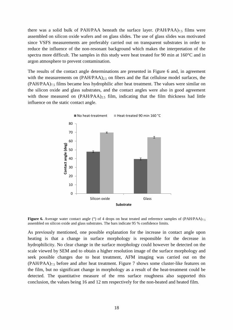
The results of the contact angle determinations are presented in Figure 6 and, in agreement
with the measurements on (PAH/PAA)2.5 on fibers and the flat cellulose model surfaces, the
(PAH/PAA)7.5 films became less hydrophilic after heat treatment. The values were similar on
the silicon oxide and glass substrates, and the contact angles were also in good agreement
with those measured on (PAH/PAA)2.5 film, indicating that the film thickness had little
influence on the static contact angle.
Figure 6. Average water contact angle (°) of 4 drops on heat treated and reference samples of (PAH/PAA)7.5
assembled on silicon oxide and glass substrates. The bars indicate 95 % confidence limits.
As previously mentioned, one possible explanation for the increase in contact angle upon
heating is that a change in surface morphology is responsible for the decrease in
hydrophilicity. No clear change in the surface morphology could however be detected on the
scale viewed by SEM and to obtain a higher resolution image of the surface morphology and
seek possible changes due to heat treatment, AFM imaging was carried out on the
(PAH/PAA)7.5 before and after heat treatment. Figure 7 shows some cluster-like features on
the film, but no significant change in morphology as a result of the heat-treatment could be
detected. The quantitative measure of the rms surface roughness also supported this
conclusion, the values being 16 and 12 nm respectively for the non-heated and heated film.
0
10
20
30
40
50
60
70
80
Silicon oxide Glass
Co
nta
ct a
ngl
e (
de
g)
Substrate
No heat-treatment Heat-treated 90 min 160 °C

19
Figure 7. Tapping Mode AFM images of (PAH/PAA)7.5 on silicon oxide. Left image displays a film that was not
heat treated. The right surface was heat treated for 90 min at 160°C.
Figure 8 shows the VSF spectra of (PAH/PAA)7.5 on glass, with and without heat treatment.
The measurements were conducted in a pure nitrogen atmosphere in order to minimize the
risk of contamination. Two different VSFS active polarization combinations (SSP and PPP)
were employed in order to assist in the spectral assignments.
Figure 8. VSF spectra for the SSP and PPP polarization respectively of (PAH/PAA)7.5 on glass substrate.
In Figure 8, a CH2 symmetric stretch can be identified at 2850 cm-1
in the SSP polarization of
the non-heat-treated sample and its Fermi resonance is visible as a shoulder at around 2920
cm-1
[70] These bands originate from the back-bone of the two polyelectrolytes. Also, as the
position of the symmetric CH2 shifts when it is close to a polar group [71], the band centred at

20
2930 cm-1
includes contributions from CH2 groups located closer to the NH3 and COOH
groups of PAH and PAA respectively [72]. Furthermore, minor additions from weaker bands,
such as CH and asymmetric CH2 cannot be excluded in the region 2875-2920 cm-1
[72-74].
The PPP polarized VSF spectrum of the non-heat-treated sample show no visible peaks,
which is expected since asymmetric CH2, although more favourable in the PPP polarization
compared with SSP[75], are usually weak due to the fact that the asymmetric CH2 vibrations
have dissimilar frequencies in IR and Raman spectra in general[76]. This has also been
observed for e.g. sodium acrylate [77]. The band at 2875 cm-1
, visible in the SSP polarization
combination for the heat treated sample, corresponds to the symmetric CH3 stretch, while in
the PPP polarization combination the asymmetric CH3 stretch is seen at 2963 cm-1
[70]. The
relative strength between the polarization combinations are all in accordance with the so-
called polarization rules and supports the assignment [78]. The aforementioned weakness of
the asymmetric CH2 vibrations in VSFS, which otherwise also could have been the origin of
this band, also supports this assignment The shoulder appearing at 2940 cm-1
in SSP is
assigned to the CH3 Fermi resonance stretching vibration [79] and reside in an environment
where the centrosymmetry is broken [80-82]. In a recent VSFS investigation on PEMs made
from PAH/PSS, Silva et al. assigned the band at 2875 cm-1
to the CH-stretch [83]. However,
the CH stretch is seen at around 2890-2900 cm-1
, and is usually very weak [73, 84].
Furthermore, the use of different polarization combinations definitely excludes this
assignment. Considering the correlation between the increased contact angles, presented in
Figure 6, and the increased CH3 intensity upon heat-treatment, it is concluded that the CH3
signal originates from the polymer/air interface. VSF spectra of substantially thicker
(PAH/PAA)65.5 films (results not shown) were similar to those from (PAH/PAA)7.5 and
supports the fact that the air/polymer interface contributes strongly to the VSF signal.
For the heat-treated sample, a strong peak at 2920 cm-1
can also be observed in the VSF
spectrum in Figure 8. This may be attributed to CH2 Fermi resonance, however the reason for
its strength upon heat treatment is not known. The decrease seen for the 2930 cm-1
peak in
SSP, could be due to that polar COO- and NH3
+ withdraw from the surface, being hydrophilic
groups and thus less energetically favourable at the solid/air interface. This decreases the
number of CH2 close to these groups at the surface and subsequently the VSF signal. The
increased intensity of VSFS signals from the methyl groups clearly shows an enrichment of
CH3 groups at the solid/air interface and one interpretation is that this is due to a reorientation
of the polymer chain-ends, where the methyl groups are located. Considering the demands for
a mode to be VSFS active, it is evident that the CH3 groups form an ordered structure
Although the results of the VSFS experiments provide a plausible mechanism for the increase
in contact angle of the PEMs on flat model surfaces, the discrepancy between the contact
angle of the heat treated-sheets and the model surfaces is yet to be explained. However, Zhai
et.al [28] have reported an advancing contact angle of 115° for a heat-treated micro-structured
LbL film of PAH/PAA, where an acid treatment of a smooth film gave a porous and rough
surface (pore size 10 µm and surface roughness >400 nm). Meanwhile the contact angle for
water on the flat, non-structured and non-heat-treated conformation was 60°. This suggests
that the combination of re-conformation and crosslinking during the heat treatment, together
21
with the surface roughness which display large similarities to the structure of the fibre
network in a paper sheet, creates a hydrophobic surface with the same set of polyelectrolytes
used in this work. A similar mechanism has also been suggested by Feng et.al [85] for
nanofibers of polyvinyl alcohol (PVA) which are highly hydrophobic as in contrast to a
smooth film of the same material. They used angle-resolved XPS to analyze the surfaces and
an enrichment of hydrophobic –CH2- was observed at the solid-air interface compared to the
bulk material. The observations are however contradicted by the general view that all highly
hydrophobic or superhydrophobic surfaces result from microstructures on a surface with a
contact angle originally around 90°. Nevertheless, there is an increasing body of literature
reporting that a surface roughness of the appropriate geometry also leads to a highly
hydrophobic surface from a previously hydrophilic substrate [85-87]. This has also been
theoretically examined by Liu et.al [14], who claim that for certain surface geometries such as
mushroom-like microstructures and hierarchical micro- and nano-structures, highly
hydrophobic surfaces may be created from hydrophilic substrates. If these theories can be
applied to cellulose fiber networks, where it cannot be excluded that such geometries may
exist, a combination with the change in surface chemistry as demonstrated by the VSFS
measurements is sufficient to create a hydrophobic surface.
4.2 Dry adhesion between cellulose, lignin, and glucomannan model
surfaces Smooth and well-defined wood biopolymer model surfaces were prepared and used in dry
adhesion measurements utilizing the JKR technique. For the first time, the elastic
hemispherical PDMS cap was surface modified and coated with NFC. This was made through
the LbL film assembly of PAH(NFC/PEI)1.5 on the cap. Adhesion was measured against flat
model surfaces of cellulose (also PAH(NFC/PEI)1.5 ), hemicellulose and lignin.
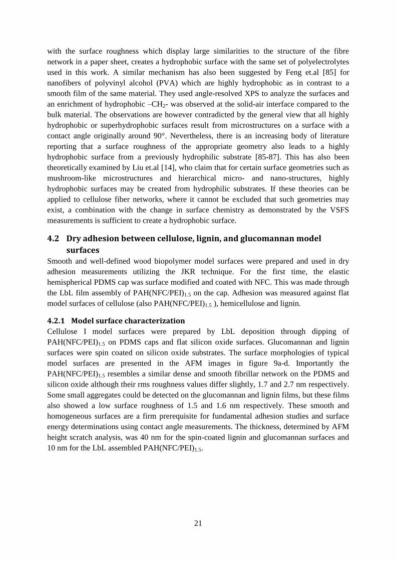
4.2.1 Model surface characterization
Cellulose I model surfaces were prepared by LbL deposition through dipping of
PAH(NFC/PEI)1.5 on PDMS caps and flat silicon oxide surfaces. Glucomannan and lignin
surfaces were spin coated on silicon oxide substrates. The surface morphologies of typical
model surfaces are presented in the AFM images in figure 9a-d. Importantly the
PAH(NFC/PEI)1.5 resembles a similar dense and smooth fibrillar network on the PDMS and
silicon oxide although their rms roughness values differ slightly, 1.7 and 2.7 nm respectively.
Some small aggregates could be detected on the glucomannan and lignin films, but these films
also showed a low surface roughness of 1.5 and 1.6 nm respectively. These smooth and
homogeneous surfaces are a firm prerequisite for fundamental adhesion studies and surface
energy determinations using contact angle measurements. The thickness, determined by AFM
height scratch analysis, was 40 nm for the spin-coated lignin and glucomannan surfaces and
10 nm for the LbL assembled PAH(NFC/PEI)1.5.

22
Figure 9. Tapping-mode AFM height images (1 x 1 µm) of wood biopolymer model surfaces: a)
PAH(NFC/PEI)1.5 on PDMS-cap, b) PAH(NFC/PEI)1.5 on flat silica, c) spin-coated glucomannan on flat silica,
and d) spin-coated lignin on flat silica.
In addition to the characterization of morphology and thickness, the wood polymer model
surfaces, including PAH(NFC/PEI)1.5 on flat PDMS, were also characterized in terms of
interfacial properties by determining the surface energy through contact angle determinations
with water, glycerol and methylene iodine (Table 3).
Table 3. Contact angles of water, glycerol, and methylene iodide on flat wood biopolymer model surfaces
Model surface Water (deg) Glycerol (deg) Methylene iodide (deg)
NFC on SiO2 22 19 34
NFC on PDMS 57 69 67
Glucomannan 19 40 32
Lignin 47 51 37
The total surface energies, as well as their dispersive and polar contributions which provide
information about the surface’s ability to interact with liquids and other solids, were
calculated according to the van Oss method [88]. The results presented in Table 4 indicate
that the total surface energies were similar for the model surfaces of all three main wood
components. The dispersive component was dominant and almost identical for the three wood
biopolymers on SiO2 substrates, all of which displayed the same surface characteristics,
although lignin had a smaller polar component than did the cellulose and glucomannan. The
total surface energy and the dispersive component for the PAH(NFC/PEI)1.5 on silica were in
good agreement with the corresponding values, 58.8 and 40.3 mJm-2
, previously reported for

23
(PEI/NFC)1 on silica [69]. The dispersive component was also similar to the 40-42 mJm-2
previously reported for various spin-coated cellulose surfaces [89]. The surface energy of the
NFC film on PDMS was substantially lower than that of the NFC film on SiO2, due mainly to
a lower dispersive contribution. Since the morphologies of the PAH(NFC/PEI)1.5 films on
PDMS and SiO2 were homogeneous and very similar, the surface energies of
PAH(NFC/PEI)1.5 were also expected to be similar on the two substrates. However, the film
was thinner on the PDMS substrate and if the test liquids can diffuse through the
PAH(NFC/PEI)1.5 film, the substrate will influence the observed contact angle and thereby the
calculated surface energy. The surface energy calculated from the contact angles on
PAH(NFC/PEI)1.5 on SiO2 is thus considered to be closer to the actual surface energy of the
film.
Table 4. Calculated values for the total surface energy and its components for the wood biopolymer model
surfaces.
Model surface γd
(mNm/m2) γ
+ (mNm/m
2) γ
- (mNm/m
2) γ
p (mNm/m
2) γ
T (mNm/m
2)
NFC SiO2 42 3.6 18 16 58
NFC PDMS 24 1.4 21 11 35
Glucomannan 43 1.5 30 14 57
Lignin 41 0.7 18 6.9 48
4.2.2 Adhesion measurements
Figures 10a-c show typical graphs of JKR adhesion measurement data from experiments
conducted under ambient conditions (25%-35% RH) between cellulose/cellulose,
cellulose/glucomannan and cellulose/lignin. The work of adhesion on loading, Wload ,and
unloading, Wunload, were obtained by fitting the data to eq 4. Eq 5 was used to calculate Wmin
from the pull-off force.
Figure 10. The cube of the contact radius (a3) as a function of load for a) cellulose/cellulose, b)
cellulose/glucomannan, and c) cellulose/lignin. The lines are fitted to the data points according to the inverse of
eq 4.
24
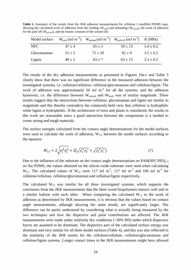
Table 5. Summary of the results from the JKR adhesion measurements for cellulose I–modified PDMS caps,
showing the calculated work of adhesion from the loading (Wload) and unloading (Wunload), the work of adhesion
for the pull-off (Wpull-off), and the elastic constant of the system (K)
Model surface Wload (mJ m–2
) Wunload (mJ m–2
) Wpull-off (mJ m-2
) K (MPa)
NFC 47 ± 4 65 ± 5 59 ± 13 3.4 ± 0.2
Glucomannan 51 ± 5 72 ± 10 62 ± 9 3.5 ± 0.2
Lignin 49 ± 5 63 ± 7 63 ± 13 3.3 ± 0.3
The results of the dry adhesion measurements as presented in Figures 10a-c and Table 5
clearly show that there was no significant difference in the measured adhesion between the
investigated systems, i.e. cellulose/cellulose, cellulose/glucomannan and cellulose/lignin. The
work of adhesion was approximately 50 mJ m-2
for all the systems, and the adhesion
hysteresis, i.e. the difference between Wunload and Wload, was of similar magnitude. These
results suggest that the interactions between cellulose, glucomannan and lignin are similar in
magnitude and this thereby contradicts the commonly-held view that cellulose is hydrophilic
while lignin is hydrophobic. If the architecture of trees and plants is considered, the results in
this work are reasonable since a good interaction between the components is a needed to
create strong and tough materials.
The surface energies calculated from the contact angle determinations for the model surfaces
were used to calculate the work of adhesion, W12, between the model surfaces according to
the equation
√
(√
√
) (7)
Due to the influence of the substrate on the contact angle determinations on PAH(NFC/PEI)1.5
on flat PDMS, the values obtained on the silicon oxide substrate were used when calculating
W12. The calculated values of W12 were 117 mJ m-2
, 117 mJ m-2
and 106 mJ m-2
for
cellulose/cellulose, cellulose/glucomannan and cellulose/lignin respectively.
The calculated W12 was similar for all three investigated systems, which supports the
conclusion from the JKR measurements that the three wood biopolymers interact well and in
a similar fashion with each other. When comparing the calculated W12 to the work of
adhesion as determined by JKR measurements, it is obvious that the values based on contact
angle measurements, although showing the same trends, are significantly larger. The
difference can be partly understood by considering what is actually being measured by the
two techniques and how the dispersive and polar contributions are affected. The JKR
measurements were made under relatively dry conditions (~30% RH) under which dispersive
forces are assumed to be dominant. The dispersive part of the calculated surface energy was
dominant and very similar for all three model surfaces (Table 4), and this was also reflected in
the similarity of the JKR results for the cellulose/cellulose, cellulose/glucomannan and
cellulose/lignin systems. Longer contact times in the JKR measurements might have allowed
25
a reorientation of polymer chains near the interface [90, 91] enabling more polar interactions,
even though the mobility of the wood polymers was probably very limited at that low
humidity. An important comment is that it is rather well established that the test liquid affects
the surfaces in contact angle determinations, leading to a larger measured polar contribution
than is actually the case for the dry substrate [92]. This leads to an overestimation of W12
when calculated from surface energies.
Although the surface roughness of the model surfaces was very low, it cannot be excluded
that the adhesion measured in the JKR measurements was affected. It is well established that
surface roughness leads to a lower adhesion due to a decrease in the true contact area, and
roughness on the nm level might influence the measurements [93, 94]. Nolte [95] also
demonstrated a clear effect of surface roughness when performing JKR adhesion
measurement with LbL-coated PDMS caps. Nevertheless, the measurements in the current
work show clear trends and suggest that there are no great differences in the adhesive
interactions between the investigated wood biopolymers.
26
5 Conclusions Fiber modification using PEMs of PAH/PAA and paraffin wax nano-particles resulted in
highly hydrophobic paper sheets with a contact angle of 150 °. The adsorption of wax
impaired the mechanical properties of the sheets, but at an adsorption of 8 mg wax per gram
fiber there was still a 37 % increase in tensile strength index compared to the untreated
reference pulp. This demonstrates the flexibility and robustness of the LbL technique in
combining the known adhesive effect of (PAH/PAA) with the functionality of the wax
nanoparticles, creating a stronger and highly hydrophobic paper.
Without the addition of wax nanoparticles, heat treatment of sheets made of PAH/PAA
modified fibers resulted in hydrophobic papers displaying a contact angle for water of 113°.
No obvious change in the surface structure was observed, leading to the conclusion that the
change in wetting properties was a result of a change in the surface chemistry of the treated
fibres. This was further supported by contact angle determinations on PAH/PAA films
assembled on well-defined flat cellulose model surfaces, which also exhibited a larger contact
angle for water after heat treatment.
Interfacial sensitive Vibrational Sum Frequency Spectroscopy measurements on PAH/PAA
films on glass substrates showed that heat treatment resulted in an increased intensity of the
CH3 signals at the solid/air interface. This indicated a heat-induced reorientation of the
polymer chain ends, where the methyl groups are located, migrate and reorient towards the
interface. Given the hydrophobicity of these groups, it is suggested that a molecular migration
and reorientation is the cause of the increase in hydrophobicity on heat treatment.
Successful surface modification of PDMS caps using LbL deposition of NFC and PEI,
together with the preparation of smooth homogeneous lignin and glucomannan model
surfaces, made it possible to perform the first known all wood biopolymer JKR adhesion
measurements, measuring the adhesion between cellulose/cellulose, cellulose/lignin and the
cellulose/glucomannan surfaces. The work of adhesion on loading and the adhesion hysteresis
were very similar for all three systems, suggesting that the adhesion between the different
wood biopolymers does not differ greatly. Compared to adhesion values calculated from the
surface energies of the surfaces, obtained from contact angle determinations, the adhesion
values from the JKR measurements were lower than expected. Although on different levels,
there was nothing contradicting in the trends observed from the two methods, indicating that
the mutual adhesive interactions between cellulose/cellulose, cellulose/lignin and
cellulose/hemicellulose are on the same level. It is suggested that these differences are caused
by an overestimation of the surface energy of the surfaces from the contact angle
determinations due to an influence of the liquids on the characterized surfaces and due to a
low but nevertheless existing surface roughness of the model surfaces.
The results create a framework for further fundamental JKR adhesion measurements of these
systems, where it would be interesting to study the influence of variables such as contact time
and humidity as well as chemical and physical modifications of the model surfaces.

27
6 Acknowledgements First and foremost I want to thank my supervisor Professor Lars Wågberg for good
supervision and for always being positive and enthusiastic.
Lars Ödberg is appreciatively acknowledged for fruitful scientific discussions and support
with manuscript writing.
All my co-authors: Erik Johansson, Per Larsson, Jonas Hedberg and Magnus Johnson are
thanked for good collaboration.
I would also like to thank all members of the Fibre Technology group and my co-workers at
Wallenberg Wood Science Center at KTH for contributing to a fun and creative work
environment.
Inga, Mia, Ewa and Mona, thanks for making administrative and practical matters run
smoothly.
The Wallenberg Wood Science Center is thanked for financial support.
28
7 List of abbreviations AFM Atomic force microscopy
AKD Alkyl ketene dimer
CNC Cellulose nanocrystals
DMSO Dimethylsulfoxide
DMAc Dimethyldiacetamine
GGM Galactoglucomannan
JKR Johnson-Kendall-Roberts
LbL Layer-by-layer
NFC Nanofibrillated cellulose
NMMO N-methylmorpholine-N-oxide
PAA Polyacrylic acid
PAH Polyallylamine hydrochloride
PDMS Polydimethylsiloxane
PEI Polyelethylene imine
PEM Polyelectrolyte multilayers
RH Relative humidity
Rms Root mean square
SEM Scanning electron microscopy
SFA Surface force apparatus
VSFS Vibrational sum frequency spectroscopy
29
8 References 1. Israelachvili, J.N., Intermolecular and Surface Forces. FIELD Full Journal Title:1991. 291 pp.
2. Page, D.H., Theory for the tensile strength of paper. Tappi, 1969. 52: p. 674-681.
3. Davison, R.W., Weak link in paper dry strength. Tappi, 1972. 55: p. 567-573.
4. Page, D.H., The mechanism of strength development of dried pulps by beating. Svensk
Papperstidning, 1985. 88(3): p. 30-35.
5. Eriksson, M., A. Torgnysdotter, and L. Wågberg, Surface Modification of Wood Fibers Using
the Polyelectrolyte Multilayer Technique: Effects on Fiber Joint and Paper Strength
Properties. Industrial & Engineering Chemistry Research, 2006. 45(15): p. 5279-5286.
6. Lindström, T., L. Wågberg, and T. Larsson, On the nature of joint strength in paper - a review
of dry and wet strength resins used in paper manufacturing. Advances in Paper Science and
Technology: Transactions of the 13th Fundamental Research Symposium, Vols 1-3, ed. S.J.
Ianson2005, Bury: Pulp & Paper Fundamental Research Society. 457-562.
7. Wågberg, L., S. Forsberg, A. Johansson, and P. Juntti, Engineering of fibre surface properties
by application of the polyelectrolyte multilayer concept. Part I. Modification of paper
strength. J. Pulp Pap. Sci., 2002. 28: p. 222-228.
8. Roberts, J. and Editor, Paper Chemistry, Second Edition1996. 320 pp.
9. Von Bahr, M., R. Seppaenen, F. Tiberg, and B. Zhmud, Dynamic wetting of AKD-sized
papers. Journal of Pulp and Paper Science, 2004. 30(3): p. 74-81.
10. Werner, O., L. Wagberg, and T. Lindstrom, Wetting of structured hydrophobic surfaces by
water droplets. Langmuir, 2005. 21(26): p. 12235-12243.
11. Öner, D. and T.J. McCarthy, Ultrahydrophobic surfaces. Effects of topography length scales
on wettability. Langmuir, 2000. 16(20): p. 7777-7782.
12. Nishino, T., M. Meguro, K. Nakamae, M. Matsushita, and Y. Ueda, The lowest surface free
energy based on -CF3 alignment. Langmuir, 1999. 15(13): p. 4321-4323.
13. Dorrer, C. and J. Rühe, Some thoughts on superhydrophobic wetting. Soft Matter, 2009. 5(1):
p. 51-61.
14. Liu, J.L., X.Q. Feng, G.F. Wang, and S.W. Yu, Mechanisms of superhydrophobicity on
hydrophilic substrates. Journal of Physics-Condensed Matter, 2007. 19(35).
15. Decher, G., Single- and multilayer structures formed on substrates and methods for forming
them. Ger. Offen., 1992: p. 23 pp.
16. Decher, G. and J.B. Schlenoff, eds. Multilayer Thin Films: Sequential Assembly of
Nanocomposite Materials. 2003, WHILEY-VCH Verlag GmbH & Co.
17. Sun, Y.P., X. Zhang, C.Q. Sun, B. Wang, and J.C. Shen, Fabrication of ultrathin film
containing bienzyme of glucose oxidase and glucoamylase based on electrostatic interaction
and its potential application as a maltose sensor. Macromolecular Chemistry and Physics,
1996. 197(1): p. 147-153.
18. Westman, E.H., M. Ek, and L. Wagberg, Antimicrobial activity of polyelectrolyte multilayer-
treated cellulose films. Holzforschung, 2009. 63(1): p. 33-39.
19. Sukhorukov, G.B., A.A. Antipov, A. Voigt, E. Donath, and H. Mohwald, pH-controlled
macromolecule encapsulation in and release from polyelectrolyte multilayer nanocapsules.
Macromolecular Rapid Communications, 2001. 22(1): p. 44-46.
20. Eriksson, M., S.M. Notley, and L. Wågberg, The influence on paper strength properties when
building multilayers of weak polyelectrolytes onto wood fibres. Journal of Colloid and
Interface Science, 2005. 292(1): p. 38-45.
21. Lingstrom, R., L. Wagberg, and P.T. Larsson, Formation of polyelectrolyte multilayers on
fibres: Influence on wettability and fibre/fibre interaction. Journal of Colloid and Interface
Science, 2006. 296(2): p. 396-408.
22. Johansson, E., E. Blomberg, R. Lingström, and L. Wågberg, Adhesive Interaction between
Polyelectrolyte Multilayers of Polyallylamine Hydrochloride and Polyacrylic Acid Studied
Using Atomic Force Microscopy and Surface Force Apparatus. Langmuir, 2009. 25(5): p.
2887-2894.

30
23. Wågberg, L., G. Decher, M. Norgren, T. Lindström, M. Ankerfors, and K. Axnäs, The Build-
Up of Polyelectrolyte Multilayers of Microfibrillated Cellulose and Cationic Polyelectrolytes.
Langmuir, 2008. 24(3): p. 784-795.
24. Aulin, C., E. Johansson, L. Wågberg, and T. Lindström, Self-Organized Films from Cellulose
I Nanofibrils Using the Layer-by-Layer Technique. Biomacromolecules, 2010. 11(4): p. 872-
882.
25. Cranston, E.D. and D.G. Gray, Morphological and optical characterization of polyelectrolyte
multilayers incorporating nanocrystalline cellulose. Biomacromolecules, 2006. 7(9): p. 2522-
2530.
26. Karabulut, E. and L. Wagberg, Design and characterization of cellulose nanofibril-based
freestanding films prepared by layer-by-layer deposition technique. Soft Matter, 2011. 7(7): p.
3467-3474.
27. Glinel, K., M. Prevot, R. Krustev, G.B. Sukhorukov, A.M. Jonas, and H. Moehwald, Control
of the Water Permeability of Polyelectrolyte Multilayers by Deposition of Charged Paraffin
Particles. Langmuir, 2004. 20(12): p. 4898-4902.
28. Zhai, L., F.C. Cebeci, R.E. Cohen, and M.F. Rubner, Stable superhydrophobic coatings from
polyelectrolyte multilayers. Nano Letters, 2004. 4(7): p. 1349-1353.
29. Lingström, R., S.M. Notley, and L. Wågberg, Wettability changes in the formation of
polymeric multilayers on cellulose fibres and their influence on wet adhesion. Journal of
Colloid and Interface Science, 2007. 314(1): p. 1-9.
30. Yoo, D., S.S. Shiratori, and M.F. Rubner, Controlling bilayer composition and surface
wettability of sequentially adsorbed multilayers of weak polyelectrolytes. Macromolecules,
1998. 31(13): p. 4309-4318.
31. Choi, J.Y. and M.F. Rubner, Selective adsorption of amphiphilic block copolymers on weak
polyelectrolyte multilayers. Journal of Macromolecular Science-Pure and Applied Chemistry,
2001. 38(12): p. 1191-1206.
32. Israelachvili, J.N. and G.E. Adams, Measurement of forces between 2 mica surfaces in
aqueous electrolyte solutions in range 0-100 nm. Journal of the Chemical Society-Faraday
Transactions I, 1978. 74: p. 975-&.
33. Ducker, W.A., T.J. Senden, and R.M. Pashley, Direct measurement of colloidal forces using
an atomic force microscope. Nature, 1991. 353(6341): p. 239-241.
34. Johnson, K.L., K. Kendall, and A.D. Roberts, Surface energy and the contact of elastic solid.
Proceedings of the Royal Society of London, Series A: Mathematical, Physical and
Engineering Sciences, 1971. 324(1558): p. 301-13.
35. Chaudhury, M.K. and G.M. Whitesides, Direct Measurement of Interfacial Interactions
between Semispherical Lenses and Flat Sheets of Poly(Dimethylsiloxane) and Their Chemical
Derivatives. Langmuir, 1991. 7(5): p. 1013-1025.
36. Deruelle, M., L. Leger, and M. Tirrell, Adhesion at the solid-elastomer interface - influence of
the interfacial chains. Macromolecules, 1995. 28(22): p. 7419-7428.
37. Norgren, M., S.M. Notley, A. Majtnerova, and G. Gellerstedt, Smooth model surfaces from
lignin derivatives. I. Preparation and characterization. Langmuir, 2006. 22(3): p. 1209-1214.
38. Gunnars, S., L. Wågberg, and M.A. Cohen Stuart, Model films of cellulose: I. Method
development and initial results. Cellulose, 2002. 9(3): p. 239-249.
39. Eriksson, J., M. Malmsten, F. Tiberg, T.H. Callisen, T. Damhus, and K.S. Johansen,
Enzymatic degradation of model cellulose films. Journal of Colloid and Interface Science,
2005. 284(1): p. 99-106.
40. Edgar, C.D. and D.G. Gray, Smooth model cellulose I surfaces from nanocrystal suspensions.
Cellulose, 2003. 10(4): p. 299-306.
41. Ahola, S., J. Salmi, L.S. Johansson, J. Laine, and M. Osterberg, Model films from native
cellulose nanofibrils. Preparation, swelling, and surface interactions. Biomacromolecules,
2008. 9(4): p. 1273-1282.
42. Aulin, C., I. Varga, P.M. Claesson, L. Wågberg, and T. Lindström, Buildup of polyelectrolyte
multilayers of polyethyleneimine and microfibrillated cellulose studied by in situ dual-
polarization interferometry and quartz crystal microbalance with dissipation. Langmuir,
2008. 24(6): p. 2509-2518.
31
43. Holmberg, M., J. Berg, S. Stemme, L. Ödberg, J. Rasmusson, and P. Claesson, Surface Force
Studies of Langmuir-Blodgett Cellulose Films. Journal of Colloid and Interface Science, 1997.
186(2): p. 369-381.
44. Rutland, M.W., A. Carambassis, G.A. Willing, and R.D. Neuman, Surface force
measurements between cellulose surfaces using scanning probe microscopy. Colloids and
Surfaces A: Physicochemical and Engineering Aspects. Frontiers in Colloid Chemistry an
International Festschrift to Professor Stig E. Friberg, 1997. 123-124: p. 369-374.
45. Notley, S.M., B. Pettersson, and L. Wågberg, Direct measurement of attractive van der Waals'
forces between regenerated cellulose surfaces in an aqueous environment. Journal of the
American Chemical Society, 2004. 126(43): p. 13930-13931.
46. Notley, S.M. and M. Norgren, Surface Energy and Wettability of Spin-Coated Thin Films of
Lignin Isolated from Wood. Langmuir, 2010. 26(8): p. 5484-5490.
47. Stiernstedt, J., N. Nordgren, L. Wågberg, H. Brumer, III, D.G. Gray, and M.W. Rutland,
Friction and forces between cellulose model surfaces: A comparison. Journal of Colloid and
Interface Science, 2006. 303(1): p. 117-123.
48. Notley, S.M., M. Eriksson, L. Wågberg, S. Beck, and D.G. Gray, Surface forces
measurements of spin-coated cellulose thin films with different crystallinity. Langmuir, 2006.
22(7): p. 3154-3160.
49. Cranston, E.D., D.G. Gray, and M.W. Rutland, Direct Surface Force Measurements of
Polyelectrolyte Multi layer Films Containing Nanocrystalline Cellulose. Langmuir, 2010.
26(22): p. 17190-17197.
50. Lee, S.B. and P. Luner, The wetting and interfacial properties of lignin. Tappi, 1972. 55(1): p.
116-121.
51. Tammelin, T., M. Österberg, L.S. Johansson, and J. Laine, Preparation of lignin and
extractive model surfaces by using spincoating technique - Application for QCM-D studies.
Nordic Pulp & Paper Research Journal, 2006. 21(4): p. 444-450.
52. Timell, T.E., Recent progress in the chemistry of wood hemicelluloses. Wood Science and
Technology, 1967. V1(1): p. 45-70.
53. Nordgren, N., J. Eklöf, Q. Zhou, H. Brumer, and M.W. Rutland, Top-down grafting of
xyloglucan to gold monitored by QCM-D and AFM: Enzymatic activity and interactions with
cellulose. Biomacromolecules, 2008. 9(3): p. 942-948.
54. Wågberg, L. and R. Hägglund, Kinetics of polyelectrolyte adsorption on cellulosic fibers.
Langmuir, 2001. 17: p. 1096-1103.
55. Wågberg, L., L. Winter, L. Ödberg, and T. Lindström, On the Charge Stoichiometry Upon
Adsorption of a Cationic Polyelectrolyte on Cellulosic Materials. Colloids and Surfaces, 1987.
27(1-3): p. 163-173.
56. Pääkkö, M., M. Ankerfors, H. Kosonen, A. Nykänen, S. Ahola, M. Österberg, J. Ruokolainen,
J. Laine, P.T. Larsson, O. Ikkala, and T. Lindström, Enzymatic hydrolysis combined with
mechanical shearing and high-pressure homogenization for nanoscale cellulose fibrils and
strong gels. Biomacromolecules, 2007. 8(6): p. 1934-1941.
57. Walecka, J.A., Low degree of substitution carboxymethylcelluloses. Tappi FIELD Full Journal
Title:Tappi, 1956. 39: p. 458-63.
58. Norgren, M. and B. Lindstrom, Dissociation of phenolic groups in kraft lignin at elevated
temperatures. Holzforschung, 2000. 54(5): p. 519-527.
59. Zhang, Y., J. Li, M.E. Lindström, A. Stepan, and P. Gatenholm. Spruce glucomannan:
Preparation, Purification, Characterization and Derivatization. in 16th International
Symposium on Wood, Fiber and Pulping Chemistry (16th ISWFPC). 2011. Tianjin, China.
60. Harris, J.J., P.M. DeRose, and M.L. Bruening, Synthesis of passivating, nylon-like coatings
through cross-linking of ultrathin polyelectrolyte films. Journal of the American Chemical
Society, 1999. 121(9): p. 1978-1979.
61. Schultz-Eklund, O., C. Fellers, and P.Å. Johansson, Method for the local determination of the
thickness and density of paper. Nordic Pulp and Paper Research Journal, 1992. 7: p. 133-9,
154.
32
62. Johnson, K.L., K. Kendall, and A.D. Roberts, Surface Energy and the Contact of Elastic
Solids. Proceedings of the Royal Society of London. A. Mathematical and Physical Sciences,
1971. 324(1558): p. 301-313.
63. Vanoss, C.J., M.K. Chaudhury, and R.J. Good, Monopolar Surfaces. Advances in Colloid and
Interface Science, 1987. 28(1): p. 35-64.
64. Della Volpe, C. and S. Siboni, Some reflections on acid-base solid surface free energy
theories. Journal of Colloid and Interface Science, 1997. 195(1): p. 121-136.
65. Johnson, C.M., E. Tyrode, S. Baldelli, M.W. Rutland, and C. Leygraf, A vibrational sum
frequency spectroscopy study of the liquid-gas interface of acetic acid-water mixtures: 1.
Surface speciation. Journal Of Physical Chemistry B, 2005. 109(1): p. 321-328.
66. Zheng, Z.G., J. McDonald, R. Khillan, Y. Su, T. Shutava, G. Grozdits, and Y.M. Lvov, Layer-
by-layer nanocoating of lignocellulose fibers for enhanced paper properties. Journal of
Nanoscience and Nanotechnology, 2006. 6(3): p. 624-632.
67. Larsson, P.A. and L. Wågberg, Influence of fibre-fibre joint properties on the dimensional
stability of paper. Cellulose, 2008. 15(4): p. 515-525.
68. Larsson, P.A. and L. Wagberg, Diffusion-induced dimensional changes in papers and fibrillar
films: influence of hydrophobicity and fibre-wall cross-linking. Cellulose, 2010. 17(5): p. 891-
901.
69. Aulin, C., S. Ahola, P. Josefsson, T. Nishino, Y. Hirose, M. Österberg, and L. Wågberg,
Nanoscale Cellulose Films with Different Crystallinities and Mesostructures-Their Surface
Properties and Interaction with Water. Langmuir, 2009. 25(13): p. 7675-7685.
70. Snyder, R.G., H.L. Strauss, and C.A. Elliger, C-H stretching modes and the structure of
normal-aklyl chains.1. long, disordered chains Journal of Physical Chemistry, 1982. 86(26):
p. 5145-5150.
71. Hsi, S.C., A.P. Tulloch, H.H. Mantsch, and D.G. Cameron, A vibrational study of the CD2
stretching bands of selectively deuterated palmitic and stearic acids. Chemistry and Physics
of Lipids, 1982. 31(1): p. 97-103.
72. Murli, C. and Y. Song, Pressure-Induced Polymerization of Acrylic Acid: A Raman
Spectroscopic Study. The Journal of Physical Chemistry B, 2010. 114(30): p. 9744-9750.
73. Fox, J.J. and A.E. Martin, Investigations of Infra-Red Spectra. Determination of C-H
Frequencies ($\sim $ 3000 cm.$^-1 $) in Paraffins and Olefins, with Some Observations on
"Polythenes". Proceedings of the Royal Society of London. Series A. Mathematical and
Physical Sciences, 1940. 175(961): p. 208-233.
74. Lu, R., W. Gan, B.-h. Wu, Z. Zhang, Y. Guo, and H.-f. Wang, C−H Stretching Vibrations of
Methyl, Methylene and Methine Groups at the Vapor/Alcohol (n = 1−8) Interfaces. The
Journal of Physical Chemistry B, 2005. 109(29): p. 14118-14129.
75. Wang, H.-F., W. Gan, R. Lu, Y. Rao, and B.-H. Wu, Quantitative spectral and orientational
analysis in surface sum frequency generation vibrational spectroscopy (SFG-VS).
International Reviews In Physical Chemistry, 2005. 24(2): p. 191-256.
76. Tyrode, E. and J. Hedberg, A Comparative Study of the CD and CH Stretching Spectral
Regions of Typical Surfactants Systems Using VSFS: Orientation Analysis of the Terminal
CH3 and CD3 Groups. The Journal of Physical Chemistry C, 2011. 116(1): p. 1080-1091.
77. Feairheller Jr, W.R. and J.E. Katon, The vibrational spectra of acrylic acid and sodium
acrylate. Spectrochimica Acta Part A: Molecular Spectroscopy, 1967. 23(8): p. 2225-2232.
78. Wang, H.F., W. Gan, R. Lu, Y. Rao, and B.H. Wu, Quantitative spectral and orientational
analysis in surface sum frequency generation vibrational spectroscopy (SFG-VS).
International Reviews In Physical Chemistry, 2005. 24(2): p. 191-256.
79. Snyder, R.G., H.L. Strauss, and C.A. Elliger, Carbon-hydrogen stretching modes and the
structure of n-alkyl chains. 1. Long, disordered chains. The Journal of Physical Chemistry,
1982. 86(26): p. 5145-5150.
80. Bain, C.D., Sum-frequency vibrational spectroscopy of the solid-liquid interface. Journal of
the Chemical Society-Faraday Transactions, 1995. 91(9): p. 1281-1296.
81. Shen, Y.R., Optical 2nd harmonic generation at interfaces. Annual Review of Physical
Chemistry, 1989. 40: p. 327-350.
82. Shen, Y.R., Surfaces probed by nonlinear optics. Surface Science, 1994. 299(1-3): p. 551-562.

33
83. Silva, H.S. and P.B. Miranda, Molecular Ordering of Layer-by-Layer Polyelectrolyte Films
Studied by Sum-Frequency Vibrational Spectroscopy. Journal Of Physical Chemistry B, 2009.
113(30): p. 10068-10071.
84. Lu, R., W. Gan, B.H. Wu, Z. Zhang, Y. Guo, and H.F. Wang, C-H stretching vibrations of
methyl, methylene and methine groups at the vapor/alcohol (n=1-8) interfaces. Journal Of
Physical Chemistry B, 2005. 109(29): p. 14118-14129.
85. Feng, L., Y.L. Song, J. Zhai, B.Q. Liu, J. Xu, L. Jiang, and D.B. Zhu, Creation of a
superhydrophobic surface from an amphiphilic polymer. Angewandte Chemie-International
Edition, 2003. 42(7): p. 800-802.
86. Herminghaus, S., Roughness-induced non-wetting. Europhysics Letters, 2000. 52(2): p. 165-
170.
87. Zhu, M.F., W.W. Zuo, H. Yu, W. Yang, and Y.M. Chen, Superhydrophobic surface directly
created by electrospinning based on hydrophilic material. Journal of Materials Science, 2006.
41(12): p. 3793-3797.
88. Van Oss, C.J., Interfacial Forces in Aqueous Media. FIELD Full Journal Title:1994, New
York: Marcel Dekker. 432 pp.
89. Eriksson, M., S.M. Notley, and L. Wågberg, Cellulose Thin Films: Degree of Cellulose
Ordering and Its Influence on Adhesion. Biomacromolecules, 2007. 8(3): p. 912-919.
90. Choi, G.Y., S.J. Kim, and A. Ulman, Adhesion hysteresis studies of extracted
poly(dimethylsiloxane) using contact mechanics. Langmuir, 1997. 13(23): p. 6333-6338.
91. Kim, S.J., G.Y. Choi, A. Ulman, and C. Fleischer, Effect of chemical functionality on
adhesion hysteresis. Langmuir, 1997. 13(25): p. 6850-6856.
92. Berg, J.C. and Editor, Wettability. [In: Surfactant Sci. Ser., 1993; 49]. FIELD Full Journal
Title:1993. 531 pp.
93. Kendall, K., Molecular Adhesion and Its Applications: The Sticky Universe2001, New York:
Kluwer Academic/Plenum Publishers.
94. Zappone, B., K.J. Rosenberg, and J. Israelachvili, Role of nanometer roughness on the
adhesion and friction of a rough polymer surface and a molecularly smooth mica surface.
Tribology Letters, 2007. 26(3): p. 191-201.
95. Nolte, A.J., J.Y. Chung, M.L. Walker, and C.M. Stafford, In situ Adhesion Measurements
Utilizing Layer-by-Layer Functionalized Surfaces. Acs Applied Materials & Interfaces, 2009.
1(2): p. 373-380.