Systemic and Opportunistic Fungi
5
SYSTEMIC and PATHOGENIC FUNGI FUNGAL AGENT INFECTION MACROCONIDI A MICROCONIDIA FUNGAL COLONY CHARACTERISTICS TEMP. Blood- Enriched Medium Medium Lacking Blood Enrichment Blood- Enriched Medium Nonblood- Enriched Medium Blastomyces dermatitidis Blastomycosis Slow to moderate growth Young colonies tenacious, older colonies glabrous to woolly Spicules in center of colony Oval, pyriform, to globose smooth conidia borne on short, lateral hypha-like conidiosph ores Colonies are cream to tan, soft, moist, wrinkled, waxy, flat to heaped and yeaslike; “tufts” of hyphae often project upward from colonies Colonies are white to cream to tan, some with drops of exudates present, fluffy to glabrous, and adherent to the agar surface Hyphae- 1-2m in diameter are present; some are aggregat ed in ropelike clusters ; sporulat ion is rare Hyphae-1- 2m in diameter are present; single pyriform conidia are produced on short to long conidiosp hores; some cultures produce few conidia 30⁰C Slow growth White to dark tan Small, one- celled, Colonies are heaped, Colonies are white, cream, tan Hyphae- 1-2m in diameter Young cultures usually
-
Upload
ria-alcantara -
Category
Documents
-
view
218 -
download
0
description
Systemic and Opportunistic Fungi
Transcript of Systemic and Opportunistic Fungi
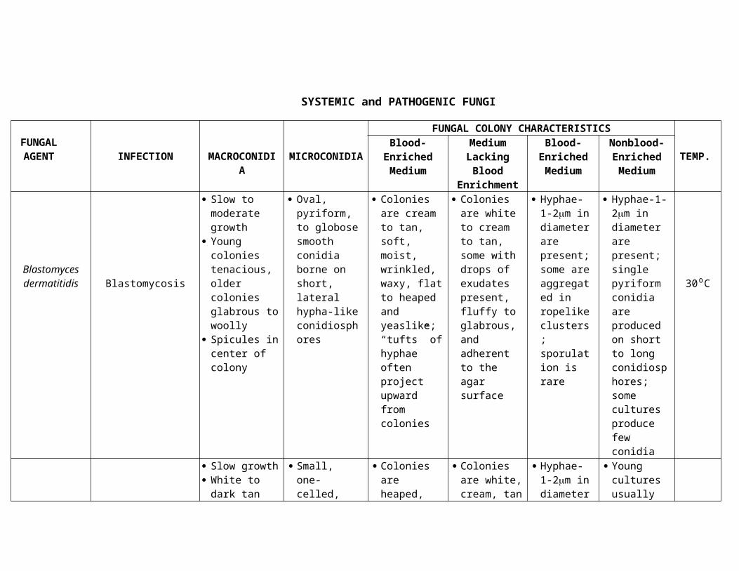
SYSTEMIC and PATHOGENIC FUNGI
FUNGAL AGENT INFECTION MACROCONIDIA MICROCONIDIA
FUNGAL COLONY CHARACTERISTICS
TEMP.Blood-Enriched
MediumMedium
Lacking Blood Enrichment
Blood-Enriched Medium
Nonblood-Enriched Medium
Blastomyces dermatitidis Blastomycosis
Slow to moderate growth
Young colonies tenacious, older colonies glabrous to woolly
Spicules in center of colony
Oval, pyriform, to globose smooth conidia borne on short, lateral hypha-like conidiosphores
Colonies are cream to tan, soft, moist, wrinkled, waxy, flat to heaped and yeaslike; “tufts” of hyphae often project upward from colonies
Colonies are white to cream to tan, some with drops of exudates present, fluffy to glabrous, and adherent to the agar surface
Hyphae-1-2m in diameter are present; some are aggregated in ropelike clusters; sporulation is rare
Hyphae-1-2m in diameter are present; single pyriform conidia are produced on short to long conidiosphores; some cultures produce few conidia
30⁰C
Histoplasma capsulatum
Histoplasmosis
Slow growth White to dark
tan with age Woolly, cottony
or granular
Small, one-celled, round, smooth
Tuberculated macroconidia large, round
Colonies are heaped, moist, wrinkled, yeastlike, soft and cream, tan or pink in color; “tufts” of hyphae often project upward from colonies
Colonies are white, cream, tan or gray, fluffy to glabrous; some colonies appear yeastlike and adherent to the agar surface; many variations in
Hyphae-1-2m in diameter are present; some are aggregated in ropelike clusters; sporulation is rare
Young cultures usually have a predominance of smooth-walled macroconidia that become tuberculate with age; macroconidia
30⁰C
colonial morphology occur
may be pyriform or spherical; some isolates produce small pyriform microconidia in the presence or absence of microconidia
Paracoccidioides brasiliensis
Paracoccidiodomycosis
Slow growth White to beige Colony
glabrous, leathery, flat to wrinkled, folded or velvety
Colonies frequently only produce sterile hyphae
Fresh isolates may produce conidia similar to those of B.dermatitidis
Colonies are heaped, wrinkled, moist, and yeastlike; with age, colonies may become covered with short aerial mycelium and may turn brown
Hyphae-1-2m in diameter are present; some isolates produce conidia similar to those of B.dermatitidis; chlamydoconidia may be numerous, and multiple budding yeast cells 10-25m in diameter may be present
30⁰C
Rhizopus spp.Mucor spp.
Zygomycetes spp.
Zygomycosis
Colonies are extremely fast growing, wooly, and gray to brown to gray-black
Rhizopus spp.- rhizoids are produced at the base of sporangiophores
Mucor spp.-no rhizoids are produced
30⁰C
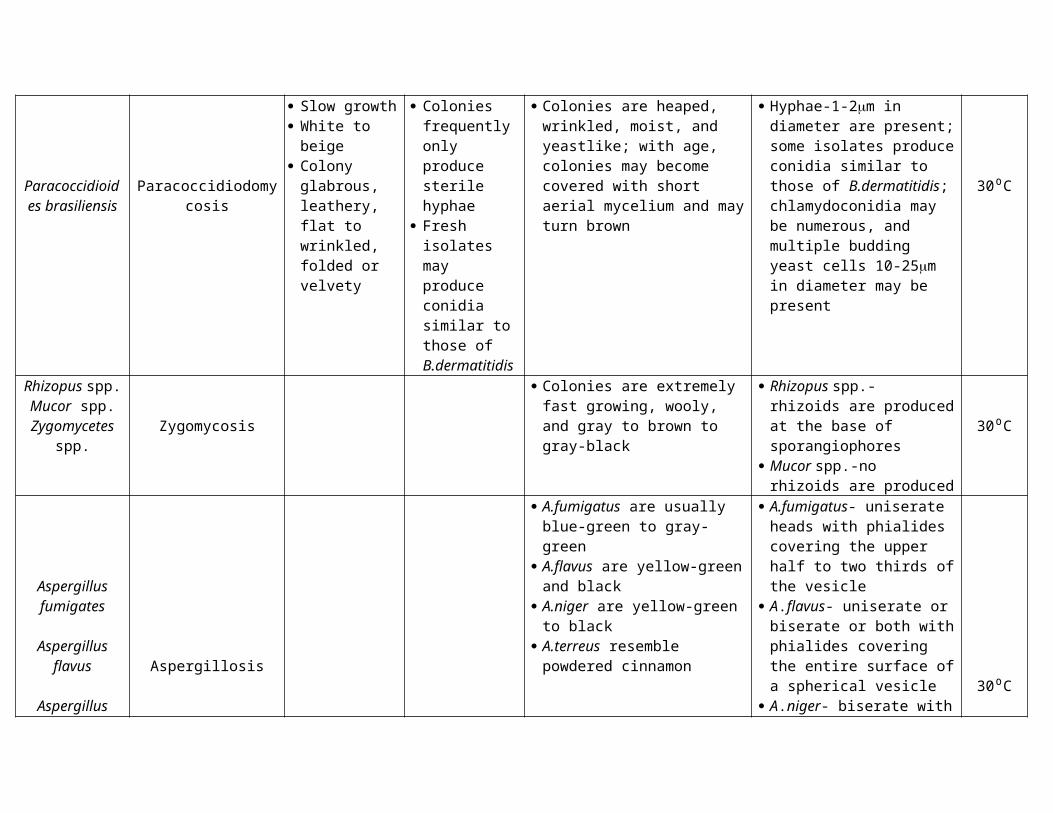
Aspergillus fumigates
Aspergillus flavus
Aspergillus niger
Aspergillus terreus
Aspergillosis
A.fumigatus are usually blue-green to gray-green
A.flavus are yellow-green and black
A.niger are yellow-green to black A.terreus resemble powdered
cinnamon
A.fumigatus- uniserate heads with phialides covering the upper half to two thirds of the vesicle
A.flavus- uniserate or biserate or both with phialides covering the entire surface of a spherical vesicle
A.niger- biserate with phialides covering the entire surface of a spherical vesicle; conidia are black
A.terreus-biserate with phialides covering the entire surface of a hemispherical vesicle; aleurioconidia are formed on submerged hyphae
30⁰C
Coccidioides immitis
Coccidioidomycosis
Rapid growth White to tan to
dark gray Young colonies
tenacious, older colonies cottony
Tend to grow in concentric rings
Alternating one-celled, “barrel-shaped” arthroconidia with disjunctor cells
Colonies may be white and fluffy to greenish on blood-enriched media; some isolates are yeastlike, heaped, wrinkled, and membranous
Colonies usually are fluffy white but may be pigmented gray, orange, brown or yellow; mycelium is adherent to the agar surface in some portions of the colony
Chains of alternate, barrel-shaped arthroconidia are characteristic; some arthroconidia may be elongated; hyphae are small and often arranged in ropelike strands, and racquet forms are seen in young cultures 30⁰C