Synthesis of Tyrian purple precursors in the...
34
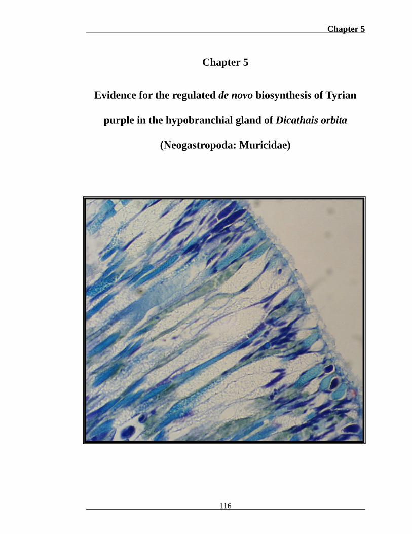
Chapter 5 Chapter 5 Evidence for the regulated de novo biosynthesis of Tyrian purple in the hypobranchial gland of Dicathais orbita (Neogastropoda: Muricidae) 116
Transcript of Synthesis of Tyrian purple precursors in the...
Chapter 5
Chapter 5
Evidence for the regulated de novo biosynthesis of Tyrian
purple in the hypobranchial gland of Dicathais orbita
(Neogastropoda: Muricidae)
116
Chapter 5
5. 0 Abstract
Investigations into Tyrian purple genesis within hypobranchial glands of
muricid molluscs have provided a wealth of information on dye composition and
biosynthesis from colourless prochromogens. However, little is known on the origin
of the precursor, tryptophan, and regulatory processes governing prochromogen and
bioactive intermediate synthesis. As an alternative to radioisotopes, novel
histochemical techniques for the demonstration of bromoperoxidase and the
brominated prochromogen, tyrindoxyl sulphate, were applied in conjunction with
standard stains for tryptophan and arylsulphatase. Two sites of prochromogen
synthesis were identified within the hypobranchial gland of the muricid, Dicathais
orbita. Endocytosed tryptophan sourced from rectum waste appears to be stored
within secretory cells of the branchial and rectal hypobranchial epithelium, which
upon liberation onto the epithelial surface, is united with bromoperoxidase to form
crystals of tyrindoxyl sulphate. Prochromogen synthesis also occurs in the vascular
sinus immediately dorsal of the medial hypobranchial gland for storage and secretion
by medial epithelial cells. Hydrolysis of tyrindoxyl sulphate and subsequent
production of bioactive intermediates appears to be regulated, and only occurs once
arylsulphatase is exocytosed onto the surface by adjacent supportive cells. This
histochemical approach identified the origin of tryptophan, provided evidence for
post-translational precursor bromination, the constitutive and controlled genesis of
prochromogens and the regulated synthesis of bioactive intermediates in D. orbita.
Together these findings strongly suggest the de novo synthesis of Tyrian purple
117
Chapter 5
precursors in the Muricidae, which further implies a naturally selected function for
the synthesis of these bioactive metabolites.
5. 1 Introduction
Examination of stable isotope incorporation by mass spectrometry and high
field NMR has been traditionally employed in determining the biosynthetic origin of
secondary metabolites in marine molluscs (Cimino et al., 2004). However, low
incorporation rates often render questionable results (Cimino et al., 2004) and labeled
precursor diversion into extraneous metabolic pathways (Garson, 1993) can be
misleading. A reliance on radioisotope incorporation is also insensitive to the
progressively recognized role of symbiotic microorganisms in natural product
biosynthesis (Cimino et al., 2004). Furthermore, this technique fails to provide
anatomical information, which is particularly useful to subsequent genetic
investigations, as natural products are often stored in a different cellular environment
to where they are synthesized (Garson, 1993). Although rarely employed, a
histochemical approach has the potential to address some of these limitations and
ultimately enhance our understanding of secondary metabolite biosynthesis.
Histochemical techniques are available for various amino acids, proteins,
glycoproteins, carbohydrates, lipids and enzymes (see Thompson 1966). The
application of staining reactions for proposed precursors and enzymes can not only
establish the primary metabolic origin of natural products, but identify sites of active
biosynthesis and qualitatively define the comparative concentration and activity of
precursors and enzymes, respectively. The biochemical and morphological properties
of associated biosynthetic tissues can also provide information on the transport,
118
Chapter 5
storage and secretion biosynthetic constituents and highlight potential regulatory
mechanisms. One highly renowned marine natural product, and an ideal model for
histochemical biosynthetic investigations, is Tyrian purple.
Tyrian purple, also known as Royal purple, Shellfish purple or Purple of the
Ancients, is an historically important dye obtained exclusively from marine molluscs
of the family Muricidae. Evidence for the commercial production of Tyrian purple
since the 13th century B.C. (Naegel and Cooksey, 2002) has prompted investigation
into the genesis of this dye within muricid hypobranchial glands. In 1909, Friedländer
investigated the chemical composition of Tyrian purple from Murex brandaris, and
elucidated the purple pigment as the brominated indole, 6,6’-dibromoindigo (5, Fig.
1). Identification of four prochromogens (Fig. 1) within the hypobranchial gland of
M. trunculus (Fouquet and Bielig, 1971; Baker, 1974), and a fifth (1, Fig. 1) from
Dicathais orbita (Baker and Sutherland, 1968) followed this discovery. These
colourless compounds were found to be 6-bromoindoxyl and indoxyl sulphate salts of
choline esters, substituted with methylthio or methylsulphonyl (McGovern and
Michel, 1990). Enzymatic hydrolysis of prochromogens by purpurase
(arylsulphatase) (Erspamer, 1946) liberates associated choline esters (X+, Fig. 1) and
leads to the formation of yellow indoxyl intermediate precursors. Substituted
bromoindoxyl intermediates (e.g. 2, Fig. 1) are then oxidized to red indoleninones
(e.g. 3, Fig. 1) and dimerize to give green tyriverdin (4, Fig. 1) (Christophersen et al.,
1978; McGovern and Michel, 1990). External to the mollusc, tyriverdin undergoes
photolytic cleavage to yield dimethyl sulphide and 6,6’-dibromoindigo (Baker and
Sutherland, 1968).
119
Chapter 5
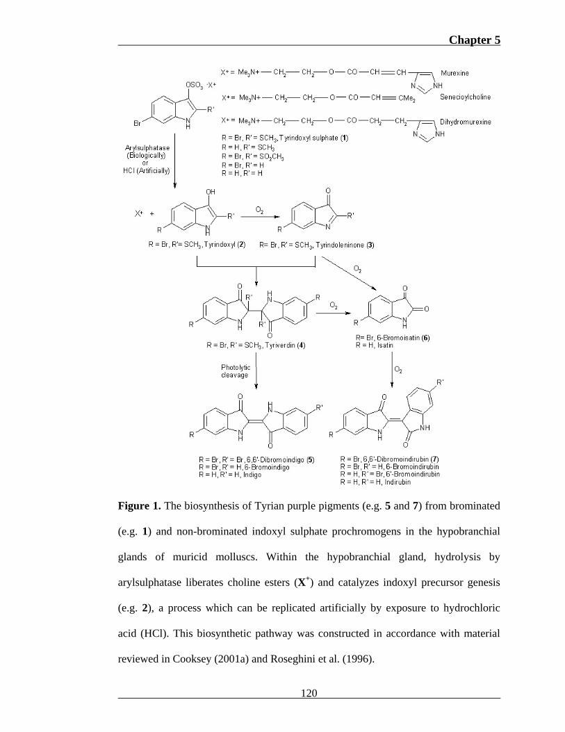
Figure 1. The biosynthesis of Tyrian purple pigments (e.g. 5 and 7) from brominated
(e.g. 1) and non-brominated indoxyl sulphate prochromogens in the hypobranchial
glands of muricid molluscs. Within the hypobranchial gland, hydrolysis by
arylsulphatase liberates choline esters (X+) and catalyzes indoxyl precursor genesis
(e.g. 2), a process which can be replicated artificially by exposure to hydrochloric
acid (HCl). This biosynthetic pathway was constructed in accordance with material
reviewed in Cooksey (2001a) and Roseghini et al. (1996).
120
Chapter 5
Depending on species prochromogen diversity, dibromoindirubin (7), mono-
and non-brominated indoles and indirubins may also evolve as dye components
(Wouters and Verhecken, 1991; Wouters, 1992; Koren, 1995; Cooksey, 2001a;
Cooksey and Withnall, 2001; Karapanagiotis and de Villemereuil, 2006). Despite the
wealth of information that exists on the chemical composition of dye pigments and
their synthesis from colourless prochromogens, little is known on the biosynthetic
origin of prochromogens and the regulatory processes governing genesis.
Indole biosynthesis commences with the essential amino acid tryptophan.
Tryptophan synthesis from the phosphorylated sugar, erythrose-4-phosphate, is
almost completely restricted to plants (Crawford, 1989; Bentley, 1990; Herrmann et
al., 1992). Consequently, de novo conversion of dietary derived tryptophan to indole
or directly to indoxyl sulphate remains the most plausible explanation for the
presence of prochromogens in the Muricidae (Westley et al., 2006). Tryptophan has
been detected in hypobranchial secretory cells of the muricids M. brandaris
(Bolognani-Fantin and Ottaviani, 1981), M. granulata (Srilakshmi, 1991) and P.
pansa (Naegel and Aguilar-Cruz, 2006). However, the origin of this amino acid and
the mode of transportation and concentration within these cells have not been
addressed. A recent investigation into the histomorphology of D. orbita proposed the
rectum and lateral hypobranchial secretory cells as the source and site of tryptophan
storage, respectively (Chapter 3). It has also been assumed that histochemical
demonstration of tryptophan is synonymous with Tyrian purple precursor localization
(Naegel and Aguilar-Cruz, 2006). However, it is unlikely that the methods employed
(Adams, 1957; Davenport, 1960) would react with indole precursors due to the lack
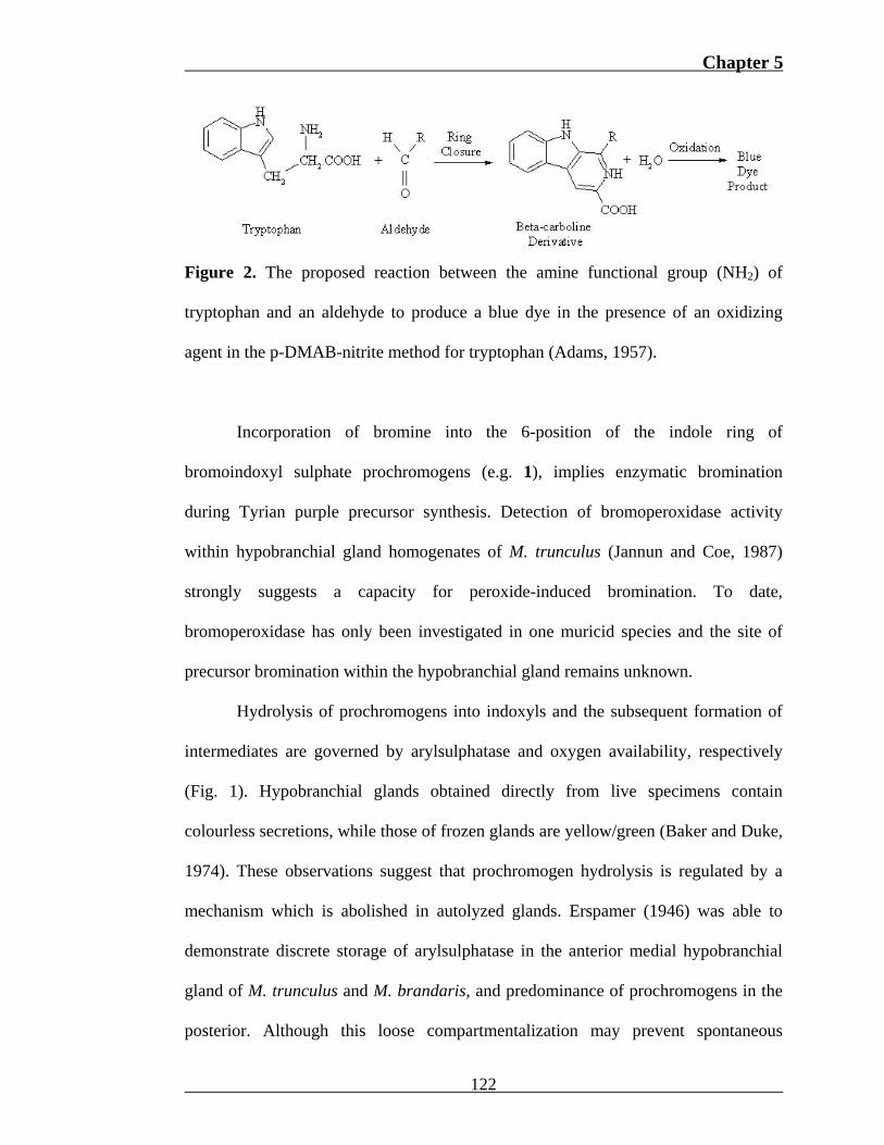
of an available amine functional group (Fig. 2).
121
Chapter 5
Figure 2. The proposed reaction between the amine functional group (NH2) of
tryptophan and an aldehyde to produce a blue dye in the presence of an oxidizing
agent in the p-DMAB-nitrite method for tryptophan (Adams, 1957).
Incorporation of bromine into the 6-position of the indole ring of
bromoindoxyl sulphate prochromogens (e.g. 1), implies enzymatic bromination
during Tyrian purple precursor synthesis. Detection of bromoperoxidase activity
within hypobranchial gland homogenates of M. trunculus (Jannun and Coe, 1987)
strongly suggests a capacity for peroxide-induced bromination. To date,
bromoperoxidase has only been investigated in one muricid species and the site of
precursor bromination within the hypobranchial gland remains unknown.
Hydrolysis of prochromogens into indoxyls and the subsequent formation of
intermediates are governed by arylsulphatase and oxygen availability, respectively
(Fig. 1). Hypobranchial glands obtained directly from live specimens contain
colourless secretions, while those of frozen glands are yellow/green (Baker and Duke,
1974). These observations suggest that prochromogen hydrolysis is regulated by a
mechanism which is abolished in autolyzed glands. Erspamer (1946) was able to
demonstrate discrete storage of arylsulphatase in the anterior medial hypobranchial
gland of M. trunculus and M. brandaris, and predominance of prochromogens in the
posterior. Although this loose compartmentalization may prevent spontaneous
122
Chapter 5
prochromogen cleavage, the coincidence of prochromogens and arylsulphatase in the
anterior medial region in M. brandaris (Erspamper, 1946), suggests the presence of a
more complex mechanism. Furthermore, prochromogen hydrolysis results in the
liberation and subsequent synthesis of a suite of compounds (Fig. 1), which may be of
selective benefit to the Muricidae (see Westley et al., 2006). The choline esters (X+),
murexine (Erspamer and Dordoni, 1947), senecioylcholine (Whittaker, 1957) and
dihydromurexine (Roseghini, 1971), display neuromuscular blocking and nicotinic
actions (reviewed in Roseghini et al., 1996) and intermediates, tyrindoleninone (3), 6-
bromoisatin (6) and tyriverdin (4) possess cytotoxic properties (Benkendorff et al.,
2000; Westley et al., 2006; Vine et al., 2007). The demonstrated activity of these
compounds together with the apparent regulation of their synthesis strongly suggests
an intricate mechanism for instigating prochromogen cleavage within muricid
hypobranchial glands.
D. orbita contains a single prochromogen, tyrindoxyl sulphate (1) (Baker and
Sutherland, 1968) and consequently, possesses the most simplistic biosynthetic
pathway to Tyrian purple. Thus, D. orbita is an ideal species for investigating
prochromogen biosynthesis and hydrolysis within the Muricidae. Through the
application of novel histochemical techniques for the localization of precursors and
enzymes required for Tyrian purple synthesis, this investigation aims provide insight
into the biosynthetic origin and regulatory mechanisms governing dye synthesis.
5. 2 Methods and materials
A total of 24 female D. orbita specimens were collected from the rocky
intertidal and subtidal regions of the Adelaide metropolitan coast, Fleurieu and Eyre
123

Chapter 5
peninsulas of South Australia. As hypobranchial gland secretory activity is thought to
elevate during the breeding season (Fretter and Graham 1994), prochromogen
synthesis and enzyme activity may become more evident during the copulating or
egg-laying seasons. Consequently, six females representing each of four reproductive
phases were collected over the annual cycles of 2005 and 2006. These phases
included; 1) post-reproductive, March; 2) pre-reproductive, early July; 3) copulating,
August-September; and 4) egg-laying, late November-December.
The shell of each live specimen was removed by cracking with a vice at the
junction of the primary body whorl and spire, and the soft body removed by severing
the columnar muscle. The soft body was then transferred to a dissecting tray and
submersed in filtered (0.22µm) seawater to reduce osmotic stress. The dorsal mantle
and pallial gonoduct were separated from the rest of the visceral mass by an incision
along the lateral margins of the columnar muscle. The mantle was then folded back
and pinned with the ventral surface facing up. Longitudinal and transverse incisions
were made along the junction between the ctenidium and branchial hypobranchial
epithelium, and the posterior gonoduct and digestive gland, respectively. Integrity
between the hypobranchial gland and gonoduct was maintained to prevent damage to
the rectal hypobranchial region and to allow histochemical examination the rectum
and rectal gland as possible sites of dietary tryptophan.
Of the 24 specimens, 12 were fixed in 10% neutral buffered formalin (6h),
dehydrated through an ethanol series, cleared in chloroform, embedded in paraffin
and sectioned at 5µm. Tissue from the remaining 12 specimens was immediately
fresh-frozen in O.C.T. compound (Tissue-Tek®) at -20°C, sectioned at 15µm and
124
Chapter 5
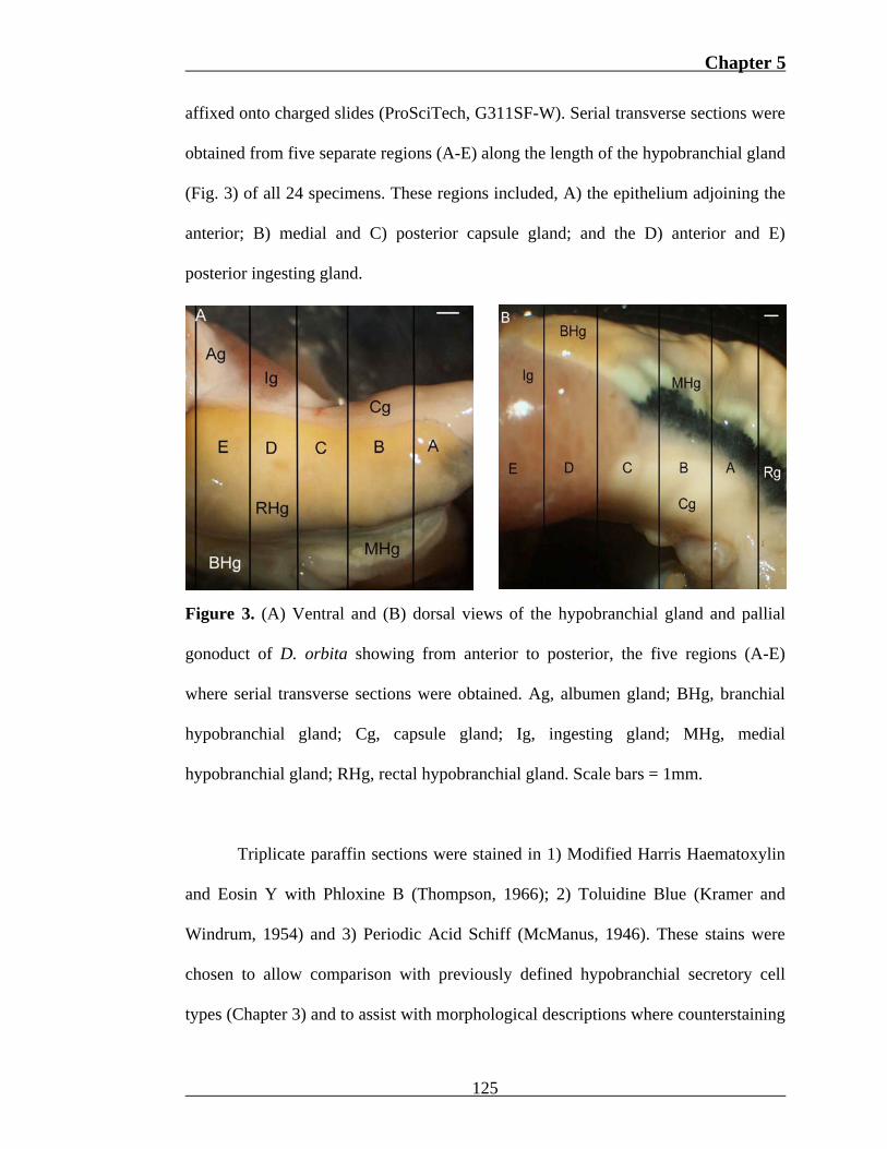
affixed onto charged slides (ProSciTech, G311SF-W). Serial transverse sections were
obtained from five separate regions (A-E) along the length of the hypobranchial gland
(Fig. 3) of all 24 specimens. These regions included, A) the epithelium adjoining the
anterior; B) medial and C) posterior capsule gland; and the D) anterior and E)
posterior ingesting gland.
Figure 3. (A) Ventral and (B) dorsal views of the hypobranchial gland and pallial
gonoduct of D. orbita showing from anterior to posterior, the five regions (A-E)
where serial transverse sections were obtained. Ag, albumen gland; BHg, branchial
hypobranchial gland; Cg, capsule gland; Ig, ingesting gland; MHg, medial
hypobranchial gland; RHg, rectal hypobranchial gland. Scale bars = 1mm.
Triplicate paraffin sections were stained in 1) Modified Harris Haematoxylin
and Eosin Y with Phloxine B (Thompson, 1966); 2) Toluidine Blue (Kramer and
Windrum, 1954) and 3) Periodic Acid Schiff (McManus, 1946). These stains were
chosen to allow comparison with previously defined hypobranchial secretory cell
types (Chapter 3) and to assist with morphological descriptions where counterstaining
125
Chapter 5
was detrimental to enzyme localization. Triplicate sections were also stained with the
p-DMAB-nitrite method for tryptophan (Adams, 1957) and counter stained in
Nuclear Fast Red (C.I. 60760).
Triplicate cryostat sections from each region (Fig. 3) were stained with an
optimized bromoperhnol-red method for bromoperoxidase (Appendix II), an acid-
hydrolysis method developed for tyrindoxyl sulphate (Appendix III) and the post-
coupling method for arylsulphatase (Rutenburg et al., 1952). The bromophenol-red
method is a histochemical technique adapted from Krenn et al. (1989) and Wever et
al. (1991) based on the enzymatic conversion of phenol red (50 µM) to bromophenol
blue in the presence of hydrogen peroxide (20µM) and potassium bromide (100Mm)
(Fig. 4). The acid-hydrolysis is novel staining reaction based on a thin layer
chromatography (TLC) technique by Baker and Duke (1976). This histochemical
procedure employs HCl incubation as an alternative to arylsulphatase, to promote
prochromogen hydrolysis and subsequent Tyrian purple genesis (Fig. 1). At high
prochromogen concentrations, the resulting indole pigments form pink polymers,
while at low concentrations, monomers and dimers appear blue-purple (Appendix
III). All sections were examined under a compound light microscope (Olympus, BH-
2).
126

Chapter 5
Figure 4. The bromoperoxidase-assisted conversion of phenol red to bromophenol
blue proposed for the bromophenol-red method for bromoperoxidase. This reaction
scheme was constructed in accordance with material presented by Butler and Walker,
(1993) and Sels et al. (1999).
5. 3 Results
The ventral mantle of D. orbita is characterized by a hypobranchial gland,
which extends from the ctenidium on the left to surround the ventral surface of the
pallial gonoduct on the right (Fig. 3a). A vascular sinus separates the hypobranchial
gland from the adjacent gonoduct and contains a prominent rectal gland (Fig. 3b) and
the rectum. The hypobranchial gland is divided into three distinct regions, a left
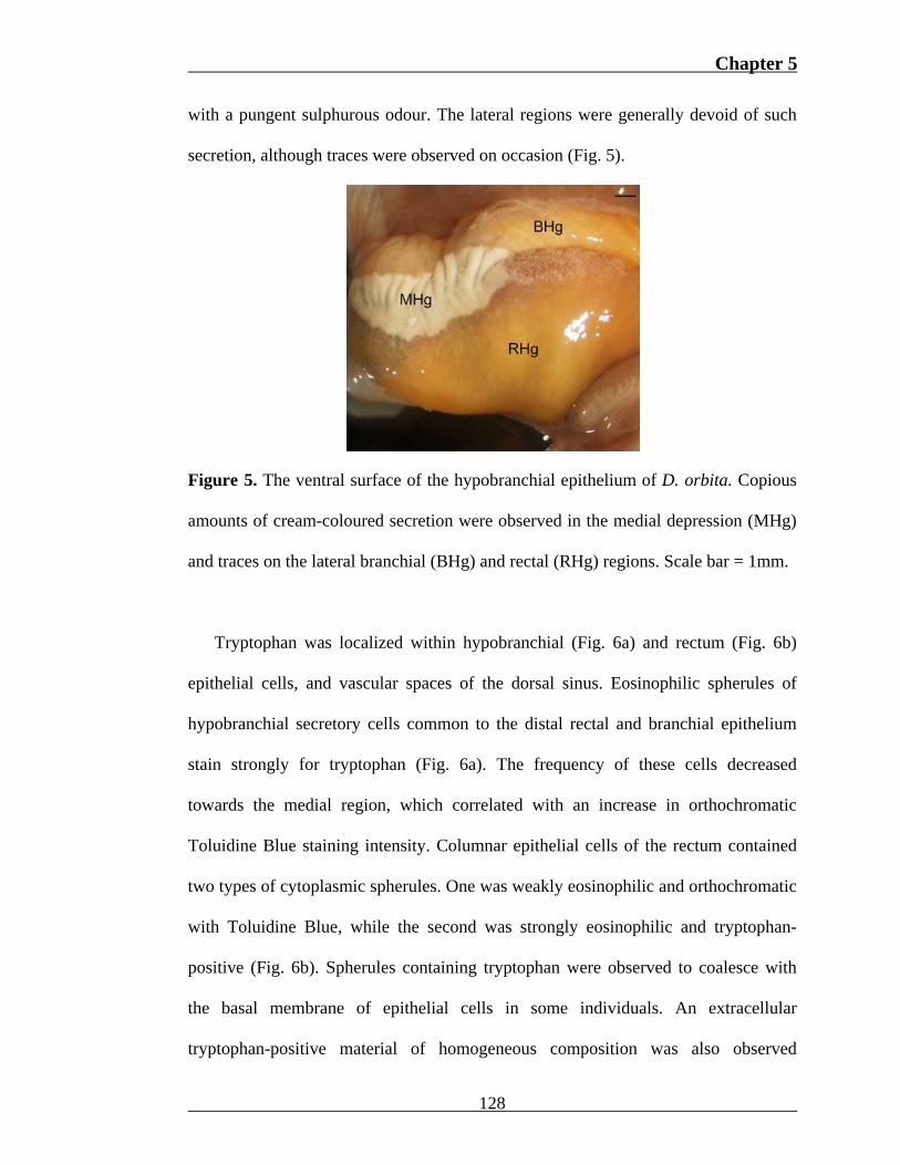
lateral branchial region, a medial depression, and a right lateral rectal region (Fig. 5).
Upon dissection, the initially cream coloured secretion within the medial epithelium
(Fig. 5) rapidly developed yellow, red, green and finally purple pigmentation, along
127
Chapter 5
with a pungent sulphurous odour. The lateral regions were generally devoid of such
secretion, although traces were observed on occasion (Fig. 5).
Figure 5. The ventral surface of the hypobranchial epithelium of D. orbita. Copious
amounts of cream-coloured secretion were observed in the medial depression (MHg)
and traces on the lateral branchial (BHg) and rectal (RHg) regions. Scale bar = 1mm.
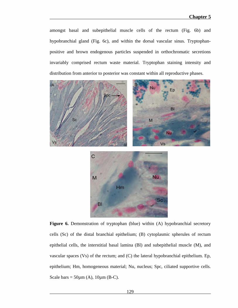
Tryptophan was localized within hypobranchial (Fig. 6a) and rectum (Fig. 6b)
epithelial cells, and vascular spaces of the dorsal sinus. Eosinophilic spherules of
hypobranchial secretory cells common to the distal rectal and branchial epithelium
stain strongly for tryptophan (Fig. 6a). The frequency of these cells decreased
towards the medial region, which correlated with an increase in orthochromatic
Toluidine Blue staining intensity. Columnar epithelial cells of the rectum contained
two types of cytoplasmic spherules. One was weakly eosinophilic and orthochromatic
with Toluidine Blue, while the second was strongly eosinophilic and tryptophan-
positive (Fig. 6b). Spherules containing tryptophan were observed to coalesce with
the basal membrane of epithelial cells in some individuals. An extracellular
tryptophan-positive material of homogeneous composition was also observed
128
Chapter 5
amongst basal and subepithelial muscle cells of the rectum (Fig. 6b) and
hypobranchial gland (Fig. 6c), and within the dorsal vascular sinus. Tryptophan-
positive and brown endogenous particles suspended in orthochromatic secretions
invariably comprised rectum waste material. Tryptophan staining intensity and
distribution from anterior to posterior was constant within all reproductive phases.
Figure 6. Demonstration of tryptophan (blue) within (A) hypobranchial secretory
cells (Sc) of the distal branchial epithelium; (B) cytoplasmic spherules of rectum
epithelial cells, the interstitial basal lamina (Bl) and subepithelial muscle (M), and
vascular spaces (Vs) of the rectum; and (C) the lateral hypobranchial epithelium. Ep,
epithelium; Hm, homogeneous material; Nu, nucleus; Spc, ciliated supportive cells.
Scale bars = 50µm (A), 10µm (B-C).
129
Chapter 5
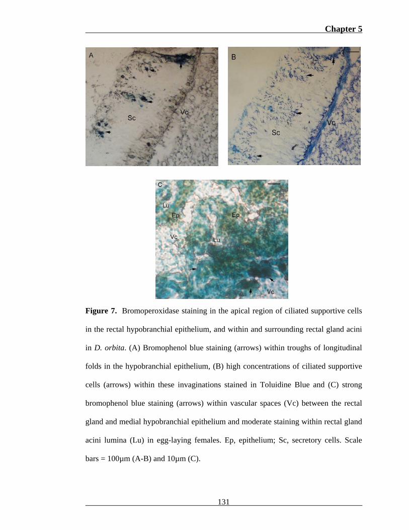
The bromo-phenol red staining reaction identified sites of bromoperoxidase
activity within the vascular sinus, hypobranchial epithelium and rectal gland of D.
orbita. Intermittent deposits of bromophenol blue were observed within the apical
(Fig. 7a) and basal region of lateral hypobranchial epithelial cells in all specimens.
Apical mucous on the surface of these cells also stained for bromoperoxidase.
Troughs formed by transverse folds in the epithelium stained more intensely than
adjacent ridges in the majority of specimens (Fig. 7a). Ciliated supportive cells were
visibly more abundant within these invaginations (Fig. 7b) due to higher epithelial
cell concentrations. Weak cytoplasmic staining was observed on two occasions
within metachromatic branchial hypobranchial cells and unstained medial cells.
Staining intensity did not vary between anterior, medial and posterior hypobranchial
sections or reproductive phases. A strong positive reaction for bromoperoxidase was
also observed within subepithelial vascular spaces, interstitial basal cells of the
medial hypobranchial gland, and the lumina of rectal gland acini in egg-laying
specimens (Fig. 7c). Bromoperoxidase activity was also occasionally detected within
the subepithelial vascular sinus of the branchial and rectal hypobranchial epithelium.
130
Chapter 5
Figure 7. Bromoperoxidase staining in the apical region of ciliated supportive cells
in the rectal hypobranchial epithelium, and within and surrounding rectal gland acini
in D. orbita. (A) Bromophenol blue staining (arrows) within troughs of longitudinal
folds in the hypobranchial epithelium, (B) high concentrations of ciliated supportive
cells (arrows) within these invaginations stained in Toluidine Blue and (C) strong
bromophenol blue staining (arrows) within vascular spaces (Vc) between the rectal
gland and medial hypobranchial epithelium and moderate staining within rectal gland
acini lumina (Lu) in egg-laying females. Ep, epithelium; Sc, secretory cells. Scale
bars = 100µm (A-B) and 10µm (C).
131
Chapter 5
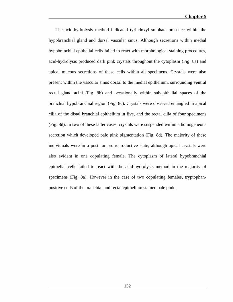
The acid-hydrolysis method indicated tyrindoxyl sulphate presence within the
hypobranchial gland and dorsal vascular sinus. Although secretions within medial
hypobranchial epithelial cells failed to react with morphological staining procedures,
acid-hydrolysis produced dark pink crystals throughout the cytoplasm (Fig. 8a) and
apical mucous secretions of these cells within all specimens. Crystals were also
present within the vascular sinus dorsal to the medial epithelium, surrounding ventral
rectal gland acini (Fig. 8b) and occasionally within subepithelial spaces of the
branchial hypobranchial region (Fig. 8c). Crystals were observed entangled in apical
cilia of the distal branchial epithelium in five, and the rectal cilia of four specimens
(Fig. 8d). In two of these latter cases, crystals were suspended within a homogeneous
secretion which developed pale pink pigmentation (Fig. 8d). The majority of these
individuals were in a post- or pre-reproductive state, although apical crystals were
also evident in one copulating female. The cytoplasm of lateral hypobranchial
epithelial cells failed to react with the acid-hydrolysis method in the majority of
specimens (Fig. 8a). However in the case of two copulating females, tryptophan-
positive cells of the branchial and rectal epithelium stained pale pink.
132
Chapter 5
Figure 8. Tyrindoxyl sulphate localization within the hypobranchial gland, vascular
sinus and rectal gland of D. orbita. (A) Medial hypobranchial (MHg) secretory cells
(Sc) showing pink crystalline staining after application of the acid-hydrolysis method
and negative lateral secretory cells (BHg) stained in haematoxylin. (B) Pink crystals
(arrow) within the lumen (Lu) of rectal gland acini, (C) the subepithelial vascular
sinus (arrows) of the branchial hypobranchial region; and (D) apical mucous
secretions (Mu) of rectal hypobranchial epithelium. Cry, crystals; Ep, epithelial cells;
M, muscle; Pig, endogenous pigments; Sc, secretory cells; Spc, ciliated supportive
cells; Vs, vascular spaces. Scale bars = 50µm (A, C-D) and 10µm (B).
133
Chapter 5
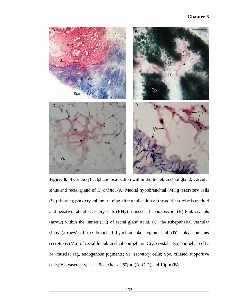
Arylsulphatase was invariably detected within the hypobranchial gland, rectum
and vascular sinus of D. orbita. Enzyme distribution appeared consistent from
anterior to posterior across all reproductive phases. Cytoplasmic products within all
hypobranchial secretory cells remained unstained with the post-coupling method for
arylsulphatase. Deposition of blue-purple azo dye was observed in the basal region of
branchial (Fig. 9a), rectal and medial hypobranchial epithelial cells, the apical domain
of branchial epithelial cells and the mucous entangled within cilia (Fig. 9a). In
contrast, rectal cell apices stained red in the majority of egg-laying and post-
reproductive females, and remained unstained in pre-reproductive and two copulating
females. A feint line of purple staining was also produced between many secretory
cells of the branchial and rectal hypobranchial gland, and occasionally between
medial secretory cells. Within sections stained for morphological comparison, these
regions correlate with the distribution of supportive cells, which rapidly taper from
the epithelial surface and basal lamina due to constriction by adjacent secretory cells.
Red staining was characteristic of all positive hypobranchial regions in pre-
reproductive females.
Spherules within rectum epithelial cells (Fig. 9b) of all specimens and particles
comprising waste occupying the lumen displayed intense purple staining after
application of the post-coupling method for arylsulphatase. Homogeneous material
within vascular spaces dorsal to the rectal, branchial (Fig. 9a) and medial
hypobranchial gland also produced a weak purple stain. Granules in the lumina of
rectal gland acini of five females from various reproductive phases also gained purple
pigmentation. In the case of two egg-laying females, cytoplasmic granules stained
bright blue (Fig. 9c).
134
Chapter 5
Figure 9. Localization of arylsulphatase by the post-coupling method within the
rectum, hypobranchial and rectal glands. (A) High activity arylsulphatase within the
apical (Ar) and basal region (Br) of the branchial hypobranchial epithelium and
homogeneous material (Hm) within subepithelial vascular spaces (Vs), (B) the
cytoplasmic contents of rectum epithelial cells (Ep), and (C) the lumen (Lu) of rectal
gland acini. Mu, mucous; Nu, nucleus; Pig, endogenous pigments; Sc, secretory cells;
Wm, waste material. Scale bars = 100µm (A-B), 10µm (C).
135
Chapter 5
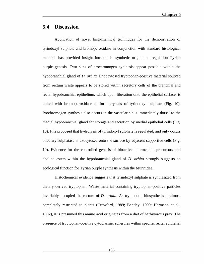
5.4 Discussion
Application of novel histochemical techniques for the demonstration of
tyrindoxyl sulphate and bromoperoxidase in conjunction with standard histological
methods has provided insight into the biosynthetic origin and regulation Tyrian
purple genesis. Two sites of prochromogen synthesis appear possible within the
hypobranchial gland of D. orbita. Endocytosed tryptophan-positive material sourced
from rectum waste appears to be stored within secretory cells of the branchial and
rectal hypobranchial epithelium, which upon liberation onto the epithelial surface, is
united with bromoperoxidase to form crystals of tyrindoxyl sulphate (Fig. 10).
Prochromogen synthesis also occurs in the vascular sinus immediately dorsal to the
medial hypobranchial gland for storage and secretion by medial epithelial cells (Fig.
10). It is proposed that hydrolysis of tyrindoxyl sulphate is regulated, and only occurs
once arylsulphatase is exocytosed onto the surface by adjacent supportive cells (Fig.
10). Evidence for the controlled genesis of bioactive intermediate precursors and
choline esters within the hypobranchial gland of D. orbita strongly suggests an
ecological function for Tyrian purple synthesis within the Muricidae.
Histochemical evidence suggests that tyrindoxyl sulphate is synthesized from
dietary derived tryptophan. Waste material containing tryptophan-positive particles
invariably occupied the rectum of D. orbita. As tryptophan biosynthesis is almost
completely restricted to plants (Crawford, 1989; Bentley, 1990; Hermann et al.,
1992), it is presumed this amino acid originates from a diet of herbivorous prey. The
presence of tryptophan-positive cytoplasmic spherules within specific rectal epithelial
136
Chapter 5
cells implies that dietary tryptophan is endocytosed and concentrated by these cells
(Fig. 10).
The distribution of tryptophan-positive material suggests this amino acid is
transported from the rectum to the vascular sinus for endocytosis by the
hypobranchial epithelial cells (Fig. 10). Homogeneous tryptophan-positive material
was observed within extracellular spaces of the rectum basal lamina, subepithelial
muscle and the vascular sinus adjoining the hypobranchial gland. Although
molluscan haemocyanin contains tryptophan residues (Waxman, 1975; Avissar et al.,
1986), localization in the vascular sinus is not likely due to haemocyanin as the N-
terminal amino acid of muricid haemocyanin is serine (Idakieva et al., 1993), which
fails to form a blue pigment with p-DMAB-nitrite (Adams, 1957). However,
haemocyanin may function as a transport protein through the temporary condensation
of tryptophan and serine, thereby resulting in p-DMAB-nitrite staining. Alternatively,
free tryptophan may be transported in the haemolymph plasma. Tryptophan-positive
material was also observed amongst basal cells of hypobranchial secretory cells
containing tryptophan-positive spherules (Fig. 6c). These cells dominate the distal
lateral epithelium (Fig. 6a), but reduce in frequency towards the medial epithelium.
Correlations in biochemistry and distribution indicate that these cells are those
previously thought be involved in the sequestration of tryptophan in D. orbita
(Chapter 3). Thus, it appears that dietary tryptophan is mobilized in the haemolymph,
accumulated by these prominent hypobranchial secretory cells and stored as a
membrane-bound mucopolysaccharide-protein complex until exocytosis (Fig. 10).
137
Chapter 5
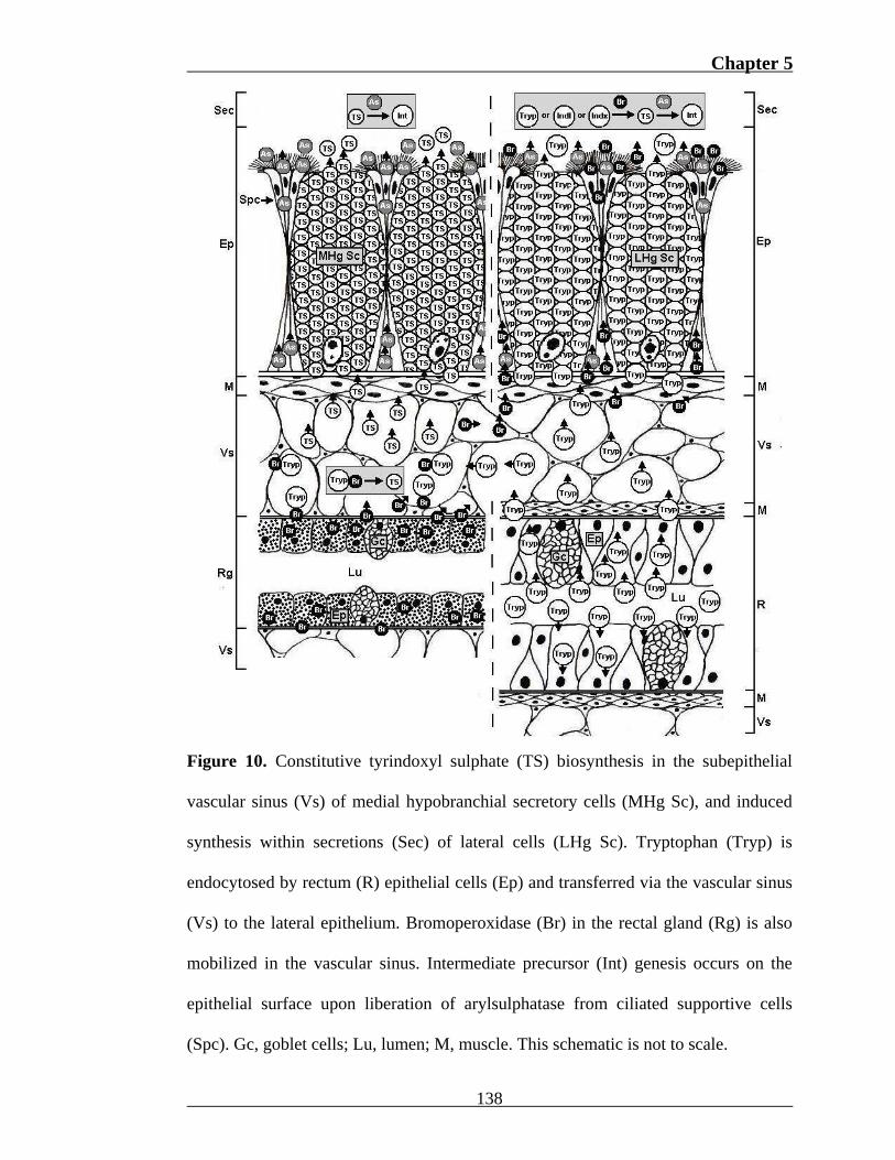
Figure 10. Constitutive tyrindoxyl sulphate (TS) biosynthesis in the subepithelial
vascular sinus (Vs) of medial hypobranchial secretory cells (MHg Sc), and induced
synthesis within secretions (Sec) of lateral cells (LHg Sc). Tryptophan (Tryp) is
endocytosed by rectum (R) epithelial cells (Ep) and transferred via the vascular sinus
(Vs) to the lateral epithelium. Bromoperoxidase (Br) in the rectal gland (Rg) is also
mobilized in the vascular sinus. Intermediate precursor (Int) genesis occurs on the
epithelial surface upon liberation of arylsulphatase from ciliated supportive cells
(Spc). Gc, goblet cells; Lu, lumen; M, muscle. This schematic is not to scale.
138
Chapter 5
Tryptophan-containing cells have been identified in hypobranchial glands of
the muricids M. brandaris (Bolognani-Fantin and Ottaviani, 1981), M. granulata
(Srilakshmi, 1991) and P. pansa (Naegel and Aguilar-Cruz, 2006). As tryptophan is
the indole precursor, detection of this amino acid has been previously assumed to
double for localization of Tyrian purple precursors (Naegel and Aguilar-Cruz, 2006).
However, as indole precursors (Fig. 1) lack the functional group to form the ß-
carboline derivative (Fig. 2) in the histochemical techniques applied (Adams, 1957;
Davenport, 1960), simultaneous demonstration is unlikely. Application of the p-
DMAB-nitrite method (Adams, 1957) in conjunction with the acid-hydrolysis
technique in this investigation highlights the error of this previous assumption.
Production of dark pink crystals after acid-hydrolysis identified secretory cells of the
medial hypobranchial epithelium as the primary region of tyrindoxyl sulphate storage
(Fig. 8a). The absence of tryptophan within these cells coupled with detection of this
amino acid in morphologically distinct lateral secretory cells (Fig. 6a), indicates that
the prochromogen and precursor are stored separately in D. orbita (Fig. 10).
The distribution of these tyrindoxyl sulphate-containing cells in D. orbita
correlates well with the findings of Erspamer (1946), which indicate prochromogen
presence exclusively within the medial hypobranchial gland of Murex trunculus and
M. brandaris. Cells, termed “clear” or “empty” due a lack of cytoplasmic staining,
have been reported in all muricid hypobranchial glands studied to date (Bolognani-
Fantin and Ottaviani, 1981; Srilakshmi, 1991; Roller et al., 1995; Naegel and
Aguilar-Cruz, 2006). Similarly, the medial cells of D. orbita failed to stain with any
of the morphological stains applied (Chapter 3). The apparent lack of proteins and
carbohydrates within these cells, coupled with the crystalline morphology of dye
139
Chapter 5
pigments after acid-hydrolysis implies that the prochromogen, an indoxyl sulphate
salt, may be present in a pure state. Furthermore, the apparent absence of these cells
in non-muricid species documented to date (Tarao, 1935; Ronkin, 1952; Hunt, 1973;
Ottaviani, 1978) suggests that the “empty” cells of other muricids also contain Tyrian
purple prochromogens.
Histochemical evidence suggests that two mechanisms of prochromogen
synthesis occur within the hypobranchial gland of D. orbita. In addition to the
cytoplasm of medial epithelial cells, crystals of tyrindoxyl sulphate were detected
within the vascular sinus dorsal to these cells and in apical secretions of the rectal
(Fig. 8d) and branchial epithelium. The invariable presence of crystals adjacent to the
proposed site of prochromogen storage in medial hypobranchial epithelial cells
suggests that synthesis occurs within these vascular spaces prior to epithelial
sequestration (Fig. 10). The absence of crystals in the cytoplasm of lateral cells (Fig.
8a) indicates that tyrindoxyl sulphate is also synthesized on the epithelial surface
upon exocytosis of tryptophan (Fig. 10).
The genesis of an unusual pale pink dye product after acid-hydrolysis suggest
that tryptophan is converted to indole or indoxyl within the cytoplasm prior to
prochromogen synthesis on the epithelial surface (Fig. 10). On two occasions
tryptophan-positive cells of the lateral hypobranchial epithelium gained pale pink
pigmentation after acid-hydrolysis, while in another two individuals, apical crystals
were suspended within a homogeneous substance which also stained pale pink (Fig.
8d). During method development, trials indicated that tryptophan fails to produce a
coloured product after exposure of HCl (Appendix III). Nevertheless, it is possible
that prior to bromoindoxyl sulphate synthesis on the surface, tryptophan degradation
140
Chapter 5
to indole or indoxyl occurs within these lateral cells, generating intermediates which
stain for tryptophan and have the potential to develop pink pigmentation. Of these
intermediates, 3-indoleacetic acid is known to react with the p-DMAB nitrite method
(Adams, 1957) and may be converted via indoxyl to pink indirubin (see Allegri et al.,
2006). The infrequent occurrence of pale pink staining further suggests that indoxyl
synthesis from tryptophan within these cells is regulated. Although cytoplasmic
indoxyl synthesis may theoretically explain the staining of some lateral cells and
apical secretions by both acid-hydrolysis and the p-DMAB nitrite method, the
location of enzymes involved in indole synthesis and the influence of HCl on indoxyl
intermediates requires further investigation.
Bromination of tryptophan, indole, 3-indoleacetic acid or indoxyl is required
for the genesis of tyrindoxyl sulphate. Application of the bromophenol-red method
identified comparatively high concentrations of bromoperoxidase amongst rectal
gland acini (Fig. 7c), subepithelial vascular spaces and basal cells of the medial
hypobranchial epithelium where tyrindoxyl sulphate is most abundant (Fig. 10). In D.
orbita, this conspicuous rectal gland commences and terminates with the medial
hypobranchial epithelium (Fig. 3b), which suggests it may be associated with dye
synthesis. Bacteria are known to produce bromoperoxidase (Butler and Walker, 1993;
Gribble, 1999) and synthesize brominated natural products (Garson, 1993). Thus, it is
possible that bromoperoxidase is acquired from symbiotic bacteria for precursor
bromination.
Bacteria have been observed within specialized invaginations of the rectal
gland epidermis in the muricid Nucella lapillus (Andrews, 1992). As the rectal gland
functions in the catabolism of haemolymph macromolecules (Andrews, 1992), this
141
Chapter 5
gland is an ideal environment for the bacterial synthesis of bromoperoxidase.
Bromoperoxidase contains tryptophan residues among other amino acids (Wever et
al., 1988), which would be made readily available during degradation of haemolymph
proteins such as haemocyanin. Furthermore, active transport is known to occur across
the rectal gland basal lamina (Andrews, 1992). Thus, it is possible that
bromoperoxidase is synthesized by symbiotic bacteria, released into the vascular
sinus and mobilized by the haemolymph for bromoindoxyl sulphate synthesis (Fig.
10). Twelve neogastropod families possess a rectal gland; the Muricidae, Conidae,
Volutidae, Olividae, Mitridae, Columbariidae, Vexillidae, Volutomitridae,
Marginellidae, Turbinellidae, Cancellariidae and Terebridae (see Ponder, 1973;
Taylor and Morris, 1988). Of these, species of the Muricidae and Conidae synthesize
metabolites which contain tryptophan residues brominated in the 6-position (Jimenez
et al., 1997; Westley et al. 2006). Furthermore, chromatic reactions have been
observed in hypobranchial secretions of the Volutidae (Weaver and Du Port, 1970),
Olividae (Marcus and Marcus, 1959) and Mitridae (Schimelman, 1982). Thus, it
would be of interest to investigate such correlations in the remaining families, as the
rectal gland may be of greater functional significance than previously thought.
It should be noted that bromoperoxidase activity was only detected within
rectal gland lumina of egg-laying females, which may by due to an increased demand
for Tyrian purple precursors during capsule manufacture. Bioactive precursors occur
in muricid egg masses, where they are thought to function in the chemical defence of
encapsulated larvae (Benkendorff et al., 2000, 2001, 2004). It has been proposed that
precursors are incorporated into egg masses as a form of maternal investment
(Westley et al., 2006). If the rectal gland is the source of bromoperoxidase in the
142
Chapter 5
Muricidae, activity or enzyme concentration may become heightened within egg-
laying females to provide for the incorporation of bioactive intermediate precursors
into egg masses.
Bromoperoxidase activity was also detected within the basal and apical region
of lateral hypobranchial secretory cells. In comparison to adjacent ridges, staining
was more intense in troughs of transverse epithelial folds where ciliated supportive
cells occur in high concentration. Although intracellular localization was not possible,
the close correlation of staining with ciliated supportive cell distribution (Fig. 7a-b)
suggests they secrete bromoperoxidase onto the epithelium surface (Fig. 10). Ciliated
cells between groups of secretory cells appear common to buccinid and muricid
hypobranchial glands (Hunt, 1973; Bolognani-Fantin and Ottoviani, 1981;
Srilakshimi, 1991; Roller et al., 1995). These cells are thought to function in
providing support, combining secretions released from adjacent cells, and moving
secretory products across the epithelial surface (Hunt, 1973; Bolognani-Fantin and
Ottaviani, 1981; Roller et al., 1993). Although cytoplasmic secretory granules are not
visible by light microscopy, ultrastructural investigation has previously revealed the
presence of vacuoles containing structures resembling cytosomes, lysosomes,
microtubules and filaments (Hunt, 1973). It is possible that enzymes such as
bromoperoxidase could be stored in these vacuoles and transported to the surface
with the assistance of microtubules. The intermittent nature of apical and basal
bromophenol staining indicates that bromoperoxidase was activated in the process of
liberation, or in comparatively high concentration in some cells, but not all supportive
cells, at the time of sectioning. This suggests that enzyme release or activation is
regulated, which may be a mechanism controlling prochromogen bromination.
143
Chapter 5
Once secreted, bromoperoxidase may be combined with tryptophan, indole 3-
indoleacetic acid or indoxyl from adjacent secretory cells through ciliary action to
facilitate precursor bromination (Fig. 10). Like all haloperoxidases, bromination by
bromoperoxidase requires hydrogen peroxide (Fig. 4). There are several potential
sources of hydrogen peroxide, the most obvious being seawater where it is produced
photochemically from dissolved organic matter (Miller et al., 2005). Seawater
introduced to the mantle cavity is in constant contact with the hypobranchial
epithelium, which would theoretically unite dissolved hydrogen peroxide with
liberated bromoperoxidase to promote precursor bromination. Oxidative enzymes
may also induce hydrogen peroxide production on the epithelial surface or within
peroxisomes. Peroxisomes are ubiquitous organelles, which contain up to 60 different
enzymes and function in fatty acid metabolism (Cancio et al., 2000). Of these
enzymes, peroxisomal oxidases catalyze the oxidation of various organic substrates,
producing hydrogen peroxide (Fahl et al., 1984). As peroxisomes are part of the
secretory pathway, supportive cells may release either hydrogen peroxide or oxidase
onto the hypobranchial epithelium in conjunction with bromoperoxidase for the
bromination of precursors liberated by adjacent secretory cells.
In addition to bromoperoxidase, ciliated supportive cells also appear to secrete
arylsulphatase (Fig. 10). Morphological comparisons indicate that arylsulphatase
staining correlates with the visible distribution of supportive cells, which rapidly
taper from the epithelial surface and basal lamina due to constriction by adjacent
secretory cells. The presence of this enzyme within these cells is also supported by
the genesis of Tyrian purple along the apical and basal region of medial and some
rectal hypobranchial cells in sections exposed to sunlight during trials for the acid-
144
Chapter 5
hydrolysis hydrolysis method (Appendix III). This mechanism of enzyme delivery
would restrict prochromogen hydrolysis to the surface of the hypobranchial
epithelium, and ultimately regulate the genesis of bioactive intermediates to Tyrian
purple.
Arylsulphatase activity varied with hypobranchial region and reproductive status.
Enzyme activity was consistently low within all pre-reproductive females. This
would theoretically result in reduced prochromogen hydrolysis and subsequent
Tyrian purple synthesis, which correlates well with previous observations of
decreased hypobranchial secretory activity outside the reproductive season (Fretter
and Graham 1994). Enzyme activity was generally high within the basal region of
supportive cells, but low in the apical domain of those in the rectal region of some
egg-laying and post-reproductive females, and absent in two copulating females.
Reductions in the arylsulphatase activity of supportive cells in the rectal
hypobranchial epithelium may be related to differences in enzyme packaging or
specificity. Six (A-F) substrate-specific arylsulphatases occur in mammalian tissues
(Tobacman, 2003). Of these, arylsulphatase B is an acid hydrolase which functions in
the hydrolysis of sulphated polysaccharides (Bhattacharyya et al., 2007). As cells
containing sulphated acid mucopolysaccharide are characteristic of the branchial
hypobranchial epithelium of D. orbita (Chapter 3), an enzyme similar to
arylsulphatase B may be associated with this region. Furthermore, liberated sulphate
(SO4-2) or the reduction of sulphuric acid (H2SO4), may then be utilized in the
synthesis of the indoxyl sulphate prochromogen (e.g. 1). The post-coupling method
applied in this investigation (Rutenburg et al., 1952) is not specific to arylsulphatase
class. Consequently, it is possible that the enzyme required for prochromogen
145
Chapter 5
hydrolysis is of consistently low activity, but appears high in the branchial region due
to staining of a high activity arylsulphatase for polysaccharide desulphation.
Alternatively, arylsulphatase is known to possess high activity when present in the
endoplasmic reticulum where synthesis occurs, but low activity once packaged into
lysosomes for transport (Bhattacharyya et al., 2007). Consequently, variations in
arylsulphatase activity may depend on the stage of enzyme synthesis. These reasons
may also explain failure to detect arylsulphatase outside of the anterior medial
hypobranchial gland of Murex trunculus and M. brandaris (Erspamer, 1946).
Cytoplasmic spherules in rectum epithelial cells (Fig. 9b), granules within lumina
of rectal gland acini (Fig. 9c) and sinus plasma, also demonstrated various levels of
arylsulphatase activity. Arylsulphatase within these regions is most likely associated
with normal cellular metabolism and cell mediated immune responses, as lysosomal
arylsulphatases are involved in macromolecule catabolism (Tobacman, 2003) and
pathogen degradation (Wootton and Pipe, 2003).
Coincidence of tryptophan-positive material, bromoperoxidase and tyrindoxyl
sulphate suggests there are two possible sites where prochromogen synthesis occurs.
The most active is within the vascular sinus immediately dorsal of the medial
hypobranchial epithelium (Fig. 10). Once synthesized, crystals of bromoindoxyl
sulphate appear to be concentrated by medial epithelial cells for secretion onto the
epithelial surface (Fig. 10). As both bromoperoxidase and a tryptophan-positive
material occur in abundance within this region, regulation of prochromogen synthesis
may depend on the availability of tryptophanase and decarboxylase for indole
biosynthesis (see Allegri et al., 2006). Alternatively, medial secretions may be
unregulated in a similar manner to the constitutive secretion of goblet cells, which
146
Chapter 5
maintain the protective mucous coat of intestinal tissues (Ho and Shekels, 2003). The
copious amount of secretion invariably observed within the medial surface depression
and epithelial cells indicates that constant prochromogen synthesis and secretion is
likely. To be metabolically viable, prochromogens must therefore be required to
maintain a specific environment or function on the epithelial surface or mantle cavity,
which is of significant selective benefit to the Muricidae.
In contrast, prochromogen synthesis on the lateral epithelial surface appears to
be induced. A previous electron microscopy study of the hypobranchial surface in
Thais haemastoma suggested that lateral secretions are directed towards the medial
depression for storage prior to release (Roller et al., 1995). However, supplementing a
region of constitutive secretion with biochemically identical secretions from another
source appears to be an unusual strategy. Furthermore, declines in the frequency of
tryptophan-positive cells towards the medial depression suggests the demand for
regulated prochromogen synthesis also declines due to constitutive synthesis within
this region. In general, controlled mucous synthesis occurs in response to certain
chemical or mechanical stimuli, such as hormones, neurotransmitters, chemical and
physical irritants (Ho and Shekels, 2003). Thus, it appears likely that Tyrian purple
precursor synthesis and secretion is induced within the lateral hypobranchial
epithelium of the Muricidae.
The significance of synthesizing precursors to bioactive metabolites in two
distinct manners is unclear as the functional role of hypobranchial gland remains
unresolved. Nevertheless, the constitutive and regulated synthesis of prochromogens
in the medial and lateral hypobranchial epithelium, respectively, appears common to
the Muricidae. Choline esters associated with prochromogens (Fig. 1) have been
147
Chapter 5
isolated to the medial region (Vincent and Jullien, 1938; Erspamer and Dordoni,
1947l, Erspamer, 1948; Keyl et al., 1957; Andrews et al., 1991) or found to be 50
times more abundant in comparison to the lateral region (Erspamer, 1948; Roseghini
and Fichman, 1973). Originally, it was thought that Tyrian purple precursors were
dietary-derived and bioactive intermediates were a coincidence of detoxification
(Verhecken, 1989). One agreed upon functional role of the hypobranchial gland
involves the trapping and cementing of particles introduced by the inhalant stream for
subsequent expulsion (Fretter and Graham, 1994). However, this role fails to explain
the significance of Tyrian purple secondary metabolites and the division of secretory
pathways. Another proposed function, which could theoretically be mediated by the
regulated synthesis and hydrolysis of prochromogens in the lateral hypobranchial
epithelium, is the use of physiologically-active choline esters in prey capture (Keyl et
al., 1957; Whittaker, 1960; Roseghini et al., 1970; Huang and Mir, 1971; Ottaviani,
1978; Bolognani-Frantin and Ottaviani, 1981; Roller et al., 1984; Srilankshmi, 1991;
Roseghini et al., 1996). However, this hypothesis neglects the fact that cytotoxic
precursors, of no apparent use during prey capture, are simultaneously generated
during prochromogen hydrolysis. Thus, it is clear that future investigation into the
evolutionary significance of hypobranchial secondary metabolites should strongly
consider the heterogeneous activity and mechanisms of synthesis associated with this
intriguing biosynthetic pathway.
5. 5 Conclusion
Histochemical evidence for the regulated and constitutive de novo synthesis of
prochromogens indicates that these natural products are of selective benefit to the
148
Chapter 5
149
Muricidae. The findings presented here also suggest that the liberation of
physiologically active choline esters and subsequent genesis of antimicrobial
precursors by arylsulphatase requires induction. It is anticipated that the insight
provided by this investigation into the biosynthetic origin and regulation of Tyrian
purple prochromogen and precursor synthesis will aid future attempts to define the
functional significance of this biosynthetic pathway and the gastropod hypobranchial
gland. It is also hoped that the advantages of adopting a histochemical approach to
natural product research will be considered in subsequent investigations aimed at
elucidating the biosynthetic origin of marine natural products.
5. 6 Acknowledgments
I would like to thank Dr. R. Wever for advice during development of the
bromophenol-red staining reaction, Ms. M. Lewis for technical advice during
histochemical analyses and Mr. W. Nobel for assistance in specimen collection.
Thanks are also due to Dr. K. Benkendorff for guidance throughout, especially during
histochemical method development, and manuscript preparation. The provision of a
Flinders University Postgraduate Scholarship is greatly appreciated. This research
was supported by a Philanthropic research grant to Dr. K. Benkendorff.