Synthesis of an IgG fragment decapeptide exhibiting phagocytosis stimulating activity of...
6
Int. J. PeptideProtein Res. 22, 1983, 119-124 Synthesis of an IgG fragment decapeptide exhibiting phagocy tosis stimulating activity of polymorphonuclear leucocy tes JEAN MARTINEZ, JEANINE LAUR and FRANCOIS WINTERNITZ Center for Endocrinology and Pharmacology, Montpellier, France Received 5 October, accepted for publication 23 November 1982 Fragment 335-344 of human IgG was synthesized stepwise, using active esters of N-protected amino acids and benzotriazolyloxytris(dimethy1amino)phos- phonium hexafluorophosphate for coupling. This fragment could be liberated from the carrier molecule by the two specific enzymes responsible for the release of tuftsin from the same carrier molecule: tuftsin endocarboxy-peptidase cleaves at the Arg-Glu bond and leukokininase at the Lys-Thr bond. This decapeptide fragment, Thr-Ile-Ser-Lys-Ala-Lys-Gly-Gln-Pro-Arg showed significant activity in stimulating phagocytosis of human polymorphonuclear leucocytes. Key words: decapeptide fragment of I@; phagocytosis stimulation;tuftsin-like activity It has been found that a specific fraction of immunoglobulin G binds to polymorphonuclear neutrophils and stimulates their phagocytic activity. This phagocytosis stimulating activity resides in a small peptide termed tuftsin, with the sequence Thr-Lys-Pro-Arg, which has been isolated from the leucophilic immunoglobulin G fraction (1). It was shown that tuftsin stimu- lates the phagocytic activity of both PMN leucocytes and macrophages. This property allowed its isolation, sequence determination and synthesis (2, 3). Bacterial as well as yeast cells have been used as target particles (1, 2,4). Among this basal activity, tuftsin stimulates all known activities of the macrophage and granu- locyte. These are as follows. Motility (2,3). Tuftsin enhances migration of granulocytes upwards in erect glass capillary tubes, this rate being dose dependent. Tuftsin appears to increase the viability of cells: after 2-3 hours, control cells without tuftsin cease to migrate on glass, remain quiescent for a few hours and finally drop off the glass or lyse. In the presence of tuftsin, they survive much longer and have been observed to maintain their motility and viability after 16 hours (5, Hexosemonophosphate shunt. It has been postulated that in the act of engulfing particles, there is an activation of the hexosemono- phosphate shunt (7). This phenomenon can be studied by reduction of nitroblue tetrazolium. Tuftsin has been shown to stimulate the reduc- tion of this dye (8). Chemotactic effect. Tuftsin exhibits signi- ficant stimulation of chemotaxis in the E. coli system (9). Effect on MuL V production. Tuftsin added to culture of MuLV infected cells resulted in a threefold increase in virion associated reverse transcriptase (10). It was concluded that tuftsin, which is present in the protein P12, has a membrane function that somehow leads to increased virion production. 6). 119
-
Upload
jean-martinez -
Category
Documents
-
view
214 -
download
1
Transcript of Synthesis of an IgG fragment decapeptide exhibiting phagocytosis stimulating activity of...
Int. J. PeptideProtein Res. 22, 1983, 119-124
Synthesis of an IgG fragment decapeptide exhibiting phagocy tosis stimulating activity of polymorphonuclear leucocy tes
JEAN MARTINEZ, JEANINE LAUR and FRANCOIS WINTERNITZ
Center for Endocrinology and Pharmacology, Montpellier, France
Received 5 October, accepted for publication 23 November 1982
Fragment 335-344 of human IgG was synthesized stepwise, using active esters of N-protected amino acids and benzotriazolyloxytris(dimethy1amino)phos- phonium hexafluorophosphate for coupling. This fragment could be liberated from the carrier molecule by the two specific enzymes responsible for the release of tuftsin from the same carrier molecule: tuftsin endocarboxy-peptidase cleaves at the Arg-Glu bond and leukokininase at the Lys-Thr bond. This decapeptide fragment, Thr-Ile-Ser-Lys-Ala-Lys-Gly-Gln-Pro-Arg showed significant activity in stimulating phagocytosis of human polymorphonuclear leucocytes.
Key words: decapeptide fragment of I@; phagocytosis stimulation; tuftsin-like activity
It has been found that a specific fraction of immunoglobulin G binds to polymorphonuclear neutrophils and stimulates their phagocytic activity. This phagocytosis stimulating activity resides in a small peptide termed tuftsin, with the sequence Thr-Lys-Pro-Arg, which has been isolated from the leucophilic immunoglobulin G fraction (1). It was shown that tuftsin stimu- lates the phagocytic activity of both PMN leucocytes and macrophages. This property allowed its isolation, sequence determination and synthesis (2, 3). Bacterial as well as yeast cells have been used as target particles (1, 2 , 4 ) . Among this basal activity, tuftsin stimulates all known activities of the macrophage and granu- locyte. These are as follows.
Motility (2 ,3) . Tuftsin enhances migration of granulocytes upwards in erect glass capillary tubes, this rate being dose dependent. Tuftsin appears to increase the viability of cells: after 2-3 hours, control cells without tuftsin cease to migrate on glass, remain quiescent for a few
hours and finally drop off the glass or lyse. In the presence of tuftsin, they survive much longer and have been observed to maintain their motility and viability after 16 hours (5,
Hexosemonophosphate shunt. It has been postulated that in the act of engulfing particles, there is an activation of the hexosemono- phosphate shunt (7). This phenomenon can be studied by reduction of nitroblue tetrazolium. Tuftsin has been shown to stimulate the reduc- tion of this dye (8).
Chemotactic effect. Tuftsin exhibits signi- ficant stimulation of chemotaxis in the E. coli system (9).
Effect on MuL V production. Tuftsin added to culture of MuLV infected cells resulted in a threefold increase in virion associated reverse transcriptase (10). It was concluded that tuftsin, which is present in the protein P12, has a membrane function that somehow leads to increased virion production.
6).
119
J . Martinez et al.
Anti-infectious effect. Tuftsin stimulates bac- tericidal activity of macrophages (1 1, 12) as well as blood clearing in vivo in normal and leukemic mice infected by various bacteria ( ; i , 12).
Immunogenic function. Tuftsin augments considerably the antigene specific macrophage dependent “education” of T-lymphocytes (1 3). Stimulation of immunogenic functions by tuftsin received strong support from works of Florentin et al. (14, 15) on immunocompetent young mice and of Bruley-Rosset et al. (16) on aged mice.
lhmoricidal activity. Stimulation of the cyto- toxicity of the macrophage against tumoral cells has been reported (15, 16) as well as it was shown by Nishioka ( 1 7) and Najjar (1 8, 19) that tuftsin activates peritoneal and body macrophages to result in allogeneic and singeneic tumoricidal activity in vitro and in vivo.
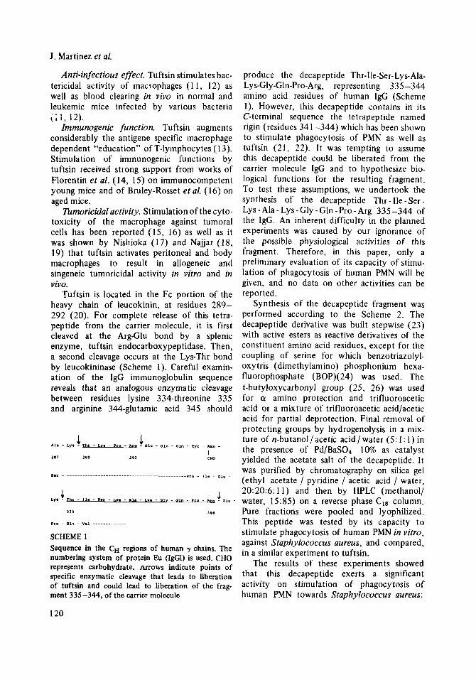
Tuftsin is located in the Fc portion of the heavy chain of leucokinin, at residues 289- 292 (20). For complete release of this tetra- peptide from the carrier molecule, it is first cleaved at the Arg-Glu bond by a splenic enzyme, tuftsin endocarboxypeptidase. Then, a second cleavage occurs at the Lys-Thr bond by leucokininase (Scheme 1). Careful examin- ation of the IgG immunoglobulin sequence reveals that an analogous enzymatic cleavage between residues lysine 334-threonine 335 and arginine 344-glutamic acid 345 should
.1- 01“ - 4 Ly. - mr - *I. - s*t - L”* . Al. . L”, - 01”
3 3 5 311
pro - 01, - V.1 ----.__ ....
SCHEME 1 Sequence in the CH regions of human y chains. The numbering system of protein Eu (IgGI) is used. CHO represents carbohydrate. Arrows indicate points of specific enzymatic cleavage that leads to liberation of tuftsin and could lead to liberation of the frag- ment 335-344, of the carrier molecule
produce the decapeptide Thr-Ile-Ser-Lys-Ala- Lys-Gly-Gln-Pro-Arg, representing 335-344 amino acid residues of human 1gG (Scheme 1). However, this decapeptide contains in its C-terminal sequence the tetrapeptide named rigin (residues 34 1 -344) which has been shown to stimulate phagocytosis of PMN as well as tuftsin (21, 22). I t was tempting to assume this decapeptide could be liberated from the carrier molecule IgG and to hypothesize bio- logical functions for the resulting fragment. To test these assumptions, we undertook the synthesis of the decapeptide Thr - Ile - Ser - Lys - Ala - Lys - Gly - Gln - Pro - Arg 335-344 of the IgG. An inherent difficulty in the planned experiments was caused by our ignorance of the possible physiological activities of this fragment. Therefore, in this paper, only a preliminary evaluation of its capacity of stimu- lation of phagocytosis of human PMN will be given, and no data on other activities can be reported.
Synthesis of the decapeptide fragment was performed according to the Scheme 2. The decapeptide derivative was built stepwise (23) with active esters as reactive derivatives of the constituent amino acid residues, except for the coupling of serine for which benzotriazolyl- oxytris (dimethylamino) phosphonium hexa- fluorophosphate (BOP)(24) was used. The t-butyloxycarbonyl group (25, 26) was used for a amino protection and trifluoroacetic acid or a mixture of trifluoroacetic acid/acetic acid for partial deprotection. Final removal of protecting groups by hydrogenolysis in a mix- ture of n-butanol/ acetic acid / water (5: 1 : 1) in the presence of Pd/BaS04 10% as catalyst yielded the acetate salt of the decapeptide. It was purified by chromatography on silica gel (ethyl acetate / pyridine / acetic acid / water, 20:20:6:11) and then by HPLC (methanol/ water, 15:85) on a reverse phase C18 column. Pure fractions were pooled and lyophilized. This peptide was tested by its capacity to stimulate phagocytosis of human PMN in virro, against Staphylococcus aureus, and compared, in a similar experiment to tuftsin.
The results of these experiments showed that this decapeptide exerts a significant activity on stimulation of phagocytosis of human PMN towards Staphylococcus aureus:
120

Phagocytosis stimulating activity
Au LYS GLY GLN PRO ARC
lW2 I I I B O C ~ O S U H-OBZ~
B o c
THR
B o c - O N p H
ILE SER LYS
B o c
BOG
"P HI-,
SCHEME 2
A* tuftsin = 1.7, A decapeptide = 1.4 at con- centration of lo-' M. This decapeptide is being tested in various immunological experiments in the hope that more light will be shed on its possible biological role. One of the purposes of this paper would be to call attention to the possible biological activity of this fragment of IgC and to its availability, in the expectation that this will stimulate experiments which could eventually reveal its yet unknown physio- logical properties.
EXPERIMENTAL PROCEDURES
Capillary melting points were determined on a Buchi apparatus and are reported uncorrected. Thin-layer chromatography (t.1.c.) was carried
*lndex A = % of phagocyte cells containing particles with peptide/% of phagocyte cells containing particles without peptide.
out on Merck silica gel plates GF2% in the fol- lowing solvent systems: A, dichloromethane/ methanol, 9: 1 ; B, ethyl acetate/pyridine/ acetic acid/H,O, 80:20:5: 10. Spots were re- vealed by their U.V. absorption, by charring and with ninhydrin. Column chromatography was conduced with Merck silica gel, 60-230 mesh ASTM. Elemental analyses were performed by "le service central de microanalyse du CNRS de Montpellier". For amino acid analysis, samples were hydrolysed with 6 N HCI in evacuated sealed ampules at 110" for 16 h and analyzed by the Stein-Moore-Spackman method on a Beckman-Spinco instrument.
tert.-Butyloxycarbonyl-L -glu taminyl-L -prolyl- N -nitro-L arginine benzyl ester 1 A solution of the di-trifluoroacetate of L- prolyl-NG-nitro-L-arginine benzyl ester (6.34 g, 10 mmol)( 1 1) in dimethylformamide (30 ml)
121

J. Martinez et al.
TABLE 1 Physicochemical properties of peptides 1-8
Compound Melting point Elemental analysisa (" C) RFA RFB ("If! (c, solvent) C H N
74-79 dec.
78-84 dec.
82-87 dec.
80-85 dec.
130-136 dec.
120 dec.
180 dec.
218-222
0.33 0.77 -45.5
0.30 0.70 -35.4
0.30 0.70 -31.9
0.28 0.70 -32.8
0.30 0.75 -27.5
0.28 0.70 -22.0
0.25 0.70 -25.5
0.23 0.70 -25.9
(1.12 DMF)
(1.07 DMF)
(1.06 DMF)
(0.91 DMF)
(1.00 DMF)
(1 .OO DMF)
(0.92 DMF)
(1.08 DMF)
52.98 52.62 52.08 51.66 55.38 55.20 55.06 54.75 56.90 56.62 55.91 55.60 56.50 56.08 56.98 56.48
6.68 6.72 6.57 6.47 6.67 6.50 6.70 6.65 6.75 6.66 6.69 6.51 6.92 6.78 6.66 6.58
17.66 17.20 18.23 18.02 16.15 15.82 16.40 15.92 15.23 14.72 15.29 14.72 15.07 14.75 14.67 14.12
'Upper figure: calculated. Lower fgure: found.
containing diisopropylethylamine (3.8 ml) was treated with tert.-butyloxycarbonyl-L-glu tamine N-hydroxysuccinimide ester (3.26 g, 0.95 mmol) (Bachem). After 12 h, the solvent was removed in vacuo and the residue was dissolved in ethyl acetate (300ml), which was washed with a 10% citric acid solution(2 x lOOml), water(lOOml), saturated sodium bicarbonate solution (2 x lOOml), water (2 x 100ml). The organic layer was dried over sodium sulfate and the solvent concentrated in vacuo at 35'. Trituration of the residue with ether afforded a white powder (5.3 g, 88%).
tert -Butyloxycarbonyl-glycyl-L -glutaminyl-L - prolyl-N -nitro-L arginine benzyl ester 2 Compound 1 (5 g, 7.9 mmol) was treated with trifluoroacetic acid (30 ml) for 30 min. Ether (200ml) was then added slowly under stirring. The resulting white precipitate was filtered, washed several times with ether and dried in vacuo over KOH pellets. It was allowed to react with tert.-butyloxycarbonyl-glycine N-hydroxy- succinimide ester (2.04 g, 7.5 mmol)(Bachem), and treated as described for compound 1.
Trituration of the residue with ether afforded a solid (4.36 g, 84%).
N" - tert. -Butyloxycarbonyl-N'-benzyloxy- carbonyl-L -lysyl-glycyI-L -glutaminy I-L -proly l- N G-nitro-L arginine benzyl ester 3 Compound 2 (4.1 g, 5.9 mmol) was treated with trifluoroacetic acid (20 ml) during 30 mn. Ether (200 ml) was then added slowly under stirring and the resulting white precipitate was filtered, washed with ether (4 x l00ml) and dried in vacuo over KOH pellets. It was dissolved in dimethylformamide (30 ml) containing di- isopropylethylamine (2.1 ml) and treated with N"-tert. - butyloxycarbonyl - NE - benzyloxy - carbonyl-L-lysine p-nitrophenyl ester (3.2 1 g, 6.3 mmo1)(27), hydroxybenzotriazole (0.96 g, 6.3 mmol)(28) and diisopropylethylamine (1.1 ml) in dimethylformamide (20 ml). After 12h , the reaction mixture was treated as previously described for compound 1 . The residue (5.26g, 93%) was purified by chro- matography on silica gel (200g) with ethyl acetate, methanol 85:15 as eluent. Pure frac-
122

Phagocytosis stimulating activity
hexafluorophosphate (1.33 g, 3.0 mmol) and diisopropylethylamine (1 S O ml) were added. After 12 h, the reaction mixture was worked up as previously described. Trituration of the residue with a mixture of ethyl acetate/ether (1: 1) yielded a white powder (3.1 g, 81%).
tert .-Butyloxycarbonyl-L -isoleucyl-L -setyl- NE-benzyloxycarbonyl-L -1ysyl-L -alanyl-NE- benzyloxycarbonyl-L -I syl glycyl-L -
benzyl ester 7 Compound 6 (2.9g, 2.1 mmol) was treated as described for 5. The resulting salt was dis- solved in dimethylformamide (20 ml) con- taining diisopropylethylamine (0.72 ml) and treated with tert. butyloxycarbonyl - L - is0 - leucine p-nitrophenyl ester (0.77 g, 2.2 mmol) (Bachem), as above. Yield 83% (2.6 9).
glutaminyl-L -prolyl-N lt -nitro-L arginine-
tions were collected and solvent removed in vacuo (temp. < 35'). Trituration of the residue with ether afforded 3 (4.5 g, 83%).
tert -Butyloxycarbonyl-L-alanyl-Ne-benzyloxy- carbony I-L -lysyl-glycyl-L -glu tam iny I- L -proly I- N '-nitro-L urginine benzyl ester 4 Compound 3 (4.3 g, 4.5mmol) was dissolved in a mixture of trifluoroacetic acid/acetic acid (7:3)(20ml) and treated as previously des- cribed. It was then dissolved in dimethyl- formamide (30ml) and treated with tert.- butyloxycarbonyl-L-alanine p-nitrophenyl ester (1.61 g, 5 mmol)(Bachem) as described for compound 3, unless dichloromethane is used instead of ethyl acetate during washings. Trituration of the resulting residue with a mixture of ethyl acetate/ether (1: 1) yielded a solid compound (4.0 g, 87%).
Nu -tert.-Butyloxycarbonyl-NE-benzyloxy- carbony I-L -1ysyl-L -alanyl-NE-benzyloxy- carbonyl-L -lysyl-glycyl-L -glutaminyl-L - prolyl-N ' 4tro-L arginine benzyl ester 5 Compound 4 (3.8 g, 3.7mmol) was treated with a mixture of trifluoroacetic acid and acetic acid (7:3)(30 ml) as described pre- viously. The resulting salt was dissolved in dimethylforrnamide (30 ml) containing di- isopropylethylamine (1.37 ml) and treated with NQ-tert.-bu tyloxycarbonyl-NE - benzyloxy- carbonyl-L-lysine p-nitrophenyl ester (2.10 g, 4.2 mmol), and treated as described for com- pound 3. The semi-crystalline residue was added with ether (300ml) and stirred during 2 h , then filtered, washed with ether (2 x l00ml) and dried in vacuo. Yield: 3.81 g, 80%; R,A 0.30; RFB 0.75; slight impurity RFA 0; RF B 0.3.
ter t .-Butyloxycarbonyl-L -seryl-N'-benzyloxy- carbony I-L -1ysyl-L ulany 1- Ne - benzy loxy - carbonyl-L -lysyl-glycyl-L -glutaminyI-L -prolyl- N'-nitro-Ldrginine benzyl ester 6 Compound 5 (3.6 g, 2.8 mmol) was partially deprotected in a mixture of trifluoroacetic acid/acetic acid (7:3)(30 ml) in 40 min. The resulting salt was dissolved in dimethylfor- mamide (30 ml). tert.-Butyloxycarbonyl-L- serine (Bachem)(0.65 g, 3.0 mmol), benzotri- azolyloxytris (dimethylamino) phosphonium
Benzy loxycarbony l-L -threony I-L - isoleucyl-L -seryl-NE-benzyloxycarbonyl-L - lysyl-L -alanyl-NE-benzyloxycarbonyl-L - Iysyl-gIycy1-L -glutaminyl-L -prolyl-N C-nitro- ~ u r g i n i n e benzyl ester 8 Compound 7 (2.4g, 1.6mmol) was partially deprotected as described for 5. The resulting salt was dissolved in dimethylformamide (20 ml) containing diisopropylethylamine (0.55 ml) and tert.-butyloxycarbonyl-L- threonine p-nitrophenyl ester (0.64 g, 1.7 mmol) (29) as already described and worked up in the usual manner. Yield, 2.1 g, 8 1%.
L-Threonyl-r,-isolatcyl-L-seryl-L - lysyl-~- alanyl-L -1ysyl-glycyl-L -glutaminyl-L -prolyl-L - arginine 9 Compound 8 (1 g, 0.6 mmol) was hydrogenated in a mixture of n-butanol/acetic acid/water (5:1:1)(70ml) containing 0.7g of 10% Pd/ BaS04 as catalyst, during 24h. The catalyst was then filtered off and washed with water (15 ml). The solvents were removed in vacuo and the residue chromatographed on a column of silica gel (loop) with a mixture of ethyl acetate / pyridine / acetic acid / water (20:20:5: 10) as eluent. Fractions containing the major pure compound were pooled and concentrated in vacuo (temp. <35') to about 10-20ml. Ether (1 50 ml) was then added, the precipitate collected by centrifugation and rinsed with
123

J. Martinez et nl.
ether (3 x 50ml). The white powder was collected: 0.35 g. It was finally passed through a C18 column on HPLC, with water/methanol (85: 15) as eluent. Fractions were collected and lyophilized to yield a white powder (0.28 g). RF 0.25 (ethyl acetate/pyridine/acetic acid/ water, 20:20: 5: 10); single peak on analytical HPLC in the same solvent system (water/ methanol, 85 : 15). M.p. 175-180”; (a): -62.0 (c 0.85, H20); amino acid analysis: Thr 0.98, Ser 1.00, Gln 0.99, Gly 1.02, Ala 0.98, Ile 1 .OO, Lys 1.96, Arg 0.97.
ACKNOWLEDGMENTS
This work has been supported in part by grants from “Le Centre National de la Recherche Scienti- fique” and from “PIRMED”.
REFERENCES
1 . Najjar, V.A. & Nishioka, K. (1970) Nature
2. Nishioka, K., Costantopoulos, A., Satoh, P.S., Mitchell, W.M. & Najjar, V.A. (1973) Biochim. Biophys. Acta 310,217-229
3. Nishioka, K., Satoh, P.S., Costantopoulos, A. & Najjar, V.A. (1973) Biochim. Biophys. Acta
4. Fridkin, M., Stabinsky, Y., Zakuth, V. & Spirer, Z . (1677) Biochim. Biophys. Acta 496, 203- 21 1
5. Najjar, V.A. (1974) Adv. Enzymol. 41, 129- 218
6. Najjar, V.A. (1976) in Biological membranes (Chapman, D. & Wallach, D.F.H., eds.), pp. 191 -240, Academic Press, New York
7. Humbert, J.R., Gross, G.P., Vatter, A.E. & Hathaway, W.E. (1973) J. Lab. Clin. Med. 82,
8. Spirer, Z., Zakuth, V., Colander, A., Boair, N. & Fridkin, M. (1975) J. Clin. Invest. 55, 198- 200
9. Yajima, H., Ogawa, H., Watanabe, H., Fujii, N., Kurobe, M. & Miyamoto, S. (1975) Chem. Pharm. Bull. 23,371-374
10. Luftig, R.B., Yoshinaka, Y. & Oroszlan, S. (1977) J. Cell Biol. 25, 397a
11. Martinez, J., Winternitz, F. & Vindel, J. (1977) European J. Med. Chem. 12,511-516
12. Martinez, J. & Winternitz, F. (1981) Mol. Cell. Biochem. 41, 123-136
228,672-673
310,230-237
20-30
13. Tzehoval, E., Segal, S., Stabinsky, Y., Fridkin, M., Spirer, Z. & Feldman, M. (1978) Proc. Natl. Acad. Sci. US 75,3400-3404
14. Florentin, I., Bruley-Rosset, M., Kiger, N., Imbach, J.L., Winternitz, F. & Mathe, G. (1978) Cancer Immunol. Jmmunother 5 , 211-217
15. Florentin, I., Schulz, J., Bruley-Rosset, M., Kiger, N., Martinez, J. & Mathe, G. (1980) Rec. Results Cancer Res. 75,153-161
16. Bruley-Rosset, M., Hercend, T., Martinez, J., Rappaport, H. & Mathe, G. (1981) J. Natl. CancerInst. 66,1113-1119
17. Nishioka, K. (1979) Brit. J. Cancer 39, 342- 344
18. Najjar, V.A. (1980) in The reticuloendothelial system (Sbarra, A.J. & Strauss, R., eds.), vol. 2, pp. 45-71, Plenum Press, New York
19. Najjar, V.A. (1981) in Macrophages and lympho- cytes (Escobar, M.R. & Friedman, H., eds.) pp. 131-147, Plenum Press, New York
20. Edelman, G.M., Cunningham, B.A., Gall, W.E., Gottlieb, P.D., Rutishauser, U. & Waxdal, M.J. (1969)Proc. Natl. Acad. Sci US 63, 78-85
21. Chipens, G.I., Veretennikova, N.I., Zalitis, G.M., Atare, Z.A., Afanasyeva, G.A. & Kukaine, E.M. (1980) 3rd International Colloquium on Physical and Chemical Information Transfer in Regulation of Reproduction and Ageing, Varna, Bulgaria,
22. Veretennikova, N.I., Chipens, G.I., Nikifirovich, G.V. & Betinsh, Y.R. (1981) Jnf. J. Peptide Protein Res. 17,430-435
23. Bodanszky, M. (1960) Ann. N. Y. Acad. Sci. 88,
24. Castro, B., Dormoy, J.R., Dourtoglou, B., Evin, G., Selve, C. & Ziegler, J.C. (1976) Synthesis,
25. McKay, F.C. & Albertson, N.F. (1957) J. Am. Chem. Soc. 79,4686-4690
26. Anderson, G.W. & McGregor, A.C. (1957) J. Am. Chem. Soc. 79,6180-6183
27. Lanzilotti, A.E., Benz, E. & Goldman, L. (1964) J. Am. Chem. Soc. 86,1880-1882
28. Konig, W. & Geiger, R. (1973) Chem. Ber. 106, 3626-3635
29. Bodanszky, M. & Ondetti, M.A. (1966) Chem. Ind., 1268-1269
Address: Jean Martinez Centre de Pharmacologie-Endocrinologie Rue de la Cardonille B.P. 5055 34033 Montpellier Cedex France
pp. 1-25
655-664
751-752
124