SYNTHESIS AND CHARACTERIZATION OF SnO2 AND PANI … · been synthesized by microwave assisted...
7
International Research Journal of Engineering and Technology (IRJET) e-ISSN: 2395-0056 Volume: 02 Issue: 09 | Dec-2015 www.irjet.net p-ISSN: 2395-0072 © 2015, IRJET ISO 9001:2008 Certified Journal Page 2634 SYNTHESIS AND CHARACTERIZATION OF SnO2 AND PANI DOPED SnO2 NANOPARTICLES BY MICROWAVE ASSISTED SOLUTION METHOD 1 Thenmozhi. C, 2 Manivannan.V, 3* Kumar.E and 4 VeeraRethinaMurugan. S. 1,2 PRIST University, Thanjavur, Tamilnadu, India 3* Department of Physics, School of Science, Tamil Nadu Open University, Chennai, Tamilnadu, India 4 Department of Physics, R.S.Govt. College, Thanjavur, Tamilnadu, India ABSTRACT: Tin oxide (SnO2) nanoparticles have been synthesized by microwave assisted solutionmethod using SnCl2·2H2O as a precursor.Polyanilinedopedtin oxide (PANI/ SnO2) nanoparticles were prepared by an in-situ polymerization of aniline in the presence ofas- synthesized SnO2 nanoparticles. The samples were characterized by X-Ray Diffraction, Scanning Electron Microscopy (SEM) and Transmission Electron Microscopy (TEM). The structure and the morphologyof the nanoparticles have been studied using XRD pattern. The X-ray analysis shows that the obtained nanoparticles are SnO2and its crystallite size was in the rangeof 10- 21nm and for PANI doped SnO2 nanoparticles, the crystallite size was in the range of 12-16nm, which was further confirmed by TEM.TEM micrograph showed that SnO2 nanoparticles were spherical in shape and embedded well in the PANI matrix. SEM micrographs indicate the presence of tin oxide nanoparticles in the PANI matrix. Keywords: Microwave assisted synthesis, Hydrothermal route, SnO2,XRD,SEM and TEM. 1. INTRODUCTION Microwaves are the electromagnetic radiations with wavelengths ranging from about 1mm to 1m in free space and frequencies between 300 GHz to 300 MHz, respectively. But, few frequency bands in this range are allowed for research work. The most common microwave frequency used for carrying out the research work is 2.45 GHz, similar to the frequency of domestic microwave oven. Microwave heating is recognized for its various advantages, such as rapid heating, energy saving, fine microstructures, better product quality, environmental friendliness [1], etc. Investigations have shown that the microwave method is an attractive choice to promote reactions andis energy effective heating method compared to conventional heat conduction methods due to the direct heating of the reaction mixture [2]. In conventional heating methods, the vessel is heated and this then this heat is transferred by convection. Microwave heating not only enhances the rate of formation, also enhances the material quality and size distributions. Additionally, the method shows rapid reaction rate, higher yield, short reaction time, small particle size, narrow particle size distribution, high purity materials, and enhanced physicochemical properties [3].Microwaves can interact uniformly throughout the material, since the wavelength of the microwaves is in agreement with the physical dimension of the sample and supply heat energy throughout thevolume of the material resulting in volumetric heating [4].Microwave assisted synthesis is being cleaner, faster and economical than the conventional methods, in the present work, high puritySnO2nanoparticles were synthesized using microwave assisted hydrothermal method. This method is environmental friendly[5] [6] [7].
Transcript of SYNTHESIS AND CHARACTERIZATION OF SnO2 AND PANI … · been synthesized by microwave assisted...

International Research Journal of Engineering and Technology (IRJET) e-ISSN: 2395-0056
Volume: 02 Issue: 09 | Dec-2015 www.irjet.net p-ISSN: 2395-0072
© 2015, IRJET ISO 9001:2008 Certified Journal Page 2634
SYNTHESIS AND CHARACTERIZATION OF SnO2 AND PANI DOPED SnO2
NANOPARTICLES BY MICROWAVE ASSISTED SOLUTION METHOD
1Thenmozhi. C, 2Manivannan.V, 3*Kumar.E and4VeeraRethinaMurugan. S.
1,2PRIST University, Thanjavur, Tamilnadu, India 3*Department of Physics, School of Science, Tamil Nadu Open University, Chennai, Tamilnadu, India
4Department of Physics, R.S.Govt. College, Thanjavur, Tamilnadu, India
ABSTRACT: Tin oxide (SnO2) nanoparticles have been synthesized by microwave assisted solutionmethod using SnCl2·2H2O as a precursor.Polyanilinedopedtin oxide (PANI/ SnO2) nanoparticles were prepared by an in-situ polymerization of aniline in the presence ofas-synthesized SnO2 nanoparticles. The samples were characterized by X-Ray Diffraction, Scanning Electron Microscopy (SEM) and Transmission Electron Microscopy (TEM). The structure and the morphologyof the nanoparticles have been studied using XRD pattern. The X-ray analysis shows that the obtained nanoparticles are SnO2and its crystallite size was in the rangeof 10-21nm and for PANI doped SnO2 nanoparticles, the crystallite size was in the range of 12-16nm, which was further confirmed by TEM.TEM micrograph showed that SnO2 nanoparticles were spherical in shape and embedded well in the PANI matrix. SEM micrographs indicate the presence of tin oxide nanoparticles in the PANI matrix. Keywords: Microwave assisted synthesis, Hydrothermal route, SnO2,XRD,SEM and TEM.
1. INTRODUCTION Microwaves are the electromagnetic radiations with wavelengths ranging from about 1mm to 1m in free space and frequencies between 300 GHz to 300 MHz, respectively. But, few frequency bands in this range are allowed for research work. The most common microwave frequency used for
carrying out the research work is 2.45 GHz, similar to the frequency of domestic microwave oven. Microwave heating is recognized for its various advantages, such as rapid heating, energy saving, fine microstructures, better product quality, environmental friendliness [1], etc. Investigations have shown that the microwave method is an attractive choice to promote reactions andis energy effective heating method compared to conventional heat conduction methods due to the direct heating of the reaction mixture [2]. In conventional heating methods, the vessel is heated and this then this heat is transferred by convection. Microwave heating not only enhances the rate of formation, also enhances the material quality and size distributions. Additionally, the method shows rapid reaction rate, higher yield, short reaction time, small particle size, narrow particle size distribution, high purity materials, and enhanced physicochemical properties [3].Microwaves can interact uniformly throughout the material, since the wavelength of the microwaves is in agreement with the physical dimension of the sample and supply heat energy throughout thevolume of the material resulting in volumetric heating [4].Microwave assisted synthesis is being cleaner, faster and economical than the conventional methods, in the present work, high puritySnO2nanoparticles were synthesized using microwave assisted hydrothermal method. This method is environmental friendly[5] [6] [7].
International Research Journal of Engineering and Technology (IRJET) e-ISSN: 2395-0056
Volume: 02 Issue: 09 | Dec-2015 www.irjet.net p-ISSN: 2395-0072
© 2015, IRJET ISO 9001:2008 Certified Journal Page 2635
SnO2 is an important N-type metal oxide semiconductor with a wide band gapof 3.6eV at room temperature. SnO2nanoparticles exhibit fascinating properties that differs from their bulk counterparts. The size and morphology of the nanomaterials affect their properties due to their high surface-to-volume ratio. Due to its outstanding properties in various applications like gas sensing properties, the preparation of SnO2 nanoparticles has attracted much attention recently. Many processes have been developed to the synthesis of SnO2 nanostructures, e.g. spray pyrolysis [8], hydrothermal methods [9–11], evaporating tin grains in air [12], chemical vapour deposition (CVD) [13], thermal evaporation of oxide powders [14], rapid oxidation of elemental tin [15] and a sol–gel method [16, 17,18] etc. 2. EXPERIMENTAL 2.1 Materials Aniline (99.5%), Tin(II) chloride dihydrate (SnCl2·2H2O) (99%)were procured from E. Merck. Ammonium persulfate (98%) and ammonia (99%) were purchased from Hi-media and used as received. All chemicals were of analytical grade and solutions were prepared with distilled water.Aniline monomer was distilled using cubic condenser. 2.2. Characterization Techniques The prepared SnO2 nanoparticles andPANI doped SnO2 nanoparticles were characterized by X-Ray Diffraction technique (XRD) and Scanning Electron Microscope (SEM) to find the structure, morphology and elemental composition. Crystallographic studies were carried out using a X-Ray diffractometer (Brucker D8 with Nickel
filtered Cu - K radiation), in the scanning range of 2θ from10° - 80° using Cu - K radiations of wavelength 1.5406 Å. TEM micrographs of the prepared samples were taken from Transmission Electron Microscope (Model JEM 100 CX II ) and HRTEM were taken using the Model JEOL – J2000. SEM images of the sample were recorded using the model HITACHI SEM, to study the morphology of the samples and their elemental analysis. UV spectra of the PANI doped SnO2 samples were recorded using UV-1800 series spectrophotometer in the absorption mode and the intensity of the absorption peaks of the sampleswere examined by it. Synthesis of SnO2 nanoparticles All the chemicals used in microwave assisted hydrothermal synthesis have been used as received from the chemical suppliers without any further purification and processing. For the synthesis of SnO2nanoparticles, 0.03 M solution of stannouschloride dihydrate SnCl2·2H2O (AR grade) was prepared and 5ml ammonia solution was added with it, drop by drop, till its pH value raises upto10. Then the mixture was kept in a magnetic stirrer for half an hour, at 80oC. This solution is sealed and kept in a microwave oven and heated for 6 minutes at 800 Watts, till the volume of the mixture becomes one fourth of its initial value. The colloidal solution obtained was filtered with whattsman filter paper. In order to remove the impurities, the solution was washed with deionized water 2-3 times. The wet nanoparticles were dried in air for 3 days, to remove the moisture content andthe dried powder was ground using pestle and mortar. Synthesis of PANI doped SnO2 nanoparticles PANI doped SnO2 nanoparticleswere synthesized by an in-situ polymerization [19] of aniline in the

International Research Journal of Engineering and Technology (IRJET) e-ISSN: 2395-0056
Volume: 02 Issue: 09 | Dec-2015 www.irjet.net p-ISSN: 2395-0072
© 2015, IRJET ISO 9001:2008 Certified Journal Page 2636
presence of as-synthesized SnO2 nanoparticles using AmmoniumPersulphate (APS) as an oxidant.Aniline monomer was distilled using cubic condenser for purification. Aniline (0.1M) and APS were dissolved separately in 2M HCl solution and stirred for 1 hour. Aniline was addeddrop by drop with 2M HCl and the mixture was kept in a magnetic stirrer for 30 minutes at 400C. Then the mixture was added with prepared SnO2 nanoparticles. The solution was stirred continuously for 10 min. Then APS solution was added gently with the mixture with continuous stirring for half an hour. Due to the polymerization of Aniline, Polyaniline (PANI) was obtained. The colloidal solution obtained was cooled for one day. After cooling, it was filtered with whattsman filter paper and washed with deionized water 2-3 times. The wet nanoparticles were dried at room temperature for 3 days, to remove the moisture content andthe dried powder was ground using pestle and mortar. 2. RESULTS AND DISCUSSION
3.1 X-Ray Diffraction studies The X-ray diffraction pattern of SnO2nanoparticles and PANI doped SnO2
nanoparticles are shown in Figure1.The crystallite size of SnO2nanoparticles and PANI doped SnO2 nanoparticles were calculated using the value of FWHM() from the most intense XRD peaks using Debye-Scherrer formula [20]
D =
cos
K nm
where D is the crystallite size, K is the shape factor(0.94), is the wavelength of X-rays ( = 1.54059A0), is the full width at half maximum(FWHM) of the diffraction peaks and is the angle of diffraction.
Figure 1. XRD spectra of Pure SnO2 and PANI doped SnO2 nanoparticles The XRD result shows that the sharp diffraction peaks formed at 260, 340 and 510 confirms the formation of SnO2 nanoparticles which matches with that of PANI doped SnO2 nanoparticles.All the peaks were indexed as tetragonal structure for both the samples, which are in good agreement with other reportedvalues[21-22].The sharpness of the peaks shows that SnO2nanoparticlesare highly crystalline.The crystallite size for SnO2 nanoparticles synthesized in this method was found to be 10-21nm and that of PANI doped SnO2 nanoparticles was calculated as 12-16nm.The peaks formed at 260, 340 and 510can be indexedto(1 1 0), (1 0 1), (2 1 1) planes of SnO2and PANI doped SnO2 nanoparticleswhich matches well with JCPDS card # 41–1445.The lattice parameters for the tetragonal phase structure were calculated using the following relation [23]
2
1
d = (
2
22
a
kh ) +
2
2
c
l
The lattice parameters of the SnO2 nanoparticles were calculated as a = 4.7615Å and c =
International Research Journal of Engineering and Technology (IRJET) e-ISSN: 2395-0056
Volume: 02 Issue: 09 | Dec-2015 www.irjet.net p-ISSN: 2395-0072
© 2015, IRJET ISO 9001:2008 Certified Journal Page 2637
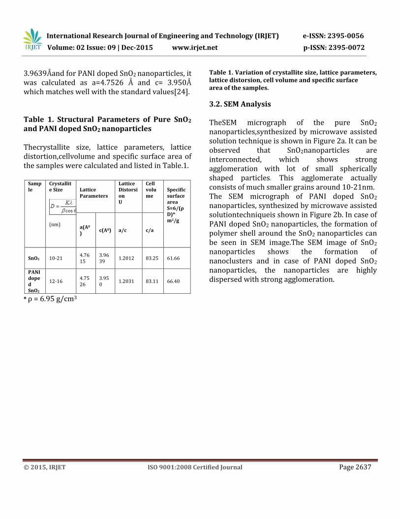
3.9639Åand for PANI doped SnO2 nanoparticles, it was calculated as a=4.7526 Å and c= 3.950Å which matches well with the standard values[24]. Table 1. Structural Parameters of Pure SnO2 and PANI doped SnO2 nanoparticles Thecrystallite size, lattice parameters, lattice distortion,cellvolume and specific surface area of the samples were calculated and listed in Table.1.
Sample
Crystallite Size
(nm)
Lattice Parameters
Lattice Distorsion U
Cell volume
Specific surface area S=6/(ρD)* m2/g
a(A0
) c(A0) a/c c/a
SnO2 10-21 4.7615
3.9639
1.2012 83.25 61.66
PANI doped SnO2
12-16 4.7526
3.950
1.2031 83.11 66.40
* ρ = 6.95 g/cm3
Table 1. Variation of crystallite size, lattice parameters, lattice distorsion, cell volume and specific surface area of the samples.
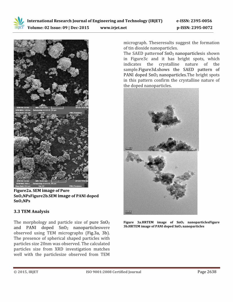
3.2. SEM Analysis TheSEM micrograph of the pure SnO2 nanoparticles,synthesized by microwave assisted solution technique is shown in Figure 2a. It can be observed that SnO2nanoparticles are interconnected, which shows strong agglomeration with lot of small spherically shaped particles. This agglomerate actually consists of much smaller grains around 10-21nm. The SEM micrograph of PANI doped SnO2
nanoparticles, synthesized by microwave assisted solutiontechniqueis shown in Figure 2b. In case of PANI doped SnO2 nanoparticles, the formation of polymer shell around the SnO2 nanoparticles can be seen in SEM image.The SEM image of SnO2
nanoparticles shows the formation of nanoclusters and in case of PANI doped SnO2
nanoparticles, the nanoparticles are highly dispersed with strong agglomeration.
International Research Journal of Engineering and Technology (IRJET) e-ISSN: 2395-0056
Volume: 02 Issue: 09 | Dec-2015 www.irjet.net p-ISSN: 2395-0072
© 2015, IRJET ISO 9001:2008 Certified Journal Page 2638
Figure2a. SEM image of Pure
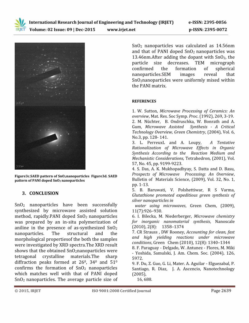
SnO2NPsFigure2b.SEM image of PANI doped SnO2NPs 3.3 TEM Analysis The morphology and particle size of pure SnO2
and PANI doped SnO2 nanoparticleswere observed using TEM micrographs (Fig.3a, 3b). The presence of spherical shaped particles with particles size 20nm was observed. The calculated particles size from XRD investigation matches well with the particlesize observed from TEM
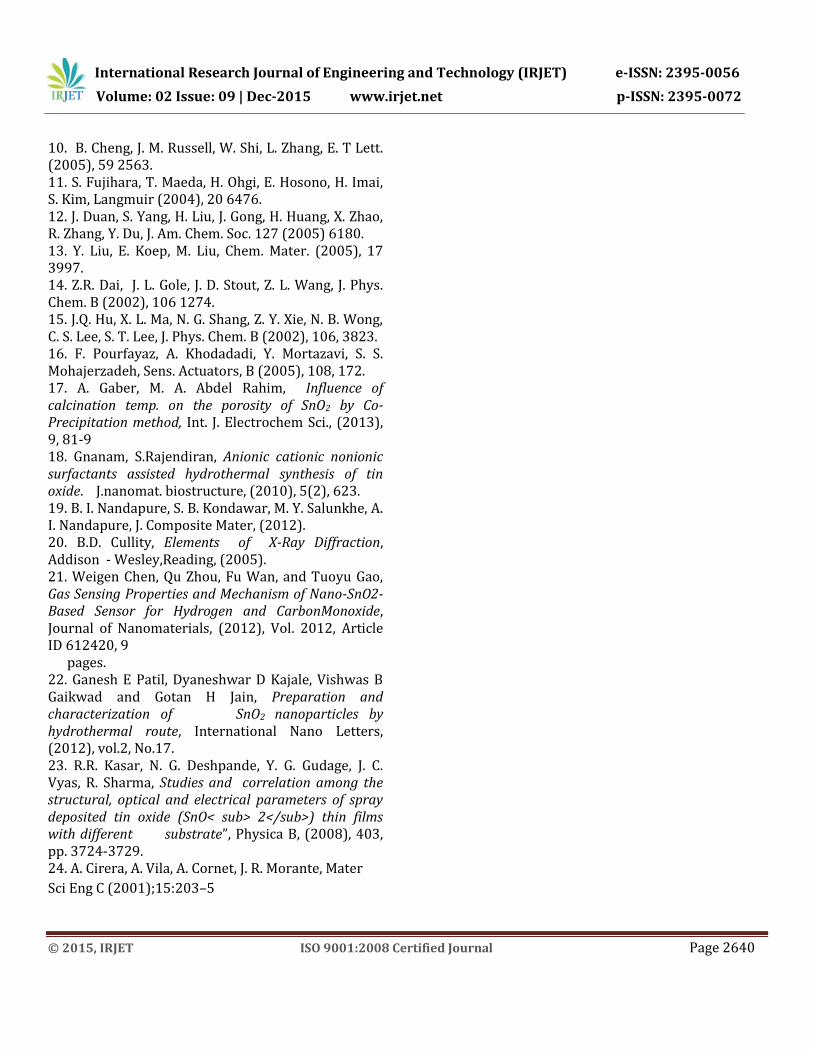
micrograph. Theseresults suggest the formation of tin dioxide nanoparticles. The SAED patternof SnO2 nanoparticlesis shown in Figure3c and it has bright spots, which indicates the crystalline nature of the sample.Figure3d.shows the SAED pattern of PANI doped SnO2 nanoparticles.The bright spots in this pattern confirm the crystalline nature of the doped nanoparticles.
Figure 3a.HRTEM image of SnO2 nanoparticlesFigure 3b.HRTEM image of PANI doped SnO2 nanoparticles
International Research Journal of Engineering and Technology (IRJET) e-ISSN: 2395-0056
Volume: 02 Issue: 09 | Dec-2015 www.irjet.net p-ISSN: 2395-0072
© 2015, IRJET ISO 9001:2008 Certified Journal Page 2639
Figure3c.SAED pattern of SnO2nanoparticles Figure3d. SAED pattern of PANI doped SnO2 nanoparticles
3. CONCLUSION
SnO2 nanoparticles have been successfully synthesized by microwave assisted solution method, rapidly.PANI doped SnO2 nanoparticles was prepared by an in-situ polymerization of aniline in the presence of as-synthesized SnO2 nanoparticles. The structural and the morphological propertiesof the both the samples were investigated by XRD spectra.The XRD result shows that the obtained SnO2nanoparticles were tetragonal crystalline materials.The sharp diffraction peaks formed at 260, 340 and 510 confirms the formation of SnO2 nanoparticles which matches well with that of PANI doped SnO2 nanoparticles. The average particle size of
SnO2 nanoparticles was calculated as 14.56nm and that of PANI doped SnO2 nanoparticles was 13.46nm.After adding the dopant with SnO2, the particle size decreases. TEM micrograph confirmed the formation of spherical nanoparticles.SEM images reveal that SnO2nanoparticles were uniformly mixed within the PANI matrix.
REFERENCES
1. W. Sutton, Microwave Processing of Ceramics: An overview, Mat. Res. Soc Symp. Proc. (1992), 269, 3-19. 2. M. Nüchter, B. Ondruschka, W. Bonrath and A. Gum, Microwave Assisted Synthesis - A Critical Technology Overview, Green Chemistry, (2004), Vol. 6, No.3, pp. 128- 141. 3. L. PerreuxL and A. Loupy, A Tentative Rationalization of Microwave Effects in Organic Synthesis According to the Reaction Medium and Mechanistic Considerations, Tetrahedron, (2001), Vol. 57, No. 45, pp. 9199-9223. 4. S. Das, A. K. Mukhopadhyay, S. Datta and D. Basu, Prospects of Microwave Processing: An Overview, Bulletin of Materials Science, (2009), Vol. 32, No. 1, pp. 1-13. 5. B. Baruwati, V. Polshettiwar, R S Varma, Glutathione promoted expeditious green synthesis of silver nanoparticles in water using microwaves, Green Chem, (2009), 11(7):926–930. 6. I. Bilecka, M. Niederberger, Microwave chemistry for inorganic nanomaterial synthesis, Nanoscale (2010), 2(8): 1358–1374 7. CR Strauss , DW Rooney, Accounting for clean, fast and high yielding reactions under microwave conditions, Green Chem (2010), 12(8): 1340–1344 8. F. Paraguay - Delgado, W. Antunez - Flores, M. Miki - Yoshida, Samulski, J. Am. Chem. Soc. (2004), 126, 5972. 9. F. Du, Z. Guo, G. Li, Mater. A. Aguilar - Elguezabal, P. Santiago, R. Diaz, J. A. Ascencio, Nanotechnology (2005), 16, 688.
International Research Journal of Engineering and Technology (IRJET) e-ISSN: 2395-0056
Volume: 02 Issue: 09 | Dec-2015 www.irjet.net p-ISSN: 2395-0072
© 2015, IRJET ISO 9001:2008 Certified Journal Page 2640
10. B. Cheng, J. M. Russell, W. Shi, L. Zhang, E. T Lett. (2005), 59 2563. 11. S. Fujihara, T. Maeda, H. Ohgi, E. Hosono, H. Imai, S. Kim, Langmuir (2004), 20 6476. 12. J. Duan, S. Yang, H. Liu, J. Gong, H. Huang, X. Zhao, R. Zhang, Y. Du, J. Am. Chem. Soc. 127 (2005) 6180. 13. Y. Liu, E. Koep, M. Liu, Chem. Mater. (2005), 17 3997. 14. Z.R. Dai, J. L. Gole, J. D. Stout, Z. L. Wang, J. Phys. Chem. B (2002), 106 1274. 15. J.Q. Hu, X. L. Ma, N. G. Shang, Z. Y. Xie, N. B. Wong, C. S. Lee, S. T. Lee, J. Phys. Chem. B (2002), 106, 3823. 16. F. Pourfayaz, A. Khodadadi, Y. Mortazavi, S. S. Mohajerzadeh, Sens. Actuators, B (2005), 108, 172. 17. A. Gaber, M. A. Abdel Rahim, Influence of calcination temp. on the porosity of SnO2 by Co-Precipitation method, Int. J. Electrochem Sci., (2013), 9, 81-9 18. Gnanam, S.Rajendiran, Anionic cationic nonionic surfactants assisted hydrothermal synthesis of tin oxide. J.nanomat. biostructure, (2010), 5(2), 623. 19. B. I. Nandapure, S. B. Kondawar, M. Y. Salunkhe, A. I. Nandapure, J. Composite Mater, (2012). 20. B.D. Cullity, Elements of X-Ray Diffraction, Addison - Wesley,Reading, (2005). 21. Weigen Chen, Qu Zhou, Fu Wan, and Tuoyu Gao, Gas Sensing Properties and Mechanism of Nano-SnO2-Based Sensor for Hydrogen and CarbonMonoxide, Journal of Nanomaterials, (2012), Vol. 2012, Article ID 612420, 9 pages. 22. Ganesh E Patil, Dyaneshwar D Kajale, Vishwas B Gaikwad and Gotan H Jain, Preparation and characterization of SnO2 nanoparticles by hydrothermal route, International Nano Letters, (2012), vol.2, No.17. 23. R.R. Kasar, N. G. Deshpande, Y. G. Gudage, J. C. Vyas, R. Sharma, Studies and correlation among the structural, optical and electrical parameters of spray deposited tin oxide (SnO< sub> 2</sub>) thin films with different substrate”, Physica B, (2008), 403, pp. 3724-3729. 24. A. Cirera, A. Vila, A. Cornet, J. R. Morante, Mater
Sci Eng C (2001);15:203–5