Syncope - FOMA District 2 · The Significance of Syncope • 500,000 new syncope patients each year...
140
Transcript of Syncope - FOMA District 2 · The Significance of Syncope • 500,000 new syncope patients each year...


Syncope
A Diagnostic and Treatment Strategy
DR DINESH PUBBI MD FACC
DIRECTOR ELECTROPHYSIOLOGY

Presentation Overview
I. Prevalence & Impact
II. Etiology
III. Diagnosis & Evaluation Options
IV. Specific Conditions
V. Treatment Options
VI. Insights into more efficient and effective diagnosis and treatment of patients with syncope

Section I:
Prevalence and Impact

The Significance of Syncope
The only difference between
syncope and sudden death
is that in one you wake up.1
1 Engel GL. Psychologic stress, vasodepressor syncope, and sudden death. Ann Intern Med 1978; 89: 403-412.

Case Presentation
• 82-year-old male was found by son,
unresponsive
• When ambulance arrived, his pulse
was 70 and BP was 160/98

Case Presentation
82-Year-Old Male
• History: HTN on HCTZ
• Exam: Facial
contusion, unable to
move (L) wrist
• ECG: SR, LBBB, PVCs
• X-ray: (L) wrist fracture

Case Presentation
82-Year-Old Male • What to do?
1) Holter as outpatient
2) Echo
3 ) Admit for EP studies
4) Admit for 23° monitoring

The Significance of Syncope
1 National Disease and Therapeutic Index on Syncope and Collapse, ICD-9-CM 780.2, IMS America, 1997
2 Blanc J-J, L’her C, Touiza A, et al. Eur Heart J, 2002; 23: 815-820.
3 Day SC, et al, AM J of Med 1982
4 Kapoor W. Evaluation and outcome of patients with syncope. Medicine 1990;69:160-175

• Individuals <18 yrs
• Military Population 17- 46 yrs
• Individuals 40-59 yrs*
• Individuals >70 yrs*
15%
20-25%
16-19%
23%
Syncope Reported Frequency
*during a 10-year period Brignole M, Alboni P, Benditt DG, et al. Eur Heart J, 2001; 22: 1256-1306.

The Significance of Syncope
• 500,000 new syncope patients each year 5
• 170,000 have recurrent syncope 6
• 70,000 have recurrent, infrequent, unexplained syncope 1-4
explained: 53% to 62%
infrequent,
unexplained:
38% to 47% 1-4
1 Kapoor W, Med. 1990;69:160-175.
2 Silverstein M, et al. JAMA. 1982;248:1185-1189.
3 Martin G, et al. Ann Emerg. Med. 1984;12:499-504.
4 Kapoor W, et al. N Eng J Med. 1983;309:197-204.
5 National Disease and Therapeutic Index, IMS America, Syncope and Collapse #780.2; Jan 1997-Dec 1997.
6 Kapoor W, et al. Am J Med. 1987;83:700-708.

1 Day SC, et al. Am J of Med 1982;73:15-23.
2 Kapoor W. Medicine 1990;69:160-175. 3 Silverstein M, Sager D, Mulley A. JAMA. 1982;248:1185-1189. 4 Martin G, Adams S, Martin H. Ann Emerg Med. 1984;13:499-504.
• Some causes of syncope are potentially fatal • Cardiac causes of syncope have the highest mortality rates
The Significance of Syncope
0%
5%
10%
15%
20%
25%
Overall Due to Cardiac Causes
Syn
co
pe
Mo
rta
lity

Impact of Syncope
1Linzer, J Clin Epidemiol, 1991. 2Linzer, J Gen Int Med, 1994.
0%
20%
40%
60%
80%
100%
Anxiety/
Depression
Alter Daily
Activities
Restricted
Driving
Change
Employment
73% 1 71% 2
60% 2
37% 2
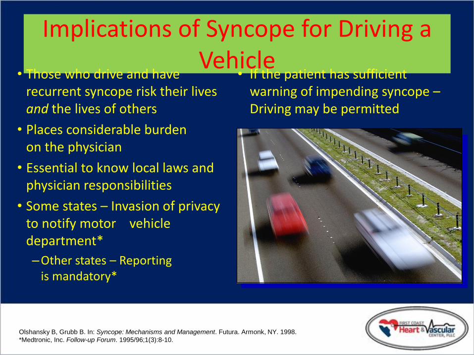
Implications of Syncope for Driving a Vehicle
• Those who drive and have recurrent syncope risk their lives and the lives of others
• Places considerable burden on the physician
• Essential to know local laws and physician responsibilities
• Some states – Invasion of privacy to notify motor vehicle department*
–Other states – Reporting is mandatory*
• If the patient has sufficient warning of impending syncope – Driving may be permitted
Olshansky B, Grubb B. In: Syncope: Mechanisms and Management. Futura. Armonk, NY. 1998.
*Medtronic, Inc. Follow-up Forum. 1995/96;1(3):8-10.

Section II:
Etiology

Syncope: A Symptom…Not a Diagnosis
• Self-limited loss of consciousness and postural tone
• Relatively rapid onset
• Variable warning symptoms
• Spontaneous complete recovery

Cause Prevalence
(Mean) %
Prevalence
(Range) %
Reflex-mediated:
Vasovagal 18 8-37
Situational 5 1-8
Carotid Sinus 1 0-4
Orthostatic hypotension 8 4-10
Medications 3 1-7
Psychiatric 2 1-7
Neurological 10 3-32
Organic Heart Disease 4 1-8
Cardiac Arrhythmias 14 4-38
Unknown 34 13-41
Causes of Syncope1
1Kapoor W. In Grubb B, Olshansky B (eds) Syncope: Mechanisms and Management. Armonk NY; Futura Publishing Co, Inc: 1998; 1-13.

Syncope: Etiology
Orthostatic Cardiac
Arrhythmia
Structural Cardio-
Pulmonary
*
1
• Vasovagal
• Carotid
Sinus
• Situational Cough
Post-
micturition
2
• Drug
Induced
• ANS
Failure Primary
Secondary
3
• Brady Sick sinus
AV block
• Tachy VT
SVT
• Long QT
Syndrome
4
• Aortic
Stenosis
• HOCM
• Pulmonary
Hypertension
5
• Psychogenic
• Metabolic
e.g. hyper-
ventilation
• Neurological
Non- Cardio- vascular
Neurally- Mediated
Unknown Cause = 34%
24% 11% 14% 4% 12%
DG Benditt, UM Cardiac Arrhythmia Center
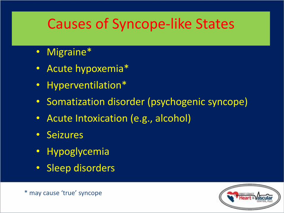
Causes of Syncope-like States
• Migraine*
• Acute hypoxemia*
• Hyperventilation*
• Somatization disorder (psychogenic syncope)
• Acute Intoxication (e.g., alcohol)
• Seizures
• Hypoglycemia
• Sleep disorders
* may cause ‘true’ syncope

Section III:
Diagnosis and Evaluation Options

Syncope Diagnostic Objectives
• Distinguish ‘True’ Syncope from other ‘Loss of Consciousness’ spells: – Seizures
– Psychiatric disturbances
• Establish the cause of syncope with sufficient certainty to: – Assess prognosis confidently
– Initiate effective preventive treatment

Initial Evaluation (Clinic/Emergency Dept.)
• Detailed history
• Physical examination
• 12-lead ECG
• Echocardiogram (as available)

Discord in the Evaluation of Syncope
Neurologist Cardiologist

Syncope Basic Diagnostic Steps
• Detailed History & Physical – Document details of events
– Assess frequency, severity
– Obtain careful family history
• Heart disease present? – Physical exam
– ECG: long QT, WPW, conduction system disease
– Echo: LV function, valve status, HOCM
• Follow a diagnostic plan...

Conventional Diagnostic Methods/Yield Test/Procedure Yield
(based on mean time to diagnosis of 5.1 months7
History and Physical
(including carotid sinus massage)
49-85% 1, 2
ECG 2-11% 2
Electrophysiology Study without SHD* 11% 3
Electrophysiology Study with SHD 49% 3
Tilt Table Test (without SHD) 11-87% 4, 5
Ambulatory ECG Monitors:
Holter 2% 7
External Loop Recorder
(2-3 weeks duration)
20% 7
Insertable Loop Recorder
(up to 14 months duration)
65-88% 6, 7
Neurological †
(Head CT Scan, Carotid Doppler)
0-4% 4,5,8,9,10
* Structural Heart Disease † MRI not studied
1 Kapoor, et al N Eng J Med, 1983. 2 Kapoor, Am J Med, 1991. 3 Linzer, et al. Ann Int. Med, 1997. 4 Kapoor, Medicine, 1990.
5 Kapoor, JAMA, 1992 6 Krahn, Circulation, 1995 7 Krahn, Cardiology Clinics, 1997. 8 Eagle K,, et al. The Yale J Biol and Medicine. 1983; 56: 1-8.
9 Day S, et al. Am J Med. 1982; 73: 15-23. 10 Stetson P, et al. PACE. 1999; 22 (part II): 782.

Syncope Evaluation and Differential Diagnosis
• Complete Description – From patient and observers
• Type of Onset
• Duration of Attacks
• Posture
• Associated Symptoms
• Sequelae
History – What to Look for

12-Lead ECG
• Normal or Abnormal?
– Acute MI
– Severe Sinus Bradycardia/pause
– AV Block
– Tachyarrhythmia (SVT, VT)
– Preexcitation (WPW), Long QT, Brugada
• Short sampling window (approx. 12 sec)

Carotid Sinus Massage
• Site: – Carotid arterial pulse just below thyroid
cartilage
• Method: – Right followed by left, pause between
– Massage, NOT occlusion
– Duration: 5-10 sec
– Posture – supine & erect

Carotid Sinus Massage
• Outcome: – 3 sec asystole and/or 50 mmHg fall in systolic blood pressure with
reproduction of symptoms =
Carotid Sinus Syndrome (CSS)
• Contraindications – Carotid bruit, known significant carotid arterial disease, previous
CVA, MI last 3 months
• Risks – 1 in 5000 massages complicated by TIA

Conventional AECG
Low Yield, Poor Symptom / Arrhythmia Concordance* • 8 studies, 2612 patients
• 19% pts had symptoms with AECG – Only 4% had arrhythmia with symptoms
• 79% pts were without symptoms – 14% had arrhythmia despite absence of
symptoms
* ACC/AHA Task Force, JACC 1999;912-948

Method
Comments
Holter (24-48 hours) Useful for infrequent events
Event Recorder Useful for infrequent events
Limited value in sudden LOC
Loop Recorder Useful for infrequent events
Implantable type more
convenient (ILR)
Wireless (internet)
Event Monitoring
In development
Ambulatory ECG
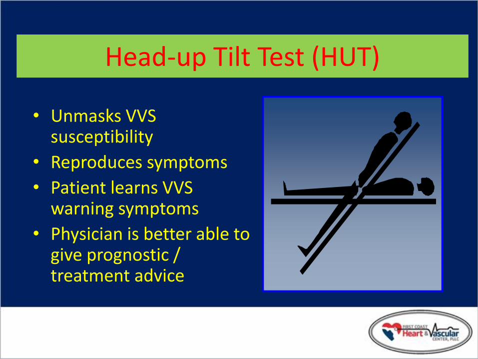
Head-up Tilt Test (HUT)
• Unmasks VVS susceptibility
• Reproduces symptoms
• Patient learns VVS warning symptoms
• Physician is better able to give prognostic / treatment advice

Head-Up Tilt Test (HUT)
DG Benditt, UM Cardiac Arrhythmia Center

Electroencephalogram
• Not a first line of testing
• Syncope from Seizures
• Abnormal in the interval between two attacks – Epilepsy
• Normal – Syncope

Value of Event
Recorder in Syncope
Linzer M. Am J Cardiol. 1990;66:214-219.
*Asterisk denotes event marker

Patient Activator Reveal® Plus ILR New LINQ
Insertable Loop Recorder

ILR Recordings*
56 yo woman with syncope accompanied with seizures. Infra-Hisian AV Block: Dual chamber pacemaker
65 yo man with syncope accompanied with brief retrograde amnesia. VT and VF: ICD and meds
*Medtronic data on file

Randomized Assessment of Syncope Trial (RAST)
Results:
Combining primary strategy with crossover, the diagnostic yield is 43% ILR only vs. 20% conventional only1
Cost/diagnosis is 26% less than conventional testing2
1Krahn AD, et al. Circ. 2001;104:46-51. 2Krahn AD, et al. JACC. 2003;42:495-501.
Unexplained Syncope
EF > 35%
60 Patients
AECG, Tilt, EP Study
Diagnosis
ILR
+
–
+
–
ILR Conventional
Testing (AECG, Tilt, EPS)
30 Patients 30 Patients
Primary Strategy
Crossover
14 6
1 8
+ +

RAST Methods
• Prospective randomized trial – 60 patients with unexplained syncope referred for cardiac
investigation
• Inclusion: – Recurrent unexplained syncope
– Referred to the arrhythmia service for cardiac investigation
– No clinical diagnosis after history, physical, ECG and at least 24 hours of cardiac monitoring
• Exclusion: – LVEF < 35%
– Unable to give informed consent
– Major morbidity precluding one year of follow-up
Krahn A, Klein GJ, Skanes Y. Circulation 2001; 104:46-51.

RAST Results
Unexplained Syncope
n=60
ILR
n=30
Conventional
n=30
In Follow-up
n=3
Diagnosed
n=14
Undiagnosed
n=13
Diagnosed
n=6
Undiagnosed
n=24
Krahn A, Klein GJ, Skanes Y. Circulation 2001; 104:46-51.

RAST Crossover Results
Krahn A, Klein GJ, Skanes Y. Circulation 2001; 104:46-51.
Unexplained Syncope
n=60
13/30
Undiagnosed after monitoring
6 accepted crossover to conventional
24/30
Undiagnosed after conventional
21 accepted crossover to ILR
Diagnosed
n=1
Undiagnosed
n=5
Diagnosed
n=8
Undiagnosed
n=5
In follow-up
n=8

RAST - Diagnoses
0
2
4
6
8
10
12
14
Bradycardia Tachycardia Vasovagal Seizures
ILR Conventional
nu
mb
er
of
pat
ien
ts
Krahn A, Klein GJ, Skanes Y. Circulation 2001; 104:46-51.

Conventional EP Testing in Syncope
• Limited utility in syncope evaluation
• Most useful in patients with structural heart disease – Heart disease……..50-80%
– No Heart disease…18-50%
• Relatively ineffective for assessing bradyarrhythmias
Brignole M, Alboni P, Benditt DG, et al. Eur Heart Journal 2001; 22: 1256-1306.

EP Testing in Syncope: Useful Diagnostic Observations
• Inducible monomorphic VT
• SNRT > 3000 ms or CSRT > 600 ms
• Inducible SVT with hypotension
• HV interval ≥ 100 ms (especially in absence of inducible VT)
• Pacing induced infra-nodal block

• Objectives: • Understand the mechanism of syncope in tilt-positive and tilt-negative
(isolated) patients • Use the ILR to assess the correlation of rhythms captured during tilt
testing and spontaneous recurrent episodes
• Inclusion Criteria: • Patients with three or more syncopal episodes in the last 2 years
• Groups matched in age, sex, history of syncope, ECG, Echo
abnormalities, SHD and arrhythmias
ISSUE Study International Study of Syncope of Uncertain Etiology
Moya A. Circulation. 2001; 104:1261-1267

ISSUE Study Design
• Multicenter, prospective
111 syncope patients 3 episodes in 2 years, first and last episode >6 months apart
History, physical exam, ECG, CSM, echo, Holter (24 hr), other tests as appropriate
Tilt test followed by implant of Reveal Insertable Loop Recorder
Follow-up to recurrent spontaneous episode
Moya A. Circulation. 2001; 104:1261-1267

ISSUE Study Results
Results
Tilt-Negative
Syncope (Isolated)
n=82
Tilt-Positive
Syncope
n=29
Recurrent Event Occurrence (#) 34% (28) 34% (10)
Mean Time to Recurrent Event
(range)
105 days (47-226) 59 (22-98)
ILR ECG Documented (#) 29% (24) 28% (8)
Tachyarrhythmia 2% (2)
Bradycardia 16% (13) 21% (6)
–Sinus Brady 2% (2) 3% (1)
–Sinus Arrest 12% (10) 17% (5)
–AV Block 1% (1)
Total Arrhythmic 18% (15) 21% (6)
Normal Sinus Rhythm 11% (9) 7% (2)
Moya A. Circulation. 2001; 104:1261-1267

ISSUE Study
• Conclusions:
• Homogeneous findings from tilt-negative and tilt-positive syncope patients were observed (clinical characteristics and outcomes). Most frequent finding was asystole secondary to progressive sinus bradycardia, suggesting a neuromediated origin
• In this study tilt-negative patients had as many arrhythmias (18%) as tilt-positive patients (21%)
• In tilt-positive patients the spontaneous episode ECG was more frequently asystolic than what was predicted by tilt test
Moya A. Circulation. 2001; 104:1261-1267
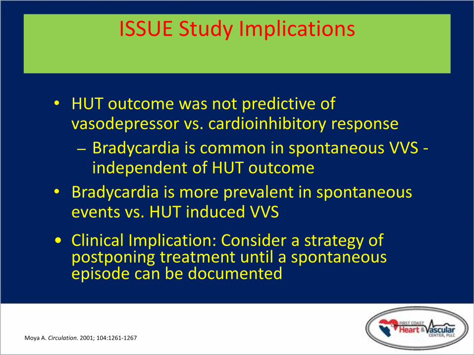
ISSUE Study Implications
• HUT outcome was not predictive of vasodepressor vs. cardioinhibitory response
– Bradycardia is common in spontaneous VVS - independent of HUT outcome
• Bradycardia is more prevalent in spontaneous events vs. HUT induced VVS
• Clinical Implication: Consider a strategy of postponing treatment until a spontaneous episode can be documented
Moya A. Circulation. 2001; 104:1261-1267

Symptom-Rhythm Correlation
Auto Activation Point
Patient Activation Point

Diagnostic Limitations
• Difficult to correlate spontaneous events and laboratory findings
• Often must settle for an attributable cause
• Unknowns remain 20-30% 1
1Kapoor W. In Grubb B, Olshansky B (eds) Syncope: Mechanisms and Management. Armonk NY; Futura Publishing Co, Inc: 1998; 1-13.

Unexplained Syncope Diagnosis History and Physical Exam
Surface ECG
Neurological Testing • Head CT Scan
• Carotid Doppler
• MRI
• Skull Films
• Brain Scan
• EEG
CV Syncope Workup
• Holter
• ELR or ILR
• Tilt Table
• Echo
• EPS
Other CV Testing • Angiogram
• Exercise Test
• SAECG
Psychological Evaluation
ENT Evaluation Endocrine Evaluation
Adapted from: W.Kapoor.An overview of the evaluation and management of syncope. From Grubb B, Olshansky B (eds) Syncope: Mechanisms and Management. Armonk, NY: Futura Publishing Co., Inc.1998.

Typical Cardiovascular Diagnostic Pathway
History and Physical, ECG
Syncope
Known SHD
No SHD
Echo
EPS
+
Treat
> 30 days; > 2 Events
Tilt ILR Tilt Holter/
ELR ILR
Tilt/ILR
< 30 days
-
Adapted from: Linzer M, et al. Annals of Int Med, 1997. 127:76-86. Syncope: Mechanisms and Management. Grubb B, Olshansky B (eds) Futura Publishing 1999 Zimetbaum P, Josephson M. Annals of Int Med, 1999. 130:848-856. Krahn A et al. ACC Current Journal Review,1999. Jan/Feb:80-84.

Section IV:
Specific Conditions

Neurally-Mediated Reflex Syncope (NMS)
• Vasovagal syncope (VVS)
• Carotid sinus syndrome (CSS)
• Situational syncope – post-micturition
– cough
– swallow
– defecation
– blood drawing
– etc.

NM Reflex Syncope: Pathophysiology
• Multiple triggers
• Variable contribution of vasodilatation and bradycardia

NMS – Basic Pathophysiology
Cerebral
Cortex
Vascular
Bed Bradycardia/
Hypotension
Baro-
receptors
Heart
Feedback via
Carotid Baroreceptors
Other Mechanoreceptors
Parasympathetic (+)
sympathetic (+) ¯ Heart Rate
¯ AV
Conduction
_
Vasodilatation
Benditt DG, Lurie KG, Adler SW, et al. Pathophysiology of vasovagal syncope. In: Neurally mediated syncope: Pathophysiology, investigations and treatment. Blanc JJ, Benditt D, Sutton R. Bakken Research Center Series, v. 10. Armonk, NY: Futura, 1996

• Neurally Mediated Physiologic Reflex Mechanism with two Components: – Cardioinhibitory ( HR )
– Vasodepressor ( BP )
• Both components are usually present
Vasovagal Syncope (VVS): Clinical Pathophysiology

Prevalence of VVS
• Prevalence is poorly known – Various studies report 8% to 37% (mean 18%)
of cases of syncope (Linzer 1997)
• In general: – VVS patients younger than CSS patients
– Ages range from adolescence to elderly (median 43 years)
– Pallor, nausea, sweating, palpitations are common
– Amnesia for warning symptoms in older patients
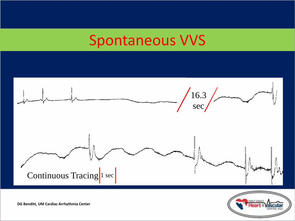
DG Benditt, UM Cardiac Arrhythmia Center
16.3 sec
Continuous Tracing 1 sec
Spontaneous VVS

Management Strategies for VVS
• Optimal management strategies for VVS are a source of debate – Patient education, reassurance, instruction
– Fluids, salt, diet
– Tilt Training
– Support hose
• Drug therapies
• Pacing – Class II indication for VVS patients with positive HUT and
cardioinhibitory or mixed reflex

Section VI:
Insights into More Efficient and Effective Diagnosis and Treatment

Principal Causes of Orthostatic Syncope
• Drug-induced (very common) – diuretics – vasodilators
• Primary autonomic failure – multiple system atrophy – Parkinsonism
• Secondary autonomic failure – diabetes – alcohol – amyloid
• Alcohol – orthostatic intolerance apart from neuropathy

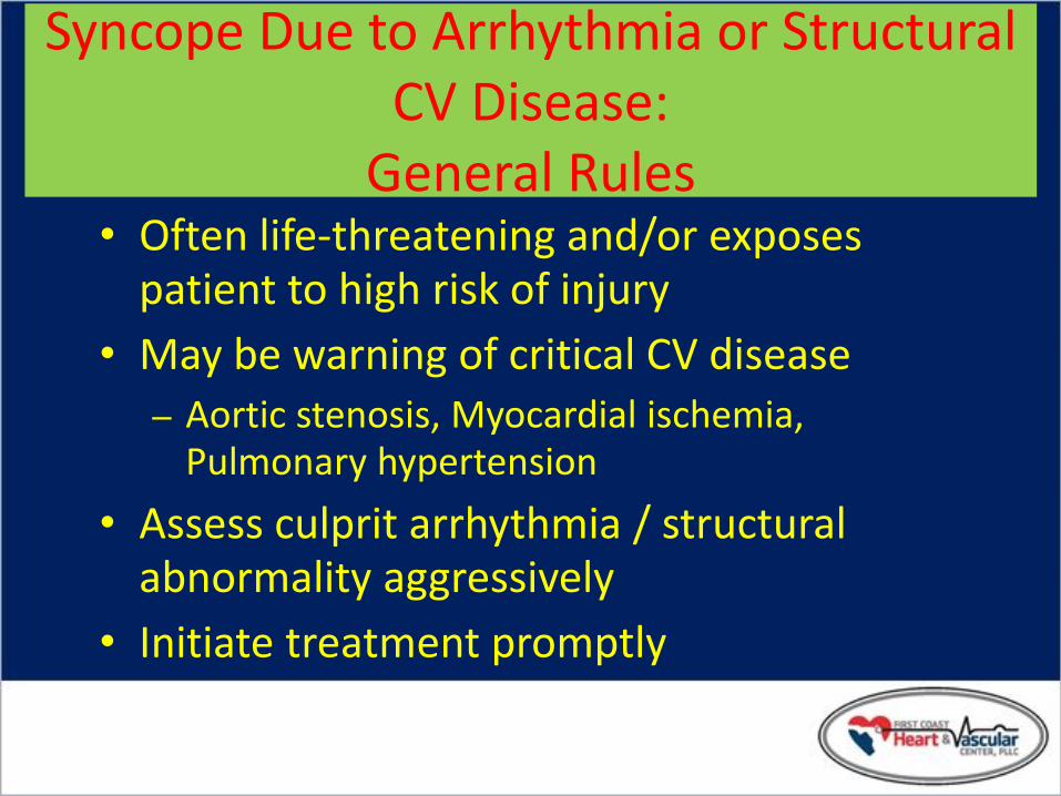
Syncope Due to Arrhythmia or Structural CV Disease:
General Rules
• Often life-threatening and/or exposes patient to high risk of injury
• May be warning of critical CV disease – Aortic stenosis, Myocardial ischemia,
Pulmonary hypertension
• Assess culprit arrhythmia / structural abnormality aggressively
• Initiate treatment promptly

Principal Causes of Syncope due to Structural Cardiovascular Disease
• Acute MI / Ischemia – Acquired coronary artery disease – Congenital coronary artery anomalies
• HOCM • Acute aortic dissection • Pericardial disease / tamponade • Pulmonary embolus / pulmonary
hypertension • Valvular abnormalities
– Aortic stenosis, Atrial myxoma

Syncope Due to Cardiac Arrhythmias
• Bradyarrhythmias – Sinus arrest, exit block
– High grade or acute complete AV block
• Tachyarrhythmias – Atrial fibrillation / flutter with rapid
ventricular rate (e.g. WPW syndrome)
– Paroxysmal SVT or VT
– Torsades de pointes

Rhythms During Recurrent Syncope
Krahn A, et al. Circulation. 1999; 99: 406-410
Normal Sinus Rhythm
58% Normal Sinus Rhythm
58%
Bradycardia
36%
Tachyarrhythmia
6%

AECG: 74 yr Male, Syncope
From the files of DG Benditt, UM Cardiac Arrhythmia Center

Syncope: Torsades
From the files of DG Benditt, UM Cardiac Arrhythmia Center

83 yo woman Bradycardia: Pacemaker implanted
28 yo man in the ER multiple times after falls resulting in trauma VT: ablated and medicated
Reveal ® ILR recordings; Medtronic data on file.

Infra-His Block
From the files of DG Benditt, UM Cardiac Arrhythmia Center

Conclusion
Syncope is a common symptom,
often with dramatic consequences,
which deserves thorough investigation
and appropriate treatment of its cause.

VVS: Tilt-Training
• Objectives – Enhance Orthostatic Tolerance
– Diminish Excessive Autonomic Reflex Activity
– Reduce Syncope Susceptibility / Recurrences
• Technique – Prescribed Periods of Upright Posture
– Progressive Increased Duration

Carotid Sinus Syndrome (CSS)
• Syncope clearly associated with carotid sinus stimulation is rare (≤1% of syncope)
• CSS may be an important cause of unexplained syncope / falls in older individuals

Etiology of CSS
• Sensory nerve endings in the carotid sinus walls respond to deformation
• “Deafferentation” of neck muscles may contribute
• Increased afferent signals to brain stem
• Reflex increase in efferent vagal activity and diminution of sympathetic tone results in bradycardia and vasodilation
Carotid Sinus

Carotid Sinus Hypersensitivity(CSH)
• Abnormal response to CSM
• Absence of symptoms attributable to CSS
• CSH reported frequent in ‘fallers’ (Kenny)
CSH CSS
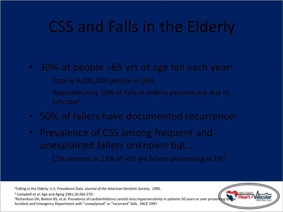
CSS and Falls in the Elderly
• 30% of people >65 yrs of age fall each year1
– Total is 9,000,000 people in USA
– Approximately 10% of falls in elderly persons are due to syncope2
• 50% of fallers have documented recurrence3
• Prevalence of CSS among frequent and unexplained fallers unknown but…
– CSH present in 23% of >50 yrs fallers presenting at ER 3
1Falling in the Elderly: U.S. Prevalence Data. Journal of the American Geriatric Society, 1995. 2 Campbell et al: Age and Aging 1981;10:264-270. 3Richardson DA, Bexton RS, et al. Prevalence of cardioinhibitory carotid sinus hypersensitivity in patients 50 years or over presenting to the Accident and Emergency Department with “unexplained” or “recurrent” falls. PACE 1997

Section V:
Treatment Options

VVS: Pharmacologic Rx
• Salt /Volume – Salt tablets, ‘sport’ drinks, fludrocortisone
• Beta-adrenergic blockers – 1 positive controlled trial (atenolol),
– 1 on-going RCT (POST)
• Disopyramide
• SSRIs – 1 controlled trial
• Vasoconstrictors (e.g., midodrine) – 1 negative controlled trial (etilephrine)

Midodrine for Neurocardiogenic Syncope
Journal of Cardiovascular Electrophysiology Vol. 12, No. 8, Perez-Lugones, et al.
Months
p < 0.001
Sym
pto
m –
Fre
e In
terv
al
180 160 140 120 100 80 60 40 20 0
100
80
60
40
20
0
Fluid
Midodrine
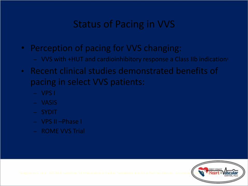
Status of Pacing in VVS
• Perception of pacing for VVS changing: – VVS with +HUT and cardioinhibitory response a Class IIb indication1
• Recent clinical studies demonstrated benefits of pacing in select VVS patients: – VPS I
– VASIS
– SYDIT
– VPS II –Phase I
– ROME VVS Trial
1Gregoratos G, et al. ACC/AHA Guidelines for Implantation of Cardiac Pacemakers and Antiarrhythmic Devices. Circulation. 1998; 97: 1325-1335.

Status of Pacing in VVS
• Benefits of specific device features evolving: – Some success with DDD/DDI hysteresis 1
• “False positives” may result in prolonged high rate intervention
• Tied to lower rate intervention
– Rate drop therapies designed for treating VVS syncope appear to be successful 2-4
1 Sutton R, et al. Circulation. 2000; 102:294-299.
2 Connolly S, et al. J Am Coll Cardiol 1999; 33:16-20.
3 Ammirati F, et al. Circulation. 2002; 104: 52-57.
4 Ammirati F, et al. NASPE Abstract #307. PACE, Vol. 24, April 2002, Part II.

VPS-I Vasovagal Pacemaker Study I
Connolly S, et al. J Am Coll Cardiol 1999; 33: 16-20.
Study Design:
54 patients randomized, prospective, single center
_ 27 DDD pacemaker with rate drop response (RDR)
_ 27 no pacemaker
Patient Inclusion Criteria:
6 syncopal events ever
+HUT
Relative bradycardia*
*a trough heart rate <60/min if no isoproterenol used, <70/min if up to 2 mcg/min isoproterenol used, or <80/min if over 2 mcg/min isoproterenol used

VPS- I
Connolly S, et al. J Am Coll Cardiol 1999; 33: 16-20.
Endpoints:
Time to first syncope
Outcome:
RESULTS
PACEMAKER
(n= 27)
CONTROL
(n=27)
Number of patients w/syncopal recurrence 6 (22%) 19 (70%)
Mean time to first recurrence (days) 112 54
Relative risk reduction of syncope* 85.4% -
*2p = 0.000022

VPS- I
Connolly S, et al. J Am Coll Cardiol 1999; 33: 16-20.
Cumulative Risk (%)
100
90
80
70
60
50
40
30
20
10
0
15 12 9 6 3 0
Control (No Pacemaker)
2P=0.000022
Pacemaker
Time in Months
Number At Risk
C 27 9 4 2 1 0
P 27 21 17 12 11 8

VPS-I
• Conclusion:
Dual-chamber pacing with rate drop response
reduces the likelihood of syncope in patients
with recurrent VVS.
Connolly S, et al. J Am Coll Cardiol 1999; 33: 16-20.

VASIS Vasovagal Syncope International Study
Sutton, R, et al. Circulation. 2000; 102:294-299.
Study Design:
42 patients, randomized, prospective, multicenter
_ 19 DDI pacemaker (80 bpm) with rate hysteresis (45 bpm)
_ 23 no pacemaker
Patient Inclusion Criteria:
> 3 syncopal events in 2 years and last event occurring within 6 months of enrollment and,
Positive VASIS type 2A or 2B cardioinhibitory response to HUT and,
Age > 40 years or drug refractory if < 40 years

VASIS
Sutton, R, et al. Circulation. 2000; 102:294-299.
Outcome:
RESULTS
Pacemaker
(n= 19)
No
Pacemaker
(n=23)
Number of patients w/syncopal recurrence 1 (5%) 14 (61%)
Median time to first recurrence (months)* 15 5
*P= 0.0006
Endpoints:
Time to first syncope

VASIS
Pacemaker
No-Pacemaker
p=0.0004
Years
% s
ynco
pe
-fre
e
100
80
60
40
20
0 2 3 4 5 6
7 12 14 15 23 31 40
# of pts
Sutton, R, et al. Circulation. 2000; 102:294-299.

VASIS
• Conclusion:
Dual-chamber pacing (at a rate of 80 bpm ) with rate hysteresis reduces the likelihood of syncope in patients with tilt-positive, cardioinhibitory syncope.
Sutton, R, et al. Circulation. 2000; 102:294-299.

SYDIT Syncope Diagnosis and Treatment Study
• Study Design:
– 93 patients randomized, prospective, multicenter
• 46 DDD pacemaker with rate drop response (RDR)
• 47 Atenolol 100 MG/D
• Patient Inclusion Criteria:
– > 55 yrs
– > 3 syncopal episodes in 2 years
– + HUT with relative bradycardia (trough HR <60 bpm)
Ammirati F, et al. Circulation. 2001; 104:52-57.

SYDIT
• Endpoints: – Time to first syncope
• Outcome:
RESULTS
PACED
(n= 46)
DRUG
(n= 47)
Number of patients w/syncopal recurrence* 2 (4%) 12 (25%)
Median time to first recurrence (days) 390 135
*P=0.004
Ammirati, et al. Circulation. 2001; 104:52-57.

Syncope-free Survival: Intention-to-Treat (n=46/paced, 47/drug).
Ammirati F, et al. Circulation. 2001; 104:52-57.
SYDIT
1.0
Time (days)
100
0.9
0.8
0.7
0.6
200 300 400 500 600 700 800 900 1000 0
P = 0.0032
drug
pacemaker
% o
f sy
nco
pe
fre
e p
ts

SYDIT
• Conclusion:
Dual-chamber pacing + RDR is superior to Atenolol in prevention of recurrent syncope in highly symptomatic patients with relative bradycardia during tilt-induced syncope.
Ammirati F, et al. Circulation. 2001; 104:52-57.

VPS-II: Phase I Vasovagal Pacemaker Study-II
• Study Design: – 100 patients, randomized, prospective, multicenter
• 50 DDD pacemaker with rate drop response (RDR)
• 50 ODO pacemaker (inactive mode)
• Patient Inclusion Criteria: – > 6 syncope events ever or > 3 syncope events in 2
years or > 1 syncope event in 6 months and,
– Positive HUT with syncope or presyncope and a heart rate blood pressure product <9000
Presented at the 23rd Annual Scientific Sessions of the North American Society of Pacing and Electrophysiology. Late Breaking Clinical Trials, May 11, 2002.

VPS-II: Phase I
• Endpoints: – Time to first syncope
• Outcome:
RESULTS
DDD Pacemaker
(n= 50)
ODO Pacemaker
(n= 50)
Number of patients w/syncopal recurrence 16 (32%) 22 (44%)
Relative Risk Reduction* 28.7% -
*P=0.153
Presented at the 23rd Annual Scientific Sessions of the North American Society of Pacing and Electrophysiology. Late Breaking Clinical Trials, May 11, 2002.

0.4
0.3
0.2
ODO DDD
P = 0.153 (one-sided)
Number at Risk ODO 40 37 35 32 31 21
DDD 39 36 34 33 33 17
0 1 2 3 4 5 6
0.1
0.0
Cu
mu
lati
ve R
isk
of
Syn
cop
e
Presented at the 23rd Annual Scientific Sessions of the North American Society of Pacing and Electrophysiology. Late Breaking Clinical Trials, May 11, 2002.
VPS-II: Phase I

VPS-II: Phase I
Conclusions:
Lower than anticipated syncope event rate in the
control arm.
Higher than anticipated event rate in the treatment
group.
Consequence: treatment effect was less than VPS-I.
Results favored pacing but the treatment effect was
not statistically significant.
Presented at the 23rd Annual Scientific Sessions of the North American Society of Pacing and Electrophysiology. Late Breaking Clinical Trials, May 11, 2002.
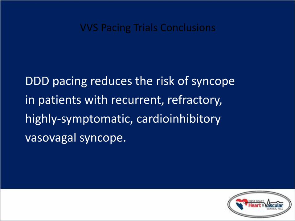
VVS Pacing Trials Conclusions
DDD pacing reduces the risk of syncope
in patients with recurrent, refractory,
highly-symptomatic, cardioinhibitory
vasovagal syncope.

SAFE PACE Study Design
• Randomized controlled trial (N=175):
– Pacing (87) vs. No Pacing (88)
• Single center: Royal Victoria Infirmary, Newcastle, UK
• Recruitment began: April 1998
• 12 month follow-up per patient
• Study concluded: May 2000
Kenny RA, J Am Coll Cardiol 2001; 38:1491-1496.

SAFE PACE Inclusion Criteria
• Consecutive adults attending accident and emergency department
• > 50 Years
- Experienced non-accidental fall
•Positive response to CSM
Kenny RA, J Am Coll Cardiol 2001; 38:1491-1496.

SAFE PACE Screening Process Accident and Emergency Attendees > 50 Yrs
Falls or Syncope
Non-accidental Fall
CSM Performed
Cardioinhibitory or Mixed CSH
RCT
Control Pacemaker
Kenny RA, J Am Coll Cardiol 2001; 38:1491-1496.

SAFE PACE Screening Results
RCT (n=175)
Control
(n=88)
Pacemaker
(n=87)
• No pacing intervention • Medtronic Thera DR
(Rate Drop Response
Algorithm)
Kenny RA, J Am Coll Cardiol 2001; 38:1491-1496.

SAFE PACE Results
Number of Falls
Control
n=87
Pacemaker
n=84
% Participants
w/Falls
60% 58%
Total Number of
Falls*
699 216
Mean Number of
Falls**
9.3 4.1
* Falls during 12 months post randomization
** Crude adjustment calculation
Kenny RA, J Am Coll Cardiol 2001; 38:1491-1496.
70%
Reduction [OR 0.42; 95%
CI: 0.23, 0.75]

Control
N=87
Pacemaker
N=84
% Participants
w/Syncopal Events
22% 11%
Total Number of
Syncopal Events
47 22
Mean Number Syncopal
Events
1.14 0.20
SAFE PACE Results Number of Syncopal Episodes
50%
Reduction [OR 0.53; 95%
CI 0.23; 1.20 ns]
* Syncopal events 12 months past randomization
** Crude adjustment calculation
Kenny RA, J Am Coll Cardiol 2001; 38:000-000.

Control
n= 87
Pacemaker
n= 84
% Participants w/Injurious
Events
41% 35%
Total Number Injury
Events
202 61
-Fractures
-Soft Tissue Injury
4
198
3
58
SAFE PACE Results Number of Injury Events
70%
Reduction
* Injurious events 12 months post randomization
Kenny RA, J Am Coll Cardiol 2001; 38:1491-1496.

SAFE PACE Conclusions
In patients with unexplained falls and a
diagnosis of Cardioinhibitory CSH, cardiac
pacing reduced the total number of:
• Falls by 70%
• Syncopal events by 53%
• Injurious events by 70%
Kenny RA, J Am Coll Cardiol 2001; 38:1491-1496.

Role of Pacing in CSS -- Syncope Recurrence Rate
Brignole et. Al. Diagnosis, natural history and treatment. Eur JCPE. 1992; 4:247-254
0%
25%
50%
75%
No Pacing Pacing
57%
%6 Class I indication for pacing (AHA and BPEG) Limit pacing to CSS that is:
•Cardioinhibitory •Mixed
DDD/DDI superior to VVI
(Mean follow-up = 6 months)

Section VI:
Insights into More Efficient and Effective Diagnosis and Treatment

Principal Causes of Orthostatic Syncope
• Drug-induced (very common) – diuretics – vasodilators
• Primary autonomic failure – multiple system atrophy – Parkinsonism
• Secondary autonomic failure – diabetes – alcohol – amyloid
• Alcohol – orthostatic intolerance apart from neuropathy
Syncope Due to Arrhythmia or Structural CV Disease:
General Rules
• Often life-threatening and/or exposes patient to high risk of injury
• May be warning of critical CV disease – Aortic stenosis, Myocardial ischemia,
Pulmonary hypertension
• Assess culprit arrhythmia / structural abnormality aggressively
• Initiate treatment promptly

Principal Causes of Syncope due to Structural Cardiovascular Disease
• Acute MI / Ischemia – Acquired coronary artery disease – Congenital coronary artery anomalies
• HOCM • Acute aortic dissection • Pericardial disease / tamponade • Pulmonary embolus / pulmonary
hypertension • Valvular abnormalities
– Aortic stenosis, Atrial myxoma

Syncope Due to Cardiac Arrhythmias
• Bradyarrhythmias – Sinus arrest, exit block
– High grade or acute complete AV block
• Tachyarrhythmias – Atrial fibrillation / flutter with rapid
ventricular rate (e.g. WPW syndrome)
– Paroxysmal SVT or VT
– Torsades de pointes

Rhythms During Recurrent Syncope
Krahn A, et al. Circulation. 1999; 99: 406-410
Normal Sinus Rhythm
58% Normal Sinus Rhythm
58%
Bradycardia
36%
Tachyarrhythmia
6%

AECG: 74 yr Male, Syncope
From the files of DG Benditt, UM Cardiac Arrhythmia Center

Syncope: Torsades
From the files of DG Benditt, UM Cardiac Arrhythmia Center

83 yo woman Bradycardia: Pacemaker implanted
28 yo man in the ER multiple times after falls resulting in trauma VT: ablated and medicated
Reveal ® ILR recordings; Medtronic data on file.

Infra-His Block
From the files of DG Benditt, UM Cardiac Arrhythmia Center

Drug-Induced QT Prolongation
• Antiarrhythmics – Class IA ...Quinidine, Procainamide, Disopyramide
– Class III…Sotalol, Ibutilide, Dofetilide, Amiodarone, (NAPA)
• Antianginal Agents – (Bepridil)
• Psychoactive Agents – Phenothiazines, Amitriptyline, Imipramine, Ziprasidone
• Antibiotics – Erythromycin, Pentamidine, Fluconazole
• Nonsedating antihistamines – (Terfenadine), Astemizole
• Others – (Cisapride), Droperidol

Treatment of Syncope Due to Bradyarrhythmia
• Class I indication for pacing using dual- chamber system wherever adequate atrial rhythm is available
• Ventricular pacing in atrial fibrillation with slow ventricular response
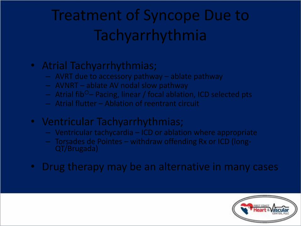
Treatment of Syncope Due to Tachyarrhythmia
• Atrial Tachyarrhythmias; – AVRT due to accessory pathway – ablate pathway – AVNRT – ablate AV nodal slow pathway – Atrial fib– Pacing, linear / focal ablation, ICD selected pts – Atrial flutter – Ablation of reentrant circuit
• Ventricular Tachyarrhythmias; – Ventricular tachycardia – ICD or ablation where appropriate – Torsades de Pointes – withdraw offending Rx or ICD (long-
QT/Brugada)
• Drug therapy may be an alternative in many cases

Conclusion
Syncope is a common symptom,
often with dramatic consequences,
which deserves thorough investigation
and appropriate treatment of its cause.
Disclaimer INDICATIONS
9526 Reveal® Plus Insertable Loop Recorder The Reveal Plus Insertable Loop Recorder (ILR) is an implantable patient activated monitoring system that records subcutaneous ECG and is indicated for patients who experience transient symptoms that may suggest a cardiac arrhythmia. 9790 Programmer The Medtronic 9790 Programmers are portable, microprocessor based instruments used to program Medtronic implantable devices. 6191 Activator The Model 6191 Activator is intended for use in combination with a Medtronic Model 9525 Reveal® and the Model 9526 Reveal Plus Insertable Loop Recorders. CONTRAINDICATIONS There are no known contraindications for the implantation of the Reveal Plus ILR. However, the patient’s particular medical condition may dictate whether or not a subcutaneous, chronically implanted device can be tolerated. WARNINGS/PRECAUTIONS 9526 Reveal Plus Insertable Loop Recorder Patients with the Reveal Plus ILR should avoid sources of magnetic resonance imaging, diathermy, high sources of radiation, electrosurgical cautery, external defibrillation, lithotripsy, and radiofrequency ablation to avoid electrical reset of the device, and/or inappropriate sensing. 6191 Activator Operation of the Model 6191 Activator near sources of electromagnetic interference, such as cellular phones, computer monitors, etc., may adversely affect the performance of this device. See the appropriate technical manual for detailed information regarding indications, contraindications, warnings, and precautions. Caution: Federal law (U.S.A.) restricts this device to sale by or on the order of a physician.

Disclaimer INDICATIONS
Medtronic.Kappa 700 Series Pacemakers
The Medtronic.Kappa 700 Series pacemakers are indicated for rate adaptive pacing in patients who may benefit from increased pacing rates concurrent with increases in activity and are also indicated for dual chamber and atrial tracking modes in patients who may benefit from maintenance of AV synchrony. Dual chamber modes are specifically indicated for treatment of conduction disorders that require restoration of both rate and AV synchrony, which include various degrees of AV block to maintain the atrial contribution to cardiac output and VVI intolerance (e.g., pacemaker syndrome) in the presence of persistent sinus rhythm.
9790 Programmer
The Medtronic 9790 Programmers are portable, microprocessor based instruments used to program Medtronic implantable devices.
9462
The Model 9462 Remote Assistant is intended for use in combination with a Medtronic implantable pacemaker with Remote Assistant diagnostic capabilities.
CONTRAINDICATIONS
The Medtronic.Kappa 700 Series pacemakers are contraindicated for the following applications:
· Dual chamber atrial pacing in patients with chronic refractory atrial tachyarrhythmias.
· Asynchronous pacing in the presence (or likelihood) of competitive paced and intrinsic rhythms.
· Unipolar pacing for patients with an implanted cardioverter-defibrillator (ICD) because it may cause unwanted delivery or inhibition of ICD therapy.
WARNINGS/PRECAUTIONS
Medtronic.Kappa 700 Series patients should avoid sources of magnetic resonance imaging, diathermy, high sources of radiation, electrosurgical cautery, external defibrillation, lithotripsy, and radiofrequency ablation to avoid electrical reset of the device, inappropriate sensing and/or therapy.
See the appropriate technical manual for detailed information regarding indications, contraindications, warnings, and precautions.
Caution: Federal law (U.S.A.) restricts this device to sale by or on the order of a physician.

Additional Slides

Falls -- Incidence, Recurrence, CHS*
1 Falling in the Elderly, 1995. 2 Richardson, PACE, 1997.
0%
25%
50%
75%
Incidence
> 65 yrs. old
Recurrence CSH* present
in fallers > 50 yrs.
presenting at ER
30% 1
50% 1
23% 2
* Carotid Sinus Hypersensitivity

VVS Pacing Trials Comparison Summary

Pacing in VVS
Two randomized, controlled trials suggest benefit in selected patients with multiple (>5 lifetime) syncope recurrences and one or more of:
– prominent cardioinhibitory features
– asystolic pause >10 seconds
– sustained HR<40/minute
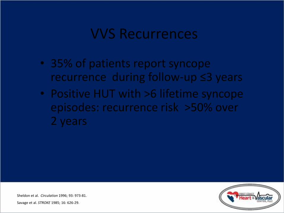
VVS Recurrences
• 35% of patients report syncope recurrence during follow-up ≤3 years
• Positive HUT with >6 lifetime syncope episodes: recurrence risk >50% over 2 years
Sheldon et al. Circulation 1996; 93: 973-81.
Savage et al. STROKE 1985; 16: 626-29.
SAFE PACE 2: Syncope and Falls in the Elderly
30% of individuals >65 yrs fall each year
5% of falls result in fractures
1% of falls result in hip fractures
SAFEPACE Pilot Study
18% prevalence of CSH in unexplained ‘fallers’
31% in ‘fallers’ >80 yrs
Kenny RA, J Am Coll Cardiol 2001; 38:1491-1496.

Both
Rate Drop Response Overview
Detection Options
Drop Detect
Low Rate Detect
Detects relative heart rate drops of a pre-determined size
Detects heart rate that falls to a user-defined lower rate
Detection occurs when either Drop Detection or Low
Rate Detection criteria are met
Rate Drop Detection in Medtronic Kappa® Series Pacemakers

Drop Detection with Intervention
Drop Detection Method: Drop Size 25, Drop Rate 70
40
50
60
70
80
90
100
110
Ven
tric
ula
r R
ate
Drop Size=25 bpm
Drop Rate
Peak Rate=90 bpm
2 consecutive beats < Drop Size and Drop Rate
Rate Drop Detection in Medtronic Kappa® Series Pacemakers

Drop Detect Peak Rate
Drop Detection Method: Drop Size 25
40
50
60
70
80
90
100
110
120
Ven
tric
ula
r R
ate
Drop Size=25
bpm
Peak Rate=90 bpm
Rate Drop Detection in Medtronic Kappa® Series Pacemakers

Low Rate Detection Method: Lower Rate 40, Detection beats 2
30
40
50
60
70
80
90
100
110
Ven
tric
ula
r R
ate
Lower Rate
2 consecutive paced
beats at Lower Rate
Low Rate Detect
Rate Drop Detection in Medtronic Kappa® Series Pacemakers

Using Both Detection Algorithms
• When both detection algorithms are used:
– Detection occurs when either Drop Detection or Low Rate Detection criteria are met
– Intervention Rate, Duration and Termination are programmed the same as when using the individual detection modes
Rate Drop Detection in Medtronic Kappa® Series Pacemakers

Rate Drop Intervention Therapy
• DDD or DDI pacing
• Pacing intervention – Paces at programmed Intervention Rate
for programmed duration
• Pacing termination – Pacing rate decreases until there are three
consecutive atrial senses or Lower Rate is reached
Rate Drop Detection in Medtronic Kappa® Series Pacemakers

Challenges of Syncope
• Cost – Cost/year – Cost/diagnosis
• Quality of Life Implications – Work/financial – Mobility (automobiles) – Psychological
• Diagnosis & Treatment – Diagnostic yield and repeatability of tests – Frequency and clustering of events – Difficulty in managing/treating/controlling future events – Appropriate risk stratification – Complex Etiology

Diagnosing VVS
• Patient history and physical exam
• Positive tilt table test (ACC Consensus Protocol) – Overnight fast
– ECG
– Blood pressure
– Supine and upright
– Tilt to 60-80 degrees
– Isoproterenol
– Re-tilt
DG Benditt, Tilt Table Testing, 1996.
60° - 80°

VVS: Treatment Overview
• Education – symptom recognition
– reassurance
– situation avoidance
• Tilt-Training – prescribed upright posture
• Pharmacologic Agents – salt/volume management
– beta-adrenergic blockers
– SSRIs
– vasoconstrictors (e.g., midodrine)
• Cardiac Pacemakers

Tilt-Training: Clinical Outcomes
• 42 HUT positive (21±13 min) VVS patients
• Home training: two 30 minute sessions daily
• Outcomes
– 41/42 pts --->45 min asymptomatic HUT
– Clinical follow-up: 15.1±7.8 mos • 36 pts syncope free
• 4 pts: presyncope
• 1 pt: syncope recurrences
Reybrouck et al. PACE 2000; 23:493-8