SVEIKATOS PRIEŽIŪROS TECHNOLOGIJOS VERTINIMAS ... · Lietuvoje bendras nereumatinių širdies...
98
SVEIKATOS PRIEŽIŪROS TECHNOLOGIJOS VERTINIMAS: TRANSKATETERINIŲ IMPLANTUOJAMŲJŲ PRIEMONIŲ TAIKYMAS MITRALINIO VOŽTUVO KOREKCIJAI SUAUGUSIEMS PACIENTIEMS, KURIEMS DIAGNOZUOTA LĖTINĖ MITRALINIO VOŽTUVO REGURGITACIJA HEALTH TECHNOLOGY ASSESSMENT: TRANSCATHETER IMPLANTABLE DEVICES FOR MITRAL VALVE REPAIR IN ADULTS WITH CHRONIC MITRAL VALVE REGURGITATION VASPVT, 2016
-
Upload
truongquynh -
Category
Documents
-
view
234 -
download
0
Transcript of SVEIKATOS PRIEŽIŪROS TECHNOLOGIJOS VERTINIMAS ... · Lietuvoje bendras nereumatinių širdies...
1
SVEIKATOS PRIEŽIŪROS TECHNOLOGIJOS VERTINIMAS:
TRANSKATETERINIŲ IMPLANTUOJAMŲJŲ PRIEMONIŲ TAIKYMAS MITRALINIO
VOŽTUVO KOREKCIJAI SUAUGUSIEMS PACIENTIEMS, KURIEMS DIAGNOZUOTA
LĖTINĖ MITRALINIO VOŽTUVO REGURGITACIJA
HEALTH TECHNOLOGY ASSESSMENT:
TRANSCATHETER IMPLANTABLE DEVICES FOR MITRAL VALVE REPAIR IN
ADULTS WITH CHRONIC MITRAL VALVE REGURGITATION
VASPVT, 2016

2
Valstybinė akreditavimo sveikatos priežiūros veiklai tarnyba prie Sveikatos apsaugos ministerijos
Autoriai: Medicinos technologijų skyriaus vyr.specialistės:
Gintarė Kelbauskaitė
Ugnė Dragūnaitė
Valstybinė akreditavimo sveikatos priežiūros veiklai tarnyba prie Sveikatos apsaugos ministerijos
Jeruzalės g. 21, LT-08420 Vilnius
Tel. (8 5) 261 5177,
Faks. (8 5) 212 7310,
El. paštas: [email protected]
Sveikatos technologijos vertinimo santrauką galima rasti interneto svetainėje:
http://www.vaspvt.gov.lt/node/486
_____________________________________________________________________________
State Health Care Accreditation Agency
under the Ministry of Health
Authors: Chief specialists of Medical Technology division:
Gintarė Kelbauskaitė
Ugnė Dragūnaitė
State Health Care Accreditation Agency under the Ministry of Health
Jeruzalės st. 21, LT-08420 Vilnius
Tel. (370 5) 261 5177,
Fax. (370 5) 212 7310,
E. mail: [email protected]
Health technology assessment is available on the website:
http://www.vaspvt.gov.lt/node/486
3
TURINYS
SANTRAUKA 6
Tikslinė populiacija (A skyrius) 6
Tikslinė būklė (A skyrius) 6
Sveikatos būklės valdymas (A skyrius) 6
Pagrindinės technologijos charakteristikos (B skyrius) 6
Pacientų saugumas (C skyrius) 7
Išgyvenamumas (D skyrius) 8
Sergamumas (D skyrius) 8
Kūno funkcijos ir gyvenimo kokybė (D skyrius) 9
IŠVADOS 10
REKOMENDACIJOS 11
SVEIKATOS TECHNOLOGIJOS VERTINIMO METODIKA 12
Tyrimų charakteristikos. 12
Pacientų charakteristikos. 13
Įrodymų kokybė 14
Vertinimo apimtis (PICO) 15
Scope (PICO) 17
SUMMARY 19
A. HEALTH PROBLEM AND CURRENT USE OF THE TECHNOLOGY 23
B.DESCRIPTION AND TECHNICAL CHARACTERISTICS OF
TECHNOLOGY 33
C.SAFETY 40
D. CLINICAL EFFECTIVENESS 44
POTENTIAL ETHICAL, ORGANISATIONAL, SOCIAL, AND LEGAL
ASPECTS 51
CONCLUSIONS 52
RECOMMENDATIONS 53
REFERENCES 54
APPENDIX 1: METHODS AND DESCRIPTION OF THE EVIDENCES
USED 61
APPENDIX 2: DESCRIPTION OF THE EVIDENCE USED 67
APPENDIX 3: QUALITY ASSESSMENT OF SELECTED STUDIES 88
APPENDIX 4. TECHNOLOGY ADAPTATION CHECKLISTS 96

4
SANTRUMPOS
6MWT 6-minute walk test (liet. 6 minučių ėjimo testas)
AATS American Association for Thoracic Surgery
ACC American College of Cardiology
ACCESS-EU MitraClip Therapy Economic and Clinical Outcomes Study Europe
ACE Angiotensin-converting-enzyme
AE Adverse event
AF Atrial fibrillation
AMADEUS CARILLON Mitral Annuloplasty Device European Union Study
ASA American Society of Anesthesiologists
BCBS Blue Cross Blue Shield Association
CCT Comparative controlled trial
CE Conformité Européene
CINAHL Cumulative Index to Nursing and Allied Health Literature
COAPT Cardiovascular Outcomes Assessment of the MitraClip Percutaneous Therapy for Heart Failure Patients With
Functional Mitral Regurgitation
CRD Centre for Reviews and Dissemination
CRT Cardiac resynchronisation therapy
CS Coronary sinus
DMR Degenerative mitral regurgitation (liet. Degeneracinė mitralinė regurgitacija)
ePTFE suture Expanded polytetrafluoroethylene suture
ERO Effective regurgitant orifice
ESC-EACTS European Society of Cardiology–-European Association for Cardio-Thoracic Surgery
EU European Union
EuroSCORE European System for Cardiac Operative Risk Evaluation
EVEREST II HRR Endovascular Valve Edge-to-Edge Repair Study II High Risk Registry
FDA United States Food and Drug Administration
FMR Functional mitral regurgitation (liet. funkcinė mitralinė regurgitacija)
GCV Great cardiac vein
GDMT Guideline-directed medical therapy
h Hours
HAS Haute Autorité de Santé
HealthPACT Health Policy Advisory Committee on Technology
HF Heart failure
HR High risk
HRQoL Health-related quality of life
HTA Health Technology Assessment
ICD International Statistical Classification of Diseases and Related Health Problems
IE Infective endocarditis
IHE Institute of Health Economics
ISRCTN International Standard Randomised Controlled Trial Number
KS Liet. Kairysis skilvelis
LA Left atrial
LBI-HTA Ludwig Boltzmann Institute for Health Technology Assessment
LES logistic Euroscore
LV Left ventricular
LVD Leaflet Verification Display
LVESD Left ventricular end-systolic dimension
LVEF Left ventricular ejection fraction
MACCE Major cardiac and cerebrovascular events
MAE Major adverse event
MATTERHORN Multicenter, Randomized, Controlled Study to Assess Mitral Valve Reconstruction for Advanced Insufficiency

5
of Functional or Ischemic Origin
MeSH Medical Subject Headings
MI Myocardial infarction
min. Minute(s)
MITRA-FR Multicentre Study of Percutaneous Mitral Valve Repair MitraClip Device in Patients With Severe Secondary
Mitral Regurgitation
MR Mitral regurgitation (liet. Mitralinė regurgitacija)
MSAC Medical Services Advisory Committee
MV Mitral valve (liet. mitralinis vožtuvas)
NHC National Health Committee
NHS National Health Service
NICE The National Institute for Health and Care Excellence
NIIR NeoChord International Independent Registry
NR Not reported
NYHA New York Heart Association
QoL Quality of life
PMVR Percutaneous mitral valve repair
R-AMSTAR Revised Assessment of Multiple Systematic Reviews
RCT Randomised controlled trial
REA Relative Effectiveness Assessment (liet. Greitas santykinio efektyvumo vertinimas)
REALISM Real World Expanded Multicenter Study of the MitraClip® System
REDUCE FMR CARILLON® Mitral Contour System® for Reducing Functional Mitral Regurgitation
RESHAPE-HF Randomized Study of the MitraClip Device in Heart Failure Patients With Clinically Significant Functional
Mitral Regurgitation
RESHAPE-HF1-FU Observational Study of Heart Failure Patients With Clinically Significant Functional Mitral Regurgitation –
Follow Up of the Former Participants in the RESHAPE-HF trial
SCAI Society for Cardiovascular Angiography and Interventions
STS The Society for Thoracic Surgeons
STV Liet. Sveikatos technologijų vertinimas
SF-36 Short-form 36 Health Survey
SGC Steerable guide catheter
SOLVD Studies Of Left Ventricular Dysfunction
STS The Society for Thoracic Surgeons
ŠN Širdies nepakankamumas
TACT Transapical Artificial Chordae Tendinae
TEE TransEsophageal Echocardiography
TOE Transoesophageal echocardiography
TITAN Transcatheter Implantation of Carillon Mitral Annuloplasty Device
TLK Tarptautinė statistinė ligų ir sveikatos sutrikimų klasifikacija
TMVR Transcatheter mitral valve repair
TRAMI Transcatheter Mitral Valve Interventions
TTE Transthoracic echocardiography
t. y. Tai yra
U Unit
VAD Ventricular assist device
VASPVT Valstybinė Akreditavimo Sveikatos Priežiūros Veiklai Tarnyba (angl. State Health Care Accreditation Agency)

6
SANTRAUKA
Tikslinė populiacija (A skyrius)
Tikslinė šio vertinimo populiacija yra suaugę asmenys, turintys vidutinę-sunkią arba sunkią degeneracinę mitralinę
regurgitaciją (DMR), galimi kandidatai chirurginiam gydymui (t.y. NeoChord DS1000 populiacija); taip pat suaugusieji su vidutine-
sunkia arba sunkia DMR arba funkcine mitraline regurgitacija (FMR), turintys didelę chirurginę riziką arba kontraindikaciją
chirurginiam gydymui (t.y.CARILLON® Mitral Contour System® ir MitraClip® System populiacijos). (A0007)
Vidutinė ar sunkesnė MR pasireiškia 11 % vyresnių nei 70 metų amžiaus asmenų ir beveik 40 % pacientų, turinčių
reikšmingą širdies nepakankamumą (ŠN). (A0006) Specifinių statistinių duomenų, tikslinės populiacijos dydžiui nustatyti nerasta.
Lietuvoje bendras nereumatinių širdies vožtuvų sutrikimų (pgl.TLK-10-AM kodai I34-I39) paplitimas 2014 metais buvo 8,4 atvejai
(1000 gyventojų). (A0023)
Tikslinė būklė (A skyrius)
Chroniška MR yra ilgalaikis sutrikimas, apibūdinamas kaip kraujo grįžimas atgal – iš kairiojo skilvelio į kairįjį prieširdį,
sistolės metu, pro nepilnai užsidarantį mitralinį vožtuvą. TLK-10 kodas, tiksliausiai apibūdinantis mitralinę regurgitaciją yra I34.0.
Pagal etiologiją MR gali būti degeneracinė (pirminė) arba funkcinė (antrinė). DMR priežastis yra vožtuvo burių anomalijos,
dažniausiai atsirandančios dėl miksomatozinės degeneracijos. FMR atsiranda kaip kairiojo skilvelio patologinės remodeliacijos
pasekmė, kartu su mitralinio vožtuvo struktūrinių elementų (žiedo, papilinių raumenų, burių) anatominiais pokyčiais. Dažniausios
MR priežastys yra degeneracinė (miksomatozinė) liga, išeminė širdies liga, reumatinė širdies liga, infekcinis endokarditas. (A0003)
Apskaičiuotas 5 metų mirštamumas dėl visų priežasčių; dėl kardialinių priežasčių; dėl kardialinių įvykių pacientams su besimptome
sunkia MR atitinkamai yra: 22,3 %; 14,3 % ir 33,3 %. (A0004) Vidutiniškai 40 % pacientų su MR turi mažiausiai 1 gretutinį
susirgimą, tai dažniausiai yra širdies nepakankamumas, prieširdžių virpėjimas ir išeminė širdies liga. (A0005)
Sveikatos būklės valdymas (A skyrius)
Detali MR gydymo rekomendacijų apžvalga, remiantis įvairių šalių klinikinėmis gairėmis, pateikta A0025.
Chroniška DMR
Medikamentinis gydymas (kraujagysles plečiantys vaistai, ACE inhibitoriai, beta blokatoriai) yra rekomenduotini
pacientams su pažengusia MR, kai pasireiškia sunkūs simptomai ir yra kontraindikuojamas chirurginis gydymas. Chirurginis
gydymas rekomenduotinas pasireiškiant ar nepasireikšiant simptomams, esant sunkiai DMR ir kairiojo skilvelio disfunkcijai.
Pirmenybė teikiama MV korekcijai, ne keitimui. Sėkminga ir ilgalaikė korekcija (95 % tikimybė) gali būti atlikta pacientams,
kuriems nepasireiškia simptomų, yra išlikusi kairiojo skilvelio funkcija, ir tikėtinas mažesnis nei 1 % mirštamumas. Transkateterinė
mitralinio vožtuvo korekcija gali būti atliekama pacientams, kuriems pasireiškia sunkūs simptomai (III–IV NYHA klasė),
draudžiama atlikti chirurginę intervenciją (dėl sunkių gretutinių susirgimų) ir kuriems sunkūs simptomai išlieka, nepaisant taikomo
optimalaus ŠN gydymo (pagal klinikines gaires). (A0025)
Chroniška FMR
Pacientai su progresuojančia ar sunkia FMR turėtų gauti optimalų gydymą (pagal klinikines gaires), skirtą ŠN gydyti arba
širdies resinhcronizacijos terapiją (jei atitinka indikacijas). Chirurginis gydymas (MV korekcija arba pakeitimas) yra taikytina
pacientams su sunkia FMR, kuriems nepaisant optimalaus ŠN gydymo išlieka sunkūs simptomai. (A0025)
Pagrindinės technologijos charakteristikos (B skyrius)
Vertinime svarstomos trys sistemos: MitraClip® System skirta vožtuvo burių korekcijai, CARILLON® Mitral Contour
System® skirta žiedo korekcijai ir NeoChord DS1000 skirta skaidulinės stygos keitimui.

7
MitraClip® System
MitraClip® yra pirma tokio tipo transkateterinė mitralinio vožtuvo taisymo (TMVT) sistema, sukurta mitralinio vožtuvo
nepakankamumo korekcijai. Šis transkateterinis gydomasis metodas įgalina vidutinio–sunkaus ir sunkaus degeneracinio (pirminio)
(DMR) ar funkcinio (antrinio) (FMR) vožtuvo nepakankamumo gydymą, pacientams, kurie nėra tinkami kandidatai įprastinei
mitralinio vožtuvo operacijai atlikti. Šios priemonės įvedimui nereikia atlikti vidinės sternotomijos ar širdies ir plaučių šuntavimo, o
poprocedūriniam gijimo laikotarpiui dauguma pacientų gali būti išrašyti namo. MitraClip® prietaisas po transeptalinės punkcijos
yra įvedamas į širdį per šlaunies veną ir implantuojamas ant vožtuvo burių, taip sudaromas dvigubas angos vožtuvas, kuriuo
sumažinamas kraujo atbulinis srautas ir įgalinamas efektyvesnis širdies kraujo pumpavimo darbas (B0001). Europos Sąjungoje
MitraClip® System yra skirta MV nepakankamumo korekcijai, atliekant audinių sugretinimą, bet Jungtinėse Amerikos Valstijose
sistema indikuota perkutaniniam pastebimų MV (> 3+) nepakankamumo simptomų mažinimui, atsiradusiems dėl pirminių
mitralinio aparato pakitimų (DMR), pacientams, kuriems yra rizikinga atlikti MV operaciją ir kuriems, nepaisant esamų gretutinių
ligų, pavyks sumažinti MR (A0020).
CARILLON® Mitral Contour System®
CARILLON® Mitral Contour System® yra perkutaninis koronariniu sinusu (CS) pagrįstas mitralinis anuloplastinis aparatas.
Tai vienintelė transkateterinė technologija, ženklinama CE (Conformité Européene) ženklu, kuri yra specialiai skirta gydyti FMR.
Šiuo būdu prietaisas implantuojamas į CS ir didžiąsias širdies venas (DŠV), ir yra patalpinamas šalia mitralinio žiedo. Prietaisas
sudaro vidinį mitralinio žiedo spaudimą, mažina žiedo dydį ir didina priekinės ir užpakalinės burės sugretinimą, taip sumažindamas
MR. CARILLON® Mitral Contour System® yra kontraindikuojama pacientams, kuriems jau yra implantuotas prietaisas CS/DŠV
srityje ir pacientams su atliktu MV šalinimu ar mitraliniu anuloplastinio žiedo implantu (B0001). Europos Sąjungoje CARILLON®
Mitral Contour System® yra indikuota pacientams su FMR (A0020).
NeoChord DS1000
NeoChord DS1000 yra vienkartinio naudojimo delninis prietaisas, skirtas išdėstyti komerciškai prieinamą išplėstą
politetrafluoretileno (ePTFE) siūlą (skirtą naudoti kaip dirbtinę skaidulinę stygą) plakant širdžiai, atliekant MV korekciją. NeoChord
DS1000 sudaro delninį tiekimo instrumentą, į kurį yra talpinamas ePTFE siūlas ir adata bei vožtuvo burės vertinimo ekranas, kuriuo
patvirtinamas vožtuvo burės užfiksavimas prietaiso distaliniuose gnybtuose dar prieš įvedant siūlą (B0001). Europos Sąjungoje,
NeoChord DS1000 yra indikuota pacientams su 3+ ar 4+ lygio MR, kurie yra kandidatai chirurginei vožtuvo korekcijai ar keitimui
(A0020). NeoChord DS1000 yra vienintelis produktas savo klasėje, kuris įgalina minimaliai invazinį būdą, nekeliantį širdies–
plaučių šuntavimo rizikos (B0002).
Dabartinės MR gydymo galimybės apima medikamentinį gydymą, chirurginį skilvelių funkcijos atkūrimą ar MV keitimą,
skilvelio darbui padedančio prietaiso implantavimą ir CRT; šiame vertinime gydymo alternatyvos pasirinktos pagal MR tipą,
medicininio prietaiso indikaciją ir klinikinių gairių rekomendacijas (B0001).
Pacientų saugumas (C skyrius)
Įrodymų nepakako išsamiai įvertinti visų trijų sistemų saugumą lyginant su gydymo alternatyvomis.
MitraClip® System
Mirštamumo ligoninėje dažnis siekė 0,1-4,2 %, o 30 dienų, 12 mėnesių ir 24 mėnesių mirštamumas siekė 1,7-6 %, 9,3-23,1
% ir 17,4-25,7 %, atitinkamai. Dalinis prietaiso atsiskyrimas įvyko 0-12,5 % ir 10 % pacientų per 30 dienų, ir 24 mėnesius.
Miokardo infarktas guvėjimo ligoninėje metu neįvyko nei vienam pacientui, o per 12 mėnesių jo dažnis siekė iki 3,4 %. Didžiausi
nepageidaujami įvykiai (DNĮ) įvyko 4,9-12,8 % pacientų gulėjimo ligoninėje metu, 4,7–26 % ir 5–41 % per 30 dienų ir 12 mėnesių,
atitinkamai (15 lentelė).
Pacientai su komplikacijomis, atsiradusiomis po MitraClip implantavimo buvo vyresni, dauguma jų buvo moterys ir
pasižymėjo bendrai silpnesne sveikatos būkle su didesniais ASA klasės ir logistiniais euroSCORE įverčiais, lyginant su pacientais,
kuriems komplikacijos nepasireiškė. Išeminė MR etiologija, kairiojo skilvelio sistolės pabaigos tūris >110 mL ir pacientų trapumo
sindromas, ypač NYHA IV klasėje, asocijuojamas su didesniu mirštamumo dažniu. (C0004)
Apsimokymo kreivės efektas pripažintas po 75 pacientų, tačiau atlikti tyrimai parodė, kad apsimokymo kreivė reikšmingai
neveikia procedūros pasisekimo. (C0004)

8
CARILLON® Mitral Contour System®
Mirties įvykių dažnis po 30 dienų stebėjimo nebuvo fiksuotas; 12 mėnesių mirštamumo dažnis siekė iki 22,2 %. Miokardo
infarktas įvyko 0-6,5 % ir 4% pacientų per 3 mėnesių ir dvylikos mėnesių stebėjimo laikotarpį, atitinkamai. (C0008; priedų 16
lentelė)
Saugiam patekimui į CS yra būdinga apsimokymo kreivė; su prietaisu susijusiai rizikai sumažinti procedūros atlikimo
įgūdžių įgijimas ir atidus rūpinimasis didelę riziką chirurginei operacijai turinčiais pacientais yra būtini. Be to, praktika grindžiami
įgūdžiai susiję su koronarinių arterijų vertinimu yra būtini tam, kad būtų galima sėkmingai ir saugiai pakartotinai surasti ir pakeisti
prietaiso poziciją. (C0007)
NeoChord DS1000
Per 30 dienų stebėjimo laikotarpį mirštamumas siekė 0-3 %, insultas ištiko 0-3% pacientų, o miokardo infarktas registruotas
0-2 % pacientų populiacijos. Operacijos tipo keitimo į įprastinę dažnis varijavo nuo 0 iki 20 %. Pakartotinė operacija, atlikta dėl
NeoChord DS1000 gedimo, varijavo nuo 13 iki 20%, per 30 dienų stebėjimo laikotarpį. DNĮ varijavo nuo 6,1 % iki 26,7 %
pacientų, 30 dienų trukmės stebėjimo laikotarpiu. (C0008; 17 lentelė)
Nebuvo rasta įrodymų, kuriais remiantis būtų galima nustatyti ar NeoChord DS1000 yra susijęs su nuo vartotojo
priklausiančia žala. Viename tyrime, autoriai pabrėžė, kad specialūs ir platūs mokymai chirurgams yra privalomi, nes tikslus
prietaiso pozicionavimas, ilgio reguliavimas ir naujos dirbtinės stygos įtempimas priklauso tik nuo chirurgo ir echokardiografuotojo
gebėjimų ir apsimokymo, ir paveikia ilgaamžiškumą, taip pat lemia ūmią proceso sėkmę. (C0007)
Kiti (kitų gamintojų) mitralinio vožtuvo gydymo implantuojamieji prietaisai nepateko į šio sveikatos priežiūros technologijos
vertinimo objektą, nes nebuvo juos aprašančių publikuotų mokslinių straipsnių.
Išgyvenamumas (D skyrius)
MitraClip® System
Bendrasis 6 mėnesių išgyvenamumas svyruoja tarp 85 ir 90 %, vienerių metų – 75-90 %, o dviejų metų – 71-75 %.
Vienintelė rasta ir į analizę įtraukta palyginamoji studija atskleidė, kad lyginant pacientus po MitraClip® implantacijos su tais,
kuriems buvo taikytas optimalus medicininis gydymas, pirmajai pacientų grupei pasireiškė statistiškai reikšmingai geresni
trumpalaikio ir ilgalaikio išgyvenamumo rezultatai: pirmaisiais metais bendras išgyvenamumas – 100 % prieš 98,3 %; antraisiais
metais – 89,7 % prieš 64,3 %, trečiaisiais metais – 61,4 % prieš 34,9 %. (D0001; D0003)
CARILLON® Mitral Contour System®
30 dienų mirštamumas, po CARILLON® implantacijos varijuoja tarp 1,9-2,2 %. Implantavus šią technologiją, būdingas
nežymiai mažesnis vienerių metų bendrojo mirštamumo rodiklis, nei pacientams, kuriems ši technologija netaikyta: 22,2 % prieš
23,5 %, atitinkamai. (D0001; D0003)
NeoChord DS1000
Trumpalaikio mirštamumo rodikliai po šios procedūros yra žemi – po 30 dienų nepranešama jokių mirties atvejų arba
pavieniai. Bendras trijų mėnesių išgyvenamumas siekia 97,2 %. Ilgalaikiai išgyvenamumo rezultatai įtrauktuose straipsniuose
nepateikiami. (D0001; D0003)
Sergamumas (D skyrius)
MitraClip® System
Praėjus vieneriems metams po implantacijos stebimas MR laipsnio ir simptomų pagerėjimas. Pacientų dalis, kuriems
nustatoma didesnė nei II NYHA klasė pirmaisiais metais sumažėja 37-83 %, taip pat 77-99 % padaugėja pacientų, turinčių ≤2+ MR
laipsnį. (D0005; D0006)
CARILLON® Mitral Contour System®

9
Vienas tyrimas pademonstravo NYHA klasės vidurkio sumažėjimą nuo 3,1 ± 0,23 (prieš implantaciją) iki 2,1 ± 0,64 (po
vienerių metų, p < 0,001). Šis statistiškai reikšmingas rezultatas išliko ir po antrų metų (2,1 ± 0,74). Taip pat tiriamųjų grupėje,
kurioje buvo implantuota technologija, labiau pagerėjo echokardiografiniai parametrai. (D0005; D0006)
NeoChord DS1000
30 dienų po implantacijos 82 % tiriamųjų NYHA klasė sumažėjo iki I, kito tyrimo duomenimis, po 3 mėnesių mažesnėje
arba II NYHA klasėje buvo 89,5 % tiriamųjų. Po 30 dienų 58,6-87,5 % pacientų išliko ≤2+ laipsnio MR. Vis dėlto, dėl skirtingo
rezultatų pateikimo įvairiose studijose, kyla sunkumų apibendrinant rezultatus. (D0005; D0006)
Kūno funkcijos ir gyvenimo kokybė (D skyrius)
MitraClip® System
Po MitraClip® implantacijos stebimas reikšmingas 6MWT rezultatų pagerėjimas. Vidutinis nueinamos distancijos
padidėjimas po 6 mėnesių svyruoja tarp 39-168 metrų, o po vienerių metų – tarp 47-115 metrų. Taip pat skirtingi tyrimuose
naudojami gyvenimo kokybės klausimynai rodo bendro gyvenimo kokybės įverčio bei įvairių sričių pagerėjimą: fizinės srities;
savarankiškumo ir savipriežiūros srities; „nerimo/depresijos“. (D0011; D0013)
CARILLON® Mitral Contour System®
Lyginant su optimaliu medicininiu gydymu, pacientams po CARILLON® implantacijos pasireiškė reikšmingai geresnė
fizinė būklė. 6MWT testu vertinta nueinama distancija tyrimo pradžioje, po 6 mėnesių ir po vienerių metų tyrimo grupėse
atitinkamai buvo: 302,5 m. prieš 337,9 m.; 429,9 m. prieš 322,2 m.; 406 m. prieš 348,1 m.
Gyvenimo kokybė, ją vertinant “Kansas City Cardiomyopathy Questionnaire“ klausimynu buvo reikšmingai geresnė
pacientams po CARILLON® implantacijos: tyrimo pradžioje bendro balo vidurkis buvo 43 prieš 40,4; po 6 mėnesių 63,4 prieš
49,6; po vienerių metų – 61,2 prieš 51. (D0011; D0013)

10
IŠVADOS
1. Dvi vertintos transkateterinių implantuojamųjų priemonių sistemos NeoChord DS1000 ir Carillon® Mitral Contour
System® laikytinos vis dar ankstyvos vystymo/ naudojimo stadijos technologijomis (CE sertifikatai išduoti 2011 m. ir 2012
m.).
2. Esančių preliminarių stebėjimo-nepalyginamųjų tyrimų rezultatų nepakanka įvertinti šioms technologijoms būdingą
santykinį klinikinį efektyvumą. Rezultatams patvirtinti yra reikalingi ilgalaikiai, aukštos kokybės palyginamieji tyrimai,
kuriuose technologija būtų lyginama su klinikinėje praktikoje taikomu gydymo standartu.
3. Pagal trumpalaikius (30 d.) nepalyginamųjų studijų rezultatus bendras mirštamumo dažnis yra žemas, tačiau nepageidaujamų
įvykių dažnis yra nevienareiškšmiškas (pagal MitraClip ir Carillon Mitral sistemų rezultatus).
4. Pavienių nepalyginamųjų tyrimų duomenimis, MitraClip® ir NeoChord DS1000 sistemos pasižymi perspektyviais bendrojo
išgyvenamumo rodikliais, po šių priemonių implantavimo sumažėja mitralinė regurgitacija. MitraClip® ir CARILON® Mitral
Contour sistemos yra siejamos su padidėjusiu fizinio krūvio toleravimu bei sumažėjusiu širdies nepakankamumu.
11
REKOMENDACIJA
Sveikatos priežiūros paslaugų teikėjams (specialistams) ir pacientams, prieš priimant sprendimus dėl konkretaus atvejo
valdymo, rekomenduojama atsižvelgti į tai, kad šiuo metu trūksta patikimos mokslinės informacijos apie transkateterinių
implantuojamųjų priemonių ilgalaikį klinikinį saugumą ir veiksmingumą. 2017 m. bus paskelbti klinikinių tyrimų rezultatai,
kurie leis objektyviau įvertinti šios priemonės ar kitos alternatyvos saugumą ir efektyvumą.
12
SVEIKATOS TECHNOLOGIJOS VERTINIMO METODIKA
Šis sveikatos technologijos vertinimas (STV) yra, Europos sveikatos technologijų vertinimo tinklo (EUnetHTA) 2015
metais atliktos, vertinimo ataskaitos adaptacija (angl. „Transcatheter implantable devices for mitral valve repair in adults
with chronic mitral valve regurgitation”). Originalaus (pirminio) STV tinkamumas, patikimumas ir pritaikomumas
Lietuvai buvo įvertintas, taikant EUnetHTA STV adaptacijos metodiką. (4 priedas)
Kaip pirminis šaltinis, parenkant vertinimo elementus, buvo naudota trečioji STV šerdinio modelio versija – greitam
santykinio efektyvumo vertinimui (REA) atlikti (angl. “HTA Core Model® for Rapid Relative Effectiveness Assessment of
Pharmaceuticals 3.0”). Papildomai buvo peržiūrėti ir įtraukti aktualūs vertinimo elementai, esantys kitose EUnetHTA šerdinio
modelio aplikacijose. Buvo naudotas REA modelio klausimynas, įvertinti potencialiems etiniams, organizaciniams, socialiniams ir
teisiniams aspektams. Technologijos vertinimo klausimai, originaliame STV atsakyti remiantis informacija, kuri pateikta gamintojų
dokumentuose ir literatūros šaltiniais, atrinktais iš sisteminės literatūros paieškos rezultatų duomenų bazėse (Ovid MEDLINE,
Embase, Cochrane Library, Cumulative Index to Nursing and Allied Health Literature (CINAHL), Centre for Reviews and
Dissemination (CRD)) bei klinikinių tyrimų registruose. [1]
Literatūros šaltinių paieška ir atranka
Kiekvienam prietaisui įvertinti, atskirai analizuoti pirminiai ir antriniai tyrimai. Originaliame EUnetHTA STV literatūros
šaltinių nuorodose atlikta kryžminė paieška (angl. cross-reference search). Siekiant atlikti platesnę apžvalgą, šaltinių atranka išplėsta
– įtraukiant STV ataskaitas, turinčias santrauką anglų kalba. Sisteminės apžvalgos vertintos, atsižvelgiant į publikacijos datą,
atlikimo laiką, apimtį ir tikslinę populiaciją, siekiant aptikti naujausias aktualias apžvalgas. Literatūros paieškos strategija atrinktose
apžvalgose išplėsta, įtraukiant straipsnius, atitinkančius atrankos kriterijus (žr. PICO) publikuotus iki 2015 m. gegužės mėnesio.
EUnetHTA STV ataskaita [1] atnaujinta atlikus sisteminę naujausios literatūros paiešką PubMed, Cochrane Library, CRD
duomenų bazėse bei klinikinių tyrimų registruose. Paieškos strategijos pateiktos 1 priede. Publikacijos atrinktos vadovaujantis PICO
atrankos kriterijais.
Pirminiame EUnetHTA vertinime, vertinant technologijos saugumo ir klinikinio veiksmingumo aspektus buvo įtraukti
pirminiai ir antriniai tyrimai. Pirmiausia buvo svarstomi antriniai tyrimai (STV, sisteminės literatūros apžvalgos) ir tik tuomet, jei
nebuvo tinkami, atrinkti pirminiai. Klinikinio veiksmingumo dalies vertinimui, iš anksto buvo planuota įtraukti tik palyginamuosius
tyrimus (klinikinius atsitiktinių imčių tyrimus, kontroliuojamus palyginamuosius tyrimus), o saugumo daliai – taip pat ir atvejų
analizės tyrimus. Tačiau šioje STV adaptacijoje, vertinant technologijos klinikinį veiksmingumą ir saugumą, buvo atrinkti
prospektyvūs palyginamieji ar stebimieji klinikiniai tyrimai, kurių imtis didesnė nei 20 pacientų arba sisteminės literatūros
apžvalgos, STV. Atnaujinta informacija apie tikslinę sveikatos būklę ir populiaciją Lietuvoje buvo atrinkta iš Lietuvos statistinių
sveikatos rodiklių duomenų bazių/registrų.
Tyrimų charakteristikos.
Pirminiame EUnetHTA STV, vertinant technologijos klinikinį veiksmingumą nebuvo rasta palyginamųjų tyrimų, tačiau
atnaujinus literatūros paiešką įtrauktas vienas atvejo-kontrolės tyrimas. Iš viso, analizuojant technologijos klinikinį veiksmingumą
šioje STV adaptacijoje, buvo remtasi 10 STV, 1 sistemine literatūros apžvalga, 12 pirminių nepalyginamųjų tyrimų (atvejų analizės,
kohortiniai tyrimai) ir 1 atvejo-kontrolės tyrimu. Saugumo vertinimui po atnaujintos literatūros paieškos naudota 1 sistematinė
apžvalga ir 28 pirminės studijos (pacientų intervalas: 20–828 pacientai).
MitraClip® System
MitraClip® buvo vertinta, remiantis 8 skirtingų institucijų 2009-2015 metais atliktais STV (2 priedas). Munkholm-Larsen et
al. sisteminė apžvalga [2] apėmė laikotarpį nuo 2000 sausio iki 2013 kovo. Visi į šią apžvalgą įtraukti 12 tyrimų buvo
prospektyvieji stebimieji tyrimai, atlikti specializuotuose tretiniuose gydymo centruose. 3/12 tyrimų buvo atlikti daugiau nei
viename centre [3-5], 3/12 tyrimų imtis buvo 100 ar daugiau pacientų (n = 202 [6]; n = 117 [7]; ir n = 100 [4]); likusiųjų tyrimų
imtis buvo mažesnė nei 100 pacientų (nuo 16 iki 85). Daugumoje tyrimų (7/12) stebėjimo laikotarpio mediana buvo vieneri metai,
3/12 tyrimų – 6 mėnesiai, viename tyrime – daugiau nei 12 mėnesių. Atsižvelgiant į tai, kad nebuvo rasta naujesnių STV (nuo 2015

13
m. gegužės), ankstesnių STV ir sisteminės apžvalgos efektyvumo domeno rezultatai atnaujinti vėliausiais įrodymais iš vieno atvejo-
kontrolės tyrimo ir 7 atvejų analizės tyrimų. Saugumo vertinimas atnaujintas vienu atvejo-kontrolės tyrimu ir 6 atvejų analizės
tyrimais. 3/8 ir 3/7naujai įtrauktų tyrimų, į efektyvumo ir saugumo domenus, atitinkamai, buvo atlikti daugiau nei viename gydymo
centre. [8-10] Visuose tyrimuose buvo tirta daugiau nei 100 pacientų (nuo 120 iki 828) ir stebėjimo laikotarpis buvo ilgesnis nei 1
metai (nuo 1 iki 5 m.).
Carillon® Mitral Contour System®
Carillon® sistemą 2010 m. vertino NICE (JK), 2012 m. HealthPACT (Australija) (ankstesnio vertinimo atnaujinimas[11]),
2013 NHC (Naujoji Zelandija) [12]. Visos trys institucijos įrodymus apie technologijos saugumą ir efektyvumą, paremtus keliais
atvejų analizės tyrimais, laikė neadekvačiais (dėl prastos kokybės ir jų kiekio); 2 iš institucijų rekomendavo, kad ši procedūra būtų
taikoma tik atliekant klinikinius tyrimus [12,13]. Naujesnės informacijos (nuo 2015 gegužės) nebuvo rasta, todėl informacija apie
Carillon® Mitral Contour System® šioje adaptacijoje nebuvo atnaujinta.
Originaliame EUnetHTA vertinime buvo plačiau aptariami ir vieno tyrimo[14], įtraukto į vėliausią STV [12] rezultatai. Šis
tyrimas [14] – tai prospektyvus nerandomizuotas, ne aklas tyrimas, atliktas daugiau nei viename gydymo centre ir suplanuotas pagal
anksčiau atliktą galimybių studiją (angl. feasibility study) (CARILLON Mitral Annuloplasty Device European Union Study
[AMADEUS] [15]), vertinusią saugumo aspektus ir funkcinius pokyčius per 24 stebėjimo mėnesius.
Neochord DS1000
Nebuvo atrinkta antrinių tyrimų taip pat ir palyginamųjų studijų NeoChord DS1000 įvertinimui. Įtraukti 3 atvejų-analizės
tyrimai [16-18], kurie neatitiko pirminio EUnetHTA vertinimo atrankos reikalavimų klinikinio veiksmingumo vertinimui, bet tai
buvo vieninteliai galimi įrodymai. Šioje adaptacijoje informacija apie NeoChord DS1000 atnaujinta vėlesnio atvejų-analizės tyrimo
rezultatais. [19]
Du tyrimai buvo atlikti su ta pačia pacientų kohorta [16,18]. Ručinsko ir bendraautorių atliktame tyrime buvo tirta 30
pacientų, anksčiau aprašytų TACT studijoje (NCT01777815), atliktoje Seeburger ir bendraautorių. [16,18]. Pacientai stebėti 30
dienų, vertinant ar MR redukcija išliko stabili. Ankstyvas procedūros pasisekimas apibūdintas kaip bent vienos chordos implantacija
ir MR redukcija iki ≤ 2+. Colli ir kt. [17] ištyrė 62 pacientus, gydytus dviejuose centruose (Padua ir Vilniuje), kurių prospektyviniai
duomenys patalpinti į Tarptautinį nepriklausomą NeoChord registrą (NIIR).
Pacientų charakteristikos.
MitraClip.
Į sisteminę literatūros apžvalgą [2] buvo įtraukti tyrimai, kurių tiriamieji turėjo sunkią DMR ir/arba FMR ir aukštą operacinę
riziką (logistinio EuroSCORE vidutinio balo intervalas tarp tyrimų: 12-36 %; STS balas: 10-24 %). Visuose tyrimuose amžiaus
vidurkis varijavo nuo 70 iki 78 metų ir daugumos pacientų (≥88 %) pradinis MR laipsnis buvo ≥2+. Pirminio EUnetHTA vertinimo
rezultatai atnaujinti įtraukus tyrimus, kuriuose tiriamieji taip pat turėjo sunkaus laipsnio DMR ir FMR bei aukštą operacinę riziką
(STS balo medianos svyravimas: 6-15, 6 %). 3/8 tyrimų [8,20,21] visi pacientai turėjo ≥3+ MR, likusiuose tyrimuose nurodyta
sunkaus laipsnio MR, neįvardijant laipsnio bei klasifikacijos sistemos. [9,10,22,23]. Pacientų amžius varijavo nuo 72 iki 76 metų.
Naujai pridėtų studijų, saugumui ir efektyvumui vertinti, pacientų charakteristikos sutapo.
Carillon® Mitral Contour System®.
Tyrimo [14] populiaciją sudarė 53 pacientai su dilatacine (ne)išemine kardiomiopatija, vidutinio ir sunkesnio laipsnio FMR;
kairiojo skilvelio išstūmimo frakcija (LVEF) < 40 %, NYHA klase II–IV, 6MWT 150–450 m ir stabiliu ŠN medikamentinio
gydymo režimu. 36/53 pacientų buvo permanentiškai implantuotas Carillon®, 17 pacientų, dėl klinikinių indikacijų, atlikta ūmi
eksplantacija (8 – dėl trumpalaikio koronarinio pavojaus ir 9 dėl to < 1 laipsnio MR redukcijos). Buvo vertintos abi pacientų grupės:
pacientai su permanentine implantacija (1, 6, 12 ir 24 mėnesių stebėjimo laikotarpiai) ir pacientai, kuriems atlikta eksplantacija
(palyginamoji grupė, stebėta: 1, 6, ir 12 mėnesių). 12 mėnesių stebėjimo laikotarpis taikytas ne visiems pacientams; priklausomai
nuo vertinamosios baigties, tiek laiko stebėta iki 25 pacientų implantacinėje grupėje ir iki 8 pacientų palyginamojoje grupėje (tai
taip pat susiję su mirštamumu, nesusijusiu su prietaiso implantacija).

14
NeoChord DS1000.
Į tyrimus [16,18] įtraukta 30 pacientų iš 7 gydymo centrų Europoje (Leipzig, Turin, Aarhus, Munich, Bad Nauheim, Milan,
Vilnius). Visiems pacientams pasireiškė sunki MR (3+ arba 4+) dėl izoliuoto užpakalinės mitralinio vožtuvo burės prolapso (DMR
pacientai), visi tiriamieji, remiantis klinikinėmis gairėmis, buvo kandidatai chirurginei operacijai. Kitame tyrime [17] visi pacientai
taip pat turėjo sunkią MR (3+ arba 4+) dėl izoliuoto priekinės ar užpakalinės vožtuvo burės prolapso arba iškritimo. Naujausiai
įtrauktame tyrime [19] buvo tiriami 49 pacientai su sunkia MR (≥3 laipsnio) ir NYHA klase ≥II, kurie stebėti 3 mėnesius. Naujai
pridėtų studijų, saugumui ir efektyvumui vertinti, pacientų charakteristikos sutapo.
Įrodymų kokybė
„Technologijos apibūdinimas ir techninės charakteristikos” bei „Sveikatos problema, dabartinis technologijos taikymas”
skyriuose pateiktos informacijos šaltinių kokybė nebuvo vertinta. Tačiau tam, kad būtų pagrįsti individualių potencialiai šališkų
šaltinių duomenys buvo remtasi dideliu kiekiu informacijos šaltinių.
Sisteminių literatūros apžvalgų metodinė kokybė tikrinta taikant „The Revised Assessment of Multiple Systematic Reviews
(R-AMSTAR)” klausimyną, Kanados Sveikatos ekonoinkos instituto sukurtu klausimynu „18-items checklist”, buvo vertinta atvejų
-analizės ir atvejo-konrolės tyrimų kokybė.
Į šį vertinimą įtrauktos sisteminės literatūros apžvalgos kokybė buvo gera [2] (R-AMSTAR balas: 30/44). Originaliame
EUnetHTA vertinime 11/20 pirminių turimų kokybė buvo priimtina (IHE balas: ≥14); 2/9 į dabartinį vertinimo atnaujinimą atrinktų
tyrimų yra priimtinos kokybės. (3 priedas)
15
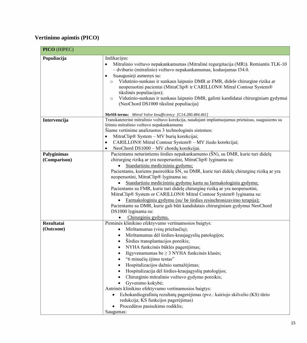
Vertinimo apimtis (PICO)
PICO (HIPEC)
Populiacija
Indikacijos:
Mitralinio vožtuvo nepakankamumas (Mitralinė regurgitacija (MR)). Remiantis TLK-10
– dviburio (mitralinio) vožtuvo nepakankamumas, koduojamas I34.0.
Suaugusieji asmenys su:
o Vidutinio-sunkaus ir sunkaus laipsnio DMR ar FMR, didele chirurgine rizika ar
neoperuotini pacientai (MitraClip® ir CARILLON® Mitral Contour System®
tikslinės populiacijos);
o Vidutinio-sunkaus ir sunkaus laipsnio DMR, galimi kandidatai chirurginiam gydymui
(NeoChord DS1000 tikslinė populiacija)
MeSH-terms: Mitral Valve Insufficiency [C14.280.484.461]
Intervencija Transkateterinė mitralinio vožtuvo korekcija, naudojant implantuojamus prietaisus, suagusiems su
lėtiniu mitralinio vožtuvo nepakankamumu Šiame vertinime analizuotos 3 technologinės sistemos:
MitraClip® System – MV burių korekcijai;
CARILLON® Mitral Contour System® – MV žiedo korekcijai;
NeoChord DS1000 – MV chordų korekcijai.
Palyginimas
(Comparison)
Pacientams neturintiems širdies nepakankamumo (ŠN), su DMR, kurie turi didelę
chirurginę riziką ar yra neoperuotini, MitraClip® lyginama su:
Standartiniu medicininiu gydymu;
Pacientams, kuriems pasireiškia ŠN, su DMR, kurie turi didelę chirurginę riziką ar yra
neoperuotini, MitraClip® lyginama su:
Standartiniu medicininiu gydymu kartu su farmakologiniu gydymu;
Pacientams su FMR, kurie turi didelę chirurginę riziką ar yra neoperuotini,
MitraClip® System or CARILLON® Mitral Contour System® lyginama su:
Farmakologiniu gydymu (su/ be širdies resinchronizavimo terapija);
Pacientams su DMR, kurie gali būti kandidatais chirurginiam gydymui NeoChord
DS1000 lyginama su:
Chirurginiu gydymu.
Rezultatai
(Outcome)
Pirminės klinikino efektyvumo vertinamosios baigtys:
Mirštamumas (visų priežasčių);
Mirštamumas dėl širdies-kraujagyslių patologijos;
Širdies transplantacijos poreikis;
NYHA funkcinės būklės pagerėjimas;
Išgyvenamumas be ≥ 3 NYHA funkcinės klasės;
“6 minučių ėjimo testas”
Hospitalizacijos dažnio sumažėjimas;
Hospitalizacija dėl širdies-kraujagyslių patologijos;
Chirurginio mitralinio vožtuvo gydymo poreikis;
Gyvenimo kokybė;
Antrinės klinikino efektyvumo vertinamosios baigtys:
Echokardiografinių rezultatų pagerėjimas (pvz.: kairiojo skilvelio (KS) tūrio
redukcija; KS funkcijos pagerėjimas)
Procedūros pasisekimo rodiklis;
Saugumas:

16
Prietaiso igaamžiškumas;
Trumpalaikiai ir ilglaikiai nepageidaujami įvykiai (NĮ) (susiję su prietaisu ir/ar
procedūra): 1) visi NĮ; 2) sunkūs NĮ; 3) dažniausi NĮ.
Vertinamieji rodikliai pasirinkti, remiantis klinikinėmis MR gydymo rekomendacijomis
[24,25] ir EUnetHTA klinikinių ir surogatinių vertinamųjų baigčių bei saugumo aspektų
rekomendacijomis [26 ] ir redaguotos, vadovaujantis išorinių ekspertų pastabomis.
Literatūros šaltininių tipas Veiksmingumas:
Sistematinės apžvalgos;
Sveikatos technologijų vertinimai;
Randomizuoti kontrolės tyrimai;
Kontroliuojami klinikiniai tyrimai.
Saugumas (kiti studijų dizainai/tipai, nei vardinti anksčiau):
Atvejo analizės;
Medicinos prietaisų nepageidaujamų įvykių registras
Kalba Anglų
Lietuvių
PICO klausimas: Ar minimaliai invaziniai gydymo metodai, naudojant MitraClip® System,CARILLON® Mitral Contour
System®, ar NeoChord DS100, pacientams sergantiems mitraline regurgitacija/dviburio vožtuvo nepakankamumu/
mitralinio vožtuvo nepakankamumu, yra saugesni ir efektyvesni, nei standartiniai gydymo metodai?

17
Scope (PICO)
PICO (HIPEC)
Population
Indication:
MR; International Statistical Classification of Diseases and Related Health
Problems (ICD)-10: I34.0 mitral (valve) insufficiency
Adults with:
o moderate-to-severe and severe DMR or FMR who are at high surgical risk or
non-surgical candidates (i.e. MitraClip® and CARILLON® Mitral Contour
System® populations)
o moderate-to-severe and severe DMR who are surgical candidates (i.e. NeoChord
DS1000 population)
The interventions assessed are proposed to treat the condition.
MeSH-terms: Mitral Valve Insufficiency [C14.280.484.461]
Intervention Transcatheter MV repair by device implantation in adults with chronic MR.
Three systems will be considered within the present assessment:
MitraClip® System for leaflet repair
CARILLON® Mitral Contour System® for annulus repair
NeoChord DS1000 for chordal repair
Comparison
In patients without HF, with DMR, who are at high surgical risk or are non-surgical
candidates, MitraClip® will be compared to:
Standard medical care
In patients with HF, with DMR, who are at high surgical risk or are non-surgical
candidates, MitraClip® will be compared to:
Standard medical care with pharmacological treatment for HF
In patients with FMR who are at high surgical risk or are non-surgical candidates,
MitraClip® System or CARILLON® Mitral Contour System® will be compared to:
Pharmacological treatment (with or without CRT)
In patients with DMR who are surgical candidates, NeoChord DS1000 will be
compared to:
Surgery
Outcome
Primary effectiveness outcomes:
Mortality (all-cause)
Cardiovascular mortality
Need for cardiac transplantation
NYHA functional status improvement
Freedom from NYHA class ≥ 3
6MWT
Reduction in rate of hospitalisation
Cardiovascular hospitalisation
Need for MV surgery
QoL
Secondary effectiveness outcomes:

18
Improvements in echocardiographic outcomes (e.g. reduction in LV volumes,
improvement in LVEF)
Procedural success rate
Safety :
Durability of the device
Short- and long-term AEs (device-related as well as procedure-related): 1) Any
AE; 2) serious AEs; 3) most frequent AEs
Outcomes were selected based on the recommendations from the clinical guidelines for
treatment of MR [24,25] and the EUnetHTA Guidelines on Clinical and Surrogate
Endpoints and Safety [26] and amended following comments from dedicated reviewers
and external experts
Study design Effectiveness:
Systematic reviews;
Health Technology Assessment (HTA) reports;
Randomised controlled trials (RCT);
Controlled clinical trials (CCT).
Safety (other than the designs already listed):
Case series;
Medical devices adverse events registries.
Language English
Lithuanian
Research question: Is minimaly invasive treatment approaches, using MitraClip® System,CARILLON® Mitral Contour
System®, or NeoChord DS1000, of patients with MR are safe and more effective treatment approaches?
19
SUMMARY
Target population (Domain A)
The target population of this assessment is adults with moderate-to-severe and severe DMR who are surgical candidates (i.e.
NeoChord DS1000 population) and adults with moderate-to-severe and severe DMR or FMR who are at high surgical risk or
surgery is contraindicated for them (i.e. CARILLON® Mitral Contour System® and MitraClip® System populations). (A0007)
Moderate or greater in severity MR is present in 11% of people over 70 years and almost 40% of patients with significant
heart failure have MR. (A0006) Specific statistical data on the size of the target population were not found. Overall prevalence of all
nonrheumatic valve disorders (I34-I39) in 2014 was 8.42 cases (per 1000 capita). (A0023)
Target condition (Domain A)
Chronic MR is a long-term disorder characterised by backward flow of blood from the left ventricle to the left atrium during
systole. The closest ICD-10 classification of MV disorders for MR is I34.0. According to the aetiology MR can be degenerative
(primary) or functional (secondary). DMR refers to abnormalities of the leaflets and is most commonly caused by myxomatous
degeneration. With FMR occurs as a consequence of adverse LV remodelling, with papillary muscle displacement, leaflet tethering,
and annular dilatation. The most frequent causes of MR are degenerative (myxomatous) disease, ischaemic heart disease, rheumatic
heart disease, and infectious endocarditis. (A0003) The estimated 5-year rates of death from any cause, death from cardiac causes,
and cardiac events for patients with asymptomatic severe MR have been reported to be 22.3%, 14.3%, and 33.3%, respectively.
(A0004) Approximately 40% of patients with MR have at least 1 comorbidity, most often it is heart failure, artrial fibrillation and
ischemic heart disease. (A0005)
Current management of the condition (Domain A)
An explicit overview of the recommendations from the latest guidelines for the management of chronic MR is presented in
A0025.
Chronic DMR
Medical therapy (vasodilators, ACE inhibitors, beta-blockers) is considered in patients with advanced MR and severe
symptoms, which are not suitable for surgery. Surgical therapy is recommended for symptomatic or asymtomatic patients with
severe DMR, left ventricle dysfunction. MV repair is suggested in preference to MV replacement when a successful and durable
repair can be accomplished (likehood 95%) for asymptomatic patients with prederved LV function and expected mortality less than
1%. Transcatheter MV repair may be considered for severely symptomatic patients (NYHA class III to IV) who have a prohibitive
surgical risk because of severe comorbidities and remain severely symptomatic despite optimal guideline-directed medical therapy
for HF. (A0025)
Chronic FMR
Patients with progresive or severe FMR should receive GDMT for HF or cardiac resynchronization therapy if satisfies the
inclusion criteria.Medical therapy MV surgery (repair or replacement) is reasonable for patients with severe FMR who have
persistent symptoms despite GDMT for HF. (A0025)
Features of the technology (Domain B)
Three systems are considered in the present assessment: MitraClip® System for leaflet repair, CARILLON® Mitral Contour
System® for annulus repair and NeoChord DS1000 for chordal repair.
MitraClip® System
MitraClip® is a first-of-its-kind transcatheter mitral valve repair (TMVR) system designed to reconstruct the insufficient
mitral valve (MV). This transcatheter therapeutic option provides a solution for patients with moderate-to-severe and severe
degenerative, or primary, mitral regurgitation (DMR) or functional, or secondary, MR (FMR) who are not considered suitable
20
candidates for conventional mitral valve surgery. It is delivered without requiring median sternotomy or cardiopulmonary bypass
and most patients can be discharged home after the post-procedure recovery period. The MitraClip® device is delivered to the heart
through the femoral vein after transseptal puncture is performed and is implanted on the valve leaflets to create a double orifice
valve that decreases the backflow of blood and allows the heart to pump blood more efficiently (B0001). In the EU, the MitraClip®
System is intended for reconstruction of the insufficient MV through tissue approximation, but in the USA it is indicated for the
percutaneous reduction of significant symptomatic MR (≥ 3+) due to primary abnormality of the mitral apparatus (DMR) in patients
who have been determined to be at prohibitive risk for MV surgery by a “heart team” (including a cardiac surgeon experienced in
MV surgery and a cardiologist experienced in MV disease), and for whom existing comorbidities would not preclude the expected
benefit from reduction of the MR (A0020).
CARILLON® Mitral Contour System®
The CARILLON® Mitral Contour System® is a percutaneous coronary sinus (CS)-based mitral annuloplasty device, and the
only transcatheter technology holding a Conformité Européene (CE) mark that was designed specifically to treat FMR. The
approach incorporates a device implanted in the CS and great cardiac vein (GCV), which lie adjacent to the mitral annulus. The
device imparts inward pressure on the mitral annulus, decreasing annular dimension, increasing anterior and posterior leaflet
coaptation and reducing MR. The CARILLON® Mitral Contour System® is contraindicated for use in patients with existing
devices in the CS/GCV and in patients who have had a MV replacement or a mitral annuloplasty ring implant (B0001). In the EU,
the CARILLON® Mitral Contour System® is indicated for use in patients with FMR (A0020).
NeoChord DS1000
The NeoChord DS1000 is a single-use, handheld device designed to deploy commercially available expanded
polytetrafluoroethylene (ePTFE) suture (labelled for use as artificial chordae tendineae) while the heart is beating, as an alternative
to the conventional surgical approach for this type of MV repair. The NeoChord DS1000 consists of the handheld delivery
instrument, into which an off-the-shelf ePTFE suture is loaded, and a needle, and includes a tethered Leaflet Verification Display
(LVD), which enables confirmation of leaflet capture in the distal clamp of the device prior to deploying the suture and knot at the
leaflet (B0001). In the EU, NeoChord DS1000 is indicated for use in patients with Grade 3+ or 4+ MR who are candidates for
surgical mitral valve repair or replacement (A0020). NeoChord DS1000 is the only-in-class product that provides a minimally
invasive approach to expand access to patients with DMR, without cardiopulmonary bypass-associated risks (B0002).
Current therapeutic options for the treatment of MR include medical management, surgical repair or replacement of the MV,
ventricular assist device implantation, heart transplantation, and CRT; in this assessment, comparators are chosen by type of MR,
medical device indication, and clinical guidelines recommendations (B0001).
Other (other manufacturers) mitral valve implantable medical devices did not fall into this health technology assessment
object, because it was not described in retrieved articles.
Patient safety (Domain C)
MitraClip® System
Mortality in hospital rate was 0.1-4.2%. 30 days, 12 months and 24 months mortality rates were 1.7-6 %, 9.3-23.1% and
17.4-25.7%, respectfuly. Partial clip detachment was observed in 0-12.5% and 10% of patients at 30 days and 24 months,
respectfuly. Miocard infarct rate during in hospital stay did not occur for any of patients and after 12 months frequency of MI was
3.4%. Major adverse events wate was 4.9-12.8%, 4.7–26% and 5–41% in hospital, at 30 days and at 12 months, respectfuly.
Patients with complications after MitraClip implantation were significantly older, more frequently women, and had a more
frail general health status with higher ASA- class and loogistic euroSCOREs at baseline than those without complications. Ischemic
MR etiology, left ventricular end-systolic volume >110 mL and frailty of patients, in particular NYHA class IV, has been associated
with higher mortality rates. (C0004)
Effects of a learning curve have been acknowledged in a series of 75 patients, whereas a later study, showed that a learning
curve does not appear to significantly affect procedural success. (C0004)
CARILLON® Mitral Contour System®
21
There were no deaths during 30 day period. Death rate after 12 months was up to 22.2%. Miocard infarct occured in 0-6,5 %
and 4% of patients durig 3 months and 12 months follow-up, respectively. (C0008)
CS; careful management of patients with high surgical risk, and acquisition of procedural skills are necessary to lower the
risks associated with this device. Furthermore, experience-based skills related to the assessment of coronary arterial flow are crucial
for recapturing and repositioning the device successfully and safely. (C0007)
NeoChord DS1000
During 30 day follow-up mortality, stroke and miocard infarct rate was 0-3%, 0-3%, and 0-2% of patients, respectfuly.
Intraoperative conversion to conventional surgery rate varied from 0 to 20%. Reoperation rate, due to NeoChord DS1000 failure,
was 13-20%, during 30 day follow-up period. MAE varied from 6.1% to 26.7% of patients at 30 day period. (C0008)
No evidence was found to help determine whether the use of NeoChord DS1000 is associated with user-dependent harms. In
one study, the authors highlighted that special and extensive training for the operators is mandatory, because the determination of
the exact positioning, length adjustment and neo-chordae tensioning depends exclusively on the ability and training of the operator
and echocardiographer, and affects the durability as well as the acute procedural success. (C0007)
Survival (Domain D)
MitraClip® System
Overall survival rates at 6 months ranges from 85-90%, at 1 year – 75-90% and at 2 year – 71-75%. As reported by the only
one comparative study included, patients after MitraClip® implantation compared to patients after optimal medical therapy, have
significantly better short-term and long-term overall survival rates, accordingly: at 1 month – 100% vs 98.3%; at 2 year – 89.7% vs
64.3%, at 3 year – 61.4% vs 34.9%. (D0001; D0003)
CARILLON® Mitral Contour System®
30 day mortality ranges from 1.9-2.2% after implantation of CARILLON®. Compared to the non-implanted group, patients
after implantation have lower 1-year mortality rates: 22.2% vs 23.5%, respectfully. (D0001; D0003)
NeoChord DS1000
Short-term mortality after the procedure is low – either no deaths or sporadic deaths were reported at 30 days after the device
implantation. Overall survival of 97.2% was reported at 3 months. No long-term evidence were found. (D0001; D0003)
Morbidity (Domain D)
MitraClip® System
Improvement in MR symptoms and severity is observable. Reduction in proportion of patients with NYHA class >II at 1-
year falls in range of 37-83%. Also at 1-year proportion of patients with MR grade ≤2+ increases 77-99%. (D0005; D0006)
CARILLON® Mitral Contour System®
One study showed improvement in NYHA class from baseline (3.1 ± 0.23) to 2.1 ± 0.64 at 12 months. The improvement
was maintained at the 24-month with NYHA class 2.1 ± 0.74 (p < 0.001). Aslo significantly better outcomes – improvemet of
echocardiographic parameters were observed after CARILLON® implantation compared to non-implanted group. (D0005; D0006)
NeoChord DS1000
In single study in 82% population reduction to NYHA class I was observed after 30 day post procedure and another study
reported reduction to ≤II NYHA class after 3 months in 89.5% population. At 30 days after NeoChord DS1000 implantation 58.6-
87.5% of patients maintained MR grade ≤2+. Although, because of a very inconsistent reporting of outcomes in different studies
and no comparative evidence it is difficult to sumarize these results. (D0005; D0006)
22
Body functions and quality of life (Domain D)
MitraClip® System
Significant improvement in 6MWT results was observed after MitraClip® implantation. Mean walking distance increcement
ranges between 39-168 metres at 6 months and between 47-115 meters at 1 year. Also different questionnaires shows improvement
in general QoL score and various areas: physical component, independency in „self-care“, „anxiety/depression“. (D0011; D0013)
CARILLON® Mitral Contour System®
Significantly bigger improvement in excercise performance had patients after CARILLON® implantation compared to
optimal medical therapy group. 6MWT disatance at baseline, 6 months and 1 year between the groups were: 302.5 m. vs 337.9 m.;
429.9 m. vs 322.2 m.; 406 m. vs 348.1 m., respectfully. Moreover QoL assessed by “Kansas City Cardiomyopathy Questionnaire“
was also significantly better in CARILLON® population: at baseline mean general score was 43 vs 40.4; at 6 months 63.4 vs 49.6;
at 1 year 61.2 vs 51. (D0011; D0013)

23
A. HEALTH PROBLEM AND CURRENT USE OF THE TECHNOLOGY
Table 1. Research questions used to formulate “Health problem and current use of the technology” domain, according to
EUnetHTA Core Model [ 27]
Element ID Research question
A0002 What is the disease in the scope of this assessment?
A0003 What are the known risk factors for developing chronic MR?
A0004 What is the natural course of chronic MR?
A0005 What are the symptoms and the burden of chronic MR for the patient?
A0006 What are the consequences of chronic MR for society?
A0024 How is chronic MR currently diagnosed according to published guidelines and in practice?
A0025 How is chronic MR currently managed according to published guidelines and in practice?
A0007 What is the target population of this assessment?
A0023 How many people belong to the target population?
A0011 How much are the MitraClip® System, CARILLON® Mitral Contour System®, and NeoChord DS1000 used?
[A0002] – What is the disease in the scope of this assessment?
MR is characterised by backward flow of blood from the left ventricle to the left atrium during systole, producing left atrial
dilatation. It also causes the left ventricle to become enlarged because of the additional workload required to maintain normal blood
flow. MR can occur because of abnormalities of the mitral valve leaflets, the annulus, the chordae tendineae or papillary muscles, or
the left ventricle (see Figure 1).
Figure 1: Mitral valve anatomy [28]
MR can be acute (leaflet perforation, chordal rupture, rupture of the papillary muscle due to MI) or chronic (long-term
disorder associated with valvular or ventricular pathology) and, according to the aetiology, primary (degenerative) or secondary
(functional) (see Table 2). DMR refers to abnormalities of the leaflets and is most commonly caused by myxomatous degeneration,
especially in developed countries. With FMR, the leaflets are usually normal, and the regurgitation occurs as a consequence of
adverse LV remodelling, with papillary muscle displacement, leaflet tethering, and annular dilatation. FMR due to ischaemic heart
disease or non-ischaemic dilated cardiomyopathy, resulting in HF. The most frequent causes of MR are degenerative (myxomatous)
disease, ischaemic heart disease, rheumatic heart disease, and infectious endocarditis [24,25,28-32].
24
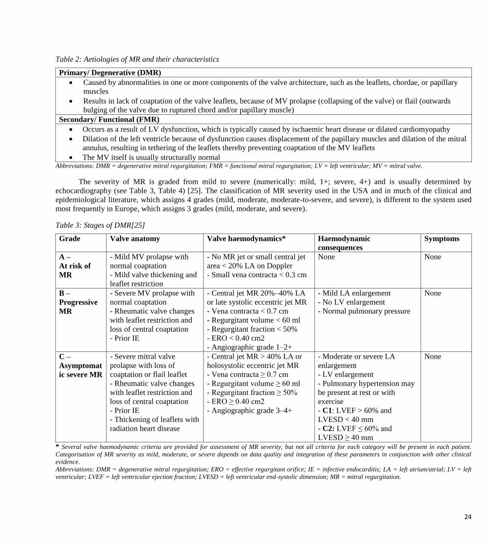
Table 2: Aetiologies of MR and their characteristics
Primary/ Degenerative (DMR)
Caused by abnormalities in one or more components of the valve architecture, such as the leaflets, chordae, or papillary
muscles
Results in lack of coaptation of the valve leaflets, because of MV prolapse (collapsing of the valve) or flail (outwards
bulging of the valve due to ruptured chord and/or papillary muscle)
Secondary/ Functional (FMR)
Occurs as a result of LV dysfunction, which is typically caused by ischaemic heart disease or dilated cardiomyopathy
Dilation of the left ventricle because of dysfunction causes displacement of the papillary muscles and dilation of the mitral
annulus, resulting in tethering of the leaflets thereby preventing coaptation of the MV leaflets
The MV itself is usually structurally normal Abbreviations: DMR = degenerative mitral regurgitation; FMR = functional mitral regurgitation; LV = left ventricular; MV = mitral valve.
The severity of MR is graded from mild to severe (numerically: mild, 1+; severe, 4+) and is usually determined by
echocardiography (see Table 3, Table 4) [25]. The classification of MR severity used in the USA and in much of the clinical and
epidemiological literature, which assigns 4 grades (mild, moderate, moderate-to-severe, and severe), is different to the system used
most frequently in Europe, which assigns 3 grades (mild, moderate, and severe).
Table 3: Stages of DMR[25]
Grade Valve anatomy Valve haemodynamics* Haemodynamic
consequences
Symptoms
A –
At risk of
MR
- Mild MV prolapse with
normal coaptation
- Mild valve thickening and
leaflet restriction
- No MR jet or small central jet
area < 20% LA on Doppler
- Small vena contracta < 0.3 cm
None None
B –
Progressive
MR
- Severe MV prolapse with
normal coaptation
- Rheumatic valve changes
with leaflet restriction and
loss of central coaptation
- Prior IE
- Central jet MR 20%–40% LA
or late systolic eccentric jet MR
- Vena contracta < 0.7 cm
- Regurgitant volume < 60 ml
- Regurgitant fraction < 50%
- ERO < 0.40 cm2
- Angiographic grade 1–2+
- Mild LA enlargement
- No LV enlargement
- Normal pulmonary pressure
None
C –
Asymptomat
ic severe MR
- Severe mitral valve
prolapse with loss of
coaptation or flail leaflet
- Rheumatic valve changes
with leaflet restriction and
loss of central coaptation
- Prior IE
- Thickening of leaflets with
radiation heart disease
- Central jet MR > 40% LA or
holosystolic eccentric jet MR
- Vena contracta ≥ 0.7 cm
- Regurgitant volume ≥ 60 ml
- Regurgitant fraction ≥ 50%
- ERO ≥ 0.40 cm2
- Angiographic grade 3–4+
- Moderate or severe LA
enlargement
- LV enlargement
- Pulmonary hypertension may
be present at rest or with
exercise
- C1: LVEF > 60% and
LVESD < 40 mm
- C2: LVEF ≤ 60% and
LVESD ≥ 40 mm
None
* Several valve haemodynamic criteria are provided for assessment of MR severity, but not all criteria for each category will be present in each patient.
Categorisation of MR severity as mild, moderate, or severe depends on data quality and integration of these parameters in conjunction with other clinical
evidence.
Abbreviations: DMR = degenerative mitral regurgitation; ERO = effective regurgitant orifice; IE = infective endocarditis; LA = left atrium/atrial; LV = left
ventricular; LVEF = left ventricular ejection fraction; LVESD = left ventricular end-systolic dimension; MR = mitral regurgitation.
25
Table 4: Stages of FMR[25]
Grade Valve anatomy Valve haemodynamics* Associated cardiac
findings
Symptoms
A –
At risk of
MR
- Normal valve leaflets,
chords, and annulus in a
patient with coronary
disease or
cardiomyopathy
- No MR jet or small
central jet area < 20% LA
on Doppler
- Small vena contracta <
0.3 cm
- Normal or mildly dilated
LV size with fixed
(infarction) or Inducible
(ischaemia) regional wall
motion abnormalities
- Primary myocardial
disease with LV dilation
and systolic dysfunction
- Symptoms due to
coronary ischaemia or HF
may be present that
respond to
revascularisation and
appropriate medical
therapy
B –
Progressive
MR
- Regional wall motion
abnormalities with mild
tethering of mitral leaflet
- Annular dilation with
mild loss of central
coaptation of the mitral
leaflets
- Regurgitant volume < 30
ml
- ERO < 0.20 cm2
- Regurgitant fraction <
50%
- Regional wall motion
abnormalities with reduced
LV systolic function
- LV dilation and systolic
dysfunction due to primary
myocardial disease
- Symptoms due to
coronary ischaemia or HF
may be present that
respond to
revascularisation and
appropriate medical
therapy
C –
Asymptomat
ic severe MR
- Regional wall motion
abnormalities and/or LV
dilation with severe
tethering of mitral leaflet
- Annular dilation with
severe loss of central
coaptation of the mitral
leaflets
- Regurgitant volume ≥30
mL
- ERO ≥ 0.20 cm2
- Regurgitant fraction ≥
50%
- Regional wall motion
abnormalities with reduced
LV systolic function
- LV dilation and systolic
dysfunction due to primary
myocardial disease
- Symptoms due to
coronary ischaemia or HF
may be present that
respond to
revascularisation and
appropriate medical
therapy
D –
Symptomatic
severe MR
- Regional wall motion
abnormalities and/or LV
dilation with severe
tethering of mitral leaflet
- Annular dilation with
severe loss of central
coaptation of the mitral
leaflets
- Regurgitant volume ≥ 30
ml
- ERO ≥ 0.20 cm2
- Regurgitant fraction ≥
50%
- Regional wall motion
abnormalities with reduced
LV systolic function
- LV dilation and systolic
dysfunction due to primary
myocardial disease
- HF symptoms due to MR
persist even after
revascularisation and
optimisation of medical
therapy
- Decreased exercise
tolerance
- Exertional dyspnoea *Several valve haemodynamic criteria are provided for assessment of MR severity, but not all criteria for each category will be present in each patient.
Categorisation of MR severity as mild, moderate, or severe depends on data quality and integration of these parameters in conjunction with other clinical
evidence.
The measurement of the proximal isovelocity surface area by 2D TTE in patients with FMR underestimates the true ERO due to the crescentic shape of the
proximal convergence.
Abbreviations: ERO = effective regurgitant orifice; FMR = functional mitral regurgitation; HF = heart failure; LA = left atrium; LV = left ventricular; MR =
mitral regurgitation; TTE = transthoracic echocardiogram.
The closest ICD-10 classification of MV disorders for MR is I34.0. Although, such codes as I05.1, I34.1; Q23.3 also can indicate
target condition (see table 5).

26
Table 5: ICD-10 classification of MV disorders [28,33,34]
I05 RHEUMATIC MITRAL VALVE DISEASES
I05.1 Rheumatic insufficiency (incompetence or regurgitation)
I34 NON RHEUMATIC MITRAL VALVE DISORDERS
I34.0 Mitral valve insufficiency (incompetence or regurgitation)
I34.1 Mitral valve prolapse (Floppy mitral valve syndrome) (exclude Marfans syndrome)
I34.8 Other non-rheumatic mitral valve disorders
I34.9 Non-rheumatic mitral valve disorders, unspecified
I39.0* Mitral valve disorders in diseases classified elsewhere
Q23.3 Congenital mitral insufficiency
[A0003] – What are the known risk factors for developing chronic MR?
The known risk factors for developing chronic MR are age, hypertension, low body mass index, coronary systolic blood
pressure, increased left atrium size and LV diastolic diameter, low LVEF, and female gender. In addition to HF being a
complication of MR, HF is a major risk factor for the development of MR, having been detected in 56% of patients with LVEF <
40% and clinical HF (70% mild, 30% moderate/severe) in a US cohort [28-31,35].
[A0004] – What is the natural course of chronic MR?
MR can be present many years before any symptom occurs. If untreated, moderate-to-severe MR can cause progressive
congestive HF, and lead, eventually, to death. The risk of mortality for those with severe MR that is left untreated is higher than for
the general population: 1-year and 5-year mortality rates of 20% and 50%, respectively, have been reported. According to further
evidence on asymptomatic severe chronic MR, the estimated 5-year rates of death from any cause, death from cardiac causes, and
cardiac events (death from cardiac causes, HF, or new AF with medical management) have been reported to be 22.3%, 14.3%, and
33.3%, respectively. In addition to symptoms, age, AF, severity of MR (particularly ERO area, pulmonary hypertension, LA
dilatation, increased LVESD, and low LVEF were all found to be predictors of poor outcome [24,28,30,31,36,37].
[A0005] – What are the symptoms and the burden of chronic MR for the patient?
Symptoms
Symptoms of chronic MR include palpitation, dyspnoea, orthopnoea, fatigue, lethargy, cardiac cachexia,
thromboembolism, and subacute infective endocarditis. MR confers a substantial physical, emotional, and social burden to
patients. Severe symptoms may prevent patients from performing everyday tasks and simple activities, such as getting out of bed.
The inability to perform activities of daily of living and be independent can lead to feelings of loss of independence, distress, and
depression. Patients feel like they are a burden to their family and they worry about the future. Patients may need to adjust their
houses in order to cope with the condition, for example installing a stairlift or rails. Given the statements above, HF can impact
upon all aspects of a patient’s QoL. Patients with HF have significant impairments in all aspects of their health compared with
the general population, and have significantly greater physical QoL impairment than patients with chronic lung disease or
arthritis [28-31,38].
Hospitalisation
A study of patients with severe MR who were not considered suitable for valve surgery reported that hospitalisations due to
HF exacerbations rose from 41% in the first year to 90% after 5 years; another study showed that a greater proportion of
hospitalisations in patients with MR before surgery were associated with congestive HF compared to the period after surgical
intervention (p < 0.001) [28,39]. Baskett et al. performed a retrospective analysis of mortality and hospitalisation for HF in 301
patients from the Studies Of Left Ventricular Dysfunction (SOLVD). The authors concluded that the presence of mitral

27
insufficiency in patients with LV dysfunction is independently associated with adverse outcomes, including death and
hospitalisations for HF [29,40].
Comorbidities
Approximately 40% of patients with MR have at least 1 comorbidity including, but not limited to, advanced age, frailty, and
prior cardiac surgery. The presence of comorbidities may influence the treatment that a patient can receive based on the impact of
the surgical risk–benefit profile. Cardiac comorbidities include: HF (almost 40% of patients with significant HF have MR), AF
(occurs as a result of increased LA pressure associated with the backflow of blood in MR), ischaemic heart disease/history of MI (a
cause of FMR), LV dysfunction (a cause of FMR), cardiomyopathy (a cause of FMR), concomitant valvular heart disease, and
atherosclerosis. Non-cardiac comorbidities include: pulmonary hypertension (a consequence of increased pressure in the left atrium,
resulting in increased pressure in the pulmonary vasculature), renal dysfunction (along with haemodialysis, may cause mechanical
stress on the valves resulting in mitral annular calcification), chronic obstructive pulmonary disease (chronic inflammation of the
airways resulting in decreased tolerance of symptoms secondary to MR), vasculopathy (atherosclerosis of the peripheral or cerebral
circulation increases the risk of stroke or embolic phenomenon), malignancy (advanced stage malignancy decreases life expectancy
of the patient and immunosuppressive treatment may increase the patient’s risk potential for complications if treated for MR),
neurological impairment (prior history of stroke or transient ischaemic attack may increase the risk for complications and increase
mortality risk associated with cardiac surgery), and frailty (will affect the patient’s morbidity and mortality risks associated with
surgery because of the postoperative recovery needed) [24,28,41 ].
[A0006] – What are the consequences of chronic MR for society?
The prevalence of MR is high among the general population, with approximately 19% having MR of at least mild severity.
The prevalence of MR increases with age: clinically meaningful MR (moderate or greater in severity) is present in < 1% of people
younger than 50 years, but in 11% of people over 70 years. MR accounts for the vast majority (97%) of all MV diseases and, in
Europe, is the second most common type of heart valve disease requiring surgery, after aortic stenosis. The incidence of MR is high
amongst patients with HF: almost 40% of patients with significant HF have MR [28,42,43 ].
In France, the annual cost-per-patient for the treatment of MR has been estimated at € 24,581 for patients receiving surgery,
and € 12,177 for patients receiving non-surgical management. MR is often associated with HF, with recurrent hospitalisations and
need for multiple medications for management placing a substantial cost burden on the health system: in the UK, the cost of
managing HF accounts for 2% of the total (NHS) budget (£625 million), of which 61% of which is accounted for by inpatient
hospital stays [28,44,45 ].
The cost of HF in the USA was also estimated to account for 2% of the total healthcare budget ($32.9 billion), with
hospitalisations accounting for 60% of this cost. The burden of MR is increasing; a study conducted in the USA reported an
increase in the number of hospitalisations due to valvular heart disease from 1983 to 2000, with a 1.5-fold greater increase among
patients with MV disease compared with aortic valve disease (p < 0.001) [28,46].
[A0024] – How is chronic MR currently diagnosed according to published guidelines and in practice?
An overview of the latest guidelines for the diagnosis of chronic MR is presented in Appendix 1, Table 10.
According to the recent US Guideline from 2014 [25], further steps should be performed to diagnose and assess the severity
of chronic MR:
“A careful history, a detailed physical examination should be performed to diagnose and assess the severity of valve lesions
based on a compilation of all findings made by inspection, palpation, and auscultation. The use of an electrocardiogram (ECG) to
confirm heart rhythm and use of a chest x-ray to assess the presence or absence of pulmonary congestion and other lung pathology
may be helpful in the initial assessment of patients with known or suspected valvular heart disease (VHD). A comprehensive
transthoracic echocardiogram (TTE) with 2–dimensional (2D) imaging and Doppler interrogation should then be performed to
correlate findings with initial impressions based on the initial clinical evaluation. The TTE will also be able to provide additional
information, such as the effect of the valve lesion on the cardiac chambers and great vessels, and to assess for other concomitant
valve lesions. Other ancillary testing such as transoesophageal echocardiography (TOE, computed tomography (CT) or cardiac
magnetic resonance (CMR) imaging, stress testing, and diagnostic haemodynamic cardiac catheterisation may be required to deter-
28
mine the optimal treatment for a patient with VHD. An evaluation of the possible surgical risk for each individual patient should be
performed if intervention is contemplated, as well as other contributing factors such as the presence and extent of comorbidities and
frailty. Follow-up of these patients is important and should consist of an annual history and physical examination in most stable
patients.”
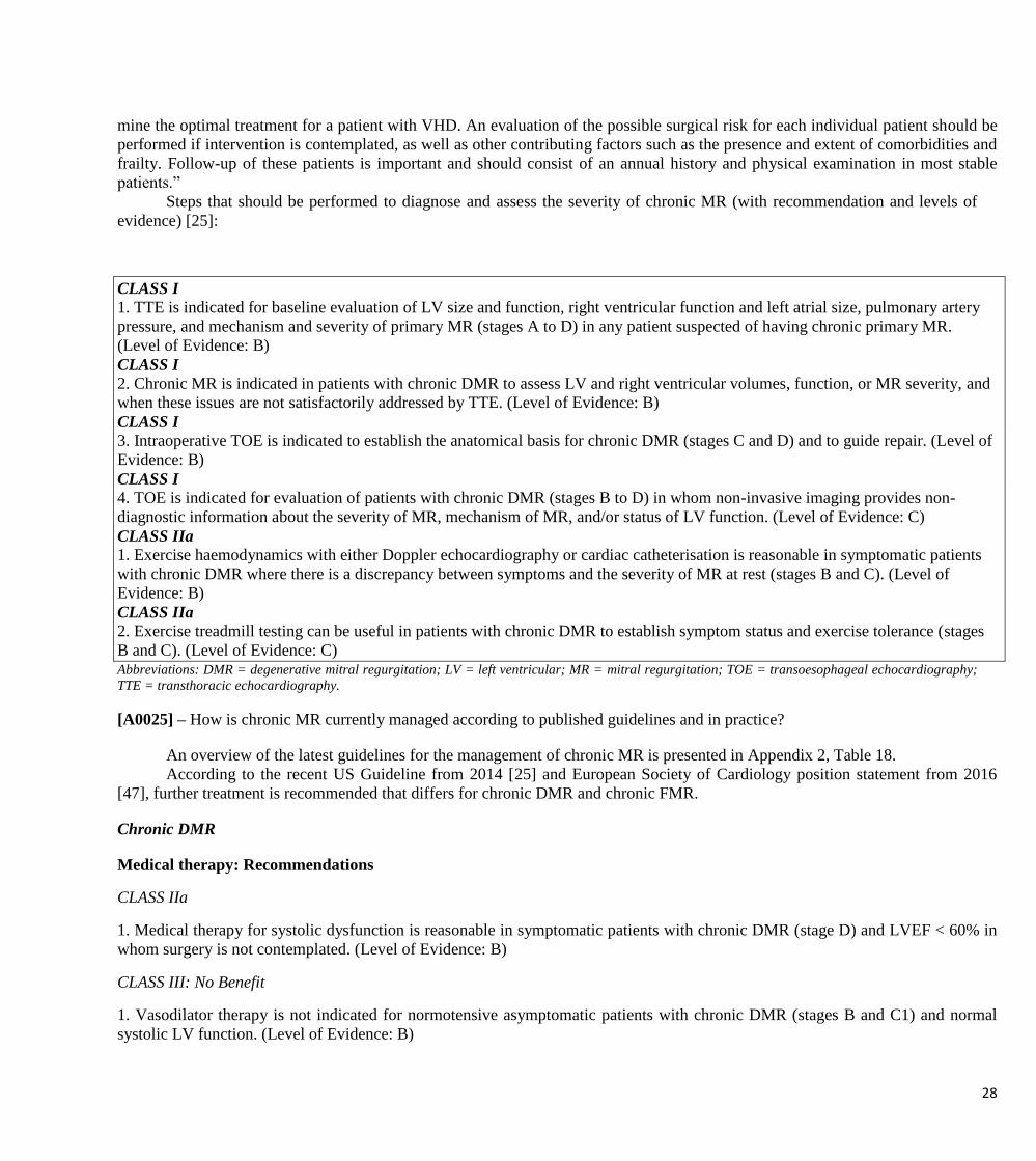
Steps that should be performed to diagnose and assess the severity of chronic MR (with recommendation and levels of
evidence) [25]:
CLASS I 1. TTE is indicated for baseline evaluation of LV size and function, right ventricular function and left atrial size, pulmonary artery
pressure, and mechanism and severity of primary MR (stages A to D) in any patient suspected of having chronic primary MR.
(Level of Evidence: B)
CLASS I 2. Chronic MR is indicated in patients with chronic DMR to assess LV and right ventricular volumes, function, or MR severity, and
when these issues are not satisfactorily addressed by TTE. (Level of Evidence: B)
CLASS I 3. Intraoperative TOE is indicated to establish the anatomical basis for chronic DMR (stages C and D) and to guide repair. (Level of
Evidence: B)
CLASS I 4. TOE is indicated for evaluation of patients with chronic DMR (stages B to D) in whom non-invasive imaging provides non-
diagnostic information about the severity of MR, mechanism of MR, and/or status of LV function. (Level of Evidence: C)
CLASS IIa 1. Exercise haemodynamics with either Doppler echocardiography or cardiac catheterisation is reasonable in symptomatic patients
with chronic DMR where there is a discrepancy between symptoms and the severity of MR at rest (stages B and C). (Level of
Evidence: B)
CLASS IIa
2. Exercise treadmill testing can be useful in patients with chronic DMR to establish symptom status and exercise tolerance (stages
B and C). (Level of Evidence: C) Abbreviations: DMR = degenerative mitral regurgitation; LV = left ventricular; MR = mitral regurgitation; TOE = transoesophageal echocardiography;
TTE = transthoracic echocardiography.
[A0025] – How is chronic MR currently managed according to published guidelines and in practice?
An overview of the latest guidelines for the management of chronic MR is presented in Appendix 2, Table 18.
According to the recent US Guideline from 2014 [25] and European Society of Cardiology position statement from 2016
[47], further treatment is recommended that differs for chronic DMR and chronic FMR.
Chronic DMR
Medical therapy: Recommendations
CLASS IIa
1. Medical therapy for systolic dysfunction is reasonable in symptomatic patients with chronic DMR (stage D) and LVEF < 60% in
whom surgery is not contemplated. (Level of Evidence: B)
CLASS III: No Benefit
1. Vasodilator therapy is not indicated for normotensive asymptomatic patients with chronic DMR (stages B and C1) and normal
systolic LV function. (Level of Evidence: B)

29
According to the EU Guideline from 2012 [24], there is no evidence to support the use of vasodilators, including ACE
inhibitors, in chronic MR without HF, and they are therefore not recommended in this group of patients. However, when HF has
developed, ACE inhibitors are beneficial and should be considered in patients with advanced MR and severe symptoms, who are not
suitable for surgery or when there are still residual symptoms following surgery. Beta-blockers and spironolactone should also be
considered as appropriate.
Intervention: Recommendations
CLASS I
1. MV surgery is recommended for symptomatic patients with chronic severe DMR (stage D) and LVEF > 30%. (Level of
Evidence: B)
CLASS I
2. MV surgery is recommended for asymptomatic patients with chronic severe DMR and LV dysfunction (LVEF 30–60% and/or
LVESD ≥ 40 mm, stage C2). (Level of Evidence: B)
CLASS I
3. MV repair is recommended in preference to MV replacement when surgical treatment is indicated for patients with chronic severe
DMR limited to the posterior leaflet. (Level of Evidence: B)
CLASS I
4. MV repair is recommended in preference to MV replacement when surgical treatment is indicated for patients with chronic severe
DMR involving the anterior leaflet or both leaflets when a successful and durable repair can be accomplished. (Level of Evidence:
B)
CLASS I
5. Concomitant MV repair or MV replacement is indicated in patients with chronic severe DMR undergoing cardiac surgery for
other indications. (Level of Evidence: B)
CLASS IIa
1. MV repair is reasonable in asymptomatic patients with chronic severe DMR (stage C1) with preserved LV function (LVEF >
60% and LVESD < 40 mm) in whom the likelihood of a successful and durable repair without residual MR is greater than
95% with an expected mortality rate of less than 1% when performed at a Heart Valve Centre of Excellence. (Level of
Evidence: B)
CLASS IIa
2. MV repair is reasonable for asymptomatic patients with chronic severe nonrheumatic DMR (stage C1) and preserved LV function
(LVEF > 60% and LVESD < 40 mm) in whom there is a high likelihood of a successful and durable repair with: 1) new onset of
AF, or 2) resting pulmonary hypertension (pulmonary artery systolic arterial pressure > 50 mm Hg). (Level of Evidence: B)
CLASS IIa
3. Concomitant MV repair is reasonable in patients with chronic moderate DMR (stage B) when undergoing cardiac surgery for
other indications. (Level of Evidence: C)
CLASS IIb

30
1. MV surgery may be considered in symptomatic patients with chronic severe DMR and LVEF ≤ 30% (stage D). (Level of
Evidence: C)
CLASS IIb
2. MV repair may be considered in patients with rheumatic MV disease when surgical treatment is indicated if a durable and
successful repair is likely or when the reliability of long-term anticoagulation management is questionable. (Level of Evidence: B)
CLASS IIb
3. TMVR may be considered for severely symptomatic patients (NYHA class III to IV) with chronic severe DMR (stage D) who
have favourable anatomy for the repair procedure and a reasonable life expectancy, but who have a prohibitive surgical risk because
of severe comorbidities and remain severely symptomatic despite optimal guideline-directed medical therapy (GDMT) for HF.
(Level of Evidence: B)
CLASS III: Harm
1. MVR should not be performed for the treatment of isolated severe DMR limited to less than one half of the posterior leaflet
unless MV repair has been attempted and was unsuccessful. (Level of Evidence: B)
Chronic FMR
Medical Therapy: Recommendations
CLASS I
1. Patients with chronic FMR (stages B to D) and HF with reduced LVEF should receive standard GDMT therapy for HF, including
ACE inhibitors, angiotensin II receptor blockers, beta blockers, and/or aldosterone antagonists as indicated. (Level of Evidence: A)
CLASS I
2. CRT with biventricular pacing is recommended for symptomatic patients with chronic severe FMR (stages B to D) who meet the
indications for device therapy. (Level of Evidence: A)
Intervention: Recommendations
CLASS IIa
1. MV surgery is reasonable for patients with chronic severe FMR (stages C and D) who are undergoing coronary artery bypass
grafting or aortic valve replacement. (Level of Evidence: C)
CLASS IIb
1. MV repair or replacement may be considered for severely symptomatic patients (NYHA class III to IV) with chronic severe FMR
(stage D) who have persistent symptoms despite optimal GDMT for HF. (Level of Evidence: B)
CLASS IIb
2. MV repair may be considered for patients with chronic moderate FMR (stage B) who are undergoing other cardiac surgery.
(Level of Evidence: C)
[A0007] – What is the target population of this assessment?
The target population of this assessment is adults with chronic DMR or FMR. More specifically, according to the Project
Plan, the target population is defined as adults with moderate-to-severe and severe DMR who are surgical candidates (i.e. NeoChord
DS1000 population) and adults with moderate-to-severe and severe DMR or FMR who are at high surgical risk or are non-surgical
31
candidates (i.e. CARILLON® Mitral Contour System® and MitraClip® System populations); the current CE mark indication is
broader and can be found in Appendix 1 (Table 19) [28,29,47-49].
MitraClip® System
According to the CE mark indication (reconstruction of the insufficient MV through tissue approximation), the target
population is patients with chronic DMR or FMR.
Manufacturer positioning: the MitraClip® System is indicated for patients suffering from moderate-to-severe and severe MR
with a high risk or contraindication for surgery [28].
Patients are thus candidates for the MitraClip® System treatment when they have severe DMR or FMR (according to the
ESC-EACTS guidelines classification), they are refractory to medical treatment, and when their risk for surgery is judged to be too
high or when surgery is contraindicated.
CARILLON® Mitral Contour System®
The CE mark indication states that: The CARILLON® Mitral Contour System® is indicated for use in patients with FMR.
According to the manufacturer, the clinical focus of the treatment modality is patients with advanced systolic HF due to
dilated ischaemic or non-ischaemic cardiomyopathy and FMR of grades 2+, 3+, or 4+ [29].
NeoChord DS1000
According to the CE mark indication, the target population for NeoChord DS1000 comprises patients with Grade 3+ or 4+
MR who are candidates for surgical MV repair or replacement [47].
[A0023] – How many people belong to the target population?
Mitral regurgitation has a prevalence of 2% in the general population.[50] In Lithuania overall prevalence of all
nonrheumatic valve disorders (I34-I39) in 2014 was 8.42 cases (per 1000 capita), meanwhile incidence – 2.47 new cases (per 1000
capita) [51]. According to Lithuania statistics there were 133 thousand people under the age of 70, 11% of them – 14.6 thousand
could have moterate or severe MR [52], based on literature [28,42,43]. Specific statistical data on the size of the target population
were not found. The MR severity classification used in the USA, and extensively in the clinical and epidemiological literature,
assigns 4 grades (mild, moderate, moderate-to-severe, and severe), which differs from the ESC-EACTS classification used
predominantly in Europe, which assigns 3 grades (mild, moderate, and severe). Part of the “moderate-to-severe” group in the USA
classification system will, therefore, be included in the “severe” group in the European classification, while the remaining part will
form part of the “moderate” group. A correlation table to correct this was not found [24,28].
MitraClip® System
Specific statistical data for Europe or other geographical regions on the prevalence/incidence of the target group, i.e. patients
suffering from severe symptomatic MR with high surgical risk or with contraindications for surgery that could be candidates for
MitraClip® System, were not found.
CARILLON® Mitral Contour System®
The size of the target population from a prevalence perspective can be estimated based on evaluating proportions of the HF
population that present with: advanced HF symptoms (NYHA class III or IV), dilated cardiomyopathy, FMR of Grades 2+, 3+, or
4+. From this group of patients, the following should be excluded: patients with contraindications (including resident coronary sinus
lead, mitral annuloplasty ring/artificial MV, significant mitral calcification, or significant degenerative leaf-let pathology), patients
who have not been optimised on a HF regimen, and patients who are deemed to be at low risk for surgery. Giving consideration to
each of these inclusions and exclusions, the estimated size of the target population can be projected to be between 15% and 30% of
the HF population [29]. In 2014 overall prevalence of heart failure in Lithuania was 110,751 cases, which means 16,613 – 33,225
cases could be considered for eligibility to CARILLON® Mitral Contour System®.

32
NeoChord DS1000
Specific statistical data on the size of the target population were not found.
[A0011] – How much are the MitraClip® System, CARILLON® Mitral Contour System®, and NeoChord DS1000 used?
MitraClip® System
The manufacturer stated that, by December 2014 and including clinical trials, MitraClip® was implanted in 16,327 patients
in Europe, and a total of 19,148 [53] patients worldwide. The procedure is performed in 312 centres in Europe (464 centres
worldwide) [28].
CARILLON® Mitral Contour System®
The manufacturer stated that, to date, there have been approximately 300 patients implanted with CARILLON® Mitral
Contour System® in 10 countries, and this is anticipated to grow to more than 550 patients before the end of 2015 [29].
NeoChord DS1000
The manufacturer stated that, to date, the experience with NeoChord DS1000 is based on 200 patients (30 patients from
TACT trial and 170 patients from post-marketing) [54].
Discussion
MR is a complex condition with two different aetiologies present in patients with a plethora of comorbidities. The burden of
MR is very high and impacts on patients as well as on society. In assessing patients with chronic MR, it is critical to distinguish
DMR from FMR as the two conditions have more differences than similarities. While the correction of DMR is curative, the
restoration of MV competence in FMR is not by itself curative. Because of that, the best therapy for FMR is less clear than it is for
DMR. Specific statistical data on the target population size in Europe or other regions were not found.
The 3 devices considered in the present assessment have distinct utilisation rates: while Neo-Chord DS1000 and
CARILLON® Mitral Contour System® appear to be in their infancy with hundreds of patients treated worldwide, the
MitraClip® System has been implanted in approximately 23,000 patients, mainly in Europe

33
B. DESCRIPTION AND TECHNICAL CHARACTERISTICS OF TECHNOLOGY
Table 6. Research questions used to formulate “Description and technical characteristics of technology” domain,
according to EUnetHTA Core Model [27]
Element ID Research question
B0001 What are the MitraClip® System, CARILLON® Mitral Contour System®, and NeoChord DS1000, and what
are the comparators?
A0020 For which indications have the MitraClip® System, CARILLON® Mitral Contour System®, and NeoChord
DS1000 received marketing authorisation or a CE mark?
B0002 What are the claimed benefits of the MitraClip® System, CARILLON® Mitral Contour System®, and
NeoChord DS1000 in relation to the comparators?
B0004 Who administers the MitraClip® System, CARILLON® Mitral Contour System®, NeoChord DS1000, and
the comparators, and in what context and level of care are they provided?
B0008 What kind of special premises are needed for the MitraClip® System, CARILLON® Mitral Contour
System®, NeoChord DS1000, and the comparators?
B0009 What supplies are needed to use the MitraClip® System, CARILLON® Mitral Contour System®, NeoChord
DS1000, and the comparators?
A0021 What is the reimbursement status of the MitraClip® System, CARILLON® Mitral Contour System®,
NeoChord DS1000, and comparators?
[B0001] – What are the MitraClip® System, CARILLON® Mitral Contour System®, and NeoChord DS1000, and what are the
comparators?
Three systems are considered within the present assessment: MitraClip® System for leaflet repair; CARILLON® Mitral
Contour System® for annulus repair; NeoChord DS1000 for chordal repair.
Table 7: Features of the technologies [28,29,48-50,54]
Device Proprietary name Manufacturer Reference codes GMDN code
MitraClip MitraClip® System Abbott Vascular
International
MitraClip kit: SK01ST;
Clip delivery system: CDS02ST;
Steerable guide catheter: SGC01ST
56280
CARILLON CARILLON®
Mitral Contour
System®
Cardiac Dimensions,
Inc.
NA 59101
NeoChord
DS1000
NeoChord DS1000 NeoChord, Inc. NA NA
Abbreviations: NA = not applicable; SGC = steerable guide catheter.
The data provided below, on the 3 technologies under assessment (MitraClip® System, CARILLON® Mitral Contour
System® and NeoChord DS1000) were sourced from the submission files provided by the manufacturers [28,29,54], instructions for
use [48-50] and 3 further references [55-57]
34
MitraClip® System
MitraClip® System is a first-in-kind TMVR system designed to reconstruct the insufficient MV. This transcatheter
therapeutic option provides a solution for patients with severe DMR or FMR who are not considered suitable candidates for
conventional MV surgery. It is delivered without requiring median sternotomy or cardiopulmonary bypass and most patients can be
discharged directly home after the post-procedure recovery period. The MitraClip® System device is delivered to the heart through
the femoral vein after transseptal puncture is performed and is implanted on the valve leaflets to create a double orifice valve that
decreases the backflow of blood and allows the heart to pump blood more efficiently.
The MitraClip® System is contraindicated in DMR patients who: cannot tolerate procedural antico-agulation or post-
procedural antiplatelet regimen; have active endocarditis of the MV; have rheu-matic MV disease; show evidence of intracardiac,
inferior vena cava, or femoral venous thrombus. According to the instructions for use [48], evaluable data regarding safety or
effectiveness is not available for prohibitive risk DMR patients with a LVEF < 20% or a left-ventricular end-systolic di-mension
(LVESD) > 60 mm. The MitraClip® System should be used only when criteria for clip suitability for DMR have been met. The
procedure is recommended to be performed when an experi-enced heart team has determined that reduction of MR to ≤ 2+ is
expected following MitraClip® System implantation. Procedures should be performed under general anaesthetic by physicians
trained to carry out invasive endovascular and transseptal procedures and those trained in the proper use of the system. The device
should be implanted with sterile techniques using fluoroscopy and echocardiography (transoesophageal echocardiography [TOE]
and transthoracic echo-cardiography [TTE]) in a facility with on-site cardiac surgery and immediate access to a cardiac operating
room. Shortterm anticoagulant therapy may be necessary after MV repair with the MitraClip® System.
The MitraClip® System consists of 2 major parts: the clip delivery system, which includes the implantable clip, a steerable
sleeve and a delivery catheter; and the steerable guide catheter (SGC), which includes a dilator. Several accessories are used in
conjunction with the MitraClip® delivery system including: a stabiliser, a lift, a silicone pad, a support plate, and fasteners.
The MitraClip® System procedure can be described as follows: while the patient is under general anaesthetic (so that TOE
can be performed safely in order to visualise the MV leaflets), transfemoral transvenous access is obtained and transseptal puncture
is performed at the interatrial fossa to position the SGC in the left atrium. The clip delivery system is then introduced through the
SGC to orient the clip perpendicular to the MV leaflets’ line of coaptation. The MV leaflets are grasped between the corresponding
arm and gripper resulting in reduction in MR, coaptation of the leaflets, and creation of a double orifice valve. MR is assessed
throughout the entire procedure using real-time TOE (2D and/or 3D) to confirm optimal positioning and sufficient reduction in MR.
The proce-dure does not require cardiac arrest or cardiopulmonary bypass, thereby permitting real-time evaluation of the impact of
the clip implantation on MR. If reduction in MR is not sufficient, the clip can be taken safely off the leaflets, repositioned, and
reimplanted, or can be removed completely from the anatomy according to the implanting physician’s judgement. Standard surgical
options are pre-served in patients after percutaneous repair with the MitraClip® System – successful surgical repair is feasible in the
majority of patients after the MitraClip® procedure, in both the acute and de-layed setting. The MitraClip® System instructions for
use [48] includes step-by-step detailed instruc-tions.
CARILLON® Mitral Contour System®
The CARILLON® Mitral Contour System® is a percutaneous CS-based mitral annuloplasty device and the only
transcatheter technology holding a CE mark that was designed specifically to treat FMR. It is a class III medical device and consists
of the following components: a proprietary im-plant intended for permanent placement in the CS/GCV, and a catheter-based
delivery system consisting of a custom curved 9F (3.0 mm outer diameter) delivery catheter and a handle assembly. The implant is
attached to the handle assembly and is delivered through the delivery catheter to the coronary vein along the posterolateral aspect of
the mitral annulus. The implant is designed to re-shape the mitral annulus in order to reduce annular dilation and MR.
The procedure typically takes 40 minutes or less. Real-time echocardiography and angiography can be conducted during the
procedure to help evaluate efficacy and safety aspects of the procedure. Since the device is placed in the venous system (right side
of the heart), blood thinners or anticoagulants are typically not required. The procedure can be conducted using conscious sedation
or general anaesthetic. The device can be recaptured, providing the physician with significant control over the procedure, and
effectively allowing him/her to reposition the device, if necessary.
The CARILLON® Mitral Contour System® is contraindicated for use in: patients with existing devices in the CS/GCV and
patients who have had a MV replacement or a mitral annuloplasty ring implant.

35
There are approximately 30 different device sizes (lengths and anchor diameters) that allow for the placement of the device
in a variety of different patient anatomies (details on appropriate selection can be found in the instructions for use [29]). In brief,
available vein length is determined by total vein length, vein diameters, geometry, and relevant arterial anatomy: if available vein
length is ≤ 12 cm, a 60 or 70 mm length implant should be used for the first attempt; if available vein length is ≥ 13 cm, an 80 mm
length implant should be used for the first attempt; if additional implant attempts are made, either length may be chosen (implants
with 13 or 14 mm distal anchors are available only in 70 or 80 mm lengths). The venogram determines the size of the device that
should be placed. In the packaging itself, both the CARILLON® delivery catheter and CARILLON® handle assembly are provided.
Given the placement of the device in the CS/GCV and the inward pressure placed on the mitral annulus, the device is designed to
reshape the mitral annulus in order to reduce annular dilation and MR. If an additional implant procedure attempt is planned, the
implant procedure should be repeated with a new delivery catheter, handle assembly, and implant. Components of the
CARILLON® Mitral Contour System® are single use only.
NeoChord DS1000
NeoChord DS1000 is a single-use, handheld device designed to deploy commercially available ePTFE suture, labelled for
the use as artificial chordae tendineae, while the heart is beating, as an alternative to the conventional surgical approach for this type
of MV repair. The NeoChord DS1000 consists of the handheld delivery instrument, in which an off-the-shelf ePTFE suture will be
loaded, a needle, and includes a tethered LVD, which enables confirmation of leaflet capture in the distal clamp of the device prior
to deploying the suture and knot at the leaflet.
NeoChord DS1000 is contraindicated in heavily calcified valves; valvular retraction with severely reduced mobility; active
bacterial endocarditis; complex mechanism of MR (leaflet perforation, etc.); significant tethering of leaflets; inflammatory valve
disease. CAUTION: The NeoChord DS1000 has not been studied in a FMR patient population. The NeoChord DS1000 has not been
studied in pa-tients with anterior leaflet prolapse.
Comparators
Current therapeutic options for the treatment of MR, some of them defined as comparators in this assessment, include
medical management, surgical repair or replacement of the MV, ventricular assist device implantation, heart transplantation, or
CRT.
Medical management: Medical management may relieve symptoms, but does not reverse the underlying pathology of MR,
so disease progression is not prevented. There is no evidence to support the use of angiotensin-converting-enzyme (ACE) inhibitors,
beta-blockers, spironolactone, diuretics, aldosterone antagonists, and nitrates in chronic MR without HF, and these agents are,
therefore, not recommended in this group of patients. When HF has developed, ACE inhibitors are beneficial and should be
considered in patients with advanced MR and severe symptoms who are not suitable for surgery or when residual symptoms persist
following surgery. Beta-blockers and spironolactone should also be considered for relief of symptoms because no medicine is
indicated for MR. These drugs are approved in all European countries [24,25,47,58,59].
MV surgery: MV surgery is the guideline recommended standard of care for patients with symp-tomatic severe DMR or
asymptomatic severe DMR with an evidence of LV dysfunction or dilation, with MV repair generally preferred to replacement as
evidenced by lower perioperative mortality, improved survival, better preservation of LV function, and lower long-term morbidity.
MV repair is the preferred surgical treatment for severe DMR with significant advantages over MV replacement, using different
techniques according to the type and location of the mitral lesion(s) (leaflet resection, implantation of artificial chordae, chordal
transposition/transfer, edge-to-edge technique, and annuloplasty using a prosthetic ring or band). According to the literature, more
than 95% of degenerative MV lesions could be successfully repaired in expert centres. Freedom from reoperation is more than 90%
at 10 years and more than 80% at 20 years [24,25,47,60-65].
Surgical outcomes depend on factors like pre-operative status, mechanism of MR, technique of re-pair, and experience of the
centre and the surgeon; centres with extensive experience in MV repair achieve hospital mortality rates less than 1%, very low rates
of MAEs and good long-term results. After timely MV repair, long-term survival and QoL are the same as in the age-matched
general population. Late survival is reduced in patients with congestive HF, reduced LVEF (≤ 30%), pul-monary hypertension, or
AF [63-72].
Despite the benefits offered by surgical intervention, approximately 50% of patients may not be considered suitable
candidates for surgery, with surgery denied more frequently in certain groups. In older patients, surgery was denied in 58% of

36
patients aged 70–80 years and in 85% of patients aged > 80 years. In patients with a high comorbidity score, surgery was denied in
70% of patients with a Charlson Comorbidity Index of 3, and in 65% of patients with a score of > 3. (The Charlson Comorbidity
Index is used to assess whether a patient will live long enough to benefit from a spe-cific screening measure or medical
intervention). Among patients with reduced LVEF, surgery was denied in 62% of patients with LVEF of 30–40%, and in 86% of
patients with LVEF < 30%.
CRT, ventricular assist device implantation, and heart transplantation: CRT and heart trans-plantation are options that are
recommended for patients with HF who suffer from severe symp-toms, are too high risk for MV surgery, and have failed on optimal
medical management. For pa-tients with HF and prolonged QRS duration, especially if associated with left bundle branch block,
CRT has been shown to improve mortality, HF hospitalisation, QoL, functional capacity, and induce reverse remodelling [73-78].
In this assessment, comparators were chosen based on CE mark, specific indications, information in published clinical
guidelines for treatment of MR [24,25] and EUnetHTA guidelines, and were amended following comments from dedicated
reviewers and external experts:
In patients with DMR who are surgical candidates, the use of the NeoChord DS1000 device was compared to surgery.
In patients without HF, with DMR who are at high surgical risk or are non-surgical candidates, the MitraClip® System was
compared to no pharmacological treatment.
In patients with HF, with DMR who are at high surgical risk or are non-surgical candidates, the Mi-traClip® System was
compared to pharmacological treatment.
In patients with FMR who are at high surgical risk or are non-surgical candidates, the MitraClip® System or the
CARILLON® Mitral Contour System® was compared to pharmacological treatment (with or without CRT).
[A0020] – For which indications have the MitraClip® System, CARILLON® Mitral Contour System®, and NeoChord DS1000
received marketing authorisation or a CE mark?
MitraClip® System [28,48]
The first use of MitraClip® System in man occurred on 27 June, 2003, in Caracas, Venezuela. The MitraClip® System was
granted a CE mark in March 2008, and the product was commercialised for the first time in Europe in September 2008. The United
States Food and Drug Administration (FDA) approval was granted in October 2013. As of 28 February, 2015, more than 20,000
patients worldwide have undergone the MitraClip® procedure. The indication for the MitraClip® System in Europe is much broader
than in the USA:
EU: The MitraClip® System is intended for the reconstruction of the insufficient MV through tissue approximation.
USA: The MitraClip® System is indicated for the percutaneous reduction of significant symptomatic MR (≥ 3+) due to
primary abnormality of the mitral apparatus (DMR) in patients who have been determined to be at prohibitive risk for MV surgery
by a heart team, which includes a cardiac surgeon experienced in MV surgery and a cardiologist experienced in MV disease, and in
whom ex-isting comorbidities would not preclude the expected benefit from reduction of the MR.
CARILLON® Mitral Contour System® [29,49]
The CARILLON® device was granted a CE mark in Europe in August 2011; in Australia, the authorisation status is
ongoing.
EU: The CARILLON® Mitral Contour System® is indicated for use in patients with FMR.
NeoChord DS1000 [47,54]
The NeoChord DS1000 device was granted a CE mark in Europe in 2012. The intended use of the device is the repair of
chordal elongation and rupture resulting in MV prolapse.
EU: Indicated for use in patients with MR grade 3+ or 4+ who are candidates for surgical MV repair or replacement.
CAUTION: The NeoChord DS1000 has not been studied in a FMR patient population. The NeoChord DS1000 has not been
studied in patients with anterior leaflet prolapse.
Details on the regulatory status of the 3 devices can be found in Appendix 1 (Table 19).
37
[B0002] – What are the claimed benefits of the MitraClip® System, CARILLON® Mitral Contour System®, and NeoChord
DS1000 in relation to the comparators?
MitraClip® System
The MitraClip® System is a first-of-its-kind TMVR solution, providing a therapeutic option for HR or inoperable
symptomatic patients.
The MitraClip® procedure is delivered via a minimally invasive approach, thereby eliminating the need for cardiopulmonary
bypass and surgical incisions, which can include sternotomy or thora-cotomy [28].
CARILLON® Mitral Contour System®
The CARILLON® Mitral Contour System® is a percutaneous CS-based mitral annuloplasty device, and the only
transcatheter technology holding a CE mark that was designed specifically to treat FMR. FMR is a common condition that can occur
secondary to systolic HF and dilated left ven-tricular cardiomyopathy. The primary method of clinical management is medical
therapy as surgical valve replacement or repair is frequently not preferred in patients with advanced HF and con-traindications to
surgery [29].
NeoChord DS1000
The device is the only-in-class product that provides a minimally-invasive approach to expand ac-cess to patients with DMR,
without cardiopulmonary bypass- associated risks [47,54].
[B0004] – Who administers the MitraClip® System, CARILLON® Mitral Contour System®, NeoChord DS1000, and the
comparators, and in what context and level of care are they provided?
The 3 technologies under assessment are provided in secondary healthcare, in both public and pri-vate settings. According to
the European Society of Cardiology-European Association for Cardio-Thoracic Surgery (ESC-EACTS) guidelines [24] and latest
position statement from the ESC Work-ing Groups on Cardiovascular Surgery and Valvular Heart Disease [47], decision-making
should ideally be made by a multidisciplinary “heart team” with a particular expertise in valvular heart disease, including
cardiologists, cardiac surgeons, imaging specialists, HF specialists, anaesthetists, and, if needed, general practitioners, geriatricians,
or intensive care specialists. This “heart team” approach is particularly advisable in the management of HR patients and is also
important for other subgroups, such as asymptomatic patients, where the evaluation of valve reparability is a key component in
decision-making. Risk assessment is fundamental to decision-making; the risk-benefit ratio should be assessed for each possible
option, keeping in mind relevant comorbidities and individualised life expectancy [47].
As stated by the manufacturer, the decision to use the MitraClip® System is aligned with the Eu-ropean guidelines and is
usually made by a “heart team”. According to the manufacturer, the adoption of CARILLON® Mitral Contour System® requires
hospitals to have expertise in the areas of interventional cardiology, echocardiography, and HF. Cardiologists typically make
recommendations regarding the application of CARILLON® Mitral Contour System®, provide information to potential patients and
their family members, and perform the procedure within a catheterisation lab. As a fully implantable, non-active, non-electronic
device, once the device is in place, there is no administration work required for patients or their caregivers. As stated by the
manufacturer, only physicians thoroughly trained in the use of NeoChord DS1000 should use the device. Use of the device requires
a minimum of one trained physician/operator and one trained member of the operating room staff [28,29,48-50,54].
Comparators
Surgery is performed in high-level care with expertise from multiple disciplines, offering all avail-able options for diagnosis
and management; surgical procedures require anaesthesia and cardio-pulmonary bypass. The optimal care for patients with complex
heart disease is best performed in Heart Valve Centres of Excellence that offer all available treatment options and surgical
techniques. Decisions about intervention should be dependent on the centres publicly available data on mortality rates and operative
outcomes [25].
Pharmacological treatment (with or without CRT) is provided by cardiologists in secondary care.
38
[B0008] – What kind of special premises are needed for the MitraClip® System, CARILLON® Mitral Contour System®,
NeoChord DS1000, and the comparators?
[B0009] – What supplies are needed to use the MitraClip® System, CARILLON® Mitral Contour System®, NeoChord DS1000,
and the comparators?
The data provided below are based on the submission files provided by the manufacturers [28,29,54] and the instructions for
use of the 3 devices [48-50].
MitraClip® System
The MitraClip® System can be used in a standard catheterisation lab or in a hybrid room with the following equipment:
fluoroscopy; general anaesthesia; slave monitors (one for fluoroscopy, one for echocardiography); echocardiography machine
equipped with TOE probe; sterile system preparation station. The MitraClip® device should be implanted with sterile techniques
using echocardiography (e.g. TOE and TTE) and fluoroscopy. The MitraClip® System is generally performed under general
anaesthetic and should be performed according to local institutional guidelines.
CARILLON® Mitral Contour System®
The CARILLON® Mitral Contour System® is deployed in a catheterisation lab utilizing standard catheterisation techniques.
The implantable device is deployed via percutaneous means with access via the jugular vein, with the device being delivered into
the CS/GCV. The delivery catheter used for device deployment is 9F. No transseptal puncture is required. The device is deployed
under fluoroscopic guidance. Echocardiography may also be used during the procedure as a diagnostic tool (either TOE or TTE).
The procedure can be conducted under general anaesthetic or conscious sedation.
NeoChord DS1000
In addition to the standard equipment used for lateral thoracotomy, anaesthesia and procedural patient monitoring, the
NeoChord DS1000 procedure requires the following: TOE; commercially available ePTFE suture indicated for chordae tendineae
repair or replacement with a mean diame-ter of 0.307 mm (GORE™ CV-4) or 0.246 mm (GORE™ CV-5); standard prolene suture;
pledget. Additional recommended ancillary equipment includes a saline rinse tray and rubber-shod clamps. Patients who receive at
least one NeoChord using the NeoChord DS1000 should be managed according to the normal standard of care for cardiac implants.
As such, a standard anticoagulation regimen for similar cardiac implants (e.g. an annuloplasty ring) is recommended. Antibiotic
admin-istration is recommended according to institutional protocol for cardiovascular implant procedure. Patient monitoring via
telemetry should be continued as necessary.
The step-by-step procedures for all 3 technologies are described in detail in their respective instruc-tions for use documents
[48-50].
According to the Society for Cardiovascular Angiography and Interventions (SCAI), the American Association for Thoracic
Surgery (AATS), the American College of Cardiology (ACC), and The So-ciety for Thoracic Surgeons (STS) [79,80], several key
components are required for the establish-ment of a structural heart disease intervention therapy programme. Comprehensive
multidiscipli-nary teams are required for transcatheter valve therapies and structural interventional programmes. The institution
should have an active valvular heart disease surgical programme with at least 2 in-stitutionally based cardiac surgeons experienced
in valvular surgery, as well as a full range of di-agnostic imaging and therapeutic facilities including a cardiac catheterisation
laboratory or hybrid operating room/catherisation laboratory; a non-invasive imaging system; physical space – minimum room size
of 800 square feet (74.3 m2) to accommodate echocardiographic equipment, so-nographers, anaesthesia equipment, the emergency
cardiothoracic surgical team, and cardiopul-monary bypass equipment (e.g. surgeon, assistant, surgical technicians, pump
technicians), if need-ed; equipment (e.g. access kits, endovascular sheaths, interventional catheters, vascular closure devices,
drainage catheters, etc.); post-procedure intensive care facility; expert consensus docu-ment on cardiac catheterization laboratory
standards update has outlined the specifications for a hybrid catherisation laboratory [80,81].
For the comparators, please see assessment element [B0004].

39
[A0021] – What is the reimbursement status of the MitraClip® System, CARILLON® Mitral Contour System®, NeoChord
DS1000, and comparators?
The data provided below are based on the submission files provided by the manufacturers [28,29,54] and according
reimbursement information provided by EUnetHTA JA2 WP5 Strand B members.
The 3 medical devices under assessment are reimbursed with different strategies across Europe; at national or regional level
(Italy, Spain), with (e.g., in Austria, the Czech Republic, France and Switzerland) or without restrictions (see original EUnetHTA
report [1]. In some countries, a specific reimbursement fee has been set for the MitraClip® System; in other countries, the 3 devices
are re-imbursed, together with more generic procedures, as the reimbursement system is not device specific. In Austria, the 3
devices are partially covered by the LKF reimbursement system.
In France, HAS recommends limiting implantations of the MitraClip® System to patients with severe degenerative mitral
insufficiency which is symptomatic despite optimal medical treatment, who are not eligible for valve replacement or repair surgery
and who meet the echocardiographic eligibility criteria. In Spain, the MitraClip® System is within the list of reimbursed devices in
the National list of Health Services. In Switzerland, trans-catheter mitral valve repair is reimbursed under certain restrictions (only
in patients with a risk > 10% of dying within the next year; collaborating in the Swiss MItra Registry). In Germany, the
CARILLON® Mitral Contour System® is approved under the New Diagnostic and Treatment Methods (NUB) regulation for usage
in the indication described in the CE mark approval (as indication in the Instructions for Use). It is recommended that the
technology is funded as a new innovation, and individual hospitals can apply for this coverage. At present, 115 hospitals in
Germany have been approved to negotiate reimbursement under this coverage. Usage should initially be concentrated in centres of
innovation. In Italy, this medical de-vice is approved for a National Classification of Medical Devices (CND) code that maps to
mini-mally invasive cardiac surgery; reimbursement is derived from this code on a regional basis. In Turkey, it is approved for
reimbursement in public hospitals.
NeoChord DS1000 is reimbursed in Germany and Lithuania; in other EU countries, reimbursement applications are in
process.
Details on the reimbursement status are given in original EUnetHTA report [1].
Discussion
The 3 technologies considered in the present assessment address the treatment of MR, either of degenerative/primary or
functional/secondary aetiology. Different access strategies have been im-plemented: mini-thoracotomy for NeoChord DS1000,
jugular vein for CARILLON® Mitral Contour System®, femoral vein for MitraClip® System. The 3 devices provide the intended
effect by acting on different MV anatomic structures. NeoChord DS1000 is specifically designed to address DMR by providing
artificial chords; CARILLON® Mitral Contour System® only addresses FMR by reshaping annulus geometry from the CS/GCV;
MitraClip® System is designed to reduce MR by clipping the leaflets of the MV to each other, thereby replicating the suture placed
in the Alfieri technique.
The 3 devices are available in the European market under the CE mark regulation. The only de-vice that is also available in
the USA is the MitraClip® System. Differences have been noted be-tween indications for use in Europe and the USA, as FDA
approval was granted only for a subset of the population. In light of this, the authors of this assessment discussed the parameters for
as-sessing these 3 medical devices, and whether they should be based on broad CE mark indication or narrow indications and
manufacturers’ positioning of the device. The authors noted that the CE mark indications were much broader than the FDA
indication or clinical guidelines recommendations and the manufacturers’ positioning of their device. A decision on the
reimbursement status in different EU countries is already made or will be made according to the CE mark Instructions for Use, the
intended use, and indications. Clinical guidelines are not mandatory and providing rec-ommendations only, which may, or may not,
be followed by clinicians.

40
C. SAFETY
[C0008] – How safe are the technologies in relation to the comparators:
– What is the frequency of AEs (any) of the TMVR (technology and procedure) in relation to comparator(s)?
– What is the frequency of serious AEs of the TMVR (technology and procedure) in relation to comparator(s)?
– What is the frequency of serious AEs leading to death for the TMVR (technology and procedure) in relation to comparator(s)?
– What are the most frequent AEs of the TMVR (technology and procedure) in relation to comparator(s)?
MitraClip® System
Given the lack of studies with proper comparisons, an assessment of safety could not be performed in relation to the
comparators defined. The largest series referred to the combined cohort from the EVEREST II HRR and the REALISM HR
studies [82] and to the German TRAMI register [9,10,83]
For the 351 high surgical risk patients in the EVEREST II HRR and REALISM HR [82], safety outcomes were reported
at 30 days and 12 months. Mortality rate at 30 days was 4.8% (17/351) with no death related to device malfunctions. The MAE
rate was 18.8% (66/351) with blood transfusion ≥ 2 units being the most frequent event occurring at a rate of 13.4% (47/351).
None of the reported strokes (9/351) was due to device or air embolisation. Major vascular complications were experienced in 12
(3.4) patients. The mortality rate at 12 months was 22.8% (80/351). The MAE rate was 37.6% (132/351), with the most common
event being blood transfusion (22.5%; 79/351), and 3 additional strokes occurred (12/351). Events of single-leaflet device
attachment, listed as the most frequent device-related complication, occurred at a rate of 2.3% (8/351), mostly in the early phase.
A second MitraClip® procedure was necessary in 1.1% of patients (4/351) only within 30 days after the initial procedure. Mitral
valve surgery was performed in 0.9% of patients (3/351). No events of device embolisation occurred (see Appendix 2).
For the 557-828 high surgical risk patients in the TRAMI register [9,10,83] safety outcomes were reported at in-hospital
(mean hospital stay: 9-10 days; 6–17) and post-discharge follow-up (749 patients; 30 days; 307 patients; mean: 75 days; 42.0–
172.0 and 749 patient; 12 months). In-hospital mortality rate was 2,2-4.3% (18/825 and 24/554, respectively). Events of stroke
were reported in 0.7-0.8% of patients (4 and 6 pts., respectively) and no event of myocardial infarction [10,83]. MAE rate was
19.4% (108/557) with transfusion or severe bleeding as the most frequent events occurring at a rate of 13.7% (75/546) [83].
Major vascular complications were experienced in 2.2% of patients (12/546) [83]. Respiratory insufficiency and psycho
syndrome for 3 or more days, both listed among MAEs, were observed in 3.5% (19/547) and 2.4% (13/546) of patients,
respectively [83]. Mortality rate at post-discharge follow-up was 13.4-20.3% (41/307 and 152/749, respectfuly) [10,83]. Rate of
MACCE was 13.4% (41/307) [83]. 38.6-64.3% of patients (103/267 and 364/566) experienced rehospitalization for cardiac,
cardiovascular, and other reasons [10,83]. Device-related complications were partial detachment of clip from one of the leaflets.
It was seen in 2% of patients [9,83]. Procedural complications rate was 8.9% (49/550) (see Appendix 2).
CARILLON® Mitral Contour System®
Although the study by Siminiak [14] assessed the safety outcomes in 2 cohorts of patients, one with successfully
implanted with CARILLON® Mitral Contour System® and one with patients in whom the implanted device was recaptured for
clinical indications, the evidence available is not sufficient to answer this research question exhaustively. All safety findings
reported refer to the overall intention-to-treat population without distinguishing between the intervention and comparator cohorts,
except for the endpoint “death” measured at 30 days and 12 months of follow-up. The incidence of deaths was lower in the
implanted group at 30 days, with 0% (0/36 patients) vs 6% (1/17 patients), as well as at 12 months, with 22.2% (8/36) vs 23.5%
(4/17 patients) (see Appendix 2). However, the different safety findings in the 2 groups were not statistically analysed because of
the small number of complications that occurred at 30-days’ follow-up.
41
NeoChord DS1000
All the included studies assessing NeoChord DS1000 were not comparative. The availability of “absolute” safety data,
referring to the cohorts of patients who underwent MV repair using this specific device, does not allow a critical assessment of
the safety profile of NeoChord DS1000 in relation to the comparator (conventional surgery). Taking into account the safety data
related to the implantation of neo-chordae, MAEs ranged from 6,1% to 26.7% of patients at the 30-day follow-up (see Appendix
2). It is important to note that MAEs were not defined exhaustively in all the studies and did not include the same safety
endpoints.
[C0004] – Which aspects may affect the frequency and/or severity of harms?
MitraClip® System
Patient selection and organisational settings have been identified as aspects affecting frequency and severity of harms [8-
10,22,84,85 ]. Patients with complications after MitraClip implantation were significantly older, more frequently women, and had
a more frail general health status with higher ASA- class and loogistic euroSCOREs at baseline than those without complications
[Eggebracht]. Frailty of patients, in particular NYHA class IV, has been associated with higher mortality rates [84]. Predictors of
all-cause mortality or rehospitalization were: NYHA functional class IV, ischemic MR etiology, and left ventricular end-systolic
volume >110 mL [Capodanno]. Preoperative factors associated with cardiac death were advanced age, lower body mass index,
preoperative end-diastolic diameter, preoperative NYHA class IV, and estimated glomerular filtration rate (<60 mL/kg/min or
<45 ml/min). [22,86]. Significant predictors of 1-year mortality were NYHA class IV, anaemia, previous aortic valve
intervention, serum creatinine ≥ 1.5 mg/dL, peripheral artery disease, left ventricular ejection fraction <30% and severe tricuspid
regurgitation [10]. NYHA functional class IV and ischemic MR etiology were found to significantly and independently increase
the risk of all-cause death at long term [8].
A learning curve effect has been documented previously [85]. It is likely that high-volume centres, with a proper heart
team experienced in patient selection and with the specific technology, are able to perform the procedure with the lowest harm
rate for the patients [87].
CARILLON® Mitral Contour System®
Two studies [14,15] on this novel device did not address specifically this research question. However, 1 study [15]
highlighted that 2 MAEs (CS perforations) occurred early in the study (first and fourth patient) confirming that there is a learning
curve to access the CS. Therefore, risks associated with this therapy are expected to decrease with improved procedural skills and
experience. In addition, careful assessment of coronary arterial flow is important to successfully recapture or reposition the
system when a compromised coronary artery was observed.
NeoChord DS1000
The evidence currently available is insufficient to address which aspects could affect frequency and/or severity of harms
associated with NeoChord DS1000. The infancy of this device along with the fact that all included studies were performed in the
same setting (University hospitals) with a short term follow-up (30 days apart one study [18] reporting safety data about two
outcomes at 6 months) do not allow to evaluate how harms are affected by time or different settings. One study [16] reported that
safety significantly improved because of the introduction of 2 procedure refinements: the use of multiple neo-chordae per
procedure to equally distribute the mechanical stress on mitral valve leaflet and polytetrafluoroethylene sutures, and the revision
of the left ventricular access to a posterolateral approach to reduce mechanical stress due to the posterolateral fixation of neo-
chordae. Colli et al. [19] better safety result associated with improvement in 3D transesophageal echocardiography (TEE)
imaging guidance and some technical refinements. Also patient’s selection based on mitral valve leaflet morphology was refined
during the study; patients with a wide prolapse achieved better results than patients with narrow prolapsing segments (considered
to be the most suitable at the beginning of the trial).
42
[C0005] – Which patient groups are more likely to be harmed by the use of the technologies?
MitraClip® System
Subgroup analyses on specific populations were performed in the included studies. In particular, the impact of type II
diabetes mellitus [88], anaemia [89], NYHA class [84], LES and STS scores [22], and severity and type of MR [8,10] were
studied. No significant differences in terms of safety and effectiveness emerged from the study on diabetic patients [88] even
though only short-term (3 months) results for a small population (19 with type II diabetes and 39 with no diabetes) were
presented. Similarly, peri-procedural MACCE and 1-year survival did not differ between patients with anaemia (n = 41) and
those without anaemia (n = 39) [89]. In the other comparison [84], while in-hospital MACCE and re-hospitalisation rates were
similar between groups in different NYHA classes, the 30-day mortality rate was significantly higher in NYHA class IV patients:
8.0% in NYHA IV (11/137), 3.2% in NYHA III (17/526), and 4.8% in NYHA II/I (4/83) (p < 0.05). 1-year survival was
significantly reduced in patients with MR 3+/4+ (compared with patients with MR severity of ≤1+ or of 2+) [10]. MR recurrence
was remarkably higher in the 2+ MR group compared with the ≤1+ MR group, in both the FMR and DMR groups [86]. Higher
LES (30.8±11.5 vs. 21.7±14.7) and STS (14.4±7.9 vs. 13.2±8.4) score constitued strong predictors of cardiac death [22]
Traintafillis]. Compared with patients with functional nonischemic or organic MR, those with ischemic MR showed significantly
increased all-cause death at long- term follow-up [8]. Preoperative factors associated with cardiac death were advanced age
(77.2±7 y. vs. 73.7±9.9 y.), lower body mass index (BMI, 24.7±4.6 kg/m2 vs. 26.3±4.8 kg/m2), [22].
CARILLON® Mitral Contour System®
The available evidence does not allow us to analyse this research question. The cohort size in the 2 included studies were
small (range 48–53 patients) and seemed to overlap. Moreover, subgroup analyses were not undertaken. One study [15] pointed
out that neither demographic nor echocardiographic parameters were clearly predictive of procedural success. Instead, the
procedural steps of placing the device further distal in the CS/GCV and applying more traction to plicate more tissue were
associated with procedural success.
NeoChord DS1000
No evidence was found to answer this research question. The cohorts of patients were mostly small in all the included
studies (range 13–62 patients), and subgroup analyses for safety issues were not undertaken. One study observed that procedural
success at 30 days was highly dependent on the morphological characteristics of the MV [19]. Patients with ideal MV anatomy
were associated with a low risk of procedural failure (4%), patients with acceptable MV anatomy with a mild risk (8%), and
patients with challenging MV anatomy were linked to a moderate risk (29%) [17].
[C0007] – Are the technologies and comparators associated with user-dependent harms?
MitraClip® System
Effects of a learning curve have not been addressed in any of the studies included for the present safety analysis.
However, one of the included studies [83] referenced a previous study in which a learning curve effect was acknowledged and
significant differences between the earliest and latest procedures were observed [85]. In the series of 75 patients, the median total
procedure time (total time from puncture to closure of the femoral vein) decreased from 180 min to 95 min (p = 0.0001); the
median device time (total time from insertion of the SGC until removal of the clip delivery) decreased from 105 min to 55 min (p
= 0.002); safety events decreased from 16 to 3 (p = 0.0003); acute procedural success (clip successfully placed and MR Grade ≤
2+ at discharge) increased from 80% to 92% (p = 0.46). At 6 months, completeness of MV repair (MR ≤ 2+) was 89.4% for the
latest patients and 65.0% for the earliest (p = 0.03) [86]. The manufacturer, Abbott Vascular International, highlighted a more
recent analysis from the German Mitral Valve Registry (496 patients in 10 centres) that investigates the impact of the learning
curve on procedural success and complications [87]. The analysis, which is limited to centres performing at least 50 procedures
per year, showed that a learning curve does not appear to significantly affect acute MR reduction in-hospital and 30-day
mortality.

43
CARILLON® Mitral Contour System®
The analysis of the available evidence [15] showed that there is a learning curve for accessing the CS safely; careful
management of high surgical risk patients and acquisition of procedural skills are necessary to lower the risks associated with this
device. Furthermore, experience-based skills related to the assessment of coronary arterial flow are crucial for recapturing and
repositioning the device successfully and safely. No other evidence was found to answer the research question.
NeoChord DS1000
The TACT trial [16] showed that procedural refinements, arising from the learning curve, resulted in an improvement in
safety. In addition, the authors highlighted that special and extensive training for the operators is mandatory. In fact, the
determination of the exact positioning, length adjustment and neo-chordae tensioning depends exclusively on the ability and
training of the operator and echocardiographer, and affects the durability as well as the acute procedural success. No other
evidence was found to answer the research question.
Discussion
Safety data related to the MitraClip® System were retrieved from large series and registries that, overall, showed comparable
rates. However, as recognised by most of the authors, comparative analyses with longer follow-ups are deemed necessary to clarify
the benefits/harms ratio of the procedure.
In VASPVT retrieved Gianinni et al. [20] Giannini] study only cardiac-related and noncardiac related mortality was
compared between two cohort groups (percutaneous mitral valve repair (PMVR) group and optimal medical therapy group). Other
safety aspects were reported only about PMVR group.
Effects of a learning curve have been acknowledged in a series of 75 patients [85] while the analysis of 496 procedures in 10
centres performing at least 50 procedures per year, showed that a learning curve does not appear to significantly affect acute MR
reduction, in-hospital and 30-day mortality [87].
As for clinical effectiveness, the evidence of safety for CARILLON® Mitral Contour System® and NeoChord DS1000 is
still limited to small series, and little can be concluded on the transferability of the results. Available data are encouraging and both
technologies have been acknowledged to be relatively safe within the studies identified. However, the fact that the effects of a
learning curve have not been explored is an issue that should be considered carefully.
44
D. CLINICAL EFFECTIVENESS
Methods
Domain framing
No deviation was required from the general scope of the project, according to the final Project Plan.
Table 8. Research questions used to formulate “Clinical effectiveness” domain, according to EUnetHTA Core Model [27]
Element ID Research question
D0001 What is the expected beneficial effect of the technologies on mortality?
D0003 What is the effect of the technologies on the mortality due to causes other than the target disease?
D0005 How do the technologies impact on symptoms and severity of chronic MR?
D0006 How do the technologies affect progression (or recurrence) of chronic MR?
D0011 What is the effect of the technologies on patients’ body functions?
D0016 How does the use of the technologies affect activities of daily living?
D0012 What is the effect of the technologies on generic HRQoL?
D0013 What is the effect of the technologies on disease-specific QoL?
D0017 Was the use of the technologies worthwhile?
Results
[D0001] – What is the expected beneficial effect of the technologies on mortality?
[D0003] – What is the effect of the technologies on the mortality due to causes other than the target disease?
MitraClip® System
The review by Munkholm-Larsen et al. [2] showed that survival at 1 year was reported by 6 of the 12 included studies and
ranged from 75% to 90%. Long-term survival data were not available.
Overall survival was reported in three (out of eitght) of newly included primary studies [20,22,86] in which 1 year survival
falls within previously reported range. Meanwhile 2-year survival rate ranges from 71% to 74%. 1- and 2-year cardiac survival,
reported in one case series [22] was 86.7% and 77.7%, respectfuly.
Comparative study indicated significantly better 1-, 2- and 3-year overall survival after MitraClip implantation compared to
optimal therapy: 89.7% vs 64.3%, 71.2% vs 51.7% and 61.4% vs 34.9%, respectfully (HR: 2.31, CI: 1.3 to 4.09; p=0.007). Also
patients after percutaneous mitral valve repair (PMVR) have better rates of survival freedom from cardiac death, than those after
OMT, accordingly: at 1-year – 93.6 vs 68.3, at 2-year – 80.8 vs 58.6, 3-year – 76.6% vs 41.8% (HR: 3.32; CI: 1.71 to 6.45;
p=0.002) [20].
Overall 30-day mortality after MitraClip implantation ranges 1.8-3.5% [8,22]. 1-year cummulative all cause mortality ranges
between 13.8-20.3 [8,10].
CARILLON® Mitral Contour System®
In the study by Siminiak et al. [14], the 30-day mortality rate was 1.9% (1/53 patients from the non-implanted group) while
the 1-year mortality rate was 22.2% (8/36) in the implanted group and 23.5% (4/17) in the non-implanted group. The earlier
feasibility study (AMADEUS [15]) reported a 30-day mortality rate of 2.2% (1/46).

45
NeoChord DS1000
In the study by Seeburger et al. [16], 1 patient died within 30 days due to post-cardiotomy syndrome and concomitant sepsis.
No death at 30 days has been observed in the study by Colli et al. [17].
3-months overall survival of 97.2% was reported in newly included primary study. [19].
[D0005] – How do the technologies impact on symptoms and severity of chronic MR?
MitraClip® System
The review by Munkholm-Larsen et al. [2] presented an assessment of symptoms and severity of MR by the NYHA
classification. A reduction (early, at 6 months or at 12 months of follow-up) of the number of patients in NYHA class III/IV was
reported in 9/11 studies that provided information on this outcome. Five of the included studies reported data at 12 months showing
a reduction in proportion of patients in NYHA class II/IV from 98% to 35%, from 88% to 27%, from 94% to 11%, from 98% to
22%, and from 90% to 26%.
Half of included latest primary studies reported improvement in MR severity and syptoms by NYHA classification. Increase
of proportion of patiensts in NYHA class ≤II at 1-year was from 11% to to 63.3% [10]. Another study reported mean NYHA class
descrese from 3.2 to 1.5 (p<0,001) [23]. Also, there were no significant gender specific difference found: proportion of men in
NYHA class ≤II increased from 36% to 62.8%, in women – 5.3-58.2% [21]. Another case series shows, that at median 20.5 follow
up NYHA class improves in MR grade 2+ and ≤1+ (p < 0.001), but better results had those with MR ≤1+ (≤II NYHA class in 91.3%
≤1+ compared to 71% 2+ grade patient) [86]. Overall decrease in proportion of patients, with ≥III NYHA class at 1-year widely
ranges between 37-83%. [2,10,21].
CARILLON® Mitral Contour System®
The study by Siminiak et al. [14] presented an assessment of symptoms and severity of FMR, in both groups, according to
NYHA classification. At baseline, NYHA class was 3.1 ± 0.23 in the implanted group (36 patients) and 2.9 ± 0.24 in the comparison
group (17 patients) (p = 0.105). The implanted group was reassessed at 12 months and showed improvement in NYHA class from
baseline to 2.1 ± 0.64 (25 patients). The improvement was maintained at the 24-month visit with NYHA class 2.1 ± 0.74 (19
patients) (p < 0.001).
NeoChord DS1000
In the study by Colli et al. [17], 87% (55/63) of the patients were in NYHA class I at 30 days, whereas at baseline, only 3
(5%) patients were in NYHA class I.
Latest included case series reported that 89.5% of patients were in NYHA class ≤II after 3 months follow up, while at
baseline all the patiens were in ≥II class. [19]
[D0006] – How do the technologies affect progression (or recurrence) of chronic MR?
MitraClip® System
The review by Munkholm-Larsen et al. [2] reported that, after MitraClip® implantation, reduction of MR Grade to ≤ 2+ was
observed in all 11 studies that reported this outcome, and ranging from 73% to 100% of patients. In the studies that reported from 6
to 12 months of follow-up, 61–99% of patients showed an MR grade ≤ 2+. LV volume as well as LV diameter showed a reduction
from baseline in all the 6 studies that reported this outcome. LVEF was reported as improved or unchanged from baseline in 6
studies.
According to lately added primary studies, mean MR grade reduced from baseline 3.5 to 1.9 at median 1 year follow up.[23]
Proportion of patients with MR grade ≤2+ from baseline (0%) increased already at discharge (88.5%) and stayed high after 1 year
(87%) and 2 year (86.5%) [21]. After the procedure residual MR ≤2+ was observed in 91.8% of overall patients, MR 2+ was found
in 27.7% of patients affected by FMR vs 30.9% of those with DMR (p = 0.63). MR ≥3 recurrence during follow-up (median 20.5
46
months) was observed in 18.1% patients with FMR and 17.6% of DMR patients. [86] Increase in proportion (%) of patients with
MR grade ≤2+ at 1-year (range 77-99%). [2,21]
Another study reported cummulative rehospitalization due to heart failure rate, which at 1 year is 14.6%, at 2 year –
23.2%.[8]
CARILLON® Mitral Contour System®
The study by Siminiak et al. [14] presented an assessment of FMR progression and changes in cardiac structure by
echocardiographic measures. A statistically significant difference was noted between the two groups, with a continued decrease of
FMR up to 12 months noted only in the implanted group. Among 25 implanted patients, the reduction in FMR at 12 months was 3
grades in 3 patients, 2 grades in 5 patients, 1 grade in 12 patients, and less than 1 grade in 5 patients. A statistically significant
reduction of LV size was noted in the implanted group, compared with continued enlargement in the comparison group: the mean
reduction in LV end-systolic volume was 19% at 12 months. Eight of 25 patients had a > 10% reduction in LV end-systolic volume
at 12 months. The echocardiographic assessment included also the assessment of regurgitant volume, ERO area, vena contracta,
FMR jet area relative to left atrial area, and annular septal-lateral diameter; in the implanted group at 12-months’ follow-up, all of
the measures were statistically significantly reduced from baseline.
NeoChord DS1000
In the study by Seeburger et al. [16], at 30 days, 17/29 patients (58.6%) maintained performance success (defined as MR
graded ≤ 2+ on early echocardiography). Of these patients, 12 (71%) have maintained an MR Grade ≤ 1+. In the study by Colli et al.
[17], at the 30-day follow-up, 10 (16%) patients showed a reduction in MR by 2 grades, 16 (25%) patients by 3 grades, and 29
(46%) patients by 4 grades, while 8 (12.5%) patients were in MR Grade 3+ or 4+.
At baseline all the patients had 3+ or higher grade MR, at discharge and after 3 months there were 4.1% patients left with 3+
grade MR. Majority of patients had no or mild MR at discharge (73.6%) and after 3 months (64.6%) [19]
[D0011] – What is the effect of the technologies on patients’ body functions?
MitraClip® System
The review by Munkholm-Larsen et al. [2] reported that only 3 studies assessed functional status in exercise performance by
the 6MWT showing improvements for up to 6 months of follow-up: 194 ± 44 m to 300 ± 70 m (p < 0.01) [90]; 171 ± 99 to 339 ±
134 metres (p < 0.001) [91]; and 300 ± 108 m to 339 ± 120 m (p = 0.02) [92].
Significant improvement in 6MWT performance was reported in two case series, changes from mean baseline results of
305.4±108.7 m. to 352.5±125.6 (p<0.01) at median 1 year follow up [23]. Another study reported that man had significantly better
performance at baseline and after 1 year than woman: median 215 m. vs 149 m. (p=0.0011) and 345 m. vs 250 m. (<0.001). [21]
CARILLON® Mitral Contour System®
Functional changes in exercise performance were observed in the study by Siminiak et al. [14] by the 6MWT. Scores
(distance walked, in metres) were reported at baseline and at 1, 6, and 12 months for both groups. In the implanted group, 6MWT
scores were 302.5 ± 74 m at baseline (36 patients), 397.9 ± 152 m at 1 month (32 patients), 429.9 ± 209 m at 6 months (27 patients),
and 406.0 ± 180 m at 12 months (23 patients). In the comparison group, 6MWT scores were 337.9 ± 83 m at baseline (17 patients),
351.0 ± 98 m at 1 month (14 patients), 322.2 ± 105 m at 6 months (10 patients), and 348.1 ± 138 m at 12 months (8 patients). There
was a statistically significant difference between the two groups (p = 0.005).

47
NeoChord DS1000
The effects of NeoChord DS1000 on patients’ body functions were not assessed in the 2 studies [16,17].
[D0016] – How does the use of the technologies affect activities of daily living?
MitraClip® System
Only 2 studies of those included in the review by Munkholm-Larsen et al. [16,17] assessed changes in performing activities
of daily living (e.g. dressing, showering, walking, doing housework) within a general QoL assessment. This dimension is included
within the 2 tools used in the studies (the SF36 Health Survey Quality of Life Questionnaire and the innesota questionnaire). Refer
to [D0012] and [D0013].
CARILLON® Mitral Contour System®
The assessment of changes in performing activities of daily living (e.g. dressing, showering, walking, doing housework) at
baseline and at follow-up intervals was not reported separately in the study by Siminiak et al. [14], but it is included within the tool
used to assess QoL (i.e. the Kansas City Cardiomyopathy Questionnaire). Refer to [D0012] and [D0013].
NeoChord DS1000
The effects of NeoChord DS1000 on activities of daily living were not assessed in the 2 studies [16,17].
[D0012] – What is the effect of the technologies on generic HRQoL?
[D0013] – What is the effect of the technologies on disease-specific QoL?
MitraClip® System
The review by Munkholm-Larsen et al. [2] reported that QoL was assessed in 2 studies using different questionnaires. The
SF-36 Health Survey Quality of Life Questionnaire (score range 0–100) showed improvements in the physical component from a
baseline score of 31.6 ± 9.1 to 37.0 ± 9.7 at 1 month and 36.5 ± 10.6 at the 12-month follow-up (p = 0.01). The Minnesota
questionnaire (score range 105–0) also showed statistically significant improvement from 56.5 ± 21.9 pre-intervention to 39.4 ±
20.5 at the 6-month follow-up (p < 0.001).
Significant QoL improvement at 1 year after MitraClip implantation was captured compared to baseline. Self rated health
status improved from 50 to 60 scores (p<0.0001) (assessed with EQ VAS). Significantly more patients were completely
independent, considering „self care“ (58.6% vs 74%, p=0.005) and more patients reported having less problems concerning
„anxiety/depression“ (48.9% vs 66.7%). [10] Another study reported QoL improvement (assessed with MLHFQ) from baseline to 1
year without significant gender realated differences (men – from 40 to 31; women – from 42 to 32). [21]
CARILLON® Mitral Contour System®
In the study by Siminiak et al. [14], QoL was assessed using the Kansas City Cardiomyopathy Questionnaire (score range 0–
100). Scores were reported at baseline and at 1, 6, and 12 months for both groups. In the implanted group, scores were 43.0 ± 18 at
baseline (36 patients), 64.6 ± 19 at 1 month (31 patients), 63.4 ± 23 at 6 months (28 patients), and 61.2 ± 26 at 12 months (24
patients). In the comparison group, scores were 40.4 ± 19 at baseline (17 patients), 47.5 ± 25 at 1 month (14 patients), 49.6 ± 22 at 6
months (10 patients), and 51.0 ± 19 at 12 months (7 patients). There was a statistically significant difference between the two groups
(p = 0.001).

48
NeoChord DS1000
The effects of NeoChord DS1000 on generic HRQoL or disease-specific QoL were not assessed in the 2 studies [16,17].
[D0017] – Was the use of the technologies worthwhile?
MitraClip® System
Patient satisfaction was not assessed in the review by Munkholm-Larsen et al. [2].
CARILLON® Mitral Contour System®
Patient satisfaction was not assessed in the single study [14].
NeoChord DS1000
Patient satisfaction was not assessed in the either of the studies [16,17].
Discussion
The same schortcomings remain in present assessment as mentioned in previous systematic review [2] and HTA [92] of
PMVR technologies. Patients included in studies have multiple aetiologies of MR with heterogeneous baseline characteristics and
therapeutic strategies that are not identical. DMR and FMR are often combined in included studies thus efficacy cannot be assessed
by type of MR. The definition and classification system of “high surgical risk” varies significantly depending on the study. The
available literature is of low quality.
MitraClip® System
There is a lack of comparative evidence on the use of MitraClip® in high surgical risk patients with moderate-to-severe and
severe MR vs standard care (either no treatment or pharmacological therapy). However, positive results from small comparative
series (the EVEREST II HRR study enrolled only 78 patients and compared the outcomes with a retrospective cohort of 36 patients
receiving medical therapy or surgery), case series, and national registries led some institutions to recommend the procedure in a
specific subset of the potential population (patients with severe DMR who are symptomatic despite optimal medical treatment, and
are ineligible for surgery [93]). The latest European guidelines, even recognising an evidence level of “C” (consensus of opinion of
the experts and/or small studies, retrospective studies, registries) give the same recommendations (“may be considered in patients
with symptomatic severe secondary MR despite optimal medical therapy – including CRT if indicated, who fulfil the echo criteria
of eligibility, are judged inoperable or at high surgical risk by a team of cardiologists and cardiac surgeons, and who have a life
expectancy greater than 1 year”) [24]. It is not possible to make any conclusion, based on current evidence for patients with FMR.
Evidence published to date, reporting data up to 4 years [94], has been focusing on the framework defined by the EVEREST
trials in which the MitraClip® System was compared to surgery. In this context, surgery proved to be more effective in terms of
reduction of MR and reintervention rate. The MitraClip® System indications have since been updated, making high surgical risk or
nonsurgical patients the target population for this therapy. For such a subset of patients, surgery is no longer the best option and
medical therapy solely aims to reduce symptoms. The comparator in the present assessment has been defined using such
considerations and taking into account the manufacturer’s positioning of the device. Several registered studies are ongoing and will,
in the near future, be crucial for defining clear indications of MitraClip® and identifying criteria to select the population that may
benefit most from the procedure (see Appendix 2, Table 20).
For the present assessment, 4 ongoing studies are particularly relevant because they do not consider any surgical option as
comparator:
49
The RESHAPE-HF1-FU study (NCT02444286) is an observational cohort aimed to enrol 42 FMR patients in NYHA class
III/IV with chronic HF, who had previously participated in the RESHAPE-HF trial. MitraClip® System outcomes will be
compared with outcomes from optimal standard of care therapy. Results are expected in January 2017.
In October 2017, results from the MITRA-FR trial (NCT01920698) are expected to be available. MITRA-FR is a
multicentre, randomised study comparing treatment with MitraClip® implantation in addition to optimal standard medical
therapy vs optimal medical therapy alone in 288 patients with severe FMR.
Another multicentre, randomised trial (NCT02444338) is expected to be completed by September 2019; 380 patients with
chronic HF and clinically significant FMR (NYHA class II–IV) will be randomised to MitraClip® plus optimal standard of
care therapy or standard of care therapy alone.
The largest trial, the COAPT multicentre, randomised study (NCT01626079), will be completed in 2020 and expects to enrol
430 symptomatic HF subjects, treated with the standard of care, who have been deemed by the site's local heart team as
being unsuitable for MV surgery. Percutaneous MV repair using MitraClip® System will be compared to no intervention
(non-surgical management based on standard hospital clinical practice).
Another ongoing studies that deserve to be mentioned despite the surgical comparator (reconstructive MV surgery) are: the
Multicenter, Randomized, Controlled Study to Assess Mitral Valve Reconstruction for Advanced Insufficiency of Functional or
Ischemic Origin (MATTERHORN) trial (NCT02371512), aimed to assess MV repair with the MitraClip® System in the context of
a multicentre, randomised study enrolling 210 high surgical risk patients with clinically significant MR of primarily functional
pathology. Results are expected by December 2017; another one is open label randomized trial (NCT02534155) to assess outcomes
of MitraClip® therapy compared to clinical standard of surgical repair or replacement of mitral valve for high and intermediate risk
patients with degenerative MR.
CARILLON® Mitral Contour System®
There is a lack of comparative evidence on the use of CARILLON Mitral Contour System® in adults with moderate-to-
severe and severe FMR who are at high surgical risk or are non-surgical candidates. The only comparative study identified [14]
reported on a population of patients in which the surgical risk was not assessed, thus ruling out the study from formal inclusion
within the present assessment. Nevertheless, the study was described in the absence of any other evidence. Results refer to the
TITAN trial, a prospective, non-randomised, non-blinded, multicentre study designed to assess the safety and functional changes for
up to 24 months of follow-up. Although improvements have been observed in terms of FMR reduction, NYHA classification,
exercise performance, and QoL measures, some critical issues can be highlighted in this study. The comparison group was created
by implanting and acutely recapturing the device for clinical indications (i.e. transient coronary compromise or reduction in MR
Grade < 1) in 17/53 patients initially enrolled for treatment. How this procedure impacted on the outcomes observed in the
comparison group or whether this group of patients differed clinically given that the intervention had been unsuccessful for them, is
unknown.
Moreover, the mortality noticeably affected the number of patients followed at 12 and 24 months: in the implanted group, in
the best case (and depending on the outcome assessed), only 25/36 patients were observed at 12 months, and 19 patients at 24
months (follow-up was not feasible in 30.5% and 47.2% of patients, respectively). In the comparison group, follow-up was limited
to 12 months and, in the best case, was achieved in 8/17 patients (follow-up was not feasible in 52.9% of patients).
In conclusion, results from the TITAN trial, even if promising, need to be confirmed by randomised blinded studies
comparing patients receiving treated with CARILLON® Mitral Contour System® with a medically managed control group with
higher rates of follow-up.
50
The only ongoing registered study is the REDUCE FMR (NCT02325830) that plans to enrol 180 patients and provide results
by July 2017. It is a prospective, multicentre, randomised, double-blind study aimed to assess the safety and efficacy of
CARILLON® Mitral Contour System® in treating FMR associated with HF, compared to a randomised control group that is
medically managed according to HF guidelines (see Appendix 2, Table 21). REDUCE FMR does not state to formally assess the
surgical risk of the candidates but set a LVEF ≤ 40% among the inclusion criteria. Results from REDUCE FMR are anticipated, as
they may helpful to answer the research questions of the present assessment and provide further information for defining the role of
the procedure within the clinical pathway.
NeoChord DS1000
The use of NeoChord DS1000 for the treatment of DMR is in its infancy. Early results on patients who were candidates for
surgery are promising in terms of procedural success (chordae implantation and reduction of MR). However, no comparative studies
are available and no definitive conclusions can be drawn from the few case series available (a total of 62 patients have been
observed in the largest cohort and the longest follow-up did not exceed 3 months).
The only ongoing registered study is the NeoChord TACT trial (NCT01784055) that is expected to enrol 100 patients and to
provide results by July 2016. However, such results will not be sufficient to produce final recommendations, as the NeoChord
TACT study is a patient registry aimed to observe procedural success at Day 1 (i.e. reduction in MR ≤ 2+) (see Appendix 2, Table
22). Comparative studies with a larger number of patients and adequate long-term follow-up will be necessary to define the use of
NeoChord DS1000 in comparison to the standard of care (i.e. surgery).

51
POTENTIAL ETHICAL, ORGANISATIONAL, SOCIAL, AND LEGAL ASPECTS
Table 9.Research questions, according to EUnetHTA Core Model [27]
Element ID Research question
F0100 At what severity level of the disease are the technologies directed?
G0001 What kind of work flow and patient flow processes are needed?
G0003 What processes are required to ensure proper education and training for staff?
Results
[F0100] – At what severity level of the disease are the technologies directed?
Information about the severity level of MR and extent to which the patient would be considered at high risk for conventional
surgery could be important to define whether TMVR by device implantation should be considered. According to both European and
American guidelines, surgical risk needs to be assessed by a “heart team” with a particular expertise in valvular diseases, including
cardiologists, cardiac surgeons, imaging specialists, anaesthetists and, if needed, general practitioners, geriatricians, or intensive care
specialists [24,25]. The ideal tools for the risk classification of such a subset of patients are still debated: some authors [83] pointed
out that the logEuroSCORE may be inadequate in reflecting decisions based on valve morphology or aetiology of MR, and
suggested the EuroSCORE II as a more suitable tool, while still acknowledging that neither of the scores apply specifically to
minimally invasive cardiovascular interventions.
[G0001] – What kind of work flow and patient flow processes are needed?
In small countries, setting up a service requires consideration of how many procedures there are likely to be in relation to the
number required to ensure reasonable outcomes.
[G0003] – What processes are required to ensure proper education and training for staff?
Training programmes are provided in different modalities and duration by the manufacturers of the devices. Details have
been provided from the manufacturers but, recently, they have been deemed confidential.
Discussion
At present, and given their complex profile characterised by high comorbidity, the “heart team” approach for the assessment
of severity level seems to be a crucial element in the decision-making chain of moderate-to-severe and severe MR patients.
Considerations on volume/outcome relationships are needed to ensure proper service provision.
Training programmes are provided in different modalities and duration from the manufacturers of the devices.

52
CONCLUSIONS
1. Two assessed transcatheter implantable devices NeoChord DS1000 and Carillon® Mitral Contour System® are in early stage of
development (CE sertificates issued in 2011 y. and 2012 y.).
2. Retrieved preliminary observational non-comparative study results are not sufficient to evaluate relative clinical effectiveness of
these technologies It is required long-term, high-quality comparative studies in which technology would be compared with the
standard treatment.
3. According to short-term (30 d.) non-comparative study results overall frequency of mortality is low, however frequency of
adverse events is ambiguous (according to MitraClip and Carillon Mitral systems results).
4. According to scarce non-comparative study results, MitraClip® and NeoChord DS1000 systems have a promising overall
survival rates also it reduces mitral regurgitation. MitraClip® and CARILON® mitral Contour system is associated with increased
exercise tolerance and reduced heart failure.
53
RECOMMENDATION:
For health care providers (for specialists) and patients before making a decision on a specific case management, it is recommended
to take into account the lack of reliable scientific information of transcatheter implantable devices considering long-term clinical
safety and efficacy results. In 2017 y. results of trials will published, which will allow a more objective evaluation of these measures
or alternative technology.
54
REFERENCES
1. The 5th pilot rapid assessment of WP5 JA2 Strand B on “Transcatheter implantable devices for mitral valve repair in adults
with chronic mitral valve regurgitation”. Available from: https://eunethta.fedimbo.belgium.be/outputs/5th-pilot-rapid-
assessment-wp5-ja2-strand-b-transcatheter-implantable-devices-mitral-valve-r
2. Munkholm-Larsen S, Wan B, Tian DH, Kearney K, Rahnavardi M, Dixen U, et al. A systematic review on the safety and
efficacy of percutaneous edge-to-edge mitral valve repair with the MitraClip system for high surgical risk candidates. Heart.
2014;100(6):473-8.
3. Auricchio A, Schillinger W, Meyer S, Maisano F, Hoffmann R, Ussia GP, et al. Correction of mitral regurgitation in
nonresponders to cardiac resynchronization therapy by MitraClip improves symptoms and promotes reverse remodeling.
Journal of the American College of Cardiology. 2011;58(21):2183-9.
4. Sürder D, Pedrazzini G, Gaemperli O, Biaggi P, Felix C, Rufibach K, et al. Predictors for efficacy of percutaneous mitral
valve repair using the MitraClip system: The results of the MitraSwiss registry. Heart. 2013;99(14):1034-40.
5. Whitlow PL, Feldman T, Pedersen WR, Lim DS, Kipperman R, Smalling R, et al. Acute and 12-month results with catheter-
based mitral valve leaflet repair: The EVEREST II (Endovascular Valve Edge-to-Edge Repair) High Risk Study. Journal of
the American College of Cardiology. 2012;59(2):130-9.
6. Treede H, Schirmer J, Rudolph V, Franzen O, Knap M, Schluter M, et al. A heart team's perspective on interventional mitral
valve repair: Percutaneous clip implantation as an important adjunct to a surgical mitral valve program for treatment of high-
risk patients. Journal of Thoracic and Cardiovascular Surgery. 2012;143(1):78-84.
7. Paranskaya L, D'Ancona G, Bozdag-Turan I, Akin I, Kische S, Turan GR, et al. Residual mitral valve regurgitation after
percutaneous mitral valve repair with the MitraClip® system is a risk factor for adverse one-year outcome. Catheterization
and Cardiovascular Interventions. 2013;81(4):609-17.
8. Capodanno D, Adamo M, Barbanti M, Giannini C, Laudisa ML, Cannata S, et al. GRASP-IT Investigators. Predictors of
clinical outcomes after edge-to-edge percutaneous mitral valve repair. Am Heart J. 2015 Jul;170(1):187-95.
9. Eggebrecht H, Schelle S, Puls M, Plicht B, von Bardeleben RS, Butter C, et al. Risk and outcomes of complications during
and after MitraClip implantation: Experience in 828 patients from the German TRAnscatheter mitral valve interventions
(TRAMI) registry. Catheter Cardiovasc Interv. 2015 Oct;86(4):728-35.
10. Puls M, Lubos E, Boekstegers P, von Bardeleben RS, Ouarrak T, Butter C, et al. One-year outcomes and predictors of
mortality after MitraClip therapy in contemporary clinical practice: results from the German transcatheter mitral valve
interventions registry. Eur Heart J. 2016 Feb 21;37(8):703-12.
11. HealthPACT – Health Policy Advisory Committee on Technology. Technology Brief: Update Carillon Mitral Contour
System for Mitral Regurgitation. Australia and New Zealand: HealthPACT 2012 [cited 25.5.2015]; Available from:
https://www.health.qld.gov.au/healthpact/docs/briefs/WP089.pdf.
12. National Health Committee. Technology Note: Percutaneous Interventions for Mitral Regurgitation. Wellington: NHC; 2013
[cited 23.05.2015]; Available from: http://nhc.health.govt.nz/system/files/documents/publications/percutaneous-
interventions-for-mrt.pdf.
13. National Institute for Health and Care Excellence. NICE interventional procedure guidance IPG352: Percutaneous mitral
valve annuloplasty London, Manchester: NICE; 2010 [cited 23.05.2015]; Available from:
http://www.nice.org.uk/guidance/ipg352/documents.
14. Siminiak T, Wu JC, Haude M, Hoppe UC, Sadowski J, Lipiecki J, et al. Treatment of functional mitral regurgitation by
percutaneous annuloplasty: Results of the TITAN Trial. European Journal of Heart Failure. 2012;14(8):931-8.

55
15. Schofer J, Siminiak T, Haude M, Herrman JP, Vainer J, Wu JC, et al. Percutaneous mitral annuloplasty for functional mitral
regurgitation: Results of the CARILLON mitral annuloplasty device european union study. Circulation. 2009;120(4):326-33.
16. Seeburger J, Rinaldi M, Nielsen SL, Salizzoni S, Lange R, Schoenburg M, et al. Off-pump transapical implantation of
artificial neo-chordae to correct mitral regurgitation: The TACT trial (Transapical Artificial Chordae Tendinae) proof of
concept. Journal of the American College of Cardiology. 2014;63(9):914-9.
17. Colli A, Manzan E, Rucinskas K, Janusauskas V, Zucchetta F, Zakarkaite D, et al. Acute safety and efficacy of the
NeoChord procedure. Interactive Cardiovascular and Thoracic Surgery. 2015;20(5):575-81.
18. Rucinskas K, Janusauskas V, Zakarkaite D, Aidietiene S, Samalavicius R, Speziali G, et al. Off-pump transapical
implantation of artificial chordae to correct mitral regurgitation: Early results of a single-center experience. Journal of
Thoracic and Cardiovascular Surgery. 2014;147(1):95-9.
19. Colli A, Manzan E, Zucchetta F, Bizzotto E, Besola L, Bagozzi L, et al. Transapical off-pump mitral valve repair with
Neochord implantation: Early clinical results. Int J Cardiol. 2016 Feb1;204:23-8
20. Giannini C, Fiorelli F, De Carlo M, Guarracino F, Faggioni M, Giordano P, et al. Comparison of Percutaneous Mitral Valve
Repair Versus Conservative Treatment in Severe Functional Mitral Regurgitation. Am J Cardiol. 2016 Jan 15;117(2):271-7.
21. Tigges E, Kalbacher D, Thomas C, Appelbaum S, Deuschl F, Schofer N, et al. Transcatheter Mitral Valve Repair in Surgical
High-Risk Patients:Gender-Specific Acute and Long-Term Outcomes. Biomed Res Int. 2016;2016:3934842.
22. Triantafyllis AS, Kortlandt F, Bakker AL, Swaans MJ, Eefting FD, et al. Long-term survival and preprocedural predictors of
mortality in high surgical risk patients undergoing percutaneous mitral valve repair. Catheter Cardiovasc Interv. 2016 Feb
15;87(3):467-75.
23. Lesevic H, Sonne C, Braun D, Orban M, Pache J, Kastrati A, et al. Acute and Midterm Outcome After MitraClip Therapy in
Patients With Severe Mitral Regurgitation and Left Ventricular Dysfunction. Am J Cardiol. 2015 Sep 1;116(5):749-56
24. Vahanian A, Alfieri O, Andreotti F, Antunes MJ, Baron-Esquivias G, Baumgartner H, et al. Guidelines on the management
of valvular heart disease (version 2012). Eur Heart J. 2012;33(19):2451-96.
25. Nishimura RA, Otto CM, Bonow RO, Carabello BA, Erwin JP, Guyton RA, et al. 2014 AHA/ACC guideline for the
management of patients with valvular heart disease: A report of the American college of cardiology/American heart
association task force on practice guidelines – Developed in Collaboration with the American Association for Thoracic
Surgery, American Society of Echocardiography, Society for Cardiovascular Angiography and Interventions, Society of
Cardiovascular Anesthesiologists, and Society of Thoracic Surgeons. Journal of the American College of Cardiology.
2014;63(22):e57-e185.
26. European Network for Health Technology Assessment, (EUnetHTA). Endpoints used for relative effectiveness assessment
of pharmaceuticals. Available from: http://www.eunethta.eu/outputs/methodological-guideline-rea-pharmaceuticals-clinical-
endpoints.
27. Assessment element tables for HTA Core Model Application for Medical and Surgical Interventions (3.0) . Available from:
http://meka.thl.fi/htacore/model/AE-tables-interventions-3.0.pdf
28. Abbott Vascular International. Submission File Abbott Vascular. 2015.
29. Cardiac Dimensions Inc. Submission File Cardiac Dimensions. 2015.
30. Camm AJ, Bunce NH. Cardiovascular disease (Valvular heart disease). In: Kumar PJ, Clark ML, editors. Kumar and Clark's
clinical medicine. 8th ed. Edinburgh: Saunders Elsevier; 2012. p. 669-790.
31. Longo D, Fauci A, Kasper D, Hauser S, Jameson J, Loscalzo J . Valvular heart disease. Harrison’s manual of medicine. 18th
ed. New York: McGraw-Hill; 2013. p. 815-22.
32. Koelling TM, Aaronson KD, Cody RJ, Bach DS, Armstrong WF. Prognostic significance of mitral regurgitation and
tricuspid regurgitation in patients with left ventricular systolic dysfunction. American Heart Journal. 2002;144(3):524-9.
33. ICD-10 Version 2010: Chronic rheumatic heart diseases (I05-I09). [cited 23.05.2015]; Available from:
http://apps.who.int/classifications/icd10/browse/2010/en#/I05-I09.

56
34. Projekto “Asmens sveikatos priežiūros kokybės gerinimas reglamentuojant rizikingiausius pacientų saugai diagnostikos ir
gydymo protokolus. Dviburio vožtuvo nesandarumo diagnostika ir gydymas. Trapinė ataskaite Nr.4. 2015. Prieiga internete:
https://sam.lrv.lt/diagnostikos-gydymo-metodikos-ir-rekomendacijos/diagnostikos-ir-gydymo-protokolai
35. Rimington H, Weinman J, Chambers JB. Predicting outcome after valve replacement. Heart. 2010;96(2):118-23
36. Enriquez-Sarano M, Akins CW, Vahanian A. Mitral regurgitation. Lancet. 2009;373(9672):1382-94. Epub 2009/04/10.
37. Trichon BH, Felker GM, Shaw LK, Cabell CH, O'Connor CM. Relation of frequency and severity of mitral regurgitation to
survival among patients with left ventricular systolic dysfunction and heart failure. American Journal of Cardiology.
2003;91(5):538-43.
38. Robbins JD, Maniar PB, Cotts W, Parker MA, Bonow RO, Gheorghiade M. Prevalence and severity of mitral regurgitation
in chronic systolic heart failure. American Journal of Cardiology. 2003;91(3):360-2.
39. Hong KN, Iribarne A, Ascheim D, Moskowitz A, Gelijns A, Russo MJ. Resource utilization in patients with mitral
regurgitation before and after surgical intervention: An analysis of the centers for medicare and medicaid services database.
Circulation. 2011;124(21):A18075.
40. Baskett RJF, Exner DV, Hirsch GM, Ghali WA. Mitral insufficiency and morbidity and mortality in left ventricular
dysfunction. Canadian Journal of Cardiology. 2007;23(10):797-800.
41. Lindmark K, Söderberg S, Teien D, Näslund U. Long-term follow-up of mitral valve regurgitation-Importance of mitral
valve pathology and left ventricular function on survival. International Journal of Cardiology. 2009;137(2):145-50.
42. Nkomo VT, Gardin JM, Skelton TN, Gottdiener JS, Scott CG, Enriquez-Sarano M. Burden of valvular heart diseases: a
population-based study. Lancet. 2006;368(9540):1005-11.
43. Iung B, Baron G, Tornos P, Gohlke-Bärwolf C, Butchart EG, Vahanian A. Valvular Heart Disease in the Community: A
European Experience. Current Problems in Cardiology. 2007;32(11):609-61.
44. Mealing S, Feldman T, Eaton J, Singh M, Scott DA. EVEREST II high risk study based UK cost-effectiveness analysis of
MitraClip® in patients with severe mitral regurgitation ineligible for conventional repair/replacement surgery. Journal of
Medical Economics. 2013;16(11):1317-26.
45. Trochu JN, Le Tourneau T, Obadia JF, Caranhac G, Beresniak A. Economic burden of functional and organic mitral valve
regurgitation. Archives of Cardiovascular Diseases. 2014;108(2):88-96
46. Braunschweig F, Cowie MR, Auricchio A. What are the costs of heart failure? Europace. 2011;13(SUPPL. 2):ii13-ii7.
47. De Bonis M, Al-Attar N, Antunes M, Borger M, Casselman F, Falk V et al. Surgical and interventional management of
mitral valve regurgitation: a position statement from the European Society of Cardiology Working Groups on Cardiovascular
Surgery and Valvular Heart Disease. Eur Heart J. 2015;37(2):133-139Abbott Vascular International. MitraClip® System
Instructions for Use.
48. Abbott Vascular International. MitraClip® System Instructions for Use.
49. Cardiac Dimensions Inc. CARILLON® Mitral Contour System® Instructions for Use.
50. NeoChord Inc. Neochord DS1000™ Instructions for Use
51. Higienos instituto sveikatos statistinių duomenų portalas. Prieiga internete: http://stat.hi.lt/
52. Lietuvos statistikos departamentas. Oficialios statistikos portalas. Prieiga internete: http://osp.stat.gov.lt/temines-lenteles19
53. Initially, Abbott Vascular stated in the Submission file that their device was implanted in 19,148 patients worldwide. This
number was updated to approx. 23,000 by Abbott Vascular during the review of the draft assessment.
54. NeoChord Inc. Submission File NeoChord. 2015
55. Maisano F, Alfieri O, Banai S, Buchbinder M, Colombo A, Falk V, et al. The future of transcatheter mitral valve
interventions: competitive or complementary role of repair vs. replacement? Eur Heart J. 2015;36(26):1651-9. Epub
2015/04/15.
56. Maisano F, Buzzatti N, Taramasso M, Alfieri O. Mitral transcatheter technologies. Rambam Maimonides medical journal.
2013;4(3):e0015. Epub 2013/08/03.

57
57. Chiam PTL, Ruiz CE. Percutaneous transcatheter mitral valve repair. JACC: Cardiovascular Interventions. 2011;4(1):1-13.
58. Yancy CW, Jessup M, Bozkurt B, Butler J, Casey DE, Drazner MH, et al. 2013 ACCF/AHA guideline for the management
of heart failure: A report of the american college of cardiology foundation/american heart association task force on practice
guidelines. Circulation. 2013;128(16):e240-e327.
59. McMurray JJV, Adamopoulos S, Anker SD, Auricchio A, Böhm M, Dickstein K, et al. ESC Guidelines for the diagnosis and
treatment of acute and chronic heart failure 2012. European Journal of Heart Failure. 2012;14(8):803-69.
60. Bizzarri F, Tudisco A, Ricci M, Rose D, Frati G. Different ways to repair the mitral valve with artificial chordae: a
systematic review. Journal of cardiothoracic surgery. 2010;5((Bizzarri F.) Cardiac Surgery Unit, Polo Pontino, Heart and
Great Vessels Department, University of Rome Sapienza, Latina, Italy.):22.
61. Castillo JG, Anyanwu AC, Fuster V, Adams DH. A near 100% repair rate for mitral valve prolapse is achievable in a
reference center: Implications for future guidelines. Journal of Thoracic and Cardiovascular Surgery. 2012;144(2):308-12.
62. Gillinov AM, Blackstone EH, Nowicki ER, Slisatkorn W, Al-Dossari G, Johnston DR, et al. Valve repair versus valve
replacement for degenerative mitral valve disease. Journal of Thoracic and Cardiovascular Surgery. 2008;135(4):885-93.e2.
63. Braunberger E, Deloche A, Berrebi A, Abdallah F, Celestin JA, Meimoun P, et al. Very long-term results (more than 20
years) of valve repair with Carpentier's techniques in nonrheumatic mitral valve insufficiency. Circulation.
2001;104(SUPPL. 1):i8-i11.
64. David TE, Armstrong S, McCrindle BW, Manlhiot C. Late outcomes of mitral valve repair for mitral regurgitation due to
degenerative disease. Circulation. 2013;127(14):1485-92.
65. DiBardino DJ, ElBardissi AW, McClure RS, Razo-Vasquez OA, Kelly NE, Cohn LH. Four decades of experience with
mitral valve repair: Analysis of differential indications, technical evolution, and long-term outcome. Journal of Thoracic and
Cardiovascular Surgery. 2010;139(1):76-84.
66. Haan CK, Cabral CI, Conetta DA, Coombs LP, Edwards FH. Selecting patients with mitral regurgitation and left ventricular
dysfunction for isolated mitral valve surgery. Annals of Thoracic Surgery. 2004;78(3):820-5.
67. Heikkinen J, Biancari F, Satta J, Salmela E, Juvonen T, Lepojärvi M. Quality of life after mitral valve repair. Journal of
Heart Valve Disease. 2005;14(6):722-6.
68. Salvador L, Mirone S, Bianchini R, Regesta T, Patelli F, Minniti G, et al. A 20-year experience with mitral valve repair with
artificial chordae in 608 patients. Journal of Thoracic and Cardiovascular Surgery. 2008;135(6):1280-7.e1.
69. Anyanwu AC, Bridgewater B, Adams DH. The lottery of mitral valve repair surgery. Heart. 2010;96(24):1964-7.
70. Iung B, Baron G, Butchart EG, Delahaye F, Gohlke-Bärwolf C, Levang OW, et al. A prospective survey of patients with
valvular heart disease in Europe: The Euro Heart Survey on valvular heart disease. European Heart Journal.
2003;24(13):1231-43.
71. Montant P, Chenot F, Robert A, Vancraeynest D, Pasquet A, Gerber B, et al. Long-term survival in asymptomatic patients
with severe degenerative mitral regurgitation: A propensity score-based comparison between an early surgical strategy and a
conservative treatment approach. Journal of Thoracic and Cardiovascular Surgery. 2009;138(6):1339-48.
72. Nicolini F, Agostinelli A, Vezzani A, Manca T, Benassi F, Molardi A, et al. The evolution of cardiovascular surgery in
elderly patient: A review of current options and outcomes. BioMed Research International. 2014;2014((Nicolini F.,
[email protected]; Gherli T., [email protected]) Unità di Cardiochirurgia, Dipartimento di Medicina Clinica e
Sperimentale, Università Degli Studi di Parma, 43126 Parma, Italy).
73. Bristow MR, Saxon LA, Boehmer J, Krueger S, Kass DA, De Marco T, et al. Cardiacresynchronization therapy with or
without an implantable defibrillator in advanced chronic heart failure. The New England journal of medicine.
2004;350(21):2140-50. Epub 2004/05/21.
74. St. John Sutton MG, Plappert T, Abraham WT, Smith AL, DeLurgio DB, Leon AR, et al. Effect of cardiac
resynchronization therapy on left ventricular size and function in chronic heart failure. Circulation. 2003;107(15):1985-
90.
58
75. Cleland JGF, Daubert JC, Erdmann E, Freemantle N, Gras D, Kappenberger L, et al. The effect of cardiac
resynchronization on morbidity and mortality in heart failure. New England Journal of Medicine. 2005;352(15):1539-49.
76. Rao RK, Kumar UN, Schafer J, Viloria E, De Lurgio D, Foster E. Reduced ventricular volumes and improved systolic
function with cardiac resynchronization therapy: A randomized trial comparing simultaneous biventricular pacing,
sequential biventricular pacing, and left ventricular pacing. Circulation. 2007;115(16):2136-44.
77. Saxon LA, De Marco T, Schafer J, Chatterjee K, Kumar UN, Foster E. Effects of long-term biventricular stimulation for
resynchronization on echocardiographic measures of remodeling. Circulation. 2002;105(11):1304-10.
78. St. John Sutton M, Ghio S, Plappert T, Tavazzi L, Scelsi L, Daubert C, et al. Cardiac resynchronization induces major
structural and functional reverse remodeling in patients with New York heart association class I/II heart failure.
Circulation. 2009;120(19):1858-65.
79. O'Gara PT, Calhoon JH, Moon MR, Tommaso CL. Transcatheter therapies for mitral regurgitation: A professional society
overview from the American College of Cardiology, the American Association for Thoracic Surgery, Society for
Cardiovascular Angiography and Interventions Foundation, and the Society of Thoracic Surgeons. Journal of Thoracic and
Cardiovascular Surgery. 2014;147(3):837-49.
80. Tommaso CL, Fullerton DA, Feldman T, Dean LS, Hijazi ZM, Horlick E, et al. SCAI/AATS/ACC/STS operator and
institutional requirements for transcatheter valve repair and replacement. Part II. mitral valve. Journal of the American
College of Cardiology. 2014;64(14):1515-26.
81. Bashore TM, Balter S, Barac A, Byrne JG, Cavendish JJ, Chambers CE, et al. 2012 American College of Cardiology
Foundation/Society for Cardiovascular Angiography and Interventions Expert Consensus Document on Cardiac
Catheterization Laboratory Standards update: A report of the American College of Cardiology Foundation Task Force on
Expert Consensus Documents. Journal of the American College of Cardiology. 2012;59(24):2221-305.
82. Glower DD, Kar S, Trento A, Lim DS, Bajwa T, Quesada R, et al. Percutaneous mitral valve repair for mitral regurgitation
in high-risk patients: Results of the EVEREST II study. Journal of the American College of Cardiology. 2014;64(2):172-81.
83. Wiebe J, Franke J, Lubos E, Boekstegers P, Schillinger W, Ouarrak T, et al. Percutaneous mitral valve repair with the
mitraclip system according to the predicted risk by the logistic EuroSCORE: Preliminary results from the German
Transcatheter Mitral Valve Interventions (TRAMI) registry. Catheterization and Cardiovascular Interventions.
2014;84(4):591-8.
84. Rudolph V, Huntgeburth M, Von Bardeleben RS, Boekstegers P, Lubos E, Schillinger W, et al. Clinical outcome of
critically ill, not fully recompensated, patients undergoing MitraClip therapy. European Journal of Heart Failure.
2014;16(11):1223-9.
85. Schillinger W, Athanasiou T, Weicken N, Berg L, Tichelbcker T, Puls M, et al. Impact of the learning curve on outcomes
after percutaneous mitral valve repair with MitraClip® and lessons learned after the first 75 consecutive patients. European
Journal of Heart Failure. 2011;13(12):1331-9.
86. Buzzatti N, De Bonis M, Denti P, Barili F, Schiavi D, Di Giannuario G, et al. What is a "good" result after transcatheter
mitral repair? Impact of 2+ residual mitral regurgitation. J Thorac Cardiovasc Surg. 2016 Jan;151(1):88-96.
87. Ledwoch J, Franke J, Baldus S, Schillinger W, Bekeredjian R, Boekstegers P, et al. Impact of the learning curve on outcome
after transcatheter mitral valve repair: results from the German Mitral Valve Registry. Clinical Research in Cardiology.
2014;103(11):930-7.
88. Hellhammer K, Zeus T, Balzer J, Van Hall S, Rammos C, Wagstaff R, et al. Safety and efficacy of percutaneous mitral valve
repair using the MitraClip® system in patients with diabetes mellitus. PLoS ONE. 2014;9(11).
89. Hellhammer K, Balzer J, Zeus T, Rammos C, Niebel S, Kubatz L, et al. Percutaneous mitral valve repair using the
MitraClip® system in patients with anemia. International Journal of Cardiology. 2015;184((Hellhammer K.; Balzer J.; Zeus
T.; Rammos C.; Niebel S.; Kubatz L.; Wagstaff R.; Kelm M.; Rassaf T., [email protected])
University Hospital Düsseldorf, Medical Faculty, Dept. of Medicine, Division of Cardiology, Pulmonology and Vascular
Medicine, Düsseldorf, Germany):399-404.

59
90. Pleger ST, Mereles D, Schulz-Schönhagen M, Krumsdorf U, Chorianopoulos E, Rottbauer W, et al. Acute safety and 30-day
outcome after percutaneous edge-to-edge repair of mitral regurgitation in very high-risk patients. American Journal of
Cardiology. 2011;108(10):1478-82.
91. Ihlemann N, Franzen O, Jørgensen E, Hansen PB, Hassager C, Møller JE, et al. Promising results after percutaneous mitral
valve repair. Danish Medical Bulletin. 2011;58(7):A4299.
92. Van Den Branden BJL, Swaans MJ, Post MC, Rensing BJWM, Eefting FD, Jaarsma W, et al. Percutaneous edge-to-edge
mitral valve repair in high-surgical-risk patients: Do we hit the target? JACC: Cardiovascular Interventions. 2012;5(1):105-
11.
93. Haute Autorité de Santé. Evaluation d’un clip de réparation mitrale bord à bord et de son acte d’implantation – Rapport
d’évaluation technologique. Saint-Denis La Plaine: HAS; 2015 [cited 23.05.2015]; Available from: http://www.has-
sante.fr/portail/jcms/c_2028913/fr/rapport-d-evaluation-mitraclip
94. Mauri L, Foster E, Glower DD, Apruzzese P, Massaro JM, Herrmann HC, et al. 4-Year results of a randomized controlled
trial of percutaneous repair versus surgery for mitral regurgitation. Journal of the American College of Cardiology.
2013;62(4):317-28.
95. Home - ClinicalTrials.gov [Internet]. Clinicaltrials.gov. 2016 [cited 3 May 2016]. Available from: https://clinicaltrials.gov
96. HTA Core Model® for Rapid Relative Effectiveness Assessment of Pharmaceuticals. (v 3.0). Available from:
http://meka.thl.fi/htacore/model/HTA%20Core%20Model%20for%20Rapid%20REA%20of%20Pharmaceuticals%203.0.pdf
97. Alegria-Barrero E, Chan PH, Foin N, Syrseloudis D, Tavazzi G, Price S, et al. Concept of the central clip: When to use one
or two MitraClips®. EuroIntervention. 2014;9(10):1217-24.
98. Armoiry X, Brochet É, Lefevre T, Guerin P, Dumonteil N, Himbert D, et al. Initial French experience of percutaneous mitral
valve repair with the MitraClip: A multicentre national registry. Archives of Cardiovascular Diseases. 2013;106(5):287-94.
99. Attizzani GF, Ohno Y, Capodanno D, Cannata S, Dipasqua F, Immé S, et al. Extended use of percutaneous edge-to-edge
mitral valve repair beyond EVEREST (Endovascular Valve Edge-to-Edge Repair) criteria: 30-Day and 12-month clinical
and echocardiographic outcomes from the GRASP (getting reduction of mitral insufficiency by percutaneous clip
implantation) registry. JACC: Cardiovascular Interventions. 2015;8(1):74-82.
100. Bozdag-Turan I, Paranskaya L, Birkemeyer R, Turan RG, Kische S, Akin I, et al. Percutaneous mitral repair with the
MitraClip system in patients with mild-to-moderate and severe heart failure: A single-centre experience. Cardiovascular
Therapeutics. 2014;32(2):66-73.
101. Braun D, Lesevic H, Orban M, Michalk F, Barthel P, Hoppe K, et al. Percutaneous edge-to-edge repair of the mitral valve
in patients with degenerative versus functional mitral regurgitation. Catheterization and Cardiovascular Interventions.
2014;84(1):137-46.
102. Koifman E, Fefer P, Hay I, Feinberg M, Maor E, Guetta V. MitraClip implantation for high riskpatients with severe mitral
regurgitation: the Sheba experience. The Israel Medical Association journal : IMAJ. 2014;16(2):91-5. Epub 2014/03/22.
103. Reichenspurner H, Schillinger W, Baldus S, Hausleiter J, Butter C, Schäefer U, et al. Clinical outcomes through 12 months
in patients with degenerative mitral regurgitation treated with the mitraclip® device in the ACCESS-EUrope phase i trial.
European Journal of Cardio-thoracic Surgery. 2013;44(4):e280-e8.
104. Toggweiler S, Zuber M, Surder D, Biaggi P, Gstrein C, Moccetti T, et al. Two-year outcomes after percutaneous mitral
valve repair with the MitraClip system: durability of the procedure and predictors of outcome. Open heart.
2014;1(1):e000056. Epub 2014/10/22.
105. Vandendriessche T, Kotrc M, Tijskens M, Bartunek J, Delesie M, Paelinck BP, et al. Percutaneous mitral valve repair in
high-risk patients: Initial experience with the mitraclip® system in Belgium. Acta Cardiologica. 2014;69(3):265-70.
106. Yeo KK, Yap J, Yamen E, Muda N, Tay E, Walters DL, et al. Percutaneous mitral valve repair with the MitraClip: Early
results from the MitraClip Asia-Pacific Registry (MARS). EuroIntervention. 2014;10(5):620-5.
60
107. Lancellotti P, Moura L, Pierard LA, Agricola E, Popescu BA, Tribouilloy C, et al. European association of echocardiography
recommendations for the assessment of valvular regurgitation. Part 2: Mitral and tricuspid regurgitation (native valve disease).
European Journal of Echocardiography. 2010;11(4):307-32.
108. https://www.escardio.org/static_file/Escardio/Subspecialty/EACVI/position-papers/eacvi-recommendations-valvular-regurgitation-
summary.pdf
109. Boekstegers P, Hausleiter J, Baldus S, Von Bardeleben RS, Beucher H, Butter C, et al. Percutaneous interventional mitral
regurgitation treatment using the Mitra-Clip system. Clinical Research in Cardiology. 2014;103(2):85-96.
110. Nickenig G, Mohr FW, Kelm M, Kuck KH, Boekstegers P, Hausleiter J, et al. Consensus of the German cardiac society and the
German society for thoracic and cardiovascular surgery on treatment of Mitral valve insufficiency. Kardiologe. 2013;7(2):76-90.
111. Foster E, Wasserman H, Gray W, Homma S, Di Tullio M, Rodriguez L et al. Quantitative Assessment of Severity of Mitral
Regurgitation by Serial Echocardiography in a Multicenter Clinical Trial of Percutaneous Mitral Valve Repair. The
American Journal of Cardiology. 2007;100(10):1577-1583.

61
APPENDIX 1: METHODS AND DESCRIPTION OF THE EVIDENCES USED
Methods
This HTA is an adaptation of previous HTA report “Transcatheter implantable devices for mitral valve repair in
adults with chronic mitral valve regurgitation”, carried out by European Network for Health Technology Assessment
(EUnetHTA) Work Package 5 (WP5) in 2015. EUnetHTA HTA Adaptation Toolkit was used for assessment of an original report’s
relevance, reliability and transferability for being adapted in the context of Lithuania setting (Appendix 4).
To develop the assessment elements of the original EUnetHTA assessment, submission files by the manufacturers were
consulted and systematic searches were conducted using the PubMed, Ovid MEDLINE, Embase, Cochrane Library, Cumulative
Index to Nursing and Allied Health Literature (CINAHL), and Centre for Reviews and Dissemination (CRD) databases and clinical
trials databases. [1]
A preliminary working version of the HTA Core Model® for Rapid Relative Effectiveness Assessment (REA), based on the
“HTA Core Model® for Rapid Relative Effectiveness Assessment of Pharmaceuticals 3.0”, was the primary source for selecting the
assessment elements. Additionally, assessment elements from other EUnetHTA Core Model Applications were screened and
included when believed relevant to the present assessment. The REA Model Checklist was used for potential ethical, organizational,
social, and legal aspects. [1]
Search of a literature and selection of studies.
Analysis of secondary and primary studies was performed, for each device. A cross-reference search was also performed in
original EUnetHTA assessment. To allow a broader overview, searches were extended to include HTA reports having a summary in
English. HTA reports were extracted and tabulated in ascending chronological order. Only the most recent reports were discussed
qualitatively. Systematic reviews were assessed according to year of publication, time range, scope, and population to identify the
most recent review that overlapped with the scope of the present assessment. Searches from such reviews were then extended to
include May 2015, to identify further, more recent, primary studies fulfilling the inclusion criteria of the original assessment.
The EUnetHTA assessment report [1] was updated with systematic latest literature search in PubMed, Cochrane Library,
CRD databases and clinical trials databases. Search strategies are introduced in detail further in this “Appendix 1” on
“Documentation of search strategies”. References were included or excluded according to the PICO-scheme described in the scope.
In original EUnetHTA assessment, for the Clinical Effectiveness and Safety domains, secondary and primary studies were
considered for inclusion. Secondary studies (i.e. Health Technology Assessment [HTA] reports and systematic reviews published in
peer-reviewed journals) were analyzed first, and then, only where secondary studies were not available, primary studies were
considered for inclusion. Only studies with a comparative design (randomized controlled trials [RCTs] and comparative controlled
trials [CCTs]) were planned to include in the Clinical Effectiveness domain, whereas case series were also considered for the Safety
domain. Meanwhile for update of original report, SRs/HTAs, prospective comparative or observational clinical trials with sample
size bigger than 20 were considered for answering questions related to the domains “Clinical effectiveness”, and “Safety”. For the
“A. Health problem and current use of the technology” domain information was retrieved from Lithuanian health indicator
databases/registries.
Results
No comparative evidence was found for assessing effectiveness in original assessment, but it was updated with 1 cohort
study. Overall 10 HTAs, 1 systematic review, 12 primary non-comparative studies (case series, cohort studies) and 1 case control
study were used as best available evidence to attempt to summarize clinical effectiveness findings in present assessment. For
assessing safety, 1 systematic review and 28 primary studies were included (range of the number of patients: 20–828).
MitraClip® System
Study characteristics. MitraClip® was assessed by 8 different HTA institutions, from 2009 to 2015 (Table 19). The review
by Munkholm-Larsen et al. [2] covered the time frame from January 2000 to March 2013. All 12 studies included were prospective
observational studies from specialized tertiary referral centers (no comparative studies were identified). Only 3/12 studies involved

62
multiple centers [3-5] , and only 3/12 studies had 100 or more patients (n = 202 [6]; n = 117 [7]; and n = 100 [4]); the rest of the
studies included fewer than 100 patients (range 16–85). Most of the studies (7/12) had a median follow-up of 1 year; 3/12 studies
had a median follow-up of 6 months, and only one study reported outcomes beyond 12 months. As no recent HTA (from 2015 may)
were retrieved, results from previous HTAs and 1 SR were updated with latest evidence from one case control study and 7 case
series. 3/8 studies involved multiple centers [8-10], all of the studies had more than 100 patients (range: 120-828) and follow-up
longer than 1 year (range: 1-5 year).
Patient’s characteristics. In a SR [2] patients with severe DMR and/or FMR and high surgical risk (range of logistic
EuroSCORE: mean 12-36%; STS score: 10-24) candidates were included. Mean age ranged from 70 to 78 years in all studies. In all
of the studies majority (≥88%) of patients had baseline MR grade ≥2+. In newly added studies, patients with severe DMR or FMR
and high surgical risk were included (median STS score range: 6-15.6%). In 3/8 studies [8,20,21] all of the patients had MR grade
≥3+, in rest of the studies severe MR was reported although without mentioning severity classification system or grade [9,10,
22,23]. Age of patients among all the studies ranged from 72 to 76 years.
Carillon® Mitral Contour System®
CARILLON was assessed by the NICE (UK) in 2010 [13], by HealthPACT (Australia) in 2012 (up-date of an earlier
assessment [11]), and by the NHC (New Zealand) in 2013 [12]. The evidence of safety and efficacy, based only on a few case
series, was judged inadequate in quality and quantity from the 3 institutions; 2 of them recommended this procedure should be used
only in the context of research [12,13]. Evidence were used from the study [14] mentioned in the most recent HTA report [12]. No
latest evidence was detected so information about Carillon® Mitral Contour System® was not updated in present assessment.
Study characteristics. Mentioned above study [14] is a prospective, non-randomised, non-blinded, multicentre study designed
on the basis of an earlier feasibility study (CARILLON Mitral Annuloplasty Device European Union Study [AMADEUS] [15]) to
assess safety and functional changes at 24 months.
Patient’s characteristics. The population was composed of 53 patients with dilated ischaemic or non-ischaemic
cardiomyopathy, at least moderate (2+) FMR; LVEF < 40%, NYHA class II–IV, 6MWT150–450 m, and stable HF medication
regimen. Of those 53 patients, 36 underwent permanent device implantation, and 17 had the device implanted and acutely
recaptured due to clinical indications (i.e. 8 due to transient coronary compromise and 9 due to < 1 Grade MR reduction). Two
groups were then observed: patients with a permanent implant (at 1, 6, 12, and 24 months) and patients with a recaptured implant
(comparison group; followed at 1, 6, and 12 months). Follow-up at 12 months was not completed for all patients; depending on the
outcome measure, up to 25 patients in the implanted group were followed and up to 8 in the comparison group. This was also related
to the mortality in this sick patient population that was adjudicated to be not device related. Follow-up at 24 months was limited to
19 patients in the implanted group (only for patients who had paired data at both 6 and 12 months). Echocardiographic measures
assessed changes in FMR and cardiac structure.
Neochord DS1000
No secondary studies were selected as well as no comparative studies were found on the use of NeoChord DS1000. The only
evidence came from 3 case series [16-18], which were non-comparative, and hence, did not meet the inclusion criteria set to assess
clinical effectiveness within the original assessment. In present assessment information about NeoChord DS1000 was updated with
another one latest case series study. [19]
Study characteristics. Two studies [16,18] seem to report on the same cohort of patients. The study by Rucinskas et al.
presented a subset of the 30 patients described in the TACT study (NCT01777815) by Seeburger et al. [16,18]. Patients were seen at
30 days to assess whether the reduction of MR was maintained. Early procedural success was defined as placement of at least 1
chord and reduction of MR to ≤ 2+. The study by Colli et al. [17] reported on 62 patients, treated in 2 centres (Padua and Vilnius),
within the prospective data collection for the NeoChord International Independent Registry (NIIR).
Patient characteristics. A total of 30 patients were enrolled at 7 centres across Europe (Leipzig, Turin, Aarhus, Munich, Bad
Nauheim, Milan, and Vilnius). All patients presented with severe MR (Grade 3+ or 4+) due to isolated prolapse of the posterior
mitral leaflet (i.e. DMR patients) and were candidates for surgery according to guidelines. [16,18]. All patients presented with
severe MR (Grade 3+ or 4+ due to isolated prolapse or flail of the posterior, anterior or both MV leaflets) or were under medical
treatment. All patients were candidates for conventional MV repair surgery, according to current guidelines. [17] In the latest

63
included case series study 49 patients with severe MR (≥3 grade) and NYHA class ≥II were treated with the follow up of 3
months.[19]
Quality of evidence
No quality assessment tool was used for the Description and Technical Characteristics of the Technology, and Health
Problem and Current Use of Technology domains, however, multiple sources were used in order to validate individual, potentially
biased, sources. Descriptive analysis was performed on different information sources.
The Revised Assessment of Multiple Systematic Reviews (R-AMSTAR) tool was designated for quality assessment of
systematic reviews, while the 18-items checklist developed by the Institute of Health Economics (IHE) was selected for assessing
the quality of case series and cohort studies.
The review [2] was considered to be of good quality (R-AMSTAR score: 30/44). In original assessment quality of primary
11/20 studies was acceptable (IHE score: ≥14); only 2/9 of newly included studies had acceptable quality. (Appendix 3)

64
DOCUMENTATION OF THE SEARCH STRATEGIES
PubMed (1st Jan 2005 – 16th May 2015) (Update 17th May 2015 – 18th April 2016)
“Mitral Valve Insufficiency”
MESH term OR
[Title/Abstract]
“Mitral Valve Incompetence”
OR
“Failed Mitral valve” OR
“Mitral Regurgitation” OR
“Mitral Valve Insufficiency”
OR
“Mitral Valve Regurgitation”
OR
“Mitral Valve Incompetence”
OR
“Mitral Insufficiency” OR
“ mitral valve repair” OR
“Mitral Incompetence” OR
“leaflets repair” OR
“percutaneous edge-to-edge
repair” OR
“ transcatheter edge-to-edge
repair”
“percutaneous annulus repair”
OR
“transcatheter annulus repair”
OR
“transapical chordal repair” OR
“Transcatheter mitral valve” OR
“mitral valve repair” OR
“transapical mitral valve repair”
OR
“transapical chordal
replacement” OR
“ percutaneous chordal repair”
OR
“transcatheter chordal repair”
“mitral valve” and transcatheter
AND (Carillon* AND “annulus
repair”) OR
(“MitraClip Sys-tem” AND
leaflets) OR
(NeoChord*” AND chordal)
OR
neochord OR
MitraClip OR
Carillon
AND “Safety” MESH term OR
“Comparative Effectiveness
Research” MESH term OR
“quality of life” MESH term OR
“Return to work” MESH term OR
“Patient Satisfaction” MESH term
OR
“Hospitalization MESH term OR
”Patient discharge” MESH term OR
Survival Rate MESH term OR
Treatment Outcome MESH term OR
“Follow-Up Studies” MESH term
OR
“Quality of life” MESH term
[Title/Abstract]
“Length of stay OR
“Duration of inotropic support” OR
“Exercise capacity” OR
Safety OR Mortality OR
Effectiveness OR “return-to-work”
OR
“Back-to-Work” OR Complication*
OR pain OR “Adverse events” OR
“side effects” OR morbility OR
survival
EMBASE (1st Jan 2005 – 17th May 2015)
'mitral valve repair'/exp
EMTREE term OR
“mitral valve disease”/exp
EMTREE term OR
'mitral valve regurgitation'/exp
EMTREE term OR
AND 'annuloplasty ring'/exp
EMTREE term OR
'implantable clip'/exp EMTREE
term OR
“Transcatheter mi-tral valve
repair “ OR
AND EMTREE TERM: 'quality of
life'/exp OR
EMTREE TERM:”clinical
effectiveness” OR
EMTREE TERM: “comparative
effective-ness” OR

65
“Mitral Valve Incompetence”
OR
“Failed Mitral valve” OR
“Mitral Regurgitation” OR
“Mitral Valve Insufficiency” OR
“Mitral Valve Regurgitation”
OR
“Mitral Valve Incompetence”
OR
“Mitral Insufficiency” OR
“ Miitral valve repair” OR
“Mitral Incompetence” OR
“leaflets repair” OR
“percutaneous edge-to-edge
repair” OR
“ transcatheter edge-to-edge
repair”
“percutaneous annulus repair”
OR
“transcatheter annulus repair”
OR
“transapical chordal repair” OR
“Transcatheter mitral valve” OR
“mitral valve repair” OR
“transapical mitral valve repair”
OR
“transapical chordal
replacement” OR
“ percutaneous chordal repair”
OR
“transcatheter chordal repair”
“mitral valve” and transcatheter
(Carillon AND “an-nulus
repair”) OR
(“MitraClip Sys-tem” AND
leaflets) OR
(“CARILLON Mitral Contour
System” AND annulus) OR
(NeoChord*” AND DS1000
AND chordal) OR
neochord OR
MitraClip OR
Carillon
EMTREE TERM: 'device
safety'/exp OR
EMTREE TERM: 'program
effective-ness'/exp OR
EMTREE TERM: 'program
evaluation'/exp OR
EMTREE TERM: 'risk
assessment'/exp OR
EMTREE TERM: Mortality/exp
OR
EMTREE TERM: “return-to-
work”/exp OR
EMTREE TERM: “Back-to-
Work”/exp OR
EMTREE TERM: 'program
acceptabil-ity'/exp OR
EMTREE TERM: Safety/exp OR
EMTREE TERM: 'heart
failure'/exp
EMTREE TERM: Ventricular
Function, Left” OR
EMTREE TERM:" Ventricular
Dysfunction” OR
“Length of stay” OR “ “Exercise
capacity” OR Complications OR
pain OR
'device failure analysis'/exp OR
Effective-ness OR
“Comparative Effectiveness
Research”
Survival Rate OR Treatment
Outcome OR
“Postoperative Complications”
“Adverse events” OR “side
effects” OR
“quality of life” OR QoL OR
“Right Ventric-ular failure”OR
survival OR morbility OR ef-
fectiveness

66
Cochrane Library (1st Jan 2005 – 18th May 2015) (Update 17th May 2015 – 18th April 2016)
“Mitral Valve Insufficiency” MESH term
OR
[Title/Abstract]
“Mitral Valve Incompetence” : ti,ab,kw
OR
“Failed Mitral valve” : ti,ab,kw OR OR
“Mitral Regurgitation” : ti,ab,kw OR OR
“Mitral Valve Insufficiency” : ti,ab,kw OR
“Mitral Valve Regurgitation” : ti,ab,kw
OR
“Mitral Valve Incompetence” : ti,ab,kw
OR
“Mitral Insufficiency” : ti,ab,kw OR
“ mitral valve repair” : ti,ab,kw OR
“Mitral Incompetence” : ti,ab,kw OR
“leaflets repair” : ti,ab,kw OR
“percutaneous edge-to-edge repair” :
ti,ab,kw OR
“ transcatheter edge-to-edge repair” :
ti,ab,kw OR
“percutaneous annulus repair” : ti,ab,kw
OR
“transcatheter annulus repair” : ti,ab,kw
OR
“transapical chordal repair” : ti,ab,kw OR
“Transcatheter mitral valve” : ti,ab,kw OR
“mitral valve repair” : ti,ab,kw OR
“transapical mitral valve repair” : ti,ab,kw
OR
“transapical chordal replacement” :
ti,ab,kw OR
“ percutaneous chordal repair” : ti,ab,kw
OR
“transcatheter chordal repair” : ti,ab,kw
“mitral valve” and transcatheter” : ti,ab,kw
AND neochord OR
MitraClip OR
Carillon
MESH descriptor: Safety OR
MESH descriptor: Compara-tive
Effectiveness Research OR
MESH descriptor: “quality of life”
OR
MESH descriptor: “Return to
work” OR
MESH descriptor: “Patient
Satisfaction” OR
MESH descriptor: “Hospitalization
OR
MESH descriptor:”Patient
discharge” OR
MESH descriptor: Survival Rate
OR
MESH descriptor: Treatment
Outcome OR
MESH descriptor: “Postoperative
Compli-cations” OR
MESH descriptor: “Follow-Up
Studies” OR
MESH descriptor: “Heart Failure”
OR
MESH descriptor:"Ventricular
Function, Left” OR
MESH descriptor:" Ventricular
Dysfunction
“Length of stay ” : ti,ab,kw OR
“Duration of inotropic support” :
ti,ab,kw OR
“Exercise capacity” : ti,ab,kw OR
Safety: ti,ab,kw OR Mortality:
ti,ab,kw OR
Effectiveness: ti,ab,kw OR “return-
to-work” ” : ti,ab,kw OR
“Back-to-Work” ” : ti,ab,kw OR
Complica-tion: ti,ab,kw OR
Complications: ti,ab,kw OR
pain: ti,ab,kw OR “Adverse events”
: ti,ab,kw OR
“side effects” : ti,ab,kw OR
morbility” : ti,ab,kw OR
survival : ti,ab,kw OR
morbility: ti,ab,kw OR
effectiveness : ti,ab,kw

67
FLOW CHART OF STUDY SELECTION
PRISMA Flow Diagram (primary flow diagram can be found in original EUnetHTA HTA report [1])
Scre
enin
g In
clu
ded
El
igib
ility
Id
enti
fica
tio
n
Records after duplicates
removed
(n=70)
Records screened
(n= 70)
Records identified through
database searching
(n= 72)
Full-text articles excluded,
with reasons
(n = 31)
Wrong study design: n= 18
Wrong population/
indication: n= 1
Wrong outcomes: n=1
Wrong sample size: n=1
Wrong language: n= 2
Unretreivable: n= 6
Duplicate: n=1
Other:.n= 1
Studies included in qualitative
synthesis
(n=10)
CC=1
CS= 8
MA=1
Articles excluded, with
reasons (n= 29)
Wrong sample size: n=1
Wrong language: n=3
Wrong objective: n= 19
Wrong population/
indication: n= 1
Wrong study design: n= 4
Duplicate: n= 1
Full-text articles assessed for
eligibility
(n= 41 )

68
APPENDIX 2: DESCRIPTION OF THE EVIDENCE USED
Evidence tables of individual studies included
Table 10. Study characteristis and main clinical effectiveness outcomes of included case control studies.
Author, yr
[ref]
Study, baseline characteristics Clinical effectiveness outcomes
Inter
venti
on
Compa
risson
Indcati
on
(%)
Follo
w-up
N
of
pts
Age MR
grade
(%)
NYHA
(%)
Surgical
risk
%
OS
(%)
SFCD
(%)
SFRCD
(%)
Giannini
C [20],
2016
Mira
Clip
OMT FMR
(100)
Md
515
d
60
vs
60
74
vs
76
3+:
45 vs
53
4+:
55 vs
37
≥III:
73
vs
76
LES:
16 vs 17
ES II:
5 vs 7
30-d: ss
100 vs 98.3
1-yr: ss
89.7 vs 64.3
2-yr: ss
71.2 vs 51.7
3-yr:ss
61.4 vs 34.9
30-d: ss
100 vs 96.5
1-yr:ss
93.6 vs 68.3
2-yr: ss
80.8 vs 58.6
3-yr: ss
76.6 vs 41.8
30-d: ss
98.2 vs 96.4
1-yr: ss
76.5 vs 66.9
2-yr: ss
71.5 vs 48.2
3-yr: ss
57.2 vs 36.5 APS – acute procedural success; d – day; FMR – functional mitral regurgitation; MR – mitral regurgitation; N – number; NYHA – New York Heart Association Heart failure functional classification; OMT – optimal medical therapy; OS – overall survival; ref – reference; SFCD- survival freedom from cardiac death; SFRCD – survival free from readmission due to cardiac death;
SS – statistical significant difference; vs – versus; yr – year;
Table 11. Study characteristis and main clinical effectiveness outcomes of included case series studies.
Aut,
yr[ref]
Triantafyllis
AS, 2015 [22]
Capodanno
D, 2015 [8]
Puls M,
2015 [10]
Eggebrecht
H, 2015 [9]
Tigges E,
2016 [21]
Lesevic H,
2015 [23]
Buzzati N,
2016 [86]
Colli A,
2016 [19]
Study baseline characteristics
Indic.
%
DMR: 16.9
FMR: 77.9
MMR: 5.1
DMR: 21
FMR: 79
DMR: 27.8
FMR: 71.3
DMR: 30
FMR: 70 nr
DMR: 57 vs 78SS
(57 all)
FMR: 88 vs 22SS
(43 all)
DMR: 32.8 vs 29.6
(30.5 all)
FMR: 67.2 vs 70.4
(69.5 all)
DMR: 86
FMR: 11
Other: 3
Interv. MitraClip MitraClip MitraClip MitraClip MitraClip MitraClip MitraClip NeoChord
Group
of pts na na na na
Gender
♂ vs ♀
LVEF
≤30% vs >30%
Residual MR
2+ vs ≤1+ na
Follow
up 2 yr Md 366 d 1 yr 5 yr 2.13 yr Md 371 d Md 20.5 mo 3 mo
N of
pts 136 304 749 828
362 vs 230
(595 all)
42 vs 94
(136 all)
64 vs 159
(223 all) 49
Age 74.5±4.5 72±10 Md 76 Md 76 75±8.7 72.9±12
DMR: 76.7 vs 76.2
FMR: 70.7 vs 69 Md 72
MR
grade
(%)
≥3+
(100)*
4+
(69.7)**
Severe
(93.8)* III*
≥3+
(100)**
Mean
3.2 vs 3.6
Residual ≤1+ (28.7)
vs 2+ (28.7)
≥3+
(100)**
NYHA
(%) ≥III (89.7) IV (17.1) ≥III (89) ≥III (89) nr ≥III (92)
≥III
DMR:61.9 vs 61.7
FNR: 76.8 vs 79.5
≥II (100)
Surgic
al risk
%
LES:
23.1±15.7
STS: 13.2±8.2
ES II:
Md 6
LES:
Md 20
STS: Md 6
LES:
Md 20
STS: Md 6
LES:
22.9 vs. 19.5ss
Md 21(all)
STS: 4.4 vs 4.1
Md 4.3 (all)
LES:
27.7 vs 17SS
20.3±19.9 (all)
STS: 19.6 vs 13.7
15.6±15.9 (all)
LES:
DMR: 18.8 vs 11.7
FMR: 22 vs 17.1
STS-PROM:
DMR: 5.2 vs 4.1
FMR: 8 vs 4.9
ES I: Md 3.3
ES II: Md 1.8
STS: Md 1.4
LVEF
% 36.4±15.3 37±14 nr <30 (33%) nr nr nr nr
Main effectiveness outcomes
Primary

69
OM % 30d: 3.5
30d: 2.6
1yr:12.5
2yr:28.3
nr nr nr nr 30d: 1.8 (all) nr
CM % nr
30d: 3.4
1yr: 13.8
2yr: 29.8
30d: 2.4
1yr:20.3 nr nr nr nr nr
OS % 1yr: 84.6
2 yr: 74.8 nr nr nr nr nr
(all)e
2yr: 74.4±3.4
4yr: 63.1±4.4
3 mo:
97.2±2.1
CS % 1yr: 86.7
2 yr: 77.7 nr nr nr nr nr nr nr
RHF
% nr
30-d: 1a
nr nr nr nr nr nr 1-yr: 14.6a
2-yr: 23.2a
NYHA
improv
ement
%
nr nr At 1yr:
≥III d (63.3) nr
BL:96.1 vs 94.7
DC:43.6 vs 40
1yr:37.2 vs 41.8
2yr:38.9 vs
46.5hj
BL: 3.3 vs 3.2
VS
FUV: 1.6 vs 1.4
(3.2 vs 1.5 all)f
≥III d
(29 vs 8.7)SS
3 mo
≤II d (89.5)
QoL nr nr
EQ VAS; SC;
A/D scores:ss
BL:50;74;66.7
1yr:60;58.6;48.9
nr
MLHFQh
BL: 40 vs 42
DC:17 vs 55.5ss
1 yr: 31 vs 32
2 yr: 25 vs 35
nr nr nr
6-
MWT nr nr nr nr
BL:215 vs 149ss
DC:300 vs 175ss
1yr:345 vs 250ss
2yr:355 vs 270h
BL:273.9 vs312.2
VS
FUV:329 vs 357.6
(305.4 vs 352.5
all)g
nr nr
Secondary
APS % 92.3 92 nr 96 nr 88.4 nr nr
MR
grade
decrea
se
nr nr nr nr
BL: 0 vs 0
DC:89.3 vs 87.7
1yr:90.1 vs 83.9
2yr:92.1 vs
80.8c,h
BL: 3.2 vs 3.6
VS
FUV: 1.7 vs 2
(3.5 vs 1.9 all)f
nr
DC vs 3moi
None:45 vs
33.4
1+: 28.6 vs
31.2
2+: 22.4 vs 25
3+: 4.1 vs 4.1 * – classification system not reported; ** – acc. to ECG results and grading by Foster et al. [111]; a – cumulative incidence; b – Kaplan-Maier estimates; c – percentage of patients with MR grade
≤2+. No significant gender related differences; d - percentage of patients with NYHA class ≥III; e – actuarial; f - statistically significant differences between groups and results (before vs. after intervention); g - statisticaly significant differences only in LVEF>30% and overall population of baseline vs. FUV results; h - stat. sig. of differences in outcome changes in time frame not reported; i
– percentage of patients with MR grade; j – percentage of patients in NYHA class ≥III; ss – statistically significant; 6MWT – 6 Minute Walking Test; A/D – anxiety/depression; APS – acute
procedure success; Aut – author; BL – baseline; CM – cumulative mortality; CM – cumulative mortality; CS – cardiac survival; d – day; DC – discharge; DMR – degenerative mitral regurgitation; EQ VAS – EQ visual analogue scale , self-rated health status; ES I (II) – EuroSCORE I (II); FMR – functional mitral regurgitation; FUV – longest available follow up visit; Indic. – indication;
Interv. – intervention; LES – logistic EuroSCORE; LVEF – left ventricle ejection fraction; Md – median; MLHFQ – Minesota Living with Heart Failure Questionnaire.; mo – month; MR – mitral
regurgitation; N – number; na – not applicable; nr – not reported; NYHA – New York Heart Association, functional heart failure classification; OM – overall mortality; OS – overall survival; OS – overall survival; pts – patients; ref – reference; RHF – rehospitalization due to heart failure; SC – self-care; vs – versus; yr – year
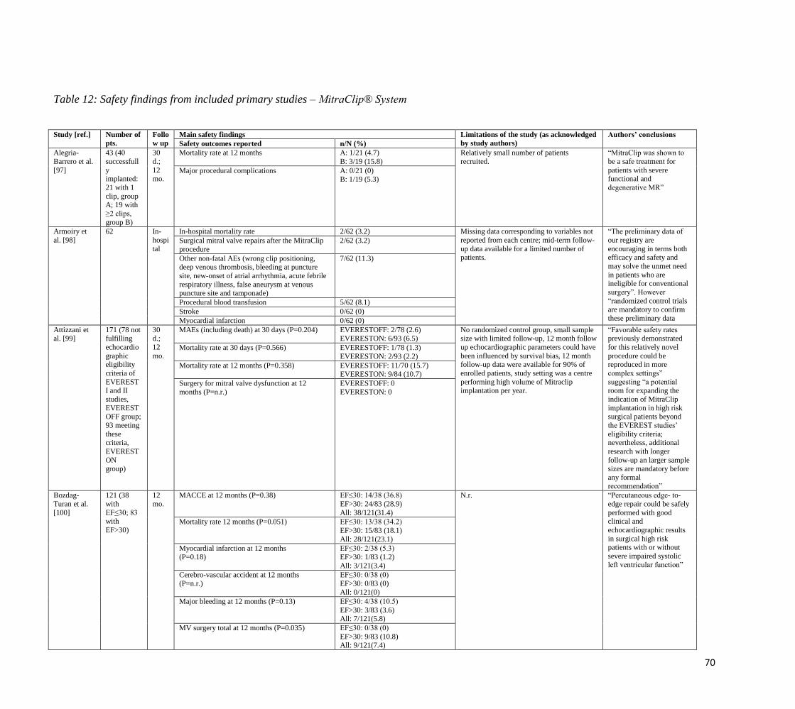
70
Table 12: Safety findings from included primary studies – MitraClip® System
Study [ref.] Number of
pts.
Follo
w up
Main safety findings Limitations of the study (as acknowledged
by study authors)
Authors’ conclusions
Safety outcomes reported n/N (%)
Alegria-
Barrero et al.
[97]
43 (40
successfull
y implanted:
21 with 1
clip, group A; 19 with
≥2 clips,
group B)
30
d.;
12 mo.
Mortality rate at 12 months A: 1/21 (4.7)
B: 3/19 (15.8)
Relatively small number of patients
recruited.
“MitraClip was shown to
be a safe treatment for
patients with severe functional and
degenerative MR”
Major procedural complications A: 0/21 (0) B: 1/19 (5.3)
Armoiry et
al. [98]
62 In-
hospi
tal
In-hospital mortality rate 2/62 (3.2) Missing data corresponding to variables not
reported from each centre; mid-term follow-
up data available for a limited number of patients.
“The preliminary data of
our registry are
encouraging in terms both efficacy and safety and
may solve the unmet need
in patients who are ineligible for conventional
surgery”. However
“randomized control trials are mandatory to confirm
these preliminary data
Surgical mitral valve repairs after the MitraClip
procedure
2/62 (3.2)
Other non-fatal AEs (wrong clip positioning, deep venous thrombosis, bleeding at puncture
site, new-onset of atrial arrhythmia, acute febrile
respiratory illness, false aneurysm at venous puncture site and tamponade)
7/62 (11.3)
Procedural blood transfusion 5/62 (8.1)
Stroke 0/62 (0)
Myocardial infarction 0/62 (0)
Attizzani et al. [99]
171 (78 not fulfilling
echocardio
graphic eligibility
criteria of
EVEREST I and II
studies,
EVERESTOFF group;
93 meeting
these criteria,
EVEREST
ON group)
30 d.;
12
mo.
MAEs (including death) at 30 days (P=0.204) EVERESTOFF: 2/78 (2.6) EVERESTON: 6/93 (6.5)
No randomized control group, small sample size with limited follow-up, 12 month follow
up echocardiographic parameters could have
been influenced by survival bias, 12 month follow-up data were available for 90% of
enrolled patients, study setting was a centre
performing high volume of Mitraclip implantation per year.
“Favorable safety rates previously demonstrated
for this relatively novel
procedure could be reproduced in more
complex settings”
suggesting “a potential room for expanding the
indication of MitraClip
implantation in high risk surgical patients beyond
the EVEREST studies’
eligibility criteria; nevertheless, additional
research with longer
follow-up an larger sample sizes are mandatory before
any formal recommendation”
Mortality rate at 30 days (P=0.566) EVERESTOFF: 1/78 (1.3)
EVERESTON: 2/93 (2.2)
Mortality rate at 12 months (P=0.358) EVERESTOFF: 11/70 (15.7) EVERESTON: 9/84 (10.7)
Surgery for mitral valve dysfunction at 12
months (P=n.r.)
EVERESTOFF: 0
EVERESTON: 0
Bozdag-
Turan et al.
[100]
121 (38
with
EF≤30; 83 with
EF>30)
12
mo.
MACCE at 12 months (P=0.38) EF≤30: 14/38 (36.8)
EF>30: 24/83 (28.9)
All: 38/121(31.4)
N.r. “Percutaneous edge- to-
edge repair could be safely
performed with good clinical and
echocardiographic results
in surgical high risk patients with or without
severe impaired systolic
left ventricular function”
Mortality rate 12 months (P=0.051) EF≤30: 13/38 (34.2)
EF>30: 15/83 (18.1)
All: 28/121(23.1)
Myocardial infarction at 12 months (P=0.18)
EF≤30: 2/38 (5.3) EF>30: 1/83 (1.2)
All: 3/121(3.4)
Cerebro-vascular accident at 12 months
(P=n.r.)
EF≤30: 0/38 (0)
EF>30: 0/83 (0)
All: 0/121(0)
Major bleeding at 12 months (P=0.13) EF≤30: 4/38 (10.5)
EF>30: 3/83 (3.6) All: 7/121(5.8)
MV surgery total at 12 months (P=0.035) EF≤30: 0/38 (0)
EF>30: 9/83 (10.8) All: 9/121(7.4)

71
Braun et al.
[101]
119 (72
with DMR, 47
with
FMR)
12
mo.
Conventional mitral surgery after Mitraclip
procedure at 12 months
DMR: 10/n.c.
FMR: 0/n.c.
Relatively small patient population, lack of
follow-up data, subjectivity of echocardiographic MR quantification after
clip implantation.
Percutaneous edge- to-
edge repair of mitral valve is feasible in patients with
degenerative as well as
functional MR”. However “Randomized controlled
trial comparing MitraClip
therapy in high risk patients to medical therapy
as well as mitral valve
surgery are necessary to clarify the future role of
this novel method”.
Second mitral valve clipping at 12 months DMR: 4/n.c.
FMR: 1/n.c.
Procedural deaths DMR: 0/n.c.
FMR: 0/n.c.
Post-procedural deaths DMR:1/n.c.
FMR: 1/n.c.
Procedure related complications DMR: 1/n.c.
FMR: 1/n.c.
Glower et al. [82]
351 (105 with
DMR and
246 with FMR)
30 d.;
1 y.
Mortality rate at 30 days All: 17/351 (4.8) DMR: 7/105 (6.7)
FMR: 10/246 (4.1)
Patient group was narrowly defined, short term data, no surgical or medical control
group, possibly placebo effect due to
medical therapy prior to device implantation.
"Mitral valve device is feasible and relatively safe
and is effective (…) in this
high-risk group of patients who are unlikely to receive
surgery and essentially
have no other option to reduce MR’’
MAEs at 30 days All: 66/351 (18.8) DMR: 19/105 (18.1)
FMR: 47/246 (19.1)
Stroke at 30 days* All: 9/351 (2.6)
Major bleeding complications at 30 days DMR: 19/105 (18.1) FMR: 47/246 (19.1)
Blood transfusions ≥2U at 30 days* All: 47/351 (13.4)
Mortality rate at 1 year All: 80/351 (22.8)
DMR: 25/105 (23.8) FMR: 55/246 (22.4)
MAEs at 1 year All: 132/351 (37.6)
DMR: 38/105 (36.2)
FMR: 94/246 (38.2)
Stroke at 1 year* All: 12/351 (3.4)
Blood transfusions ≥2U at 1 year* All: 79 /351 (22.5)
Single leaflet device attachment rate (device
related complication) at 1 year*
All: 8/351 (2.28)
Mitral valve surgery (device related complication) at 1 year*
All: 3/351 (0.9)
Second Mitraclip procedure (device related
complication) at 1 year*
All: 4/351 (1.1)
Mitraclip embolization (device related complication) at 1 year*
All: 0/351 (0)
Mitral valve stenosis (device related
complication) at 1 year*
All: 3/351 (0.9)
Hellhammer
et al. [88]
58 (19 with
diabetes
mellitus II, 39 without
diabetes
mellitus II)
30
d.;
3 mo.
Mortality rate at 30 days (P=0.672) Diabetes: 0/19 (0)
No diabetes: 1/39 (2.6)
Short follow-up, small size population,no
randomized study.
“MitraClip system is safe
and event rates are low.”
However “a prospective randomized study with
more patients and longer
follow follow-up time was necessary”.
Successful clip implantation rate (P=0.672) Diabetes: 19/19 (100)
No diabetes: 38/39 (97.4)
In-hospital complications (MACCE, peripher
vascular complications, stroke, pacemaker
damage, sepsis, ventilation>24 h, acute kidney injury stage III, major bleeding)
Diabetes: 1/19 (5.3)
No diabetes: 9/39 (23.1)
Mitral valve surgery (P=0.672) Diabetes: 0/19 (0)
No diabetes: 1/39 (2.6)
Hellhammer et al. [89]
80 (41 with anaemia,
group A; 39
with normal
erythrocyte
levels, group B)
Up to 12
mo.
Mortality rate at 30 days (P = 0.611) A: 1/41 (2.4) B: 2/39 (5.1)
Limited number of patients and unequal follow-up times.
“Mitral valve repair with the MitraClip system can
be performed safely and
efficiently in patients with anemia. Anemia does not
affect clinical outcome and
quality of life in patients undergoing mitral valve
repair.”
MACCE rate (including death, myocardial
infarction, stroke, and procedure related re-
operation; P = 0.959)
A: 2/41 (4.9)
B: 2/39 (5.1)
Koifman et
al. [102]
30 231
d. (mea
n)
Abortion of the procedure 1/20 (5) Small sample size and relatively short
follow-up duration.
“Mitral valve repair using
the MitraClip Percutaneous technique is feasible and
safe in high risk, mainly
inoperable, highly
Patients hospitalised for more than 5 days 4/20 (20)
Mortality at 7 months 2/20 (10)

72
symptomatic patients with
significant MR.”
Reichenspurn
er et al.
[103]
117 (33
high
surgical risk, group
A; 84 low
surgical risk, group
B)
30
d.;
12 mo.
Adverse events rate at 30 days 21/117 (17.9)
A: 9/33 (27.3)
B: 12/84 (14.3)
Lack of a protocol for patient selection and
determination of aetiology.
“Primarily for DMR
patients who are inoperable
or at exceedingly high risk for surgical MVR,
MitraClip therapy
represents an attractive and less-invasive treatment
option. The majority of
patients thus treated benefit significantly regarding the
severity of MR as well as
clinically, regarding NYHA functional class
and improvements in
physical capacities and quality of life.”
Mortality rate at 30 days 7/117 (6.0) A: 3/33 (9.1)
B: 4/84 (4.8)
Adverse events rate at 12 months 48/117 (41.0)
Mortality rate at 12 months 20/117 (17.1) A: 8/33 (24.2)
B: 12/84 (14.3)
Rudolph et
al. [84]
803 (143
NYHA class IV,
572 NYHA
class III, 88 NYHA
class I or
II)
30 d. Mortality
Procedural (P = 0.59) At 30 days (P < 0.05)
Procedural
1/803 (0.1) – NYHA III At 30 days
NYHA IV: 11/137 (8.0)
NYHA III: 17/526 (3.2) NYHA I/II: 4/83 (4.8)
Inhomogeneous population (but still
reflecting real-life practice) and need of randomised studies to clarify the real
therapeutic value and optimal time point of
MitraClip implantation in severely diseased group of patients (e.g., those in NYHA IV).
“Our data indicate that
percutaneous mitral valve repair with the MitraClip is
feasible and safe, and leads
to relevant clinical improvement even in
critically ill, not fully
recompensated patients, but is associated with an
elevated 30-day mortality.
The decision to perform the procedure in this group
of patients has therefore to
be individualized. While awaiting further studies
addressing this topic,
aggressive medical management of acute HF
should be considered prior
to MitraClip therapy in this patient group.”
Transfusions/severe bleeding
Procedural (P < 0.01)
At 30 days (P = 0.19)
Procedural
NYHA IV: 19/140 (13.6)
NYHA III: 35/554 (6.3) NYHA I/II: 3/87 (3.4)
At 30 days:
NYHA IV: 15/98 (15.3) NYHA III: 38/387 (9.8)
NYHA I/II: 4/57 (7.0)
Transient ischaemic attacks (TIA) Procedural (P < 0.01)
At 30 days (P < 0.01)
Procedural NYHA IV: 5/140 (3.6)
NYHA III:3/555 (0.5)
NYHA I/II: 0/88 (0) At 30 days
NYHA IV: 6/91 (6.6)
NYHA III: 5/380 (1.3) NYHA I/II: 0/55 (0)
≥3 days until mobilisation (P < 0.05) NYHA IV: 16/137 (11.7)
NYHA III: 35/546 (6.4)
NYHA I/II: 2/88 (2.3)
Toggweiler
et al. [104]
74 2
years
Intra-procedural complications making procedure
unfeasible
3/74 (4) Low number of patients and limited
experience of the centres.
“In the light of these
results, the
definition of procedural success may need to be re-
evaluated. In future,
improved patient selection, experience and maybe
concomitant utilisation
with nonsurgical mitral annuloplasty devices may
lead to even better
outcomes and a wider application of the
MitraClip procedure.”
Bleeding requiring transfusion 6/74 (8)
Pericardial tamponade after rupture of the left atrium
1/74 (1)
Strokes at 24 months 0/74 (0)
In-hospital mortality 3/74 (4)
Partial clip detachment at 24 months 7/74 (10)
Repeated procedure due to persistent 3+ or 4+ MR
5/74 (6.8)
Vandendriessche et
al. [105]
41 Up to 12
mo.
In-hospital MAEs (death, additional major bleeding need to undergo urgent cardiac
surgery).
5/41 (12) Small sample size. “In the light of these results, the definition of
procedural success may
need to be re-evaluated. In future, improved patient
selection, experience and
maybe concomitant utilisation with nonsurgical

73
mitral annuloplasty devices
may lead to even better outcomes and a wider
application of the
MitraClip procedure.”
Wiebe et al.
[83]
557 72 d.
(med
ian)
In-hospital MACCE rate (mortality, stroke, and
myocardial infarction)
27/546 (4.9) Inadequacy of the logEuroSCORE in
reflecting decisions based on valve
morphology or aetiology of MR, and absence of postprocedural results and longer
terms durability data.
“Percutaneous mitral valve
repair with the MitraClip
system is feasible in patients with a
logEuroSCORE ≥ 20.
Procedural results were similar, despite a
significant higher intra-
hospital MACCE rate compared to patients with
lower predicted cardiac
operative risk. Although mortality was four times
higher than in patients with
a logEuroSCORE < 20, mortality in high risk
patients was lower than
predicted by the logEuroSCORE. In
patients with a
logEuroSCORE ≥ 20, moderate residual mitral
valve regurgitation is more
frequent.”
Major in-hospital complications rate 108/557 (19.4)
Minor in-hospital complications rate 77/557 (13.8)
Other complications rate 97/546 (17.8)
Yeo et al.
[106]
142 Up
to 30
d.
Mortality rate at 30 days 8/142 (5.6) Non-comparative nature of the study and
short duration of followup.
“MitraClip therapy is a
safe and efficacious
therapeutic option for patients with either FMR
or DMR. In the Asia-
Pacific region. The significant proportion of
DMR. In comparison to the
commercial experience in Europe, deserves further
examination.”
In-hospital mortality rate 6/142 (4.2)
MAE rate at 30 days 18/142 (12,7)
Patients underwent mitral valve reoperation 1/142 (0.7)
Blood transfusion of ≥2 units 5/142 (3,5)
Sepsis 2/142 (1.4)
Prolonged intubation 1/142 (0.7)
Patients readmitted for HF 1/142 (0.7)
Capodanno et al. [8]
n=304 366 d.
(IQR
150-691d
.)
30 d.;
1 y.;
2y.
30 d. mortality 10/304 (3,3) Small sample size and the number of events, study lacks the report of follow-up
echocardiographic outcomes, might be
selection and referral biases, no recommended and standardized medical
therapy strategy were used across centers.
“In a cohort of patients undergoing MitraClip
therapy, those presenting at
baseline with ischemic functional etiology,
severely dilated ventricles,
or advanced heart failure and those undergoing
unsuccessful procedures
carried the worst prognosis at long term.”
1y. mortality 34 /304 (11,)
2 y. mortality 53/304 (17,4)
>2 y. 62/304 (20,4)
all cause 1 y. mortality (13.8)
Mitral stenosis 0/304 (0)
Rehospitalization due to hearth failure 43/304 (14,1)
Puls et al.
[10]
n=749 30
d.;
1y.
1-y. mortality (20,3) Non randomized evidence, registry results,
all baseline and in hospital data were site-
reported, echocardiographic data were not core-lab adjudicated and therefore of minor
quality, follow-up at 30 d. and at 1 y. did not
include an echocardiography, possible
selection bias.
“Treatment of significant
MR with MitraClip is
efficacious and results in significant clinical
improvements in a high
proportion of TRAMI
patients after 12 months. In
the TRAMI cohort, the
failure of procedural success exhibited the
highest hazard ratio
concerning the prediction of 1-year mortality.
However, randomized
Procedural failure (3,2)
In hospital/30-days outcomes
Intra-procedural death 1/749 (0,1)
MACCE: 22/712 (3.1)
in-hospital mortality 18/749 (2.4)
MACCE: In hospital/30-d.: One-year:
Death 34/749 (4,5) 152/749 (20,3)
MI 0/711 (0) 4/425 (0,9)
Stroke 6/712 (0,8) 9/423 (2,1)
Non MACCE:
TIA 6/712 (0,8) 16/426 (3,8)
Bleeding complications 56/443 (12,6)
Need for resuscitation 9/426 (2,1)

74
Respiration failure 16/711 (2,3) studies to verify the
efficacy of MitraClip therapy and prospective
studies to define
anatomical criteria that allow a better prediction of
procedural success are still
required.”
Severe bleeding, transfusion 50/711 (7,0)
Low cardiac output 9/710 (1,3)
Pericardial tamponade 12/70 (1,7)
Clip embolization 0/710 (0)
Partial clip detachment 5/749 (0,7)
Rehospitalization: 364/566 (64,3)
Cardiac decompensation 80/566 (14,1)
Other cardiac reasons 101/566 (17,8)
Non-cardiac reason 146/566 (25,8)
Additional MV procedure 11/710 (1,5) 37/436 (8,5)
Surgical 6/710 (0,8) 10/436 (2,3)
Percutaneous** 5/710 (0,7) 23/436 (5,2)
Triantafylli et al [22]
n=136 30 d; 2 y.
Reintervention 7/136 (5,1) Observational and nonrandomized study character, single centre trial, possible
selection bias, no secondary endpoints.
“The results presented herein clearly demonstrate
that MitraClip therapy in
“real-world” high surgical risk patients,
predominantly with
functional MR and multiple comorbidities, is
safe with favorable and
sustained outcomes in terms of survival which
extend to 2 years after the
procedure. In addition, advanced age, NYHA
functional class III and IV
and elevated levels of logNTproBNP are strong
preprocedural predictors of
longterm cardiac mortality which advocate careful and
restrictive selection of
MitraClip candidates.”
30 day overall mortality: 5/136 (3,5)
2 years overall mortality: 35/136 (25,7)
Cardiac mortality: 29/35
Decompensation of advanced heart failure 23/29
Hospital cardiac arrest 3/29
Sudden cardiac death 2/29
Miocardial infarction 1/29
Non-cardiac mortality: 6/35
Pneumonia 3/6
NR 3/6
Periprocedural complications: 30/136 (22)
Bleeding requiring transfusion: gastrointestinal
bleeding (1 pts.) access site bleeding (7 pts)
8/136 (5,9)
MitraClip detachment/malposition
Tamponade 6/136 (4,4)
Transient ischemic attack 3/136 (2,2)
Peripheral nerve palsy 1/136 (0,7)
Alergic shoc due to protamine 1/136 (0,7)
Reintervention 1/136 (0,7)
Lesevic et al.
[23]
n=136
n=42
(A: LVEF ≤30 %)
n=94
(B: LVEF >30%)
Medi
an
371 d.
[27-
1047]
30 d.
Overall: A: B: Small number of enrolled patients, especially
the high-risk patients with LVEF >=30%,
very heterogeneous group, hemodynamic measurements were performed under general
anesthesia, a number of patients did not have
a complete clinical follow-up.
“In conclusion, at midterm
follow-up, the pMVR
provided significant clinical benefits with
comparable results
achieved both in patients with significantly reduced
and in patients with
moderately reduced to normal LVEF. Thus,
pMVR represents a
feasible and effective treatment in high-risk
patients who otherwise
have limited therapeutic options and no safe option
to reduce MR.“
Death of any cause 16 (12) 7 (17) 9 (10)
Causes of deaths:
Cardiac deaths 7 - -
During index hospital stay 3 - -
At 1st hospital day after MV 1 - -
Because of terminal heart failure, or sudden
cardiac death
3 - -
Unknown cause 5 - -
Fulminant pneumonia 3 - -
Related to clip procedure 1 - -
Fibrosarcoma 1 - -
In-hospital death after index clipping 5 (4) 2 (5) 3 (3)
Second clip procedure 6 (4) 2 (5) 4 (4)
Switch to mitral valve surgery 6 (4) 0 6 (6)
(Major) Adverse Events at 30 days 23 (26) 4 (17) 19 (30)
Clip related perforation of mitral valve leaflet 57 (42) 20 (48) 37 (39)
Clip related chordal rupture 1 (1) 0 1 (1)
Pericardial effusion/tamponade 0 0 0
PM/ICD/CRT lead displacement 0 2/42 4/94
Stroke 0 0 0
Myocardial infarction (thromboembolic) 2* (2) 1* (2) 1 (1)
Mechanical ventilation > 48 h 1 (1) 1 (2) 0
ASD requiring closure 12 (9) 4 (10) 8 (9)
Infection 5† (4) 2 (5) 3† (3)
Transfusion of 2 IU blood 5 (4) 3 (7) 2 (2)
Pseudoaneurysm 10 (7) 2(5) 8 (9)
AV-fistula, n (%) 3 (2) 0 3 (3)
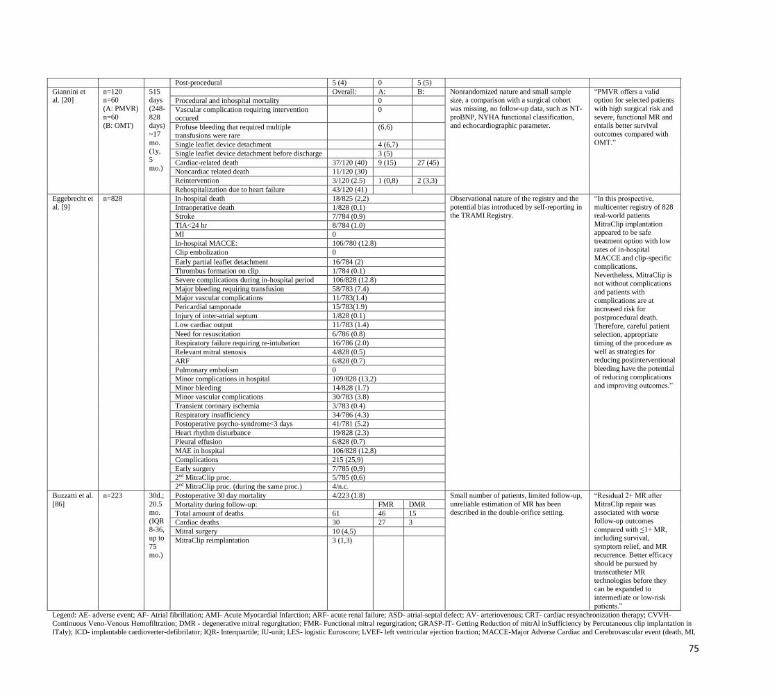
75
Legend: AE- adverse event; AF- Atrial fibrillation; AMI- Acute Myocardial Infarction; ARF- acute renal failure; ASD- atrial-septal defect; AV- arteriovenous; CRT- cardiac resynchronization therapy; CVVH-
Continuous Veno-Venous Hemofiltration; DMR - degenerative mitral regurgitation; FMR- Functional mitral regurgitation; GRASP-IT- Getting Reduction of mitrAl inSufficiency by Percutaneous clip implantation in
ITaly); ICD- implantable cardioverter-defibrilator; IQR- Interquartile; IU-unit; LES- logistic Euroscore; LVEF- left ventricular ejection fraction; MACCE-Major Adverse Cardiac and Cerebrovascular event (death, MI,
Post-procedural 5 (4) 0 5 (5)
Giannini et al. [20]
n=120 n=60
(A: PMVR)
n=60 (B: OMT)
515 days
(248-
828 days)
~17
mo. (1y,
5
mo.)
Overall: A: B: Nonrandomized nature and small sample size, a comparison with a surgical cohort
was missing, no follow-up data, such as NT-
proBNP, NYHA functional classification, and echocardiographic parameter.
“PMVR offers a valid option for selected patients
with high surgical risk and
severe, functional MR and entails better survival
outcomes compared with
OMT.”
Procedural and inhospital mortality 0
Vascular complication requiring intervention
occured
0
Profuse bleeding that required multiple transfusions were rare
(6,6)
Single leaflet device detachment 4 (6,7)
Single leaflet device detachment before discharge 3 (5)
Cardiac-related death 37/120 (40) 9 (15) 27 (45)
Noncardiac related death 11/120 (30)
Reintervention 3/120 (2.5) 1 (0,8) 2 (3,3)
Rehospitalization due to heart failure 43/120 (41)
Eggebrecht et
al. [9]
n=828 In-hospital death 18/825 (2,2) Observational nature of the registry and the
potential bias introduced by self-reporting in the TRAMI Registry.
“In this prospective,
multicenter registry of 828 real-world patients
MitraClip implantation appeared to be safe
treatment option with low
rates of in-hospital MACCE and clip-specific
complications.
Nevertheless, MitraClip is not without complications
and patients with
complications are at increased risk for
postprocedural death.
Therefore, careful patient selection, appropriate
timing of the procedure as
well as strategies for reducing postinterventional
bleeding have the potential
of reducing complications and improving outcomes.”
Intraoperative death 1/828 (0,1)
Stroke 7/784 (0.9)
TIA<24 hr 8/784 (1.0)
MI 0
In-hospital MACCE: 106/780 (12.8)
Clip embolization 0
Early partial leaflet detachment 16/784 (2)
Thrombus formation on clip 1/784 (0.1)
Severe complications during in-hospital period 106/828 (12.8)
Major bleeding requiring transfusion 58/783 (7.4)
Major vascular complications 11/783(1.4)
Pericardial tamponade 15/783(1.9)
Injury of inter-atrial septum 1/828 (0.1)
Low cardiac output 11/783 (1.4)
Need for resuscitation 6/786 (0.8)
Respiratory failure requiring re-intubation 16/786 (2.0)
Relevant mitral stenosis 4/828 (0.5)
ARF 6/828 (0.7)
Pulmonary embolism 0
Minor complications in hospital 109/828 (13,2)
Minor bleeding 14/828 (1.7)
Minor vascular complications 30/783 (3.8)
Transient coronary ischemia 3/783 (0.4)
Respiratory insufficiency 34/786 (4.3)
Postoperative psycho-syndrome<3 days 41/781 (5.2)
Heart rhythm disturbance 19/828 (2.3)
Pleural effusion 6/828 (0.7)
MAE in hospital 106/828 (12,8)
Complications 215 (25,9)
Early surgery 7/785 (0,9)
2nd MitraClip proc. 5/785 (0,6)
2nd MitraClip proc. (during the same proc.) 4/n.c.
Buzzatti et al.
[86]
n=223 30d.;
20.5 mo.
(IQR
8-36, up to
75
mo.)
Postoperative 30 day mortality 4/223 (1.8) Small number of patients, limited follow-up,
unreliable estimation of MR has been described in the double-orifice setting.
“Residual 2+ MR after
MitraClip repair was associated with worse
follow-up outcomes
compared with ≤1+ MR, including survival,
symptom relief, and MR
recurrence. Better efficacy should be pursued by
transcatheter MR
technologies before they can be expanded to
intermediate or low-risk
patients.”
Mortality during follow-up: FMR DMR
Total amount of deaths 61 46 15
Cardiac deaths 30 27 3
Mitral surgery 10 (4,5)
MitraClip reimplantation 3 (1,3)

76
stroke); MAE- major adverse event; MAE- major adverse events; MI-myocardial infarction; MV- mitral valve; NYHA- New York Heart Association; OMT-optimal medical therapy; PM- pacemaker; PMVR- percutaneous mitral valve repair; proc.-procedure; pts.- patients; TIA- transient ischemic attack; TRAMI- The transcatheter mitral valve interventions.
*Data are reported for all patients without distinguishing between DMR and FMR patients.
**Percutaneous- second MitraClip implantation
Table13 : Safety findings from included primary studies – Carillon® Mitral Contour Syste
Study
[ref.]
Number
of
patients
Follow
up
Main safety findings Limitations of the study (as
acknowledged by study
authors)
Authors’ conclusions
Safety outcomes reported n/N (%)
Schofer et
al.
(AMADE
US trial) [15]
48
patients
30 days Death 1/46 (2.2) First-in-human feasibility and
safety trial, lack of a
randomised, blinded control
group.
“Percutaneous reduction in FMR with a
novel coronary sinus– based mitral
annuloplasty device is feasible in
patients with heart failure, is associated with a low rate of major adverse events,
and is associated with improvement in
quality of life and exercise tolerance. Further studies are required to define the
long-term efficacy of the therapy,
optimal timing for intervention, and effects on survival.
Myocardial infarction§ 3/46 (6.5)
Cardiac perforation necessitating catheter based
or surgical intervention§
3/46 (6.5)
Device embolization 0/46 (0)
Device failure requiring surgical or percutaneous coronary intervention
0/46 (0)
Total major adverse events 7/46 (15.2
Siminiak et
al. (TITAN Trial) [14]
53 (36
with permane
nt
implant,
group A;
17 with
device recapture
d, group
B)
30 days;
12 months;
24
months
Death (30 days) A: 0/36 (0)
B: 1/17 (16)
Lack of a randomised and
blinded comparator.
“This study demonstrates that
percutaneous CS-based mitral annuloplasty can significantly and safely
reduce FMR severity in HF patients,
resulting in significant LV reverse
remodelling over 12 months and
improved measures of clinical outcome
over 24 months. The lack of a randomized and blinded comparator
remains the primary limitation of the
study. As such, a randomized trial comparing intervention with a medically
managed control group is warranted.”
Death (12 months) A: 8/36 (22.2) B: 4/17 (23.5)
Death (24 months*) A: 3/n.r. (n.a.)
B: n.a.
Myocardial infarction
(30 days)
(12 months)
0/53 (0)
0/53 (0)
Cardiac perforation (30 days)
(12 months)
0/53 (0)
0/53 (0)
Device embolization (30 days)
(12 months)
0/53 (0)
0/53 (0)
Surgery due to device
(30 days) (12 months)
0/53 (0) 0/53 (0)
Overall MAE rate
(30 days) (12 months)
1/53 (1.9) 14/53 (26.4)
§: not specified if the adverse event is caused by the procedure or by the device.
*Safety data at 24 months follow up refer only to the intervention group
Abbreviations: FMR, functional mitral regurgitation; n.r., not reported; n.a., not applicable; CS, coronary sinus; HF, heart failure; LV, left ventricular
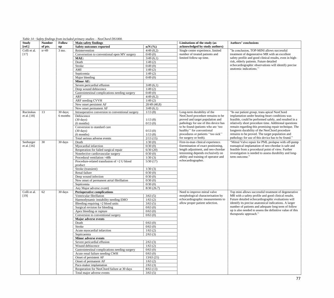
77
Table 14 : Safety findings from included primary studies – NeoChord DS1000.
Study
[ref.]
Number
of pts.
Follow
up
Main safety findings Limitations of the study (as
acknowledged by study authors)
Authors’ conclusions
Safety outcomes reported n/N (%)
Colli et al.
[17]
n=49 3 mo. Reintervention 4/49 (8,2) Single center experience, limited
number of treated patients and limited follow-up time.
”In conclusion, TOP-MINI allows successful
treatment of degenerative MR with an excellent safety profile and good clinical results, even in high-
risk, elderly patients. Future detailed
echocardiographic observations will identify precise anatomic indications.’’
Conversetion to conventional open MV surgery 0/49 (0)
MAE: 3/49 (6,1)
Death 1/49 (2)
Stroke 0/49 (0)
AMI 1/49 (2)
Septicemia 1/49 (2)
Major bleeding 0/49 (0)
Minor AE:
Severe pericardial effusion 3/49 (6,1)
Deep wound dehiscence 1/49 (2)
Gastrointestinal complications needing surgery 0/49 (0)
ARF 4/49 (8,2)
ARF needing CVVH 1/49 (2)
New onset persistent AF 20/49 (40,8)
New onset permanent AF 3/49 (6,1)
Rucinskas
et al. [18]
13 30 days;
6 months
Intraoperative conversion to conventional surgery 1/13 (8) Long-term durability of the
NeoChord procedure remains to be
proved and target population and pathology for use of this device has
to be found (patients who are ‘too
healthy’’ for conventional procedures or patients ‘‘too sick’’
for surgery or both).
“In our patient group, trans apical NeoChord
implantation under beating-heart conditions was
feasible, could be performed safely, and resulted in a relatively short procedure time. Additional questions
remain regarding this promising repair technique. The
longterm durability of the NeoChord procedure remains to be proved. The target population and
pathology for use of this device has to be found.”
Dehiscence
(30 days)
(6 months)
1/13 (8)
0/13 (0)
Conversion to standard care
(30 days)
(6 months)
0/13 (0)
1/13 (8)
Total serious adverse events 2/13 (15)
Seeburger
et al. [16]
30 30 days Death 1/30 (0) First-in-man clinical experience.
Etermination of exact positioning,
length adjustment, and neo-chordae tensioning depends exclusively on
ability and training of operator and
echocardiographer.
“Mitral Valve repair for PML prolapse with off-pump
transapical implantation of neo-chordae is safe and
feasible from a procedural point of view. Further investigation is needed to assess durability and long-
term outcome.”
Myocardial infarction 0/30 (0)
Reoperation for failed surgical repair 6/30 (20)
Nonelective cardiovascular surgery 0/30 (0)
Procedural ventilation >48h 1/30 (3)
Procedure-related transfusion of >2 U blood
product
5/30 (17)
Stroke (transient) 1/30 (3)
Renal failure 0/30 (0)
Deep wound infection 0/30 (0)
New onset of permanent atrial fibrillation 0/30 (0)
Septicemia 0/30 (0)
Any Major adverse event§ 8/30 (26.7)
Colli et al.
[19]
62 30 days Perioperative complications Need to improve mitral valve
morphological characterisation by echocardiographic measurements to
allow proper patient selection.
Top mini allows successful treatment of degenerative
MR with a safety profile and good clinical results. Future detailed echocardiographic evaluations will
identify its precise anatomical indications. A larger
number of patients and adequate long term of follow up is also needed to assess the definitive value of this
therapeutic approach.”
Ventricular fibrillation 3/63 (5)
Haemodynamic instability needing EMO 1/63 (2)
Bleeding requiring >2 blood units 3/63 (5)
Surgical revision for bleeding 0/63 (0)
Apex bleeding or rupture 0/63 (0)
Conversion to conventional surgery 0/63 (0)
Major adverse events
Death 0/63 (0)
Stroke 0/63 (0)
Acute myocardial infarction 1/63 (2)
Septicaemia 2/63 (3)
Minor adverse events
Severe pericardial effusion 2/63 (3)
Wound dehiscence 1/63 (2)
Gastrointestinal complications needing surgery 0/63 (0)
Acute renal failure needing CWH 0/63 (0)
Onset of persistent AF 13/63 (21)
Onset of permanent AF 1/63 (2)
Pace-maker implantation 2/63 (3)
Reoperation for NeoChord failure at 30 days 8/63 (13)
Total major adverse events 3/63 (5)
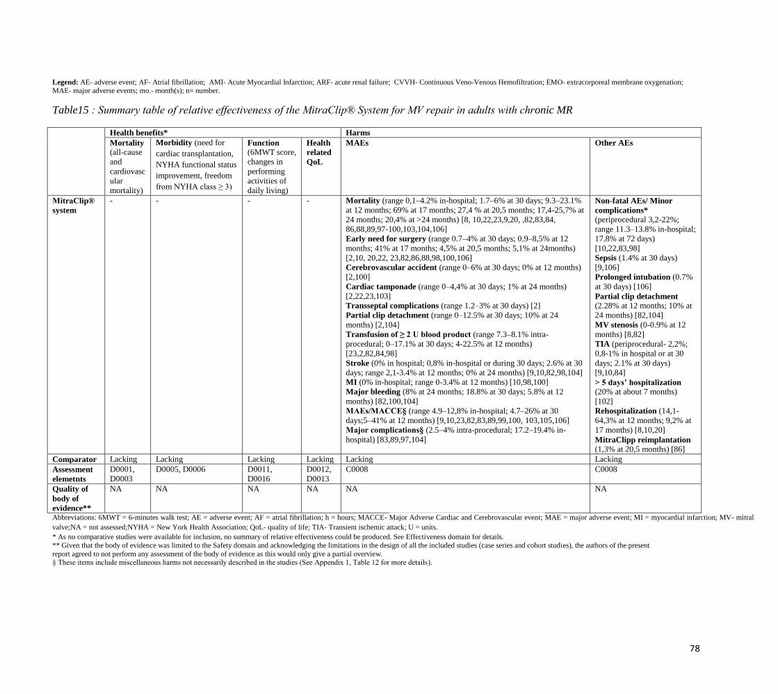
78
Legend: AE- adverse event; AF- Atrial fibrillation; AMI- Acute Myocardial Infarction; ARF- acute renal failure; CVVH- Continuous Veno-Venous Hemofiltration; EMO- extracorporeal membrane oxygenation; MAE- major adverse events; mo.- month(s); n= number.
Table15 : Summary table of relative effectiveness of the MitraClip® System for MV repair in adults with chronic MR
Health benefits* Harms
Mortality
(all-cause
and
cardiovasc
ular
mortality)
Morbidity (need for
cardiac transplantation,
NYHA functional status
improvement, freedom
from NYHA class ≥ 3)
Function (6MWT score,
changes in
performing
activities of
daily living)
Health
related
QoL
MAEs Other AEs
MitraClip®
system
- - - - Mortality (range 0,1–4.2% in-hospital; 1.7–6% at 30 days; 9.3–23.1%
at 12 months; 69% at 17 months; 27,4 % at 20,5 months; 17,4-25,7% at
24 months; 20,4% at >24 months) [8, 10,22,23,9,20, ,82,83,84,
86,88,89,97-100,103,104,106]
Early need for surgery (range 0.7–4% at 30 days; 0.9–8,5% at 12
months; 41% at 17 months; 4,5% at 20,5 months; 5,1% at 24months)
[2,10, 20,22, 23,82,86,88,98,100,106]
Cerebrovascular accident (range 0–6% at 30 days; 0% at 12 months)
[2,100]
Cardiac tamponade (range 0–4,4% at 30 days; 1% at 24 months)
[2,22,23,103]
Transseptal complications (range 1.2–3% at 30 days) [2]
Partial clip detachment (range 0–12.5% at 30 days; 10% at 24
months) [2,104]
Transfusion of ≥ 2 U blood product (range 7.3–8.1% intra-
procedural; 0–17.1% at 30 days; 4-22.5% at 12 months)
[23,2,82,84,98]
Stroke (0% in hospital; 0,8% in-hospital or during 30 days; 2.6% at 30
days; range 2,1-3.4% at 12 months; 0% at 24 months) [9,10,82,98,104]
MI (0% in-hospital; range 0-3.4% at 12 months) [10,98,100]
Major bleeding (8% at 24 months; 18.8% at 30 days; 5.8% at 12
months) [82,100,104]
MAEs/MACCE§ (range 4.9–12,8% in-hospital; 4.7–26% at 30
days;5–41% at 12 months) [9,10,23,82,83,89,99,100, 103,105,106]
Major complications§ (2.5–4% intra-procedural; 17.2–19.4% in-
hospital) [83,89,97,104]
Non-fatal AEs/ Minor
complications* (periprocedural 3,2-22%;
range 11.3–13.8% in-hospital;
17.8% at 72 days)
[10,22,83,98]
Sepsis (1.4% at 30 days)
[9,106]
Prolonged intubation (0.7%
at 30 days) [106]
Partial clip detachment (2.28% at 12 months; 10% at
24 months) [82,104]
MV stenosis (0-0.9% at 12
months) [8,82]
TIA (periprocedural- 2,2%;
0,8-1% in hospital or at 30
days; 2.1% at 30 days)
[9,10,84]
> 5 days’ hospitalization (20% at about 7 months)
[102]
Rehospitalization (14,1-
64,3% at 12 months; 9,2% at
17 months) [8,10,20] MitraClipp reimplantation (1,3% at 20,5 months) [86]
Comparator Lacking Lacking Lacking Lacking Lacking Lacking
Assessment
elemetnts
D0001,
D0003
D0005, D0006 D0011,
D0016
D0012,
D0013
C0008 C0008
Quality of
body of
evidence**
NA NA NA NA NA NA
Abbreviations: 6MWT = 6-minutes walk test; AE = adverse event; AF = atrial fibrillation; h = hours; MACCE- Major Adverse Cardiac and Cerebrovascular event; MAE = major adverse event; MI = myocardial infarction; MV- mitral
valve;NA = not assessed;NYHA = New York Health Association; QoL- quality of life; TIA- Transient ischemic attack; U = units.
* As no comparative studies were available for inclusion, no summary of relative effectiveness could be produced. See Effectiveness domain for details.
** Given that the body of evidence was limited to the Safety domain and acknowledging the limitations in the design of all the included studies (case series and cohort studies), the authors of the present
report agreed to not perform any assessment of the body of evidence as this would only give a partial overview. § These items include miscellaneous harms not necessarily described in the studies (See Appendix 1, Table 12 for more details).

79
Table 16: Summary table of relative effectiveness of the CARILLON® Mitral Contour System®for MV repair in adults with chronic MR
Health benefits* Harms
Mortality
(all-cause
and
cardiovasc
ular
mortality)
Morbidity (need for
cardiac transplantation,
NYHA functional status
improvement, freedom
from NYHA class ≥ 3)
Function (6MWT score,
changes in
performing
activities of
daily living)
Health
related
QoL
MAEs Other AEs
CARILLON
® Mitral
Contour
System®
- - - - Mortality (0 vs 16% at 30 days; 22.2% vs 23.5% at 12 months) [14]
MI (range 0–6.5% at 30 days; 4% at 12 months) [14, 15]
Cardiac perforation (range 0–6.5% at 30 days; 0% at 12 months)
[14, 15]
-
Comparator Lacking Lacking Lacking Lacking Lacking Lacking
Assessment
elemetnts
D0001,
D0003
D0005, D0006 D0011,
D0016
D0012,
D0013
C0008 C0008
Quality of
body of
evidence**
NA NA NA NA NA NA
Abbreviations: 6MWT = 6-minutes walk test; AE = adverse event; AF = atrial fibrillation; h = hours; MAE = major adverse event; MI = myocardial infarction; NYHA = New York Health
Association; U = units; NA = not assessed.
* As no comparative studies were available for inclusion, no summary of relative effectiveness could be produced. See Effectiveness domain for details.
** Given that the body of evidence was limited to the Safety domain and acknowledging the limitations in the design of all the included studies (case series and cohort studies), the authors of the
present report agreed to not perform any assessment of the body of evidence as this would only give a partial overview.
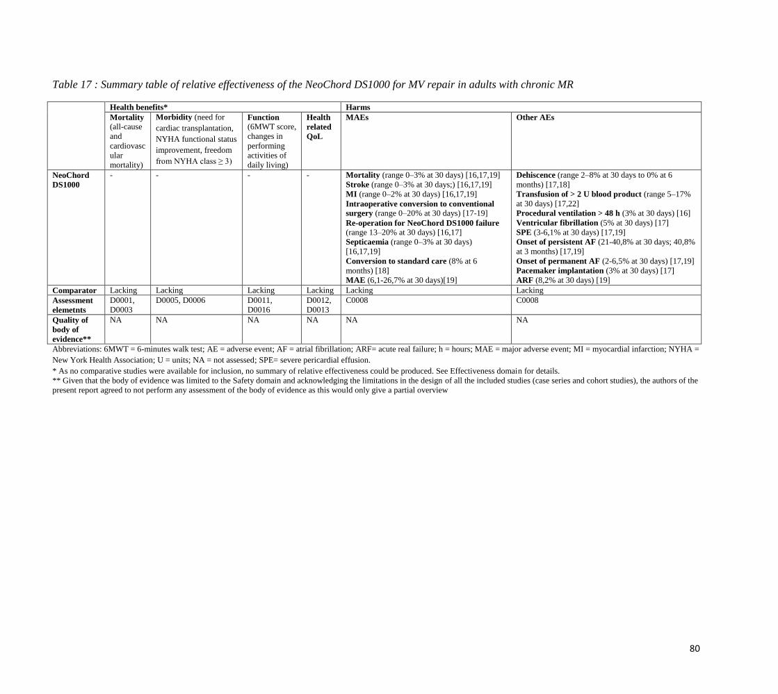
80
Table 17 : Summary table of relative effectiveness of the NeoChord DS1000 for MV repair in adults with chronic MR
Health benefits* Harms
Mortality
(all-cause
and
cardiovasc
ular
mortality)
Morbidity (need for
cardiac transplantation,
NYHA functional status
improvement, freedom
from NYHA class ≥ 3)
Function (6MWT score,
changes in
performing
activities of
daily living)
Health
related
QoL
MAEs Other AEs
NeoChord
DS1000
- - - - Mortality (range 0–3% at 30 days) [16,17,19]
Stroke (range 0–3% at 30 days;) [16,17,19]
MI (range 0–2% at 30 days) [16,17,19]
Intraoperative conversion to conventional
surgery (range 0–20% at 30 days) [17-19]
Re-operation for NeoChord DS1000 failure
(range 13–20% at 30 days) [16,17]
Septicaemia (range 0–3% at 30 days)
[16,17,19]
Conversion to standard care (8% at 6
months) [18]
MAE (6,1-26,7% at 30 days)[19]
Dehiscence (range 2–8% at 30 days to 0% at 6
months) [17,18]
Transfusion of > 2 U blood product (range 5–17%
at 30 days) [17,22]
Procedural ventilation > 48 h (3% at 30 days) [16]
Ventricular fibrillation (5% at 30 days) [17]
SPE (3-6,1% at 30 days) [17,19]
Onset of persistent AF (21-40,8% at 30 days; 40,8%
at 3 months) [17,19]
Onset of permanent AF (2-6,5% at 30 days) [17,19]
Pacemaker implantation (3% at 30 days) [17]
ARF (8,2% at 30 days) [19]
Comparator Lacking Lacking Lacking Lacking Lacking Lacking
Assessment
elemetnts
D0001,
D0003
D0005, D0006 D0011,
D0016
D0012,
D0013
C0008 C0008
Quality of
body of
evidence**
NA NA NA NA NA NA
Abbreviations: 6MWT = 6-minutes walk test; AE = adverse event; AF = atrial fibrillation; ARF= acute real failure; h = hours; MAE = major adverse event; MI = myocardial infarction; NYHA =
New York Health Association; U = units; NA = not assessed; SPE= severe pericardial effusion.
* As no comparative studies were available for inclusion, no summary of relative effectiveness could be produced. See Effectiveness domain for details.
** Given that the body of evidence was limited to the Safety domain and acknowledging the limitations in the design of all the included studies (case series and cohort studies), the authors of the
present report agreed to not perform any assessment of the body of evidence as this would only give a partial overview

81
Table 18. Overview of guidelines.
Name of
society/organization
issuing guidance
Date
of
issue
Country
to which
applicable
Summary of recommendation Class of
recommendations/
Level of evidence
ESC/EACTS
Guidelines on the
management of
valvular heart
disease (version
2012) [24]
2012 Europe • Percutaneous edge-to-edge (EE) procedure may be considered in patients
with symptomatic severe primary MR who fulfil the echo criteria of
eligibility, are judged inoperable or at high surgical risk by a ‘heart team’,
and have a life expectancy greater than 1 year
• The percutaneous mitral clip procedure may be considered in patients with
symptomatic severe secondary MR despite optimal medical therapy
(including CRT if indicated), who fulfil the echo criteria of eligibility, are
judged inoperable or at high surgical risk by a team of cardiologists and
cardiac surgeons, and who have a life expectancy greater than 1 year
IIb, C
ESC
ESC Guidelines for
the diagnosis and
treatment
of acute and chronic
heart failure 2012
[59]
2012 Europe • In patients with an indication for valve repair but judged inoperable or at
unacceptably high surgical risk, percutaneous edge-to-edge repair may be
considered in order to improve symptoms.
Not mentioned
Surgical and
interventional ma-
nagement of mitral
valve regurgitation: a
position statement
from the European
Society of
Cardiology Working
Groups on Car-
diovascular Surgery
and Valvular Heart
Disease [47]
2016 Europe Percutaneous EE repair is an alternative in symptomatic inoperable and
high-risk patients.
Percutaneous EE repair for secondary MR should only be considered in
addition to optimal medical therapy (including cardiac resynchronization
where appropriate) in patients who are symptomatic, fulfil anatomical
criteria, and judged high-risk or inoperable by the Heart Team.
Not mentioned
European
Association of
Echocardiography.
Recommendations
for the assessment of
valvular
regurgitation. Part 2:
mitral and tricuspid
regurgitation (native
valve disease) [105]
Recommendations
for the echo-
cardiographic
assessment of native
valvular regurgi-
tation: an executive
summary from the
European Asso-
ciation of Cardio-
2010
2016
Europe • TTE (Transthoracic Echocardiography) is recommended as the first-line
imaging modality for mitral valve analysis.
• TEE (Transoesophageal Echocar-diography) is advocated when TTE is of
non-diagnostic value or when further diagnostic refinement is required.
• 3D-TEE or TTE is reasonable to provide additional information in patients
with complex mitral valve lesion.
• TEE is not indicated in patients with a god-quality TTE except in the
operating room when a mitral valve surgery is performed.
Not mentioned

82
vascular Imaging
[108]
German Society for
Thoracic and
cardiovascular
Surgery on treatment
of mitral valve
insufficiency [109]
2014 Germany • DMR: Percutaneous intervention (only MitraClip has sufficient evidence)
may be considered in patients with advanced stage MR, high age or
pronounced comorbidities.
• MitraClip shows promising results for the treatment of secondary MR,
particularly in elderly patients with ischemic MI or a distinctive risk profile,
such as after coronary artery bypass surgery and represents a therapeutic
alternative in selected patients.
Not mentioned
Consensus Paper
from the focus group
‘Interven-tional
mitral valve
therapy’(Arbeitskreis
Interventionelle
Mitralklap-
pentherapie) of the
work-ing group
‘Interventional
Cardiology of the
Ger-man Society for
Cardiol-ogy’
(Arbeitsgemein-
schaft
Interventionelle
Kardiologie
(AGIK)) and Senior
Hospital Consultants
(Deutschen
Gesellschaft für
Kardiologie und der
Lei-tendenden
Kranken-hausärzte
(ALKK) [110]
2013 Germany The indication to interventional treatment of mitral valve regurgitation should
always be determined on an individual basis as currently there are no
established guidelines for this therapy. During this decision making process,
the following factors should be considered:
1. The recommendations from the current guidelines by the German and
European societies for cardiology on the treatment of cardiac valve disease.
2. The morphology of the mitral valve.
3. The cause and the severity of the MR.
4. The left ventricular function.
5. The operative risk.
Indications for the MitraClip therapy
Ideal for MitraClip treatment:
• Severe MR and
• Optimal valve morphology and
• Secondary MR with LV-EF\30 %
• Or Primary MR (with operation-indication following guidelines)
• and
• A high operative risk or other risk constellations
MitraClip to be considered: • Moderate to severe MR and
• Optimal valve morphology and
• SMR or PMR (with operation-indication following guidelines) and
• High operative risk, very high age or other risk constellations
MitraClip not recommended or only in exceptional cases:
• Moderate to severe MR and
• Conditionally suitable valve morphology
• or Life expectancy\12 months
• or LV-EF\15 % or cardiothoracic operation planned due to other indications
• or previously operated mitral valve
• or as surgical/interventional hybrid procedure
• or at low operative risk.
Not mentioned
ACC/AHA
2014 AHA/ACC
Guideline for the
Management of
Patients With
Valvular Heart
Disease [25]
2014 USA Chronic Primary MR
Transcatheter MV Repair may be considered for severely symptomatic
patients (NYHA class III/IV) with chronic severe primary MR (stage D0 who
have a reasonable life expec-tancy but a prohibitive surgical risk because of
severe comorbidities
Chronic secondary MR
Not reported
IIb, B
Abbreviations: ESC/EACTS: European Society of Cardiology and the European Association for Cardio-Thoracic Surgery (EACTS); ACC/AHA: the American College of
Cardiology/American Heart Association; MR: mitral regurgitation; DMR: de-generative mitral regurgitation
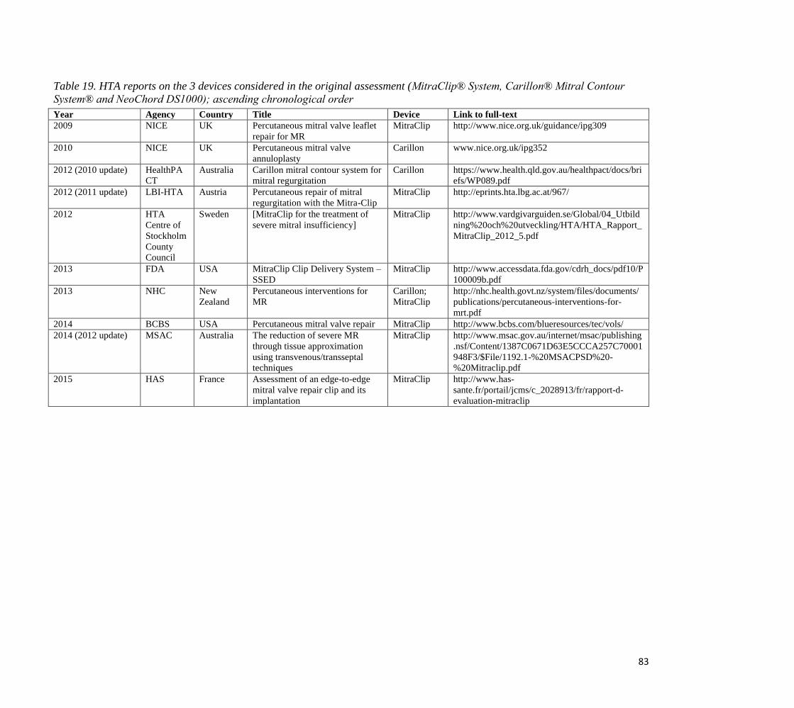
83
Table 19. HTA reports on the 3 devices considered in the original assessment (MitraClip® System, Carillon® Mitral Contour
System® and NeoChord DS1000); ascending chronological order
Year Agency Country Title Device Link to full-text
2009 NICE UK Percutaneous mitral valve leaflet
repair for MR
MitraClip http://www.nice.org.uk/guidance/ipg309
2010 NICE UK Percutaneous mitral valve
annuloplasty
Carillon www.nice.org.uk/ipg352
2012 (2010 update) HealthPA
CT
Australia Carillon mitral contour system for
mitral regurgitation
Carillon https://www.health.qld.gov.au/healthpact/docs/bri
efs/WP089.pdf
2012 (2011 update) LBI-HTA Austria Percutaneous repair of mitral
regurgitation with the Mitra-Clip
MitraClip http://eprints.hta.lbg.ac.at/967/
2012 HTA
Centre of
Stockholm
County
Council
Sweden [MitraClip for the treatment of
severe mitral insufficiency]
MitraClip http://www.vardgivarguiden.se/Global/04_Utbild
ning%20och%20utveckling/HTA/HTA_Rapport_
MitraClip_2012_5.pdf
2013 FDA USA MitraClip Clip Delivery System –
SSED
MitraClip http://www.accessdata.fda.gov/cdrh_docs/pdf10/P
100009b.pdf
2013 NHC New
Zealand
Percutaneous interventions for
MR
Carillon;
MitraClip
http://nhc.health.govt.nz/system/files/documents/
publications/percutaneous-interventions-for-
mrt.pdf
2014 BCBS USA Percutaneous mitral valve repair MitraClip http://www.bcbs.com/blueresources/tec/vols/
2014 (2012 update) MSAC Australia The reduction of severe MR
through tissue approximation
using transvenous/transseptal
techniques
MitraClip http://www.msac.gov.au/internet/msac/publishing
.nsf/Content/1387C0671D63E5CCCA257C70001
948F3/$File/1192.1-%20MSACPSD%20-
%20Mitraclip.pdf
2015 HAS France Assessment of an edge-to-edge
mitral valve repair clip and its
implantation
MitraClip http://www.has-
sante.fr/portail/jcms/c_2028913/fr/rapport-d-
evaluation-mitraclip

84
Table 20. List of ongoing studies with MitraClip® System [95]
Study
Identifier
Estimated
completion
date
[status]
Study
type
Number
of
patients
Intervention Comparator Patient
population
Endpoints
NCT01626079
COAPT
Aug 2020
[recruiting]
Interven-
tional.
Multi-
centre
rando-
mised
with
parallel
assign-
ment
430 Percutaneous
mitral valve
repair using
MitraClip
System
No Inter-
vention (non-
surgical ma-
nagement
based on
standard hos-
pital clinical
practice).
Symptomatic
heart failure
subjects who
are treated per
standard of
care and who
have been
determined by
the site's local
heart team as
not approp-
riate for mitral
valve surgery.
Primary outcomes: Composite of Single Leaflet Device Attachment
(SLDA), device embolizations, endocarditis requiring
surgery, Echocardiography Core Laboratory confirmed
mitral stenosis requiring surgery, LVAD implant, heart
transplant, and any device related com-plications
requiring non-elective cardiovascular surgery (12
months);
Recurrent heart failure (HF) hospitalizations (24
months).
Secondary outcomes: A composite of all-cause death, stroke, MI, or non-
elective cardiovascular surgery for device related
complications in the Device group (30 days)
All-cause mortality (12 months)
MR severity (12 months)
Change in distance walked on the 6MWT (12 months)
Change in quality of life (QoL) as measured by the
KCCQ (12 months)
Change in Left Ventricular End Diastolic Volume
(LVEDV) (12 months)
New York Heart Association (NYHA) Functional Class
I/II (12 months)
Recurrent hospitalizations - all cause (12 months)
Hierarchical composite of death and recurrent HF
NCT02444338 Sep 2019
[recruiting]
Intervent
ional.
Multi-
centre
ran-
domised.
380 MitraClip
device plus
optimal
standard of
care therapy
Standard of
care therapy
Patients with
chronic heart
failure and
clinically
significant
functional MR
(NYHA II to
NYHA IV).
Primary outcomes: Cardiovascular death
Secondary outcomes: MR severity reduction (24 months)
Change in 6MWT (6, 12 and 24 months)
cardiovascular hospitalizations and cardiovascular death
(24 months)
Quality of Life (QoL) overall score (12 months)
New York Heart Association (NYHA) Functional Class
(6, 12 and 24 months)
Patient-reported Global Assessment (PGA) (6, 12 and 24
months)

85
NCT02371512
MATTERHOR
N
Dec 2017
[recruiting]
Intervent
ional.
Multi-
centre
ran-
domised
with
parallel
assign-
ment.
210 Valve repair
with the
MitraClip sys-
tem
Reconstructive
mitral valve
surgery
Advanced
insufficiency
of functional
or ischemic
origin in
patients with
moderate-to-
severe MR of
primarily
functional
pathology and
reduced left
ventricular
function
considered to
be at high
surgical risk.
Primary outcomes: Composite of death, rehospitalisation for heart failure,
reintervention (repeat operation or repeat intervention),
assist device implantation and stroke (whatever is first)
(12 months)
Secondary outcomes: Recurrence of grade 3 or 4 MR (12 months)
Change in 6MWT distance (12 months)
Change in NYHA functional class (12 months)
Change in MLHFQ score (12 months)
Echocardiographic assessment of left ventricular re-
modelling (12 months)
Change in serum BNP (12 months)
Length of stay ICU / hospital
Number of patients in whom operative or interventional
mitral valve repair cannot be performed (need for mitral
valve replacement) (12 months)
NCT02033811
MitraClip®
Registry
Jan 2020
[recruiting]
Observat
ional
(Patient
Registry)
Cohort
study.
200 percutaneous
mitral valve
repair
(PMVR) with
the
MitraClip®
system
NA Patients
undergoing
percutaneous
mitral valve
repair
(PMVR) with
the
MitraClip®
system.
Primary outcomes: Major cardiac adverse events (30 days)
Secondary outcomes: Mortality (12 months)
NCT01920698
MITRA-FR
Oct 2017
[recruiting]
Intervent
ional.
Multi-
centre
ran-
domised
with
parallel
assign-
ment.
288 Percutaneous
MitraClip
device im-
plantation in
addition to
optimal
standard
medical
therapy
Optimal
medical
therapy alone
Patients with
severe FMR. Primary outcomes: All-cause mortality and unplanned hospitalizations for
heart failure (12 months).
Secondary outcomes: All-cause mortality, cardiac mortality (30 days, 6
months, 12 months, and 24 months)
Survival with no major cardiovascular events (30 days, 6
months, 12 months, and 24 months)
Serious Adverse Events (30 days, 6 months, 12 months,
and 24 months)
Change in Quality of Life score as measured by the
European Quality of Life-5 Dimensions instrument (6
and 12 months)
Change in functional evaluation (12 months)
Change in echocardiographic evaluation (6, 12 and 24
months)
Change in biomarkers (BNP levels, creatinine) (6 and 12
months)
Cost-effectiveness of each strategy (12 months)
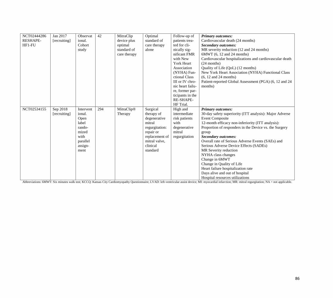
86
NCT02444286
RESHAPE-
HF1-FU
Jan 2017
[recruiting]
Observat
ional.
Cohort
study
42 MitraClip
device plus
optimal
standard of
care therapy
Optimal
standard of
care therapy
alone
Follow-up of
patients trea-
ted for cli-
nically sig-
nificant FMR
with New
York Heart
Association
(NYHA) Fun-
ctional Class
III or IV chro-
nic heart failu-
re, former par-
ticipants in the
RE-SHAPE-
HF Trial.
Primary outcomes: Cardiovascular death (24 months)
Secondary outcomes: MR severity reduction (12 and 24 months)
6MWT (6, 12 and 24 months)
Cardiovascular hospitalizations and cardiovascular death
(24 months)
Quality of Life (QoL) (12 months)
New York Heart Association (NYHA) Functional Class
(6, 12 and 24 months)
Patient-reported Global Assessment (PGA) (6, 12 and 24
months)
NCT02534155 Sep 2018
[recruiting]
Intervent
ional.
Open
label
rando-
mized
with
parallel
assign-
ment
294 MitraClip®
Therapy
Surgical
therapy of
degenerative
mitral
regurgitation:
repair or
replacement of
mitral valve,
clinical
standard
High and
intermediate
risk patients
with
degenerative
mitral
regurgitation
Primary outcomes: 30-day safety superiority (ITT analysis): Major Adverse
Event Composite
12-month efficacy non-inferiority (ITT analysis):
Proportion of responders in the Device vs. the Surgery
group
Secondary outcomes: Overall rate of Serious Adverse Events (SAEs) and
Serious Adverse Device Effects (SADEs)
MR Severity reduction
NYHA class changes
Change in 6MWT
Change in Quality of Life
Heart failure hospitalization rate
Days alive and out of hospital
Hospital resources utilizations Abbreviations: 6MWT: Six minutes walk test; KCCQ: Kansas City Cardiomyopathy Questionnaire; LVAD: left-ventricular assist device; MI: myocardial infarction; MR: mitral regurgitation; NA = not applicable.

87
Table 21. List of ongoing studies with Carillon® Mitral Contour System® [95]
Study
Identifier
Estimated
completion
date
[status]
Study
type
Number
of pa-
tients
Inter-
vention
Compa-
rator
Patient population Endpoints
NCT02325830
REDUCE FMR
Jul 2017
[recruiting]
Intervent
ional.
Multicen
tre ran-
domised
with pa-
rallel
assign-
ment.
180 Percu-
taneous
mitral
valve re-
pair with
CA-
RILLON
Mitral
Contour
Sys-tem
No
Interven-
tion
(medical
manage
ment
accordin
g to heart
failure
guide-
lines)
Diagnosis of dilated ischemic or
non-ischemic cardiomyopathy;
Functional MR: 2+ (Moderate),
3+ (Moder-ate/Severe), or 4+
(Severe);
New York Heart Association
(NYHA) II, III, or IV
Six Minute Walk distance of at
least 150 meters and no farther
than 450 meters
Left Ventricular Ejection
Fraction ≤ 40%
LV end diastolic dimension
(LVEDD) > 55mm or
LVEDD/Body Surface Area
(BSA) > 3.0cm/m2
Stable heart failure medication
regimen for at least three (3)
months prior to index
procedure.
Primary outcomes: Change in regurgitant volume associat-ed with the
CARILLON device relative to the Control
population (at 12 months).
Secondary outcomes: Document the difference in the rate of major
adverse events between Treat-ment (CARILLON)
and Control groups (30 days and 12 months)
Assess rate of heart failure hospitaliza-tions
between Treatment (CARILLON) and Control
groups (12 months)
Assess the change in six-minute walk distance
between Treatment (CARIL-LON) and Control
groups (12 months)
Assess the change in left ventricular volumes
between Treatment (CARIL-LON) and Control
groups (12 months
Table 22. List of ongoing studies with NeoChord DS 1000 [95]
Study Identifier Estimated
completion
date
[status]
Study type Number of
patients
Intervention Comparator Patient population Endpoints
NCT01784055
NeoChord TACT
July 2016
[Active. Not
recruiting]
Observational
(Patient
Registry).
Cohort study.
100 Artificial
chordae
placement
using DS1000
System
NA Patients with Grade 3+ or 4+ mitral
valve regurgitation who are
candidates for surgical mitral valve
repair or replacement
Procedure Success
(1 day).
Abbreviations: NA = not applicable.

88
APPENDIX 3: QUALITY ASSESSMENT OF SELECTED STUDIES
Quality assessment tables
Table 23. Quality assessment of included primary studies by the IHE 18-items checklist (quality assessment tables of studies included in
primary HTA can be found in original report [1])
Item # #1 #2 #3 #4 #5 #6 #7 #8 #9 #10 #11 #12 #13 #14 #15 #16 #17 #18 Yes
MitraClip
Triantafyl
lis AS,
2015
Y Y N Y Y N Y na Y Y Y Y Y Y Y Y Y N 14
Tigges E,
2016 []
Y Y N Y Y N N na Y Y Y Y Y Y Y N Y Y 13
Buzzatti
N, 2016 []
Y Y N N Y N N na N Y Y Y Y N Y Y Y Y 11
Capodan
no D,
2015
Y Y Y N Y Y N na Y N nr Y Y Y Y Y Y Y 13
Giannini
C, 2016
Y Y N N Y Y Y N N Y N Y Y Y Y Y Y N 12
Eggebrec
ht H, 2015
Y Y Y N N N Y na Y Y N Y Y N Y Y Y Y 12
Lesevic
H, 2015
Y Y nr Y Y N N na Y Y Y Y Y Y Y Y Y N 13
Puls M,
2015
Y Y Y N N N Y na Y Y N Y Y N Y Y Y Y 12
Neochord
Colli A,
2015
Y Y N Y nr N Y na Y Y Y Y Y Y Y Y Y Y 14
Abbreviations: nr= not reported; na= not applicable, Y= Yes, N= No.
*Adapted from: Moga C, Guo B, Schopflocher D, Harstall C. Development of a quality appraisal tool for case series studies using a modified Deplhi technique. Methodology Paper. Edmonton AB: In-stitute of Health
Economics, 2012. The threshold of ≥14 was chosen based on the threshold set by the Delphi panel in Moga et al.
List of items: 1. Is the hypothesis/aim/objective of the study clearly stated in the abstract, introduction or methods section? 2. Are the characteristics of the par-ticipants
included in the study described? 3. Were the cases collected in more than one centre? 4. Are the eligibility criteria (inclusion and exclusion criteria) to entry the study
explicit and appropriate? 5. Were participants recruited consecutively? 6. Did participants enter the study at a similar point in the disease? 7. Was the intervention clearly
described in the study? 8. Were additional interventions (co-interventions) clearly reported in the study? 9. Are the outcome measures clearly defined in the introduction
or methodology section? 10. Were relevant outcomes appropriately measured with objective and/or subjective methods? 11. Were outcomes measured before and after
intervention? 12. Were the statistical tests used to assess the relevant outcomes appropriate? 13. Was the length of follow-up reported? 14. Was the loss to follow-up
reported? 15. Does the study provide estimates of the random variability in the data analysis of relevant out-comes? 16. Are adverse events reported? 17. Are the
conclusions of the study supported by results? 18. Are both competing interest and source of support for the study reported?

89
Table 24. Quality assessment of systematic reviewsfrom original and present assessments
R-AMSTAR items
Munkholm-
Larsen et
al., 2014 [1]
(1) 4
(2) 3.5
(3) 4
(4) 2.5
(5) 3.5
(6) 4
(7) 2
(8) 2
(9) 1.5
(10) 1
(11) 2
D’ascenzo,
2015 (1) (2) (3) (4) (5) (6) (7) (8) (9) (10) (11)
Adapted from: Kung J, Chiappelli F, Cajulis OO, Avezova R, Kossan G, Chew L, Maida CA. From Systematic Reviews to Clinical Recom-mendations for
Evidence-Based Health Care: Validation of Revised Assessment of Multiple Systematic Reviews (R-AMSTAR) for Grading of Clinical Relevance. The Open
Dentistry Journal, 2010, 4, 84-91).
Total score*: 30/44 (final score calculated from judgments provided by 2 independent assessors)
* the R-AMSTAR total score has a range of 11 to 44, 11 signifying that none of the AMSTAR criteria were satisfied, and a score of 44 revealing that all of
the cri-teria of systematic review excellence were verified.
List of R-AMSTAR items: (1) Was an 'a priori' design provided? (2) Was there duplicate study selection and data extraction? (3) Was a comprehensive
literature search performed? (4) Was the status of publication (i.e. grey literature) used as an inclusion criterion? (5) Was a list of studies (included and
excluded) provid-ed? (6) Were the characteristics of the included studies provided? (7) Was the scientific quality of the included studies assessed and
documented? (8) Was the scientific quality of the included studies used appropriately in formulating conclusions? (9) Were the methods used to combine the
findings of studies appropri-ate? (10) Was the likelihood of publication bias assessed? (11) Was the conflict of interest included?
1 Munkholm-Larsen S, Wan B, Tian DH, Kearney K, Rahnavardi M, Dixen U, et al. A systematic review on the safety and efficacy of percutaneous edge-to-
edge mitral valve repair with the MitraClip system for high surgical risk candidates. Heart. 2014;100(6):473-8.
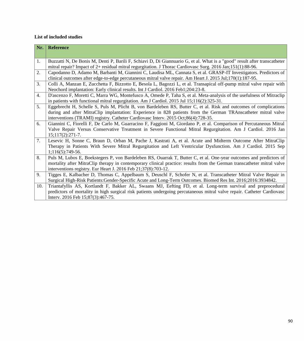
90
List of included studies
Nr. Reference
1. Buzzatti N, De Bonis M, Denti P, Barili F, Schiavi D, Di Giannuario G, et al. What is a "good" result after transcatheter
mitral repair? Impact of 2+ residual mitral regurgitation. J Thorac Cardiovasc Surg. 2016 Jan;151(1):88-96.
2. Capodanno D, Adamo M, Barbanti M, Giannini C, Laudisa ML, Cannata S, et al. GRASP-IT Investigators. Predictors of
clinical outcomes after edge-to-edge percutaneous mitral valve repair. Am Heart J. 2015 Jul;170(1):187-95.
3. Colli A, Manzan E, Zucchetta F, Bizzotto E, Besola L, Bagozzi L, et al. Transapical off-pump mitral valve repair with
Neochord implantation: Early clinical results. Int J Cardiol. 2016 Feb1;204:23-8.
4. D'ascenzo F, Moretti C, Marra WG, Montefusco A, Omede P, Taha S, et al. Meta-analysis of the usefulness of Mitraclip
in patients with functional mitral regurgitation. Am J Cardiol. 2015 Jul 15;116(2):325-31.
5. Eggebrecht H, Schelle S, Puls M, Plicht B, von Bardeleben RS, Butter C, et al. Risk and outcomes of complications
during and after MitraClip implantation: Experience in 828 patients from the German TRAnscatheter mitral valve
interventions (TRAMI) registry. Catheter Cardiovasc Interv. 2015 Oct;86(4):728-35.
6. Giannini C, Fiorelli F, De Carlo M, Guarracino F, Faggioni M, Giordano P, et al. Comparison of Percutaneous Mitral
Valve Repair Versus Conservative Treatment in Severe Functional Mitral Regurgitation. Am J Cardiol. 2016 Jan
15;117(2):271-7.
7. Lesevic H, Sonne C, Braun D, Orban M, Pache J, Kastrati A, et al. Acute and Midterm Outcome After MitraClip
Therapy in Patients With Severe Mitral Regurgitation and Left Ventricular Dysfunction. Am J Cardiol. 2015 Sep
1;116(5):749-56.
8. Puls M, Lubos E, Boekstegers P, von Bardeleben RS, Ouarrak T, Butter C, et al. One-year outcomes and predictors of
mortality after MitraClip therapy in contemporary clinical practice: results from the German transcatheter mitral valve
interventions registry. Eur Heart J. 2016 Feb 21;37(8):703-12.
9. Tigges E, Kalbacher D, Thomas C, Appelbaum S, Deuschl F, Schofer N, et al. Transcatheter Mitral Valve Repair in
Surgical High-Risk Patients:Gender-Specific Acute and Long-Term Outcomes. Biomed Res Int. 2016;2016:3934842.
10. Triantafyllis AS, Kortlandt F, Bakker AL, Swaans MJ, Eefting FD, et al. Long-term survival and preprocedural
predictors of mortality in high surgical risk patients undergoing percutaneous mitral valve repair. Catheter Cardiovasc
Interv. 2016 Feb 15;87(3):467-75.

91
List of excluded studies
Nr. Reference Reason of exclusion
1. Adamo M, Capodanno D, Cannata S, Giannini C, Laudisa M, Barbanti M et al. Comparison of
Three Contemporary Surgical Scores for Predicting All-Cause Mortality of Patients Undergoing
Percutaneous Mitral Valve Repair With the MitraClip System (from the Multicenter GRASP-IT
Registry). The American Journal of Cardiology. 2015;115(1):107-112.
Wrong objective
2. Armeni P, Boscolo PR, Tarricone R, Capodanno D, Maggioni AP, Grasso C, et al. Real-world
cost effectiveness of MitraClip combined with Medical Therapy Versus Medical therapy alone
in patients with moderate or severe mitral regurgitation. Int J Cardiol. 2016 Apr 15;209:153-60.
Wrong study design
3. Azzalini L, Millán X, Khan R, Couture P, Ducharme A, Basmadjian A, et al. Impact of left
ventricular function on clinical outcomes of functional mitral regurgitation patients undergoing
transcatheter mitral valve repair.Catheter Cardiovasc Interv. 2016 Feb 19.
Wrong study design
4. Barbanti M, Immè S, Grasso C. Transcatheter mitral valve repair: a brief review.
EuroIntervention. 2015 Sep;11 Suppl W:W42-4.
Wrong study design
5. Benito-González T, Estévez-Loureiro R, Gualis J. Chordal Rupture Following MitraClip
Implantation Resulting in Massive Mitral Regurgitation. J Invasive Cardiol. 2015
Oct;27(10):E224-5.
Wrong study design
6. Brito V, Pichon-Riviere A, Augustovski F, Garcķa Martķ S, Alcaraz A, Bardach A et al.
Percutaneous Mitral Valve Repair System (MitraClip®) for mitral valve insufficiency. Health
Technology Assessment. 2015. Available from:
http://onlinelibrary.wiley.com/o/cochrane/clhta/articles/HTA-32015001147/frame.html
Wrong language
7. Buchner S, Dreher A, Resch M, Schach C, Birner C, Luchner A. Simplified Method for
Insertion of Steerable Guide into the Left Atrium Using a Pigtail Guide Wire During the
MitraClip(®) Procedure: A Technical Tip. J Interv Cardiol. 2015 Oct;28(5):472-8.
Wrong objective
8. Cacela D, Fiarresga A, Branco L, Galrinho A, Rio P, Selas M, Ferreira R.[Percutaneous
treatment of mitral valve regurgitation: Initial experience with the MitraClip device]. Rev Port
Cardiol. 2015 Sep;34(9):515-24.
Wrong language
9. Cammalleri V, Romeo F, Ussia GP. Hemodynamic complications during transcatheter
MitraClip repair in presence of congenital atrial septal defect.Catheter Cardiovasc Interv. 2015
Sep 10.
Wrong study design
10. Candreva A, Maisano F, Taramasso M. MitraClip and Transcatheter Aortic Valve Implantation
(TAVI): State of the Art 2015. Curr Heart Fail Rep. 2015 Dec;12(6):379-88.
Wrong study design
11. Colli A, Manzan E, Rucinskas K, Janusauskas V, Zucchetta F, Zakarkaitė D et al. Acute safety
and efficacy of the NeoChord procedure. Interact CardioVasc Thorac Surg. 2015;20(5):575-
581.
Duplicate
12. De Bonis M, Taramasso M, Lapenna E, Denti P, La Canna G, Buzzatti N, et al. MitraClip
therapy and surgical edge-to-edge repair in patients with severe left ventricular dysfunction and
secondary mitral regurgitation: mid-term results of a single-centre experience†. Eur J
Cardiothorac Surg. 2016 Jan;49(1):255-62.
Wrong comparator
13. de Waha S, Seeburger J, Ender J, Desch S, Eitel I, Reinhardt A, et al. Deep sedation versus
general anesthesia in percutaneous edge-to-edge mitral valve reconstruction using the MitraClip
system. Clin Res Cardiol. 2015 Dec 18.
Wrong objective
14. Deuschl F, Schofer N, Lubos E, Blankenberg S, Schäfer U. Critical evaluation of the MitraClip
system in the management of mitral regurgitation. Vasc Health Risk Manag. 2016 Jan 11;12:1-
8.
Wrong study design
92
15. Deuschl F, Schofer N, Lubos E, Schirmer J, Conradi L, Treede H, et al. MitraClip-data analysis
of contemporary literature. J Thorac Dis. 2015 Sep;7(9):1509-17. doi: 10.3978/j.issn.2072-
1439.2015.07.38. Review.
Wrong study design
16. Downs E, Lim S, Ragosta M, Yount K, Yarboro L, Ghanta R, Kern J, Kron I, Ailawadi G. The
influence of a percutaneous mitral repair program on surgical mitral valve volume. J Thorac
Cardiovasc Surg. 2015 Nov;150(5):1093-7.
Unretrievable
17. Downs EA, Lim DS, Saji M, Ailawadi G. Current state of transcatheter mitral valve repair with
the MitraClip. Ann Cardiothorac Surg. 2015 Jul;4(4):335-40.
Wrong study design
18. Eleid MF, Sanon S, Reeder GS, Suri RM, Rihal CS. Continuous Left Atrial Pressure Monitoring
During MitraClip: Assessing the Immediate Hemodynamic Response. JACC Cardiovasc Interv.
2015 Jun;8(7):e117-9.
Wrong objective
19. Estévez-Loureiro R, Settergren M, Pighi M, Winter R, D'Allara G, Jacobsen P, et al. Effect of
advanced chronic kidney disease in clinical and echocardiographic outcomes of patients treated
with MitraClip system. Int J Cardiol. 2015 Nov 1;198:75-80.
Wrong objective.
20. Estévez-Loureiro R, Settergren M, Winter R, Jacobsen P, Dall'Ara G, Sondergaard L, et al.
Effect of gender on results of percutaneous edge-to-edge mitral valve repair with MitraClip
system. Am J Cardiol. 2015 Jul 15;116(2):275-9.
Wrong study design
21. Fam NP, Ross HJ, Verma S. Mitral clip-looking back and moving forward. Curr Opin Cardiol.
2016 Mar;31(2):169-75.
Unretrievable
22. Feldman T, Kar S, Elmariah S, Smart S, Trento A, Siegel R et al. Randomized Comparison of
Percutaneous Repair and Surgery for Mitral Regurgitation. Journal of the American College of
Cardiology. 2015;66(25):2844-2854.
Wrong population
23. Foerst J, Cardenas A, Swank G. Safety of MitraClip Implant in the Unstable Patient: Feasibility
of Concomitant Left Ventricular Support Device. JACC Cardiovasc Interv. 2016 Apr
11;9(7):e71-2.
Wrong study design
24. Frangieh AH, Gruner C, Mikulicic F, Attinger-Toller A, Tanner FC, Taramasso M et al. Impact
of percutaneous mitral valve repair using the MitraClip system on tricuspid regurgitation.
EuroIntervention. 2016 Apr 8;11(14):E1680-6.
Wrong indication
25. Frerker C, Schlüter M, Sanchez OD, Reith S, Romero ME, Ladich E, et al. Cerebral Protection
During MitraClip Implantation: Initial Experience at 2 Centers. JACC Cardiovasc Interv. 2016
Jan 25;9(2):171-9.
Wrong objective
26. Geidel S, Wohlmuth P, Schmoeckel M. Survival Prediction in Patients Undergoing Open-Heart
Mitral Valve Operation After Previous Failed MitraClip Procedures. Ann Thorac Surg. 2016
Mar;101(3):952-8.
Wrong intervention;
objective
27. Gonzalez FM, Finch AP, Armeni P, Boscolo PR, Tarricone R. Comparative effectiveness of
Mitraclip plus medical therapy versus medical therapy alone in high-risk surgical patients: a
comprehensive review. Expert Rev Med Devices. 2015 Jul;12(4):471-85.
Unretrievable
28. González-Gómez A, Fernández-Santos S, Fernández-Golfín C, Zamorano JL. Mitral valve
anatomy: pre-procedural screening and imaging techniques. EuroIntervention. 2015 Sep;11
Suppl W:W32-6.
Wrong intervention;
objective
29. Guerin P, Bourguignon S, Jamet N, Marque S. MitraClip therapy in mitral regurgitation: a
Markov model for the cost-effectiveness of a new therapeutic option. J Med Econ. 2016 Mar
22:1-6.
Wrong study design
30. Hanson ID, Hanzel GS, Shannon FL. Mitral valve repair after annuloplasty ring dehiscence
using MitraClip. Catheter Cardiovasc Interv. 2015 Oct 20.
Wrong study design
31. Horstkotte J, Kloeser C, Beucher H, Schwarzlaender E, von Bardeleben RS, Boekstegers P.
Intraprocedural assessment of mitral regurgitation during the mitraclip procedure: Impact of
continuous left atrial pressure monitoring. Catheter Cardiovasc Interv. 2016 Apr 1.
Wrong objective
32. Kaneko H, Neuss M, Schau T, Weissenborn J, Butter C. Impact of left ventricular systolic
dysfunction on the outcomes of percutaneous edge-to-edge mitral valve repair using MitraClip.
Wrong study design
93
Heart Vessels. 2016 Mar 11.
33. Ledwoch J, Matić P, Franke J, Gafoor S, Bertog S, Reinartz M, et al. Transcatheter mitral valve
repair with the MitraClip(®) can
be performed without general anesthesia and without conscious sedation. Clin Res Cardiol.
2016 Apr;105(4):297-306.
Wrong objective
34. Levi A, Sagie A, Kornowski R. MitraClip-induced systolic anterior motion complicated by
pericardial effusion: A case report. Catheter Cardiovasc Interv. 2016 Mar 4. doi:
10.1002/ccd.26476.
Wrong study design
35. Loh PH, Bourantas CV, Chan PH, Ihlemann N, Gustafsson F, Clark AL, et al. Catheter-based
intervention for symptomatic patient with severe mitral regurgitation and very poor left
ventricular systolic function - Safe but no room for complacency. World J Cardiol. 2015 Nov
26;7(11):817-21.
Wrong population size
36. New Zealand National Health Committee (NHC). Percutaneous interventions for mitral
regurgitation - technology Note. Wellington: New Zealand National Health Committee (NHC).
2013
Unretrievable
37. Nishimura RA, Vahanian A, Eleid MF, Mack MJ. Mitral valve disease—current management
and future challenges. Lancet. 2016 Mar 26;387(10025):1324-34.
Wrong study design
38. Nikendei C, Schäfer H, Weisbrod M, Huber J, Geis N, Katus HA, et al. The Effects of Mitral
Valve Repair on Memory Performance, Executive Function, and Psychological Measures in
Patients With Heart Failure. Psychosom Med. 2015 Dec 22.
Unretrievable
39. Obadia J, Armoiry X, Iung B, Lefèvre T, Mewton N, Messika-Zeitoun D et al. The MITRA-FR
study: design and rationale of a randomised study of percutaneous mitral valve repair compared
with optimal medical management alone for severe secondary mitral regurgitation.
EuroIntervention. 2015;10(11):1354-1360.
Not finished trial
40. Ohno Y, Attizzani GF, Capodanno D, Barbanti M, Cannata S, Dipasqua F, et al. Impact of
chronic kidney disease on outcomes after percutaneous mitral valve repair with the MitraClip
system: insights from the GRASP registry. EuroIntervention. 2016 Apr 8;11(14):e1649-57.
Wrong objective
41. Orban M, Braun D, Orban M, Gross L, Näbauer M, Hagl CM, et al. Established interventions
for mitral valve regurgitation : Current evidence. Herz. 2016 Feb;41(1):19-25.
Wrong study design
42. Panos A, Vlad S, Milas F, Myers PO. Is minimally invasive mitral valve repair with artificial
chords reproducible and applicable in routine surgery? Interact Cardiovasc Thorac Surg. 2015
Jun;20(6):707-11.
Wrong intervention
43. Puls M, Schillinger W. [Catheter-based mitral valve repair by MitraClip implantation :
Development, studies, and use in clinical practice]. Internist m(Berl). 2016 Apr;57(4):323-31.
Wrong language
44. Schueler R, Nickenig G, May AE, Schillinger W, Bekeredjian R, Ouarrak T, et al. Predictors for
short-term outcomes of patients undergoing transcatheter mitral valve interventions: analysis of
778 prospective patients from the German TRAMI registry focusing on baseline renal function.
EuroIntervention. 2015 Sep 8;11(5).
Wrong objective
45. Schau T, Isotani A, Neuss M, Schöpp M, Seifert M, Höpfner C, et al. Long-term survival after
MitraClip(®) therapy in patients with severe mitral regurgitation and severe congestive heart
failure: A comparison among survivals predicted by heart failure models. J Cardiol. 2016
Mar;67(3):287-94.
Wrong study design
46. Sondergaard L, Brooks M, Ihlemann N, Jonsson A, Holme S, Tang M, et al. Transcatheter
mitral valve implantation via transapical approach: an early experience. Eur J Cardiothorac
Surg. 2015 Dec;48(6):873-7; discussion 877-8.
Wrong sample size
47. Sorajja P, Mack M, Vemulapalli S, Holmes DR Jr, Stebbins A, Kar S, et al. Initial Experience
With Commercial Transcatheter Mitral Valve Repair in the United States. J Am Coll Cardiol.
2016 Mar 15;67(10):1129-40.
Wrong study design
48. Sturla F, Votta E, Onorati F, Pechlivanidis K, Pappalardo OA, Gottin L, et al. Biomechanical Wrong objective

94
drawbacks of different
techniques of mitral neochordal implantation: When an apparently optimal repair can fail. J
Thorac Cardiovasc Surg. 2015 Nov;150(5):1303-12.e4.
49. Tarsia G, Smaldone C, Costantino MF. Effective percutaneous "edge-to-edge" mitral valve
repair with mitraclip in a patient with acute post-MI regurgitation not related to papillary muscle
rupture. Catheter Cardiovasc Interv. 2016 Feb 16.
Wrong study design
50. Tay E, Muda N, Yap J, Muller DW, Santoso T, Walters DL, Liu X, et al. The MitraClip Asia-
Pacific registry: Differences in outcomes between functional and degenerative mitral
regurgitation. Catheter Cardiovasc Interv. 2015 Oct 28.
Wrong study design
51. Ussia GP, Cammalleri V, Mazzotta E, Pascuzzo G, Muscoli S, Marchei M, et al. Cardiovascular
medicine heart failure (CVM-HF) index as prognostic model for candidates to MitraClip
therapy. Heart Vessels. 2015 Dec 22.
Wrong objective
52. Van Mieghem NM, Rodríguez-Olivares R, Ren BC, van Gils L, Maugenest A, Geleijnse ML, et
al. Computed tomography optimised fluoroscopy guidance for transcatheter mitral therapies.
EuroIntervention. 2016 Mar 20;11(12):1428-31.
Wrong intervention;
objective
53. Veerbhadran SP, Pillai VV, Sasidharan B, Karunakaran J. Surgery for Congenital Tricuspid
Valve Cleft: Tricuspid Valve Repair with Neochordae and Annuloplasty. J Heart Valve Dis.
2015 Jul;24(4):525-7.
Wrong indication
54. Velazquez EJ, Samad Z, Al-Khalidi HR, Sangli C, Grayburn PA, Massaro JM, et al. The
MitraClip and survival in patients with mitral regurgitation at high risk for surgery: A
propensity-matched comparison. Am Heart J. 2015 Nov;170(5):1050-1059.e3.
Wrong study design
55. Wang A, Sangli C, Lim S, Ailawadi G, Kar S, Herrmann H et al. Evaluation of Renal Function
Before and After Percutaneous Mitral Valve Repair. Circulation: Cardiovascular Interventions.
2015;8(1):e001349-e001349.
Wrong objective
56. Wild C, Erdos, J. for the European Network for Health Technology Assessment (EUnetHTA).
Perkutane Mitralklappenreparatur bei chronischer Mitralklappeninsuffizienz (Mitraclip®,
Carillon®, NeoChord DS1000) Deutsche Kurzfassung zum gleichnamigen EUnetHTA Bericht.
Decision Support Dokument Nr. 93; 2015. Wien: Ludwig Boltzmann Institut für Health
Technology Assessment.
Wrong language
57. Wöhrle J, Karakas M, Trepte U, Seeger J, Gonska B, Koenig W, Rottbauer W. Midregional-
proAtrial Natriuretic Peptide and High Sensitive Troponin T Strongly Predict Adverse Outcome
in Patients Undergoing Percutaneous Repair of Mitral Valve Regurgitation. PLoS One. 2015
Sep 14;10(9):e0137464.
Wrong objective
58. Yanagawa B, Latter D, Verma S. Year in review: mitral valve surgery. Curr Opin Cardiol. 2016
Mar;31(2):148-53.
Unretrievable
59. Yeo KK, Yap J, Tan JW, Lim ST, Koh TH. Venous Access Closure Using the Double-ProGlide
Preclose Technique After MitraClip Implantation: Long-Term Clinical and Duplex Ultrasound
Outcomes. J Invasive Cardiol. 2016 Feb;28(2):40-3.
Wrong intervention;
objective

95
Checklist for potential ethical, organisational, social and legal aspects [96]
1. Ethical
1.1. Does the introduction of the new technology and its potential use/nonuse instead of
the defined, existing comparator(s) give rise to any new ethical issues?
Yes
1.2. Does comparing the new technology to the defined, existing comparators point to
any differences which may be ethically relevant? Yes
Information about the severity level of the disease and extent to which the patient would be consid-ered at high risk from
conventional surgery could be important to decision-makers when making decisions about whether or not to implement a
technology.
F0100: At what severity level of the disease are the technologies directed?
2. Organisational
2.1. Does the introduction of the new technology and its potential use/nonuse instead
of the defined, existing comparator(s) require organisational changes? Yes
Organisational aspects are likely to play a relevant role for those settings that will provide transcatheter mitral valve
repair for MR. Whatever is the comparator of choice (pharmacological therapy or surgery), the technology will
completely reshape the clinical pathway for both the provider and patients. Proper analyses need to be developed to
assess, for example, the impact of the technology on patient flow and the need of specialised human resources and their
training.
G0001: What kind of work flow and patient flow processes are needed?
G0003: What processes are required to ensure proper education and training for staff?
2.2. Does comparing the new technology to the defined, existing compara-tor(s) point
to any differences which may be organisationally relevant? No
3. Social
3.1. Does the introduction of the new technology and its potential use/nonuse instead
of the defined, existing comparator(s) give rise to any new social issues? No
3.2. Does comparing the new technology to the defined, existing comparator(s) point
to any differences which may be socially relevant? No
4. Legal
4.1. Does the introduction of the new technology and its potential use/nonuse
instead of the defined, existing comparator(s) give rise to any legal issues?
No
4.2. Does comparing the new technology to the defined, existing comparator(s)
point to any differences which may be legally relevant?
No

96
APPENDIX 4. TECHNOLOGY ADAPTATION CHECKLISTS
Table 25. Speedy sifting
Speedy sifting questions: Assessment of relevance Answer Pages
1. Are the policy and research questions being addressed relevant to your questions? YES 9
2. What is the language of this HTA report? Is it possible to translate this report into your
language?
YES 70
3. Is there a description of the health technology being assessed? YES 9-10
4. Is the scope of the assessment specified? YES 9, 22-23
5. Has the report been externally reviewed? YES 2
6. Is there any conflict of interest? NO 3
7. When was the work that underpins this report done? Does this make it out of date for your
purposes?
NO 2, 70-71
8. Have the methods of the assessment been described in the HTA report? YES 11, 69
Table 26. Technology use domain’s questions
a) To assess relevance: Pages
1. What is the research question considered? Is the research question considered within this
section of the report relevant to your question?
YES 9
b) To assess reliability:
2. Were conditions, target group, relevant interventions or comparisons between interventions
and relevant outcomes appropriately defined?
YES 9-10, 22-23,
24-31
3. Is the information provided on technology use and development complete and comprehensive?
Are the methods and sources used when elaborating the background information well
documented?
Partly
YES
27-28
11, 24
4. Are patterns of utilization, diffusion, indications and time trends adequately described? YES 24-29
5. Is an analysis of the regulatory status of the technology provided (market admission, status in
other countries)?
YES 16, 27-28,
30-31, 95-
98
c) To assess transferability:
6. Is there any consideration of when and how technical characteristics affect outcomes? NO -
7. Are there any differences in the use of this technology within the target setting (compared to
the uses described in the HTA report for adaptation)?
NO -
Table 27. Safety domain questions
a) To assess relevance Pages
1. Were harms or safety assessed? YES 13-15, 55-
61
2. Is the scope of the safety assessment relevant to your question? YES 22-23
b) To assess reliability The aspects that should be assessed concerning the sources of information are:
3. Was the search for studies reasonably comprehensive? YES 70-71
4. Were special sources consulted? : disease registers, routinely data collected (on utilization,
costs, adverse effects,..), consumer associations, etc..
NO -
The aspects that should be assessed concerning the sources of safety data are:
5. What are the sources of information/data? E.g. surveillance databases, declaration of incidents, YES? 11, 54, 55,

97
safety report, RCT, case reports 72
Quality of the safety assessment (i.e. appraisal of evidence)
6. Were the criteria used for deciding which studies to include in the HTA report reported? YES 22-23
7. Was bias in the selection of studies avoided? NO -
8. Did the selection of studies (in particular the choice of eligible study designs) minimize the
possibility of including studies with a high propensity for bias?
NO -
9. Were the criteria used for assessing the validity of the included studies reported? YES 92-93
10.
a) Were the inclusion criteria used for the primary studies appropriate to the study question posed
by the HTA report?
YES 11, 22-23
b) Were the criteria used to assess the validity of the primary study appropriate? PARTLY 69, 92-93
11. Which risks have been reported and how were they measured? PARTLY 13
12.
a) Were the study outcomes valid?
YES 22
b) Were the study outcomes pertinent? YES 22
13. Are the number of patients, their representativeness and the quality of the data high enough to
exclude a modest but clinically relevant rate of serious complications? i.e. what is the potential for
overlooking a possible serious adverse event?
C.A. -
14. Is there a possibility for a „class‟ effect adverse reaction or safety problem? C.A. -
c) To assess transferability
15. Does the population described for eligibility match the population to which it is targeted in the
target setting?
YES 22-23, 76-
83
16. Are there any reasons to expect differences in complication rates (e.g. epidemiology, genetic
issues, healthcare system (quality of care, surveillance))?
YES 59-60
17. Are the requirements for its use (special measures needed for use/implementation, maintenance
etc.) available in the target setting?
YES 9-10, 29-30
18. Is the necessary expertise (knowledge and skills) available in the target setting? YES 9-10, 29-30
19.
a) Is safety particularly dependent on training?
Partly 14-16, 59-
61
b) Are there types of teams to which the procedure should be limited for safety reasons? NR -
c) Is there a need for special training or certification to deliver the intervention properly? YES 15, 29-30,
60
d) Would it be possible (affordable) to organize such training, if any? YES 62
Table 28. Effectiveness (including efficacy) domain questions
a) To assess relevance Pages
1. a) What is the research question considered? YES 9 b) Is the research question considered within this section of the HTA report relevant to your HTA question? YES 9, 42 2. Are the outcome measures relevant for your HTA question? YES 22 3. Were the search methods used to find studies relevant to the main question(s) stated? PARTLY* 42, 70-71
b) To assess reliability6
4. Was the search for studies reasonably comprehensive? PARTLY 42, 70,71,
42 5. Were the criteria used for deciding which studies to include in the HTA report reported? YES 11, 42, 43 6. Was bias in the selection of studies avoided? NO - 7. Did the selection of studies (in particular the choice of eligible study designs) minimize the possibility of
including studies with a high propensity for bias? YES 11, 42-44
8. Were the criteria used for assessing the validity of the included studies reported? YES 69, 92-93

98
9. Was the validity of all studies referred to in the text assessed using appropriate criteria (either in selecting
studies for inclusion or in analyzing the studies that are cited)? YES 92-93
10. Were the methods used to combine the findings of the relevant studies (to reach a conclusion) reported? NO 94 11. Were the findings of the relevant studies combined appropriately with respect to the main question the
HTA report addresses? C.A. -
12. Were the conclusions made by the authors supported by the data and/or analysis reported in the HTA
report? YES 17
13. How likely is it that the relevance of this HTA report has changed due to additional research that had
started, completed or been published since this Health Technology Assessment report? NOT
LIKELY
87-91
c) To assess transferability
14. Would you expect the baseline risk of patients within your own setting to be the same as the baseline
risk of those patients considered within the HTA report for adaptation? (assuming that patients receive
the same treatment and same comparator)
We would expect the relative risk to be the same and baseline risk different. The user needs to consider
the impact of local epidemiological and demographic data on the baseline risk.
YES
C.A.- can’t answer.